Embed Size (px)
Citation preview
P A T T E RN S & PH ENOT Y P E S
GFP expression pattern in pituitary and gonads underthe control of nuclear progesterone receptor promoterin transgenic zebrafish
Jing Huang1 | Ting Ting Zhang1 | Ke Jiang1 | Wan Shu Hong1 |
Shi Xi Chen1,2
1State Key Laboratory of MarineEnvironmental Science, College of Oceanand Earth Sciences, Xiamen University,Xiamen, China2State-Province Joint EngineeringLaboratory of Marine Bioproducts andTechnology, Xiamen University, Xiamen,China
CorrespondenceShi Xi Chen, State Key Laboratory ofMarine Environmental Science, College ofOcean and Earth Sciences, XiamenUniversity, Xiamen 361102, China.Email: [email protected]
Funding informationProgram for New Century ExcellentTalents in Fujian Province University;National Natural Science Foundation ofChina, Grant/Award Numbers: 41976092,31672628
Abstract
Background: The nuclear progesterone receptor (Pgr) is a ligand-dependent
transcription factor primarily responsible for mediating progesterone actions
relevant for reproduction across vertebrates. Information on the cellular locali-
zation of Pgr expression in the reproductive system is required for developing
a comprehensive approach to elucidate the role of Pgr in reproduction.
Results: We generated transgenic zebrafish Tg(pgr:eGFP) that express enhanced
green fluorescent protein (eGFP) driven by promoter sequence of pgr gene. The
tissue distribution pattern of egfp mRNA is consistent with the pgr mRNA
expression in Tg(pgr:eGFP). In the pituitary, GFP signals are found in the proxi-
mal pars distalis. In order to better discern the cellular localization of GFP sig-
nals in gonads, Tg(pgr:eGFP) line was crossed with Tg(gsdf:nfsB-mCherry) line,
specifically expressing nitroreductase-mCherry fusion protein in granulosa and
Sertoli cells in ovary and testis, respectively. Imaging of testis tissue showed that
GFP expression was confined to Leydig cells. In the ovary, GFP expression col-
ocalized with the mCherry signal in granulosa cells. Intriguingly, we also identi-
fied some non-granulosa cells close to where blood vessels branched, expressing
stronger GFP signals than granulosa cells.
Conclusions: Analyzing Tg(pgr:eGFP) expression in zebrafish provided leads
toward new routes to study the role of Pgr in reproduction.
KEYWORD S
gonad, nuclear progesterone receptor, pituitary, promoter, transgenic GFP expression, zebrafish
1 | INTRODUCTION
Progestagenic sex steroid hormones play critical roles inreproduction across vertebrates. The biological activity ofprogestin is mediated via specific receptors. The classicnuclear progesterone receptor (Pgr), a member of thenuclear receptor superfamily,1 functions primarily as aligand-dependent transcription factor to regulate targetgene expression,2 while membrane-associated Pgrs are
involved in nongenomic mechanisms.3 To date, only a sin-gle pgr gene has been reported in the majority of verte-brates, except for Xenopus laevis,4 Japanese eel (Anguillajaponica),5,6 and European eel (Anguilla anguilla),7 inwhich two distinct paralogous copies of pgr genes have beendescribed. In humans, rodents, and chicken, two isoforms(A and B), encoded by a single PGR gene but originatingfrom different translational initiation sites at two in-phaseATG codons, have been reported.8,9 In teleost fish species
Received: 20 January 2020 Revised: 20 May 2020 Accepted: 25 May 2020
DOI: 10.1002/dvdy.213
Developmental Dynamics. 2020;1–12. wileyonlinelibrary.com/journal/dvdy © 2020 Wiley Periodicals LLC. 1
studied to date, only gilthead seabream (Sparus aurata)exhibit alternative splicing of the pgr.10
Studies using Pgr knockout (KO) mice, which was gen-erated using CRE-loxP gene-targeting approach, indicatedthat only PGR-A is both necessary and sufficient to elicitthe progesterone-dependent reproductive responsesrequired for female fertility, while PGR-B is needed to elicitthe normal proliferative responses of the mammary glandto progesterone.2 Considering the data in mice and the pre-dominant expression of pgr in the gonads of different spe-cies, we assume that the major function of Pgr is to mediatethe effect of the teleost progestins, 17,20β-dihydroxypregn-4-en-3-one (DHP)11 and 17,20β,21-trihydroxy-pregn-4-en-3-one (20β-S),12 on reproductive physiology. Indeed, inzebrafish (Danio rerio), pgr KO females, which was gener-ated using TALEN-mediated gene-editing technology, wasinfertile due to ovulation defects.13 The pgr KO males werefertile, but fshb transcripts levels no longer responded toDHP.14 In tilapia (Oreochromis niloticus), pgr KO males,which was generated using CRISPR/Cas9 technology,showed subfertility, reflected by significantly lower gonado-somatic indices, sperm counts and motility, reduction of allstages of germ cells.15 Although results from these pgr KOmodels provide clear evidence that the Pgr plays importantroles in fish reproduction, the molecular mechanismsunderlying these phenotypes remain to be furtherelucidated.
The identification of Pgr-expressing cell types in repro-ductive organs is a critical first step to investigate molecularmechanism regulated by Pgr. So far, research on the cellularlocalization of Pgr in vertebrate gonads often provided con-troversial results. In human testis, PGR mRNA and proteinhave been detected in spermatogenic, Sertoli, and Leydigcells,16,17 or PGR protein expression was reported in a fewperitubular and interstitial cells, but not in germ cells.18 In amurine Leydig cell, tumor line PGR was not detected,19
while Pgr promoter-driven LacZ expression was induced byintratesticular hormonal deprivation in mouse Leydigcells.20 In zebrafish testis, the pgr mRNA was detected inLeydig and Sertoli cells,21 while Pgr protein was detected inspermatogonia and spermatocytes.22 So far, most of thesestudies on the cellular localization of PGRweremainly con-ducted by immunohistochemistry (IHC) and/or in situhybridization (ISH). IHC and ISH may show inconsistentresults due to methodological issues, such as the fixationused, the specificity of antibodies or cRNA probes, or tech-niques used to detect the primary antibodies or cRNA pro-bes. Therefore, the cellular localization of PGR in thereproductive system requires further studies using otherapproaches.
In the present study, in order to investigate spatialand temporal expression patterns of the pgr gene inzebrafish, we have used the Tol2 transposon system to
generate a transgenic zebrafish strain Tg(pgr:eGFP), inwhich a �2.8-kb proximal promoter region is fused to thecoding region of enhanced green fluorescent protein(eGFP). The cloned promoter region mimicked theexpression profile of endogenous pgr transcripts inzebrafish. To better identify the gonadal cell typesexpressing GFP driven by the pgr promoter, the Tg(pgr:eGFP) strain was crossed with another transgeniczebrafish strain, Tg(gsdf:nfsB-mCherry), which expressesmCherry protein in gonads under the control of the pro-moter of zebrafish gonadal soma-derived factor (gsdf )gene, resulting in transgene expression in Sertoli andgranulosa cells.23 The results of the cellular localizationof pgr gene expression in pituitary and gonads give infor-mation on the identity of the Pgr target cells of these cen-tral tissues of the reproductive system, providing therequired basis for future, targeted studies on progestogenfunctions in these tissues.
2 | RESULTS
2.1 | Establishment of transgeniczebrafish lines
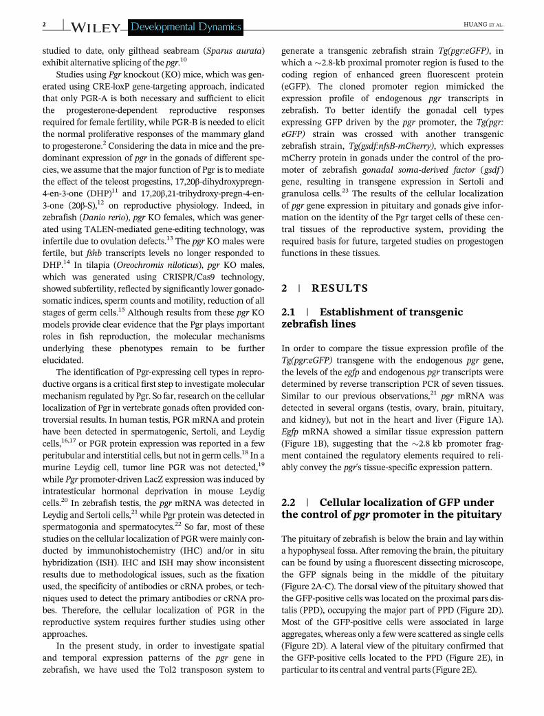
In order to compare the tissue expression profile of theTg(pgr:eGFP) transgene with the endogenous pgr gene,the levels of the egfp and endogenous pgr transcripts weredetermined by reverse transcription PCR of seven tissues.Similar to our previous observations,21 pgr mRNA wasdetected in several organs (testis, ovary, brain, pituitary,and kidney), but not in the heart and liver (Figure 1A).Egfp mRNA showed a similar tissue expression pattern(Figure 1B), suggesting that the �2.8 kb promoter frag-ment contained the regulatory elements required to reli-ably convey the pgr's tissue-specific expression pattern.
2.2 | Cellular localization of GFP underthe control of pgr promoter in the pituitary
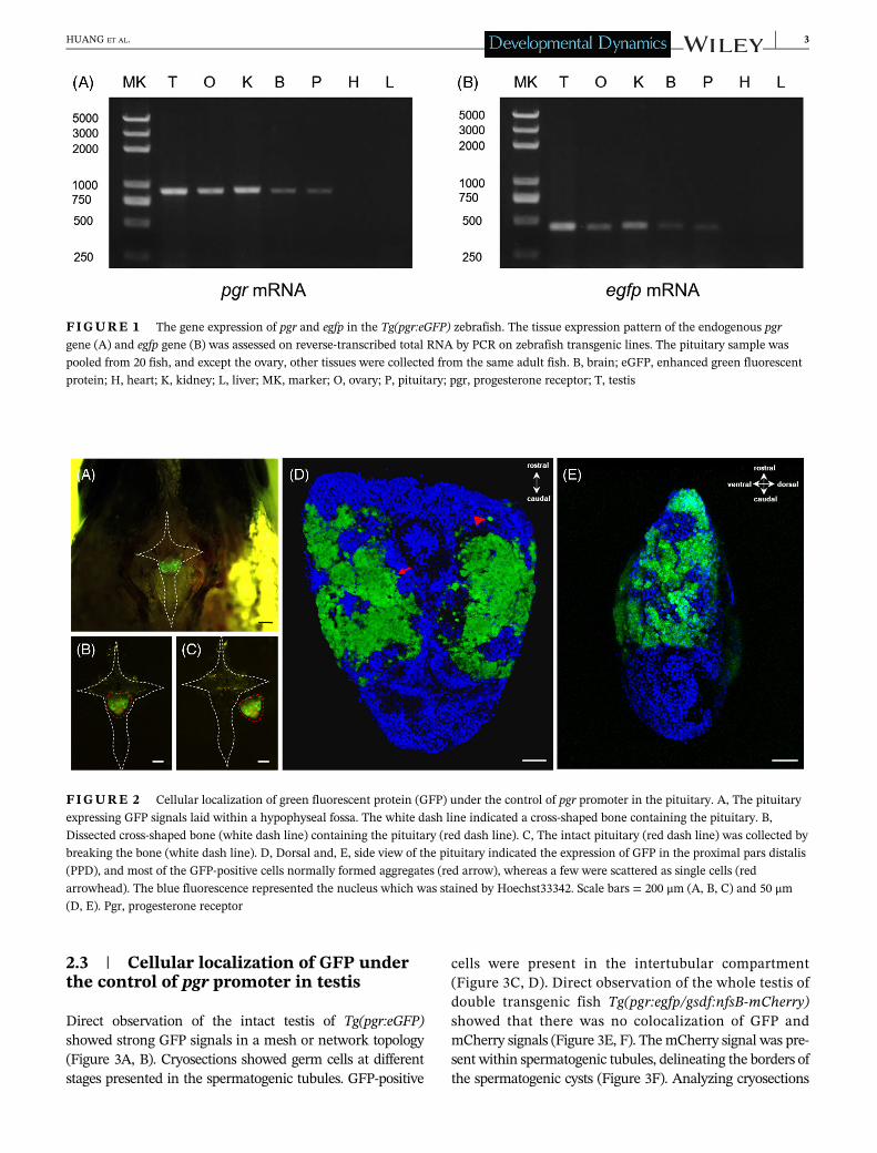
The pituitary of zebrafish is below the brain and lay withina hypophyseal fossa. After removing the brain, the pituitarycan be found by using a fluorescent dissecting microscope,the GFP signals being in the middle of the pituitary(Figure 2A-C). The dorsal view of the pituitary showed thatthe GFP-positive cells was located on the proximal pars dis-talis (PPD), occupying the major part of PPD (Figure 2D).Most of the GFP-positive cells were associated in largeaggregates, whereas only a few were scattered as single cells(Figure 2D). A lateral view of the pituitary confirmed thatthe GFP-positive cells located to the PPD (Figure 2E), inparticular to its central and ventral parts (Figure 2E).
2 HUANG ET AL.
2.3 | Cellular localization of GFP underthe control of pgr promoter in testis
Direct observation of the intact testis of Tg(pgr:eGFP)showed strong GFP signals in a mesh or network topology(Figure 3A, B). Cryosections showed germ cells at differentstages presented in the spermatogenic tubules. GFP-positive
cells were present in the intertubular compartment(Figure 3C, D). Direct observation of the whole testis ofdouble transgenic fish Tg(pgr:egfp/gsdf:nfsB-mCherry)showed that there was no colocalization of GFP andmCherry signals (Figure 3E, F). The mCherry signal was pre-sent within spermatogenic tubules, delineating the borders ofthe spermatogenic cysts (Figure 3F). Analyzing cryosections
FIGURE 1 The gene expression of pgr and egfp in the Tg(pgr:eGFP) zebrafish. The tissue expression pattern of the endogenous pgr
gene (A) and egfp gene (B) was assessed on reverse-transcribed total RNA by PCR on zebrafish transgenic lines. The pituitary sample was
pooled from 20 fish, and except the ovary, other tissues were collected from the same adult fish. B, brain; eGFP, enhanced green fluorescent
protein; H, heart; K, kidney; L, liver; MK, marker; O, ovary; P, pituitary; pgr, progesterone receptor; T, testis
FIGURE 2 Cellular localization of green fluorescent protein (GFP) under the control of pgr promoter in the pituitary. A, The pituitary
expressing GFP signals laid within a hypophyseal fossa. The white dash line indicated a cross-shaped bone containing the pituitary. B,
Dissected cross-shaped bone (white dash line) containing the pituitary (red dash line). C, The intact pituitary (red dash line) was collected by
breaking the bone (white dash line). D, Dorsal and, E, side view of the pituitary indicated the expression of GFP in the proximal pars distalis
(PPD), and most of the GFP-positive cells normally formed aggregates (red arrow), whereas a few were scattered as single cells (red
arrowhead). The blue fluorescence represented the nucleus which was stained by Hoechst33342. Scale bars = 200 μm (A, B, C) and 50 μm(D, E). Pgr, progesterone receptor
HUANG ET AL. 3
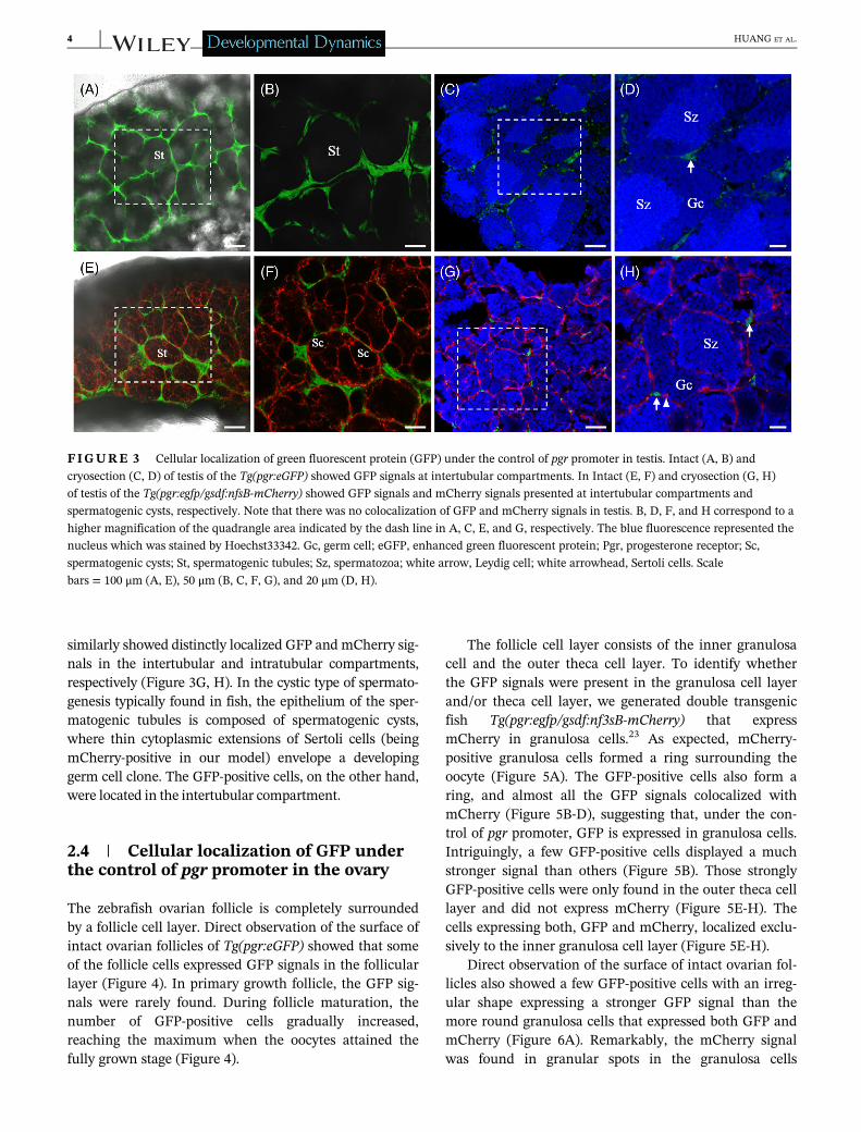
similarly showed distinctly localized GFP and mCherry sig-nals in the intertubular and intratubular compartments,respectively (Figure 3G, H). In the cystic type of spermato-genesis typically found in fish, the epithelium of the sper-matogenic tubules is composed of spermatogenic cysts,where thin cytoplasmic extensions of Sertoli cells (beingmCherry-positive in our model) envelope a developinggerm cell clone. The GFP-positive cells, on the other hand,were located in the intertubular compartment.
2.4 | Cellular localization of GFP underthe control of pgr promoter in the ovary
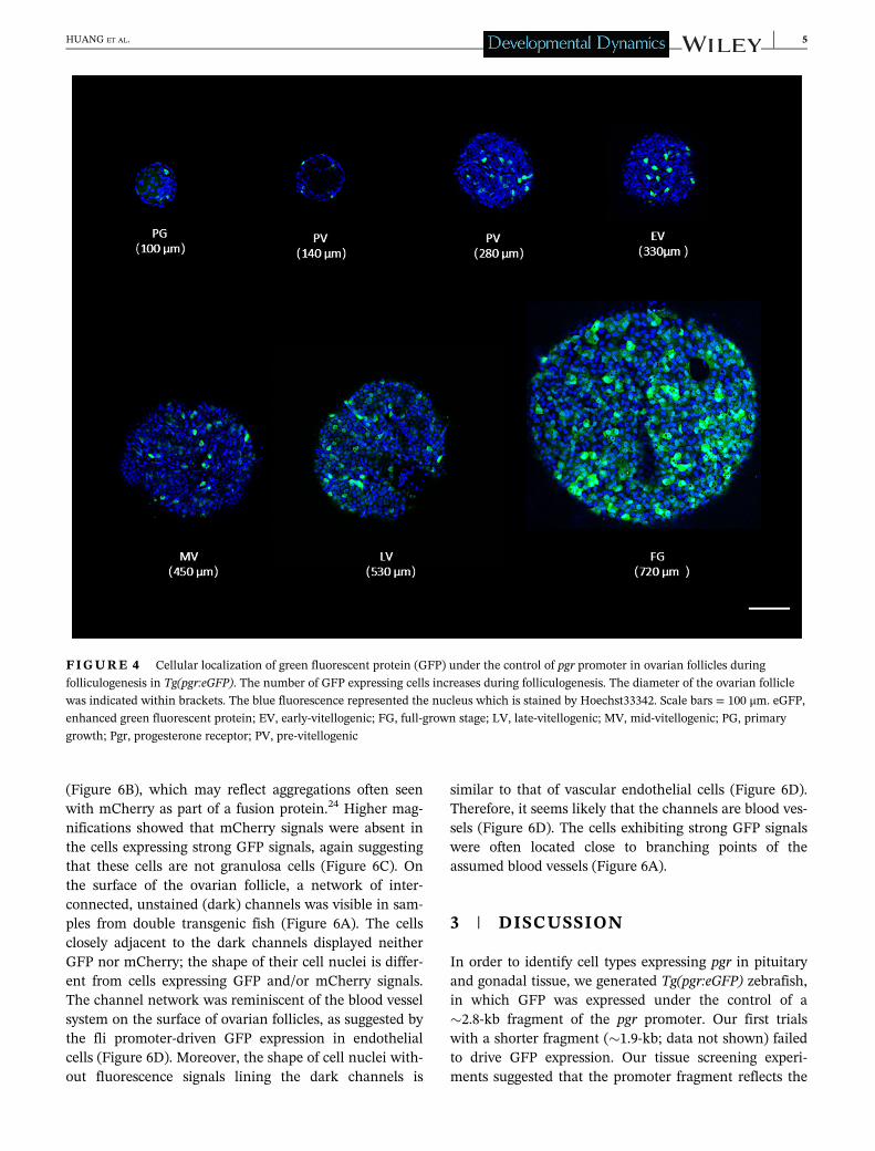
The zebrafish ovarian follicle is completely surroundedby a follicle cell layer. Direct observation of the surface ofintact ovarian follicles of Tg(pgr:eGFP) showed that someof the follicle cells expressed GFP signals in the follicularlayer (Figure 4). In primary growth follicle, the GFP sig-nals were rarely found. During follicle maturation, thenumber of GFP-positive cells gradually increased,reaching the maximum when the oocytes attained thefully grown stage (Figure 4).
The follicle cell layer consists of the inner granulosacell and the outer theca cell layer. To identify whetherthe GFP signals were present in the granulosa cell layerand/or theca cell layer, we generated double transgenicfish Tg(pgr:egfp/gsdf:nf3sB-mCherry) that expressmCherry in granulosa cells.23 As expected, mCherry-positive granulosa cells formed a ring surrounding theoocyte (Figure 5A). The GFP-positive cells also form aring, and almost all the GFP signals colocalized withmCherry (Figure 5B-D), suggesting that, under the con-trol of pgr promoter, GFP is expressed in granulosa cells.Intriguingly, a few GFP-positive cells displayed a muchstronger signal than others (Figure 5B). Those stronglyGFP-positive cells were only found in the outer theca celllayer and did not express mCherry (Figure 5E-H). Thecells expressing both, GFP and mCherry, localized exclu-sively to the inner granulosa cell layer (Figure 5E-H).
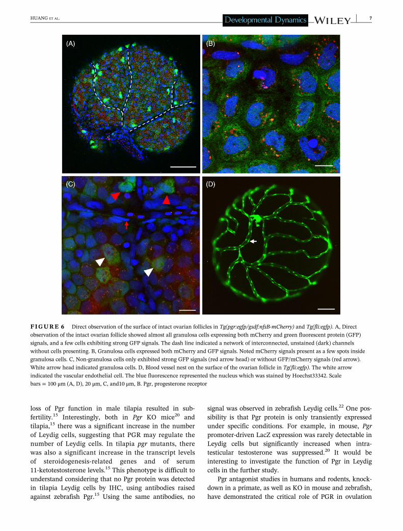
Direct observation of the surface of intact ovarian fol-licles also showed a few GFP-positive cells with an irreg-ular shape expressing a stronger GFP signal than themore round granulosa cells that expressed both GFP andmCherry (Figure 6A). Remarkably, the mCherry signalwas found in granular spots in the granulosa cells
FIGURE 3 Cellular localization of green fluorescent protein (GFP) under the control of pgr promoter in testis. Intact (A, B) and
cryosection (C, D) of testis of the Tg(pgr:eGFP) showed GFP signals at intertubular compartments. In Intact (E, F) and cryosection (G, H)
of testis of the Tg(pgr:egfp/gsdf:nfsB-mCherry) showed GFP signals and mCherry signals presented at intertubular compartments and
spermatogenic cysts, respectively. Note that there was no colocalization of GFP and mCherry signals in testis. B, D, F, and H correspond to a
higher magnification of the quadrangle area indicated by the dash line in A, C, E, and G, respectively. The blue fluorescence represented the
nucleus which was stained by Hoechst33342. Gc, germ cell; eGFP, enhanced green fluorescent protein; Pgr, progesterone receptor; Sc,
spermatogenic cysts; St, spermatogenic tubules; Sz, spermatozoa; white arrow, Leydig cell; white arrowhead, Sertoli cells. Scale
bars = 100 μm (A, E), 50 μm (B, C, F, G), and 20 μm (D, H).
4 HUANG ET AL.
(Figure 6B), which may reflect aggregations often seenwith mCherry as part of a fusion protein.24 Higher mag-nifications showed that mCherry signals were absent inthe cells expressing strong GFP signals, again suggestingthat these cells are not granulosa cells (Figure 6C). Onthe surface of the ovarian follicle, a network of inter-connected, unstained (dark) channels was visible in sam-ples from double transgenic fish (Figure 6A). The cellsclosely adjacent to the dark channels displayed neitherGFP nor mCherry; the shape of their cell nuclei is differ-ent from cells expressing GFP and/or mCherry signals.The channel network was reminiscent of the blood vesselsystem on the surface of ovarian follicles, as suggested bythe fli promoter-driven GFP expression in endothelialcells (Figure 6D). Moreover, the shape of cell nuclei with-out fluorescence signals lining the dark channels is
similar to that of vascular endothelial cells (Figure 6D).Therefore, it seems likely that the channels are blood ves-sels (Figure 6D). The cells exhibiting strong GFP signalswere often located close to branching points of theassumed blood vessels (Figure 6A).
3 | DISCUSSION
In order to identify cell types expressing pgr in pituitaryand gonadal tissue, we generated Tg(pgr:eGFP) zebrafish,in which GFP was expressed under the control of a�2.8-kb fragment of the pgr promoter. Our first trialswith a shorter fragment (�1.9-kb; data not shown) failedto drive GFP expression. Our tissue screening experi-ments suggested that the promoter fragment reflects the
FIGURE 4 Cellular localization of green fluorescent protein (GFP) under the control of pgr promoter in ovarian follicles during
folliculogenesis in Tg(pgr:eGFP). The number of GFP expressing cells increases during folliculogenesis. The diameter of the ovarian follicle
was indicated within brackets. The blue fluorescence represented the nucleus which is stained by Hoechst33342. Scale bars = 100 μm. eGFP,
enhanced green fluorescent protein; EV, early-vitellogenic; FG, full-grown stage; LV, late-vitellogenic; MV, mid-vitellogenic; PG, primary
growth; Pgr, progesterone receptor; PV, pre-vitellogenic
HUANG ET AL. 5

normal pgr expression pattern in adults. However, futureexperiments will have to show if also temporal aspectsduring ontogenesis are similar.
The adenohypophysis in fish is divided into three dis-tinct zones: rostral pars distalis (RPD), PPD, and parsintermedia. In the present study, only cells in the PPDregion of the zebrafish pituitary gland expressed GFP. Fol-lowing a general pattern in teleost fish, gonadotropes (Fshand Lh-producing cells), somatotropes (growth hormone-producing cells), and thyrotropes (Tsh-producing cells) arelocalized in the PPD.25 Previous studies in zebrafish dem-onstrated that both Fsh cells and Lh cells are localized inthe PPD.26,27 Our previous work demonstrated that DHPexerted a Pgr-mediated, direct stimulatory effect on fshbmRNA, and that pgr mRNA was expressed only in fshb-expressing cells.14 PGR protein was mainly localized togonadotrophs also in rodent and primate pituitaries.28,29
Therefore, it is very likely that Pgr is expressed in Fsh-producing gonadotrophs in the PPD of the zebrafish pitui-tary. There is little information on the localization of Pgrin vertebrate somatotropes and thyrotropes. A recent studydemonstrated that ewes with elevated serum levels of pro-gesterone exhibited greater levels of CXCR4, a receptor forchemokine ligand 12, in somatotropes.30 Further studies
using single-cell sequencing-based technologies would benecessary to identify other cell types potentially expressingPgr in zebrafish pituitary, although expression levels areexpected to be lower than in Fsh-producing gonadotrophs.
Since the cellular sites of PGR expression in the tes-tis are controversial, the physiological functions of pro-gestins mediated via PGR remain an interesting andunderexplored question. In the present study, the GFPsignals were exclusively observed in the Leydig cells.A previous study in zebrafish also reported that pgrmRNA was expressed in Leydig cells, and DHP stimu-lated 11beta-hydroxysteroid dehydrogenase activity viaa Pgr-dependent manner in adult zebrafish testis.21 Theexpression of PGR in Leydig cells has been reported inhuman, nonhuman primates, mouse, Japanese eel, andgilthead seabream.31 Therefore, the effects of progestins ontestis seem to be mediated through PGR in Leydig cells,and Leydig cell products, in turn, may affect spermatogen-esis. In mammals, progestins inhibit spermatogenesis andsperm vitality,32-34 and accordingly, the KO of the PGRincreases testicular development and sperm production.20
However, in teleosts, progestins seem to support, insteadof inhibiting, spermatogenesis (see reviews by11,35). Thisis also supported by genetic evidence, considering that
FIGURE 5 Cellular localization of green fluorescent protein (GFP) under the control of pgr promoter in ovarian follicles layer in Tg
(pgr:egfp/gsdf:nfsB-mCherry). The ovarian follicle consisted of an oocyte and a follicle cell layer (A, A0) which showed both GFP (B, B0) andmCherry (C, C0) signals. Most of GFP-positive cells expressed mCherry signals (D), and located at inner follicle layer (D0). A few of GFP-
positive cells without mCherry signals showed strong GFP signals (B0, D0) and located at outer follicle layer (D0). The blue fluorescencerepresented the nucleus which is stained by Hoechst33342. Oc, oocyte; white arrow, thecal cell; white arrowhead, granulosa cell. Scale
bars = 10 μm. Pgr, progesterone receptor
6 HUANG ET AL.
loss of Pgr function in male tilapia resulted in sub-fertility.15 Interestingly, both in Pgr KO mice20 andtilapia,15 there was a significant increase in the numberof Leydig cells, suggesting that PGR may regulate thenumber of Leydig cells. In tilapia pgr mutants, therewas also a significant increase in the transcript levelsof steroidogenesis-related genes and of serum11-ketotestosterone levels.15 This phenotype is difficult tounderstand considering that no Pgr protein was detectedin tilapia Leydig cells by IHC, using antibodies raisedagainst zebrafish Pgr.15 Using the same antibodies, no
signal was observed in zebrafish Leydig cells.22 One pos-sibility is that Pgr protein is only transiently expressedunder specific conditions. For example, in mouse, Pgrpromoter-driven LacZ expression was rarely detectable inLeydig cells but significantly increased when intra-testicular testosterone was suppressed.20 It would beinteresting to investigate the function of Pgr in Leydigcells in the further study.
Pgr antagonist studies in humans and rodents, knock-down in a primate, as well as KO in mouse and zebrafish,have demonstrated the critical role of PGR in ovulation
FIGURE 6 Direct observation of the surface of intact ovarian follicles in Tg(pgr:egfp/gsdf:nfsB-mCherry) and Tg(fli:egfp). A, Direct
observation of the intact ovarian follicle showed almost all granulosa cells expressing both mCherry and green fluorescent protein (GFP)
signals, and a few cells exhibiting strong GFP signals. The dash line indicated a network of interconnected, unstained (dark) channels
without cells presenting. B, Granulosa cells expressed both mCherry and GFP signals. Noted mCherry signals present as a few spots inside
granulosa cells. C, Non-granulosa cells only exhibited strong GFP signals (red arrow head) or without GFP/mCherry signals (red arrow).
White arrow head indicated granulosa cells. D, Blood vessel nest on the surface of the ovarian follicle in Tg(fli:egfp). The white arrow
indicated the vascular endothelial cell. The blue fluorescence represented the nucleus which was stained by Hoechst33342. Scale
bars = 100 μm (A, D), 20 μm, C, and10 μm, B. Pgr, progesterone receptor
HUANG ET AL. 7
(see36 for detailed review13,37). Inmammals, ovarian expres-sion of PGR is restricted primarily to granulosa cells of folli-cles destined to ovulate (see38 for detailed review). Inzebrafish, strong Pgr immunoreactivity was observed in thenuclei of follicular layer cells surrounding all stages ofoocytes.22 Using the same antibody, a study in tilapia rev-ealed abundant expression of Pgr in follicular cells duringthe early stages of ovarian differentiation.39 In gilthead sea-bream (S. aurata), immunohistochemical localization ofPgr showed strong immunostaining in the nuclei of granu-losa cells using a specific Pgr antibody against gilthead sea-bream Pgr.10 In the present study, our results clearly showthat the promoter of the pgr gene drives eGFP expression ingranulosa cells as well, which indicates that PGR expressionin granulosa cells is conserved among vertebrates.
Intriguingly, we also identified some non-granulosacells located at outer thecal cell layer, expressing GFP sig-nals. Some of these cells locating at the branches of theinterstitial blood vessels exhibited strong GFP signals. Weassume that these cells are theca cells, which appear out-side basement lamina associated with blood vessels, moveinto the interstitial region between oocytes, and finallyenclose the oocytes at an early vitellogenic stage (see refer-ence40 for detailed review). PGR protein has been detectedin theca cells from monkeys,41 rat,42 human,43 and dog.44
However, one study in mouse reported that there is nodetectable immunoreactivity in theca and interstitial cellsof ovary.45 Since PGR is expressed by granulosa cells andplays a critical role in ovulation, studies have focused ongranulosa cells and revealed that PGR-regulated genesunequivocally linked to the ovulatory success are theextracellular matrix proteases, for example, ADAMTS(a disintegrin and metalloproteinase with throm-bospondin motifs) family members.46 Our previous studyidentified adamts9, expressed in follicle cells, as a down-stream target of Pgr, which is essential for ovulation inzebrafish.47,48 However, it is worth noting that theca cellsare also vital to the mammalian ovulatory process.49 It hasbeen reported that Adamts1 mRNA in the theca internawas upregulated just prior to ovulation in the horse.50
Recently, studies in cattle indicate that ADAMTS familymembers produced by theca cells may play important rolesin follicle rupture and the accompanying tissue remo-deling in cattle.51,52 Therefore, further studies seemwarranted to examine whether the pgr expressed in thecacell layer participate in follicle rupture in zebrafish.
In summary, the Tg(pgr:eGFP) zebrafish line is a use-ful model to identify cellular localization of Pgr in pitui-tary and gonads. Further detailed analysis of thesequence between 1.9 and 2.8 kb pgr promoter fragmentcould reveal key regulatory elements involved in the reg-ulation of pgr gene expression. Finally, this transgeniczebrafish line will be a valuable model for the isolation of
Pgr-expressing cells to investigate molecular mechanismsmediated via Pgr in fish reproduction.
4 | EXPERIMENTAL PROCEDURES
4.1 | Animals
Tübingen strain zebrafish were propagated in our facili-ties (ESEN; Beijing, China). Fish were kept in rec-irculating aquaria at 28�C ± 0.5�C with a 14L:10Dphotoperiod (lights on at 08:00 AM). Fish were fed threetimes daily with a commercial food (Otohime B2; Mar-ubeni Nisshin Feed, Tokyo, Japan) and supplementedwith newly hatched brine shrimp. A transgenic zebrafishline Tg(fli:egfp), which expresses GFP signals in vascularendothelial cells, was kindly provided by Dr. Mingyu Li(Xiamen University, Xiamen, China). All experimentalprotocols were approved by the Institutional Animal Careand Use Committee at the Xiamen University.
4.2 | Preparation of pTol2(pgr:eGFP)Construct
The genome sequence of the pgr gene was identified in thezebrafish genome using the ENSEMBL genome browser(http://www.ensembl.org/index.html). The −2798/+26promoter fragment of the zebrafish pgr gene was amplifiedfrom zebrafish genomic DNA using specific primers (pgr-fw: 50-CCATGTGATTGGACCCGATTTCCGATTACA-30,pgr-rv: 50-CGCAATCGTCCGAGATGCGTCCTCTTT-30).The reaction was incubated for 7 cycles of 25 seconds at94�C, 3.5 minutes at 72�C, and followed by 32 cycles of25 seconds at 94�C, 3.5 minutes at 67�C. The�2.8-kb frag-ment was inserted into the upstream of the eGFP codingsequence of the pT2AL200R150G construct (kindly pro-vided by Prof. Kawakami; National Institute of Genetics,Shizuoka, Japan) between the NheI and BamHI sites togenerate the transgenic construct pTol2(pgr:eGFP)(Figure 7).
4.3 | Generation of transgenic fish lines
The transgenic zebrafish line was generated with theTol2 Transposon System.53 The animal pole of zebrafishone-cell-stage embryos was microinjected with a mixturecontaining pTol2(pgr:eGFP) vector (10 ng/μL), Tol2transposase enzyme mRNA (10 ng/μL), and phenol red(0.25%) (P0290; Sigma-Aldrich, St. Louis, Missouri). TheTol2 transposase capped mRNA was in vitro transcribedfrom the pCS-zT2TP transposase vector (kindly provided
8 HUANG ET AL.

by Prof. Kawakami) using the mMESSAGE mMACHINESP6 kit (Ambion AM1340; Austin, Texas). The DNA solu-tions were injected using a PV 820 Pneumatic PicPump(World Precision Instrument; Florida) and borosilicateglass capillaries (IB100F-4; World Precision Instrument).The F0 fish were bred to maturity and screened by PCRwith primers containing pgr promoter and egfp sequences(screen-fw: 50-ACTACAGATCACAACCTCCAAAC-30,screen-rv: 50-TTGTAGTTGTACTCCAGCTTGTGCCC-30).The injected embryos were raised to sexual maturity, andtransgenic offspring were identified by genotyping usinggenomic DNA extracted from caudal fin biopsies. Thepositive F0 fish were mated with wild-type (WT) fish togenerate F1 transgenic lines named Tg(pgr:eGFP).
The Tg(pgr:eGFP) was crossed with Tg(gsdf:nfsB-mCherry) zebrafish lines generated in our laboratory (willbe published separately) to obtain Tg(pgr:egfp/gsdf:nfsB-mCherry) double transgenic fish. Driven by the proximalpromoter region of the zebrafish gsdf gene, the Tg(gsdf:nfsB-mCherry) transgenic zebrafish line specificallyexpressed nitroreductase-mCherry fusion protein in gran-ulosa and Sertoli cells, as described previously.23
4.4 | RNA extraction and reverseTranscription-PCR
Total RNA from different tissues (brain, pituitary, heart,liver, kidney, ovary, and testis) was extracted using RNAzolreagent (RN 190; Molecular Research Center, Cincinnati,Ohio) according to the manufacturer's protocol. Reversetranscription was carried out using the RevertAid FirstStrand cDNA Synthesis Kit (K1622; Thermo Scientific, Lith-uania) with 800 ng total RNA. The PCR was conducted onthe cDNA template for 32 cycleswith an annealing tempera-ture of 58�C using pgr-specific primers (pgr-RT-fw: 50-AATC
TCATCA TGGAGCCACCG-30, pgr-RT-rv: 50-CCTCTGGCTGTGTGTTGTCG-30) and egfp-specific primers (egfp-RT-fw:50-GGACGACGGCAACTACAAGA-30, egfp-RT-rv: 50-GTCCATGCCGAGAGTGATCC-30). As a negative control, anequal volume of water was added into the PCR mixtureinstead of cDNA. The PCR products were run on a 1.5%agarose gel and photographed in the BioDocAnalyzedigital gel documentation system (Biometra GmbH,Göttingen, Germany).
4.5 | Collection of pituitary and ovarianfollicles
Adult zebrafish were anesthetized in ice water. Pituitarieswere isolated as described previously.26 In brief, the pitui-tary glands were collected under a fluorescent dissectingmicroscope (Leica M165FC; Leica Microsystems, Wetzlar,Germany) with fine forceps and eye scissors. We firstremoved the dorsal part of the skull, followed by remov-ing the whole brain. The pituitary gland, showing a clearGFP signal, lay within the hypophyseal fossa (Figure 2A).To collect the whole pituitary, we first dissected a cross-shaped bone containing the pituitary (Figure 2B) and fur-ther dissected the bone to collect the intact pituitary(Figure 2C). The pituitary was stained with Hoechst33342 (C1022; Beyotime Institute of Biotechnology,Shanghai, China), mounted on a 29-mm glass-bottomdish (D29-10-1.5-N; Cellvis, China) containing 1% low-melting agarose (A9414; Sigma-Aldrich) in phosphate-buffered saline (PBS). Images were taken with an SP8(Leica, Germany) confocal laser scanning microscope.
Isolation of ovarian follicles was conducted asdescribed previously.54 In brief, ovaries were pooled andquickly dispersed in a petri dish containing 60% LeibovitzL-15 medium (GIBCO BRL, Gaithersburg, Maryland).
FIGURE 7 Schematic representation of the zebrafish pgr gene structure and transgene construct design. The zebrafish pgr gene is
located on chromosome 18 which has eight exons. The proximal 2824 bp promoter region of the pgr gene was placed upstream to the open
reading frame of the enhanced green fluorescent protein. In addition, the transgene was franked by Tol2 sites. The black box represents
exons; pA, SV40 poly(A) signal. Pgr, progesterone receptor
HUANG ET AL. 9
Follicles in different stages of development were identi-fied and manually isolated under a stereomicroscope(Leica M165FC, Germany). The follicles were classifiedinto six stages based on the diameter, namely primarygrowth (diameter ≤ 100 μm, PG), pre-vitellogenic(100-300 μm, PV), early-vitellogenic (300-400 μm, EV),mid-vitellogenic (400-500 μm, MV), late-vitellogenic(500-600 μm, LV), and the full-grown stage (≥650 μm,FG). The follicles were stained with Hoechst 33342(Beyotime Institute of Biotechnology). Images were cap-tured using M165FC (Leica, Germany), LSM880 (CarlZeiss MicroImaging, Oberkochen, Germany), and SP8(Leica, Germany) confocal laser scanning microscope.
4.6 | Preparation of cryosections
The adult zebrafish were anesthetized, and testes werecarefully dissected and fixed in 4% paraformaldehyde(Merck, Germany) in PBS overnight. The gonads wererinsed in PBS twice (1 hour per time) and then dehydratedin 25% sucrose at 4�C. After dehydration, the whole tissueswere embedded in the Tissue Freezing Medium (LeicaBiosystems, Heidelberg, Germany) and frozen by floatingin liquid nitrogen. Cryosections of the embedded gonadwere cut using a Leica CM1850 cryostat (Leica Micro-systems, Heidelberg, Germany) at −18�C. Tissue sectionsof the testis (10 μm) or ovary (20 μm) were collected andthen mounted onto poly-L-lysine-coated slides. The slideswere air-dried for 1 hour, rinsed in PBS three times, cov-ered with VECTASHIELD Mounting Medium with DAPI(H-1200; Vector Laboratories, Burlingame, California)and coverslipped. Pictures were taken with an LSM780microscope (Carl Zeiss, Germany).
ACKNOWLEDGMENTSWe would like to express our great appreciation toDr. Rüdiger Schulz at Utrecht University for his valuablecomments and editing the English. This research wassupported by the National Natural Science Foundation ofChina (No. 31672628 and 41976092) and Program for NewCentury Excellent Talents in Fujian Province University.
AUTHOR CONTRIBUTIONSJing Huang: Conceptualization; data curation; method-ology; writing-original draft; writing-review and editing.Ting Zhang: Methodology. Ke Jiang: Methodology.Wan Hong: Supervision. Shi Chen: Conceptualization;funding acquisition; supervision; writing-original draft;writing-review and editing.
ORCIDShi Xi Chen https://orcid.org/0000-0003-1627-5378
REFERENCES1. Evans RM. The steroid and thyroid-hormone receptor super-
family. Science. 1988;240(4854):889-895.
2. Conneely OM, Mulac-Jericevic B, DeMayo F, Lydon JP,O'Malley BW. Reproductive functions of progesterone recep-tors. Recent Prog Horm Res. 2002;57:339-355.
3. Thomas P. Characteristics of membrane progestin receptoralpha (mPR alpha) and progesterone membrane receptor com-ponent 1 (PGMRC1) and their roles in mediating rapid proges-tin actions. Front Neuroendocrinol. 2008;29(2):292-312.
4. Tian JD, Kim S, Heilig E, Ruderman JV. Identification ofXPR-1, a progesterone receptor required for Xenopus oocyteactivation. Proc Natl Acad Sci USA. 2000;97(26):14358-14363.
5. Todo T, Ikeuchi T, Kobayashi T, et al. Characterization of atesticular 17 alpha,20 beta-dihydroxy-4-pregnen-3-one(a spermiation-inducing steroid in fish) receptor from a teleost,Japanese eel (Anguilla japonica). FEBS Lett. 2000;465(1):12-17.
6. Ikeuchi T, Todo T, Kobayashi T, Nagahama Y. A novel proges-togen receptor subtype in the Japanese eel, Anguilla japonica.FEBS Lett. 2002;510(1–2):77-82.
7. Morini M, Penaranda DS, Vilchez MC, et al. Nuclear and mem-brane progestin receptors in the European eel: characterizationand expression in vivo through spermatogenesis. Comp Bio-chem Phys A. 2017;207:79-92.
8. Conneely OM, Maxwell BL, Toft DO, Schrader WT,Omalley BW. The A-forms and B-forms of the chickenprogesterone-receptor Arise by alternate initiation of transla-tion of a unique messenger-RNA. Biochem Biophys ResCommun. 1987;149(2):493-501.
9. Kastner P, Krust A, Turcotte B, et al. Two distinct estrogen-regulated promoters generate transcripts encoding the twofunctionally different human progesterone receptor forms aand B. EMBO J. 1990;9(5):1603-1614.
10. Zapater C, Chauvigne F, Fernandez-Gomez B, Finn RN,Cerda J. Alternative splicing of the nuclear progestin receptorin a perciform teleost generates novel mechanisms ofdominant-negative transcriptional regulation. Gen CompEndocrinol. 2013;182:24-40.
11. Scott AP, Sumpter JP, Stacey N. The role of the maturation-inducing steroid, 17,20 beta-dihydroxypregn-4-en-3-one, inmale fishes: a review. J Fish Biol. 2010;76(1):183-224.
12. Thomas P, Trant JM. Evidence that 17-Alpha,20-Beta,21-Trihy-droxy-4-Pregnen-3-one is a maturation-inducing steroid in spottedseatrout. Fish Physiol Biochem. 1989;7(1–6):185-191.
13. Zhu Y, Liu DT, Shaner ZC, Chen SX, Hong WS, Stellwag EJ.Nuclear progestin receptor (Pgr) knockouts in zebrafish demon-strate role for Pgr in ovulation but not in rapid non-genomic ste-roid mediated meiosis resumption. Front Endocrinol. 2015;6:37.
14. Wang CL, Liu DT, Chen WT, et al. Progestin increases theexpression of gonadotropins in pituitaries of male zebrafish.J Endocrinol. 2016;230(1):143-156.
15. Fang XL, Wu LM, Yang LY, et al. Nuclear progestin receptor(Pgr) knockouts resulted in subfertility in male tilapia(Oreochromis niloticus). J Steroid Biochem. 2018;182:62-71.
16. Shah C, Modi D, Sachdeva G, Gadkar S, Puri C. Coexistence ofintracellular and membrane-bound progesterone receptors inhuman testis. J Clin Endocrinol Metab. 2005;90(1):474-483.
17. Han YB, Feng HL, Sandlow JI, Haines CJ. Comparing expres-sion of progesterone and estrogen receptors in testicular tissue
10 HUANG ET AL.
from men with obstructive and nonobstructive azoospermia.J Androl. 2009;30(2):127-133.
18. Luetjens CM, Didolkar A, Kliesch S, et al. Tissue expression ofthe nuclear progesterone receptor in male non-human pri-mates and men. J Endocrinol. 2006;189(3):529-539.
19. Schwarzenbach H, Manna PR, Stocco DM, Chakrabarti G,Mukhopadhyay AK. Stimulatory effect of progesterone on theexpression of steroidogenic acute regulatory protein in MA-10Leydig cells. Biol Reprod. 2003;68(3):1054-1063.
20. Lue YH, Wang C, Lydon JP, Leung A, Li J, Swerdloff RS. Func-tional role of progestin and the progesterone receptor in thesuppression of spermatogenesis in rodents. Andrology. 2013;1(2):308-317.
21. Chen SX, Bogerd J, Garcia-Lopez A, et al. Molecular cloningand functional characterization of a zebrafish nuclear proges-terone receptor. Biol Reprod. 2010;82(1):171-181.
22. Hanna RN, Daly SCJ, Pang YF, et al. Characterization andexpression of the nuclear progestin receptor in zebrafishgonads and brain. Biol Reprod. 2010;82(1):112-122.
23. Gautier A, Sohm F, Joly JS, Le Gac F, Lareyre JJ. The proximalpromoter region of the zebrafish gsdf gene is sufficient tomimic the Spatio-temporal expression pattern of the endoge-nous gene in Sertoli and granulosa cells. Biol Reprod. 2011;85(6):1240-1251.
24. Davidson MW, Campbell RE. Engineered fluorescent proteins:innovations and applications. Nat Methods. 2009;6(10):713-717.
25. Laiz-Carrion R, Segura-Noguera MD, del Rio MDM,Mancera JM. Ontogeny of adenohypophyseal cells in the pitui-tary of the American shad (Alosa sapidissima). Gen CompEndocrinol. 2003;132(3):454-464.
26. Chen WT, Ge W. Ontogenic expression profiles of gonadotro-pins (fshb and lhb) and growth hormone (gh) during sexual dif-ferentiation and puberty onset in female zebrafish. Biol Reprod.2012;86(3):1-11.
27. Golan M, Martin AO, Mollard P, Levavi-Sivan B. Anatomicaland functional gonadotrope networks in the teleost pituitary.Sci Rep. 2016;6:23777.
28. Sprangers SA, Brenner RM, Bethea CL. Estrogen and progestinreceptor immunocytochemistry in Lactotropes versusGonadotropes of monkey pituitary cell-cultures. Endocrinology.1989;124(3):1462-1470.
29. Fox SR, Harlan RE, Shivers BD, Pfaff DW. Chemical character-ization of neuroendocrine targets for progesterone in thefemale rat-brain and pituitary. Neuroendocrinology. 1990;51(3):276-283.
30. Sanchez NS, Quinn KE, Ashley AK, Ashley RL. In the ovinepituitary, CXCR4 is localized in gonadotropes andsomatotropes and increases with elevated serum progesterone.Domest Anim Endocrinol. 2018;62:88-97.
31. Chauvigne F, Parhi J, Olle J, Cerda J. Dual estrogenic regula-tion of the nuclear progestin receptor and spermatogonialrenewal during gilthead seabream (Sparus aurata) spermato-genesis. Comp Biochem Phys A. 2017;206:36-46.
32. Nieschlag E, Zitzmann M, Kamischke A. Use of progestins inmale contraception. Steroids. 2003;68(10–13):965-972.
33. Walton MJ, Bayne RAL, Wallace I, Baird DT, Anderson RA.Direct effect of progestogen on gene expression in the testisduring gonadotropin withdrawal and early suppression of sper-matogenesis. J Clin Endocrinol Metab. 2006;91(7):2526-2533.
34. Wang C, Swerdloff RS. Hormonal approaches to male contra-ception. Curr Opin Urol. 2010;20(6):520-524.
35. Schulz RW, de Franca LR, Lareyre JJ, et al. Spermatogenesis infish. Gen Comp Endocrinol. 2010;165(3):390-411.
36. Robker RL, Akison LK, Russell DL. Control of oocyte releaseby progesterone receptor-regulated gene expression. Nucl Rec-ept Signal. 2009;7:e012.
37. Bishop CV, Hennebold JD, Kahl CA, Stouffer RL. Knockdownof progesterone receptor (PGR) in macaque granulosa cells dis-rupts ovulation and progesterone production. Biol Reprod.2016;94(5):1-10.
38. Akison LK, Robker RL. The critical roles of progesterone recep-tor (PGR) in ovulation, oocyte developmental competence andOviductal transport in mammalian reproduction. ReprodDomest Anim. 2012;47:288-296.
39. Zhou LY, Luo F, Fang XL, et al. Blockage of progestin physiol-ogy disrupts ovarian differentiation in XX Nile tilapia(Oreochromis niloticus). Biochem Biophys Res Commun. 2016;473(1):29-34.
40. Le Menn F, Cerdà J, Babin PJ. Ultrastructural aspects of theontogeny and differentiation of ray-finned fish ovarian follicles.In: Babin PJ, Cerdà J, Lubzens E, eds. The Fish Oocyte. Dor-drecht, The Netherlands: Springer; 2007:1-37.
41. Hild-Petito S, Stouffer RL, Brenner RM. Immunocytochemicallocalization of estradiol and progesterone receptors in the mon-key ovary throughout the menstrual cycle. Endocrinology. 1988;123(6):2896-2905.
42. Telleria CM, Stocco CO, Stati AO, Deis RP. Progesterone recep-tor is not required for progesterone action in the rat corpusluteum of pregnancy. Steroids. 1999;64(11):760-766.
43. Iwai T, Nanbu Y, Iwai M, Taii S, Fujii S, Mori T. Immunohisto-chemical localization of estrogen-receptors and progesteronereceptors in the human ovary throughout the menstrual-cycle.Virchows Arch A. 1990;417(5):369-375.
44. Vermeirsch H, Simoens P, Coryn M, Van den Broeck W.Immunolocalization of progesterone receptors in the canineovary and their relation to sex steroid hormone concentrations.Reproduction. 2001;122(1):73-83.
45. Shao R, Markstrom E, Friberg PA, Johansson M, Billig H.Expression of progesterone receptor (PR) a and B isoforms inmouse granulosa cells: stage-dependent PR-mediated regula-tion of apoptosis and cell proliferation. Biol Reprod. 2003;68(3):914-921.
46. Robker RL, Russell DL, Espey LL, Lydon JP, O'Malley BW,Richards JS. Progesterone-regulated genes in the ovulation pro-cess: ADAMTS-1 and cathepsin L proteases. Proc Natl Acad SciUSA. 2000;97(9):4689-4694.
47. Liu DT, Brewer MS, Chen S, Hong W, Zhu Y. Transcriptomicsignatures for ovulation in vertebrates. Gen Comp Endocrinol.2017;247:74-86.
48. Liu DT, Carter NJ, Wu XJ, Hong WS, Chen SX, Zhu Y. Proges-tin and nuclear progestin receptor are essential forupregulation of metalloproteinase in zebrafish preovulatory fol-licles. Front Endocrinol (Lausanne). 2018;9:517.
49. Young JM, McNeilly AS. Theca: the forgotten cell of the ovar-ian follicle. Reproduction. 2010;140(4):489-504.
50. Boerboom D, Russell DL, Richards JS, Sirois J. Regulation oftranscripts encoding ADAMTS-1 (a disintegrin andmetalloproteinase with thrombospondin-like motifs-1) and
HUANG ET AL. 11
progesterone receptor by human chorionic gonadotropin inequine preovulatory follicles. J Mol Endocrinol. 2003;31(3):473-485.
51. Madan P, Bridges PJ, Komar CM, et al. Expression of messengerRNA for ADAMTS subtypes changes in the periovulatory follicleafter the gonadotropin surge and during luteal development andregression in cattle. Biol Reprod. 2003;69(5):1506-1514.
52. Willis EL, Bridges PJ, Fortune JE. Progesterone receptor andprostaglandins mediate luteinizing hormone-induced changesin messenger RNAs for ADAMTS proteases in theca cells ofbovine periovulatory follicles. Mol Reprod Dev. 2017;84(1):55-66.
53. Suster ML, Kikuta H, Urasaki A, Asakawa K, Kawakami K.Transgenesis in zebrafish with the tol2 transposon system.Methods Mol Biol. 2009;561:41-63.
54. Bai J, Gong WD, Wang CL, Gao YD, Hong WS, Chen SX.Dynamic methylation pattern of cyp19a1a core promoter dur-ing zebrafish ovarian folliculogenesis. Fish Physiol Biochem.2016;42(3):947-954.
How to cite this article: Huang J, Zhang TT,Jiang K, Hong WS, Chen SX. GFP expressionpattern in pituitary and gonads under the controlof nuclear progesterone receptor promoter intransgenic zebrafish. Developmental Dynamics.2020;1–12. https://doi.org/10.1002/dvdy.213
12 HUANG ET AL.