Embed Size (px)
Citation preview
Getting Pumped About STEMI and Cardiogenic Shock
June 20, 2017 Amy Shepard MS, RN, ACNS-BC, CCRN-K
STEMI/ Cardiogenic Shock Program Manager University Hospital
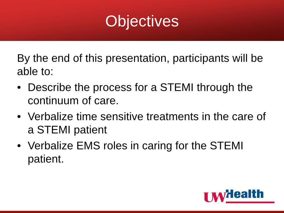
By the end of this presentation, participants will be able to: • Describe the process for a STEMI through the
continuum of care. • Verbalize time sensitive treatments in the care of
a STEMI patient • Verbalize EMS roles in caring for the STEMI
patient.
Objectives
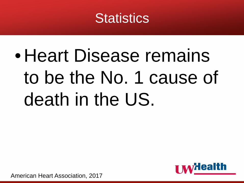
• Heart Disease remains to be the No. 1 cause of death in the US.
Statistics
American Heart Association, 2017
Every 40 seconds an American will have a heart attack.
Statistics (cont.)
American Heart Association, 2017
• About 790,000 people in the US have heart attacks each year. Of those, about 114,000 people will die.
Statistics (cont.)
American Heart Association, 2017
• Average age at the first heart attack is 65.3 years for males and 71.8 years for females.
Statistics (cont.)
American Heart Association, 2017
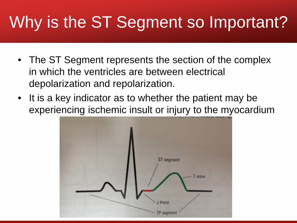
Why is the ST Segment so Important?
• The ST Segment represents the section of the complex in which the ventricles are between electrical depolarization and repolarization.
• It is a key indicator as to whether the patient may be experiencing ischemic insult or injury to the myocardium
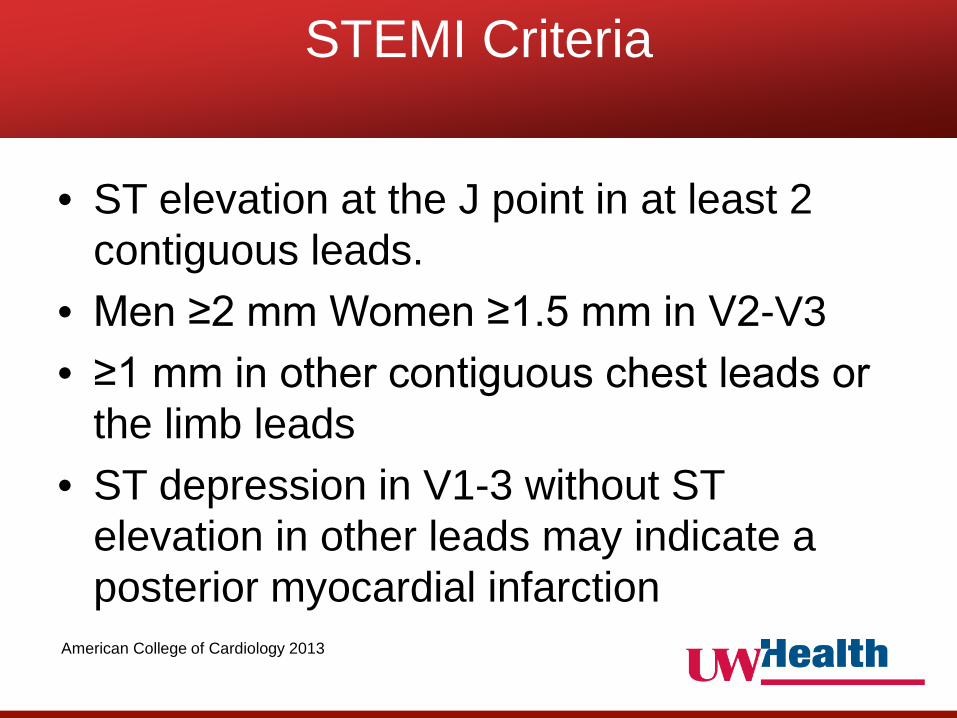
• ST elevation at the J point in at least 2 contiguous leads.
• Men ≥2 mm Women ≥1.5 mm in V2-V3 • ≥1 mm in other contiguous chest leads or
the limb leads • ST depression in V1-3 without ST
elevation in other leads may indicate a posterior myocardial infarction
American College of Cardiology 2013
STEMI Criteria
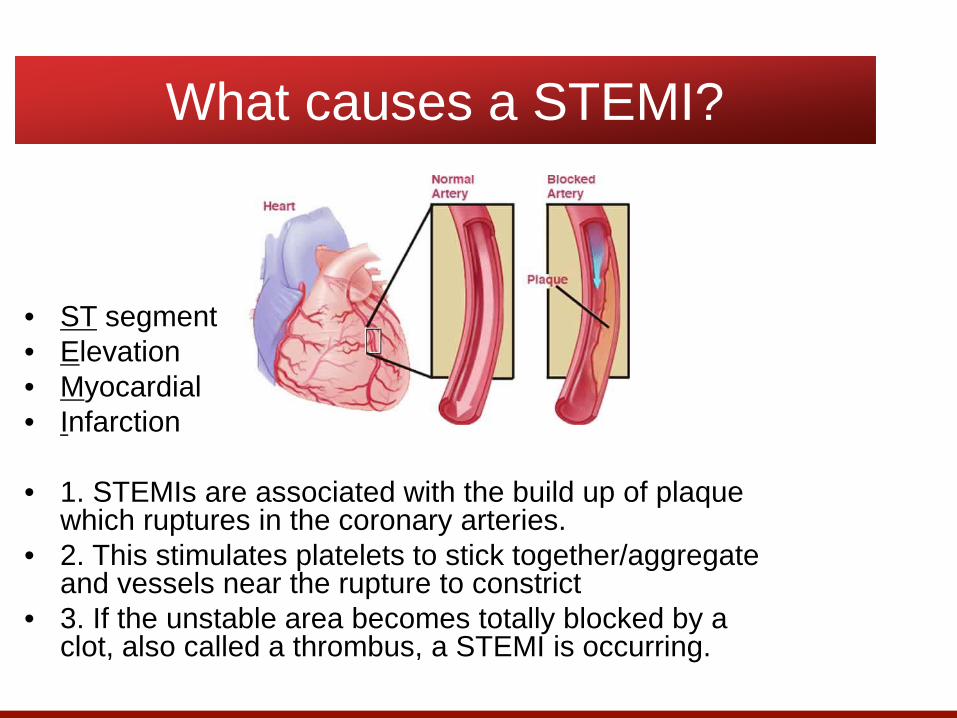
What causes a STEMI?
• ST segment • Elevation • Myocardial • Infarction
• 1. STEMIs are associated with the build up of plaque
which ruptures in the coronary arteries. • 2. This stimulates platelets to stick together/aggregate
and vessels near the rupture to constrict • 3. If the unstable area becomes totally blocked by a
clot, also called a thrombus, a STEMI is occurring.
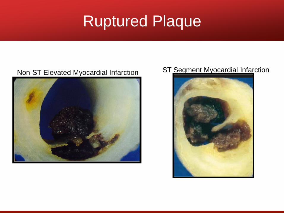
Ruptured Plaque
Non-ST Elevated Myocardial Infarction ST Segment Myocardial Infarction
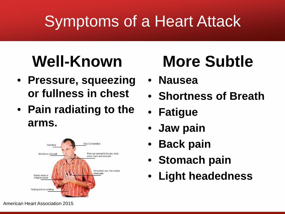
Symptoms of a Heart Attack
Well-Known • Pressure, squeezing
or fullness in chest • Pain radiating to the
arms.
More Subtle • Nausea • Shortness of Breath • Fatigue • Jaw pain • Back pain • Stomach pain • Light headedness
American Heart Association 2015
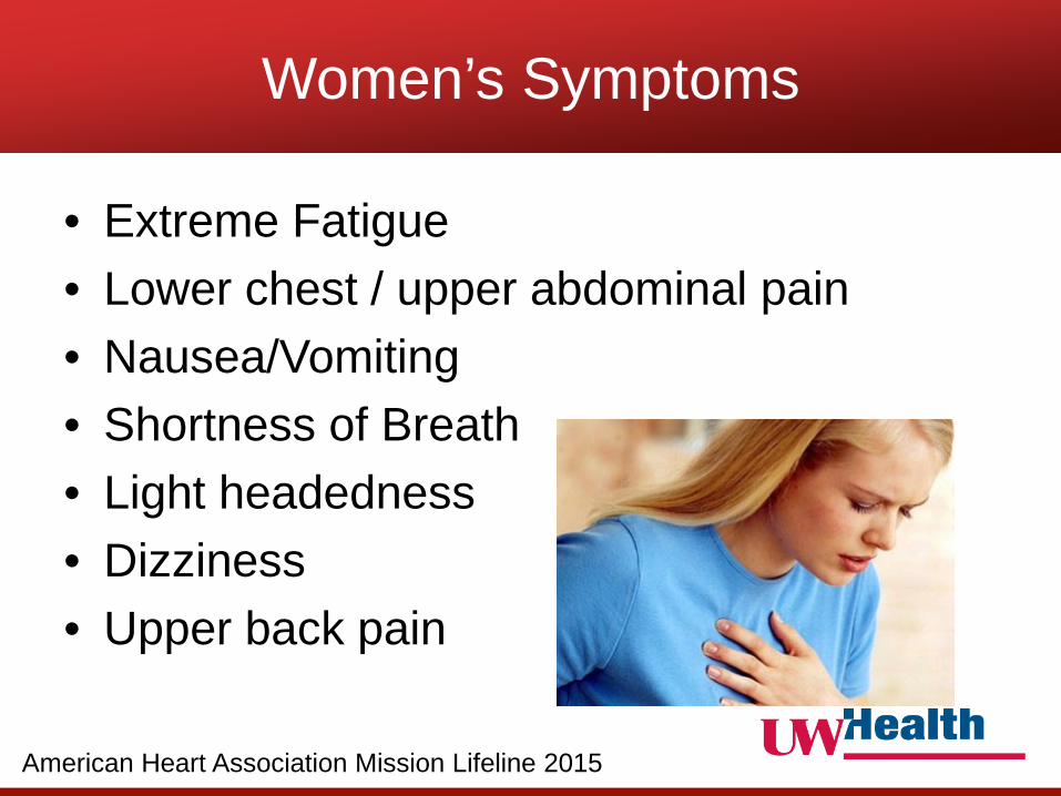
• Extreme Fatigue • Lower chest / upper abdominal pain • Nausea/Vomiting • Shortness of Breath • Light headedness • Dizziness • Upper back pain
Women’s Symptoms
American Heart Association Mission Lifeline 2015
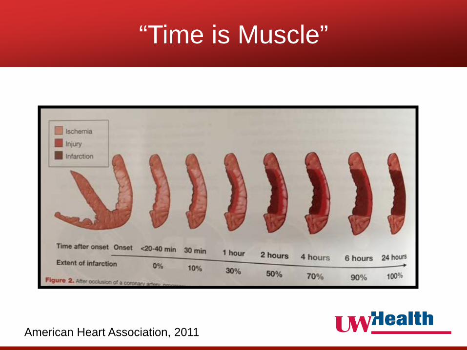
“Time is Muscle”
American Heart Association, 2011
ECG Then and Now
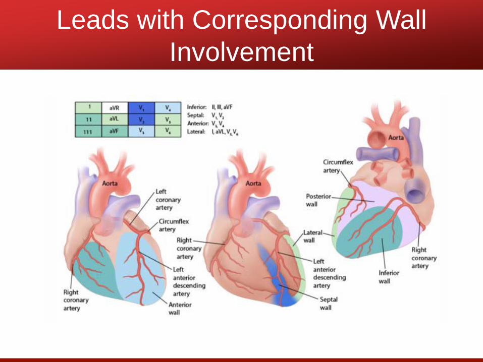
Leads with Corresponding Wall Involvement
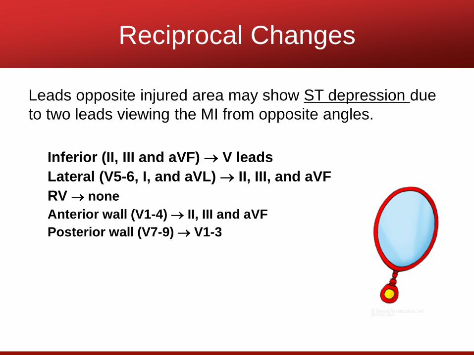
Reciprocal Changes
Leads opposite injured area may show ST depression due to two leads viewing the MI from opposite angles.
Inferior (II, III and aVF) → V leads Lateral (V5-6, I, and aVL) → II, III, and aVF RV → none Anterior wall (V1-4) → II, III and aVF Posterior wall (V7-9) → V1-3
of 12-lead when ST elevation is present is indicative of reciprocal changes.
Note: Reciprocal ST depression will have a positive T wave
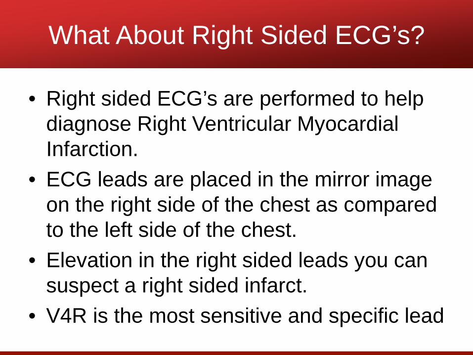
What About Right Sided ECG’s?
• Right sided ECG’s are performed to help diagnose Right Ventricular Myocardial Infarction.
• ECG leads are placed in the mirror image on the right side of the chest as compared to the left side of the chest.
• Elevation in the right sided leads you can suspect a right sided infarct.
• V4R is the most sensitive and specific lead
• If you have a patient that has definite ST-segment elevation, it is best to load them into the ambulance and get them to the Cath Lab ASAP.
• Do not delay patient transport to perform a right sided ECG.
Right Sided ECG’s (cont).
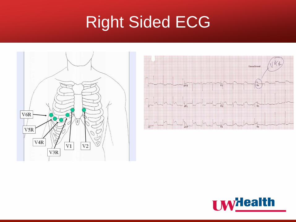
Right Sided ECG
• Posterior MI’s are associated with inferior wall MI’s and Right Ventricular Infarcts.
• With normal lead placement, a posterior ECG will present as ST segment depression in the septal leads V1 and V2.
Posterior Wall MI
• If ST segment depression is noted in V1 and V2, suspect Posterior Wall MI.
• Why ST depression and not elevation?
• Do not delay transport to obtain a posterior ECG.
Posterior ECG
Posterior ECG’s
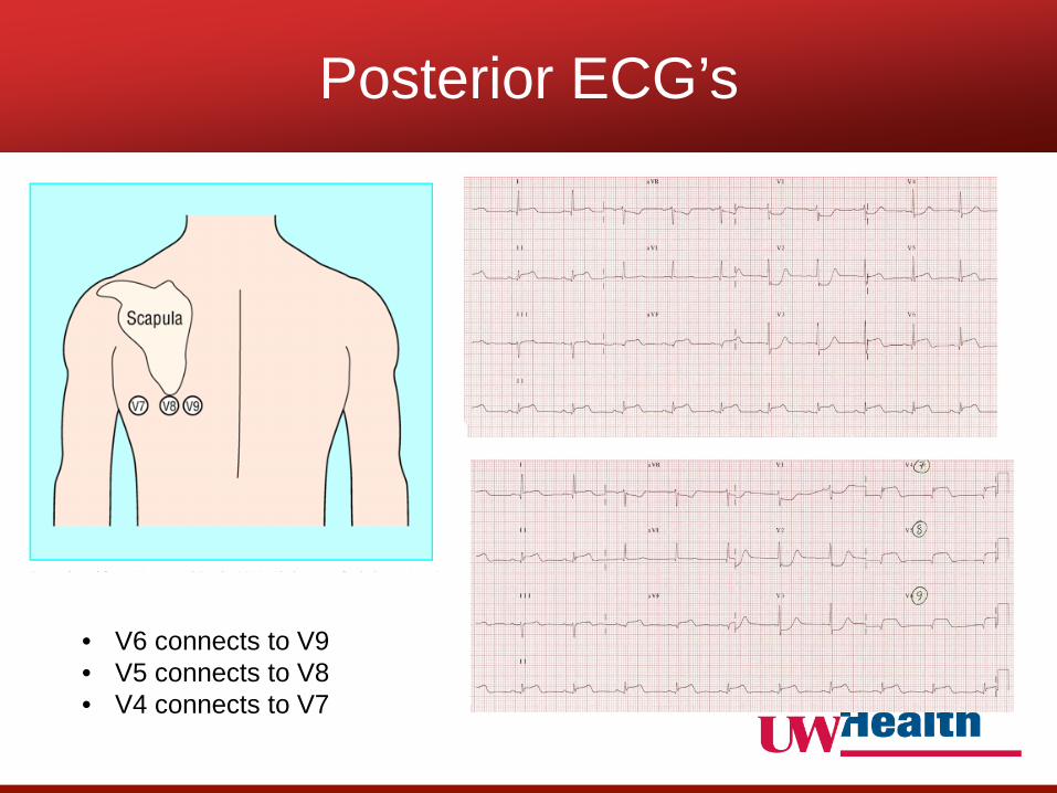
• V6 connects to V9 • V5 connects to V8 • V4 connects to V7
• Ventricular Septal Defect • Papillary muscle rupture or dysfunction, • Cardiac free wall rupture • Ventricular Aneurysm • LV outflow tract obstruction • LV or RV Failure with Cardiogenic
Shock
Complications From STEMI
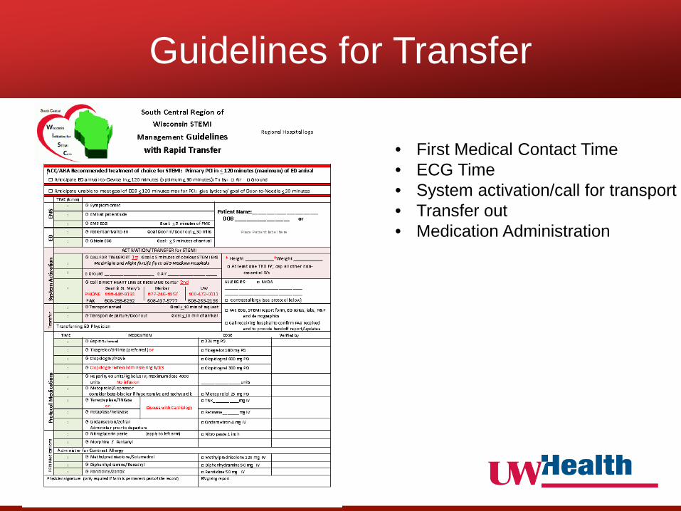
Guidelines for Transfer
• First Medical Contact Time • ECG Time • System activation/call for transport • Transfer out • Medication Administration
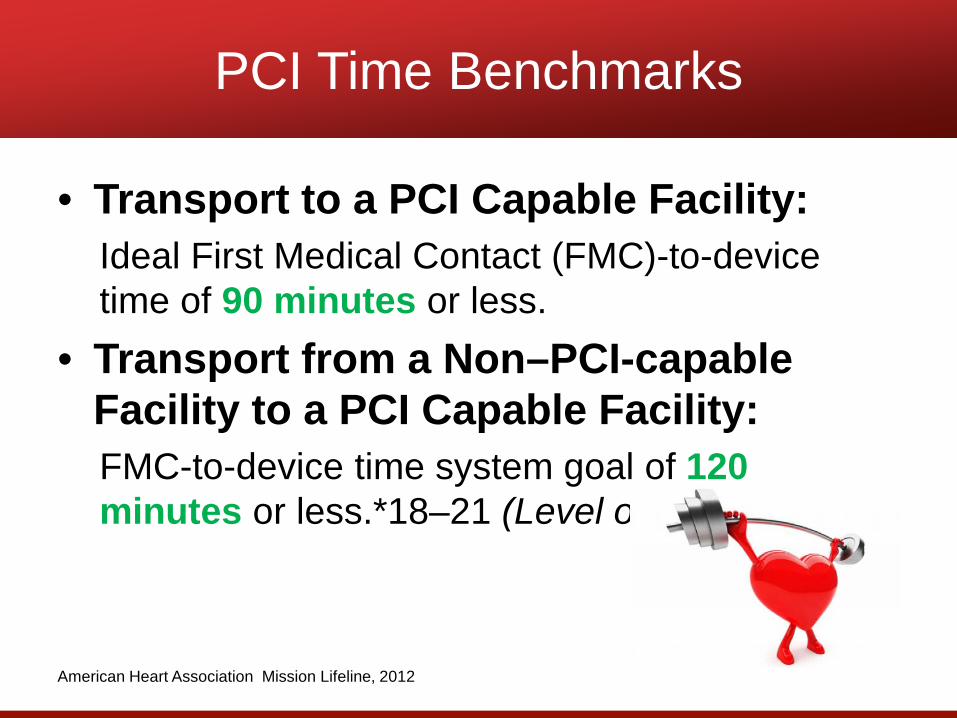
PCI Time Benchmarks
• Transport to a PCI Capable Facility: Ideal First Medical Contact (FMC)-to-device time of 90 minutes or less.
• Transport from a Non–PCI-capable Facility to a PCI Capable Facility: FMC-to-device time system goal of 120 minutes or less.*18–21 (Level of Evidence: B)
American Heart Association Mission Lifeline, 2012
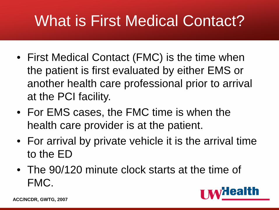
• First Medical Contact (FMC) is the time when the patient is first evaluated by either EMS or another health care professional prior to arrival at the PCI facility.
• For EMS cases, the FMC time is when the health care provider is at the patient.
• For arrival by private vehicle it is the arrival time to the ED
• The 90/120 minute clock starts at the time of FMC.
What is First Medical Contact?
ACC/NCDR, GWTG, 2007
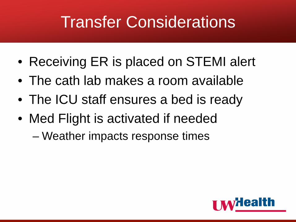
• Receiving ER is placed on STEMI alert • The cath lab makes a room available • The ICU staff ensures a bed is ready • Med Flight is activated if needed
– Weather impacts response times
Transfer Considerations

• Resources – 12 Lead ECG machine equipment and training, lack of transmission capabilities, personnel
• Not always possible to get ALS or air transport to get patient to PCI center
EMS Barriers

• Weather
EMS Barriers Continued
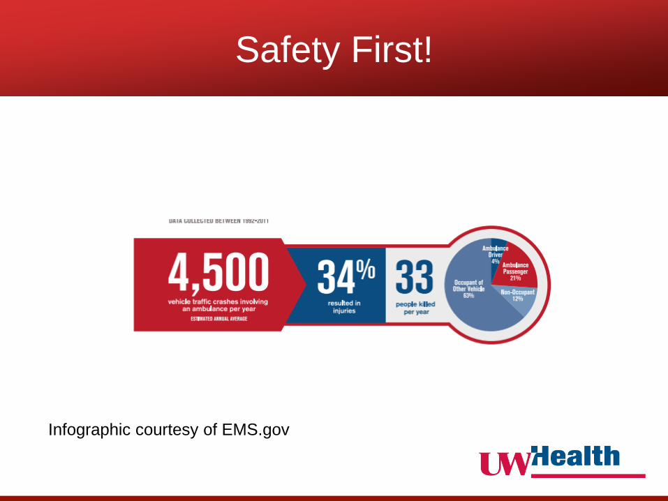
Safety First!
Infographic courtesy of EMS.gov
• Obtain 12 Lead ECG within 5 minutes – preferred, 10 minutes benchmark of first medical contact.
• If transmission capabilities exist, transmit ECG to nearest hospital IMMEDIATELY
• If transmission capabilities do not exist, notify receiving hospital that you are bringing in a suspected STEMI.
• Limit scene time to 15 minutes.
EMS Responsibilities
• Provide copies of the run report to hospital and upload to database within 24-48 hours.
• Provide hospital providers with a thorough handoff.
• Always send cardiac arrest rhythm strips and 12 leads with patient to the hospital.
EMS Responsibilities (cont.)
• Obtain ECG within 5 minutes of patient arrival • 5 Minutes STEMI ECG to decision, contact PCI
Center and transport within 5 minutes of obvious STEMI ECG.
• Goal: Door in door out = 30 Minutes
Tips for Referring Hospitals
• If ECG does not meet STEMI criteria: perform serial 12 leads every 15 minutes to monitor for evolving STEMI or if patient condition changes.
• Contact cardiology for a consult. This can be done by calling the UW Access Center.
• If transport is delayed due to weather or availability, consider thrombolytic therapy.
• Goal: Door to needle = 30 Minutes
Tips for Referring Hospitals (cont.)
• If transport is delayed due to weather or availability, consider thrombolytic therapy.
• Goal: Door to needle = 30 Minutes • Send copies of all documentation from
current ED visit and EMS documentation – Including all pre-hospital and in-hospital significant rhythms strips
Tips for Referring Hospitals (cont.)
• Discontinue IV drips • Explain to patients what is happening and
that things will be moving fast. • Send along family information if the
information is readily available.
Tips for Referring Hospitals (cont.)
• Feedback will be provided within 24 to 48 hours.
• Feedback can only be provided if documentation is available to obtain times and details of the case. Expect longer wait times for feedback if documentation is not completed in a timely manner.
• Beneficial to review cases with all staff
Quality

• Receiving ER is placed on STEMI alert • The cath lab makes a room available • The ICU staff ensures a bed is ready • Med Flight is activated if needed
– Weather impacts response times
Transfer Considerations
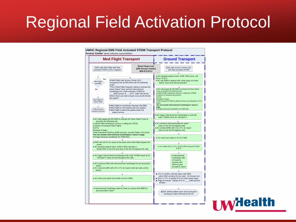
Regional Field Activation Protocol UWHC Regional EMS Field Activated STEMI Transport ProtocolAccess Center *green indicates responsibilities
Med Flight Transport Ground Transport
EMS calls Access Center (AC)with field activated STEMI
Med Flightavailable
EMS calls Med Flight with field activated STEMI via 911 dispatch
● AC calls paging @ 262-0000 to activate the Heart Attack Team & provides the following info: 1) Which EMS ambulance service is calling the STEMI2) Means of transport (Med Flight)3) ETA Example of page:“Field Activated STEMI by (EMS Service), via Med Flight, ETA (time)*Do not include Interventional Cardiologist’s name in page4) Pages Nursing Coordinator for F4M5 Bed
● AC connects EMS with Interventional Cardiologist for pre procedure report.● AC connects EMS with CCU CTL for report (Cath lab calls unit for report)
Yes
No
▲ CTL to confirm cath lab status with EMS when EMS contacts ED via radio ~10 minutes out.▲ EDC or CTL to update ETA via heart attack page. ▲ Page to include: “update ETA on ____EMS ground STEMI”.
▲EDC admits patient upon arrival using the existing pre-admit entered by the AC
Direct Heart Line(UW Access Center)
800-472-0111
● AC pages Interventional Cardiologist with code “STEMI report & AC call back #” (stay on line throughout the call).
● AC requests patient name, DOB, EMS name, call back # & ETA. ● AC ask EMS to please hold, while page out Heart Attack Team and Interventionalist.
♥ Med Flight calls Access Center (AC) emergency line @ 890-6102 with the following script: “This is Med Flight Dispatch calling to activate the Heart Attack Team and the Interventional Cardiologist for a field activation STEMI by ____EMS service & ___ETA. EMS will call the Direct Heart Line with a report once we lift off with the patient”
● Interventional Cardiology calls AC back to connect with EMS for post procedure report.
● AC enters pre-admit into Health Link for F4M5
● AC connects Interventionalist for report (stay on the line throughout call).● AC connect EMS with CCU CTL for report (stay on the line throughout call).
Follow ground transport process
Med Flight to inform EMS
● EMS will call AC for report at the latest when Med Flight departs the scene.● AC requests patient name, DOB & EMS call back #. (Keep EMS on the line and stay on the line throughout the call).
♥ Med Flight to coordinate intercept with EMS.♥ Med Flight to call hospital with eta updates. ♥ Med Flight to admit the patient when the patient arrives.
● AC pages Interventional Cardiologist on call with code “STEMI call & AC call back #”.
● AC calls paging @ 262-0000 to activate the Heart Attack Team & provides the following info: 1) Which EMS ambulance service is calling the STEMI2) Means of transport (by ground)3) ETAExample of Page:“Field Activated STEMI by (EMS Service) via ambulance, ETA (time) *Do not include Interventional Cardiologist’s name in page4) Pages Nursing Coordinator for F4M5 Bed
● AC notifies EDC or CTL @ 262-2398 of ground STEMI & ETA.
● AC enters pre-admit in HL for F4M5.
● Interventional Cardiology calls AC back to connect with EMS for post procedure report.
Cardiogenic Shock
Cardiogenic shock is when the heart is unable to pump enough blood to meet the body’s needs.
What is Cardiogenic Shock?
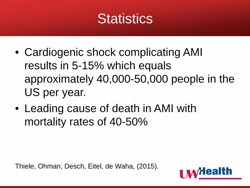
• Cardiogenic shock complicating AMI results in 5-15% which equals approximately 40,000-50,000 people in the US per year.
• Leading cause of death in AMI with mortality rates of 40-50%
Thiele, Ohman, Desch, Eitel, de Waha, (2015).
Statistics
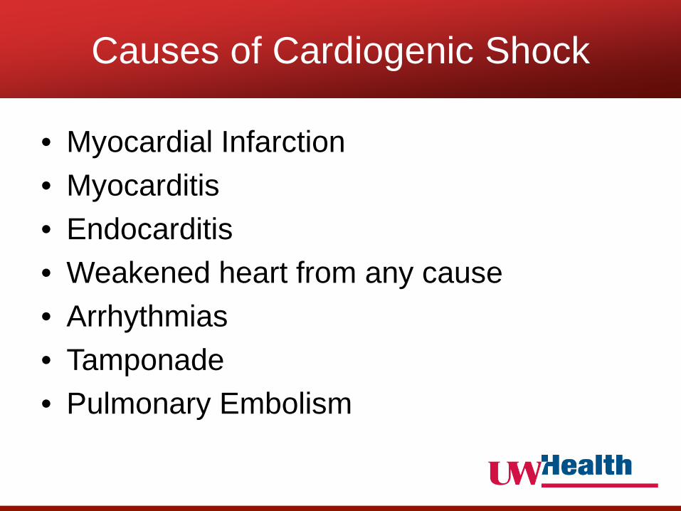
• Myocardial Infarction • Myocarditis • Endocarditis • Weakened heart from any cause • Arrhythmias • Tamponade • Pulmonary Embolism
Causes of Cardiogenic Shock
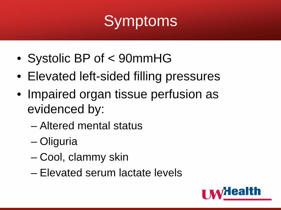
• Systolic BP of < 90mmHG • Elevated left-sided filling pressures • Impaired organ tissue perfusion as
evidenced by: – Altered mental status – Oliguria – Cool, clammy skin – Elevated serum lactate levels
Symptoms
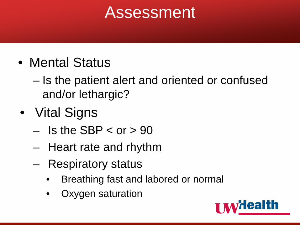
• Mental Status – Is the patient alert and oriented or confused
and/or lethargic? • Vital Signs
– Is the SBP < or > 90 – Heart rate and rhythm – Respiratory status
• Breathing fast and labored or normal • Oxygen saturation
Assessment
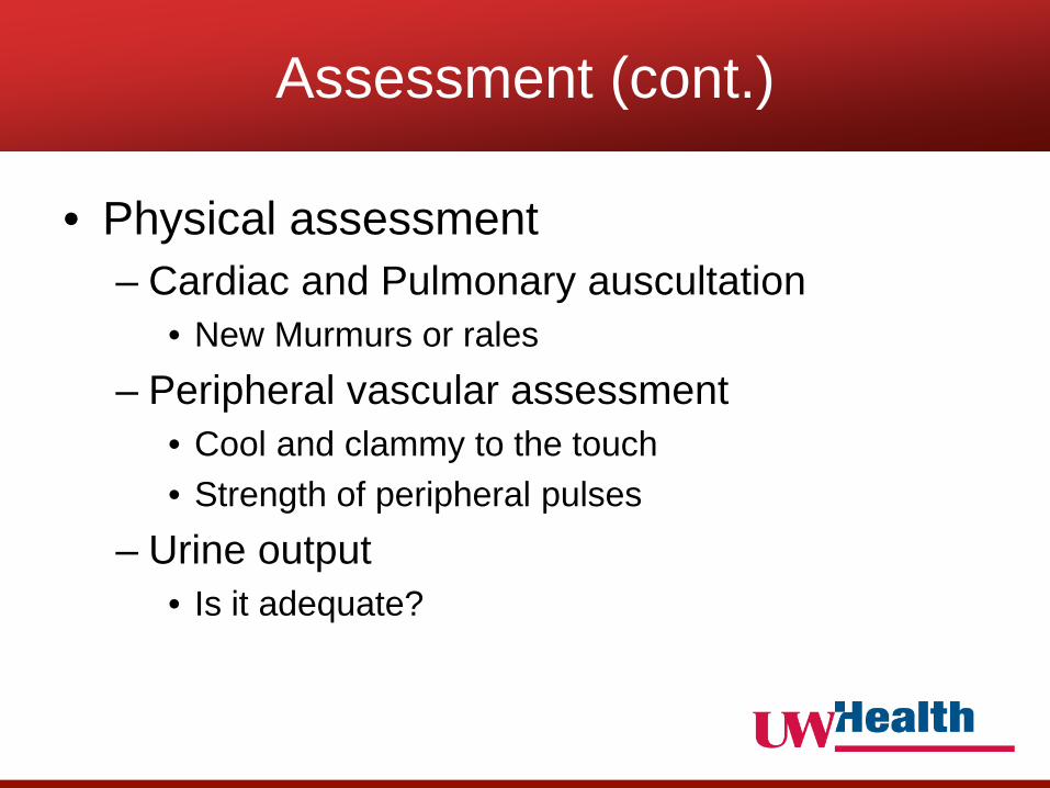
• Physical assessment – Cardiac and Pulmonary auscultation
• New Murmurs or rales – Peripheral vascular assessment
• Cool and clammy to the touch • Strength of peripheral pulses
– Urine output • Is it adequate?
Assessment (cont.)
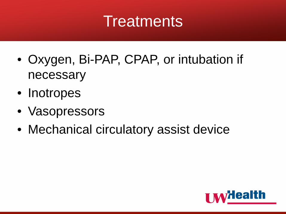
• Oxygen, Bi-PAP, CPAP, or intubation if necessary
• Inotropes • Vasopressors • Mechanical circulatory assist device
Treatments
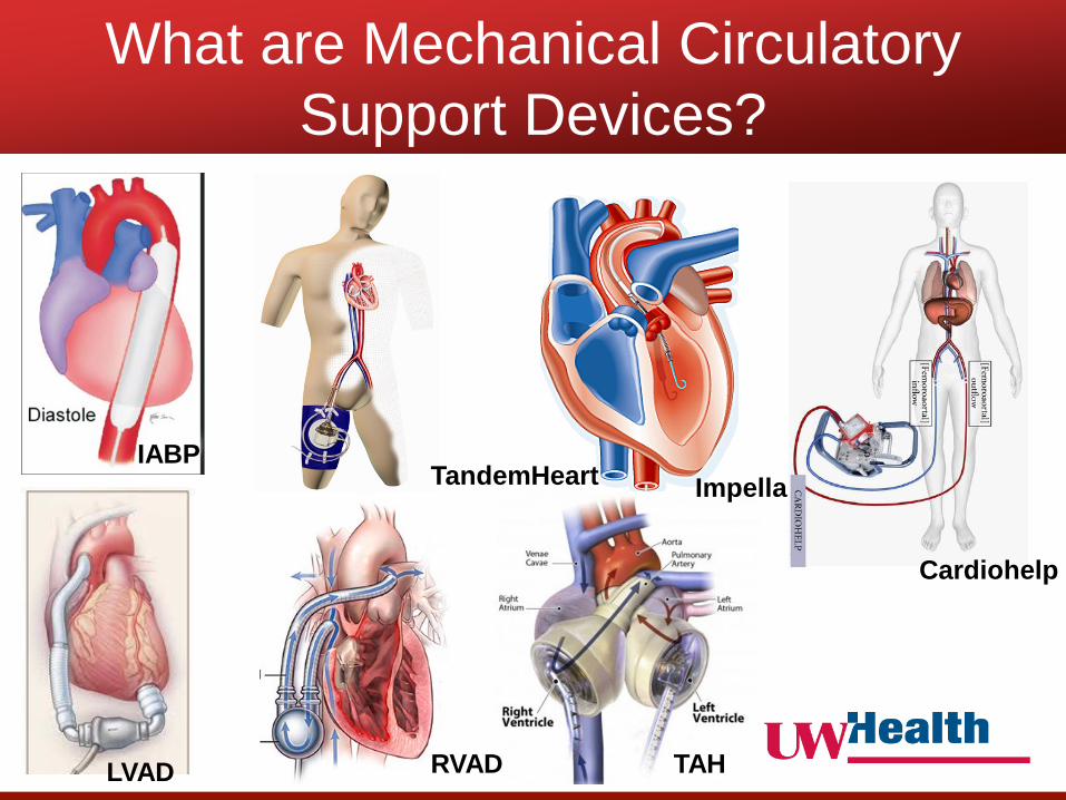
What are Mechanical Circulatory Support Devices?
IABP TandemHeart Impella
Cardiohelp
LVAD RVAD TAH
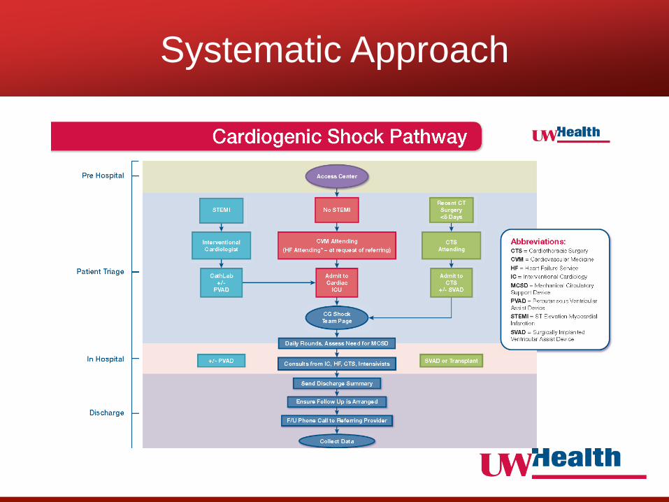
Systematic Approach
STEMI / Cardiogenic Shock Case Study
• 49 y.o. male with a positive family history of heart disease, obesity, and seizure disorder who began to experience chest pain after dinner which was associated with vomiting and diaphoresis. His mother heard him collapse, so she ran to help. Unfortunately she was unable to move him. She used her Life Alert to summon help. The patient did not receive CPR until the EMS arrived (amount of time without CPR 5-10 minutes). He was found to be in VF and was shocked once and then went into PEA.
The Story
EMS Radio Call into UW
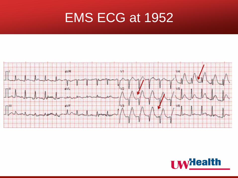
EMS ECG at 1952
• CCR upon arrival • Rhythm VF defibrillated, remained in VF
Compressions resumed • High flow oxygen via NRB • IV Left AC due to failed IO • Fluid bolus • 1mg Epi • Glucose checked
EMS Treatment
• Defib charged – patient in PEA, charged dumped, compressions resumed
• Organized rhythm with pulse, agonal respirations. Patient moved to stretcher. BVM to assist respirations.
• 12 lead done en route to UW which revealed anterior STEMI
EMS Treatment (cont.)
• 12 Lead upon arrival • Second IV placed • Intubation • Propofol • Cooling blankets • Patient sent to CT scan to rule out head
bleed.
ED Treatment
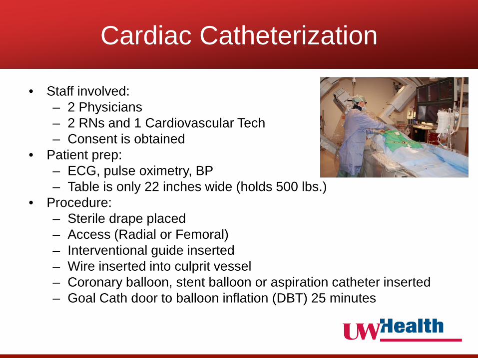
• Staff involved: – 2 Physicians – 2 RNs and 1 Cardiovascular Tech – Consent is obtained
• Patient prep: – ECG, pulse oximetry, BP – Table is only 22 inches wide (holds 500 lbs.)
• Procedure: – Sterile drape placed – Access (Radial or Femoral) – Interventional guide inserted – Wire inserted into culprit vessel – Coronary balloon, stent balloon or aspiration catheter inserted – Goal Cath door to balloon inflation (DBT) 25 minutes
Cardiac Catheterization
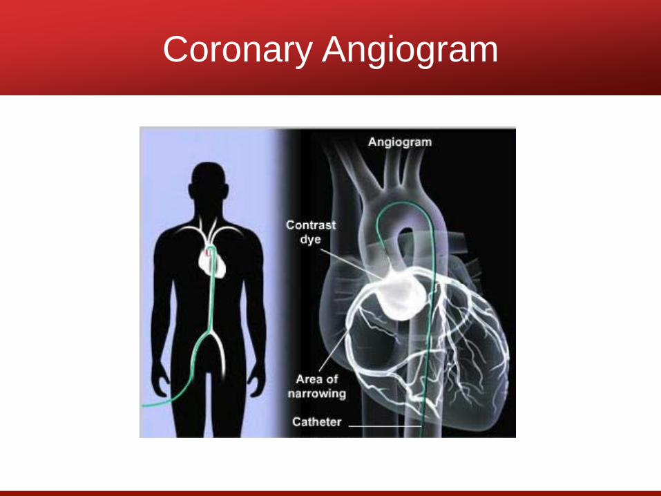
Coronary Angiogram
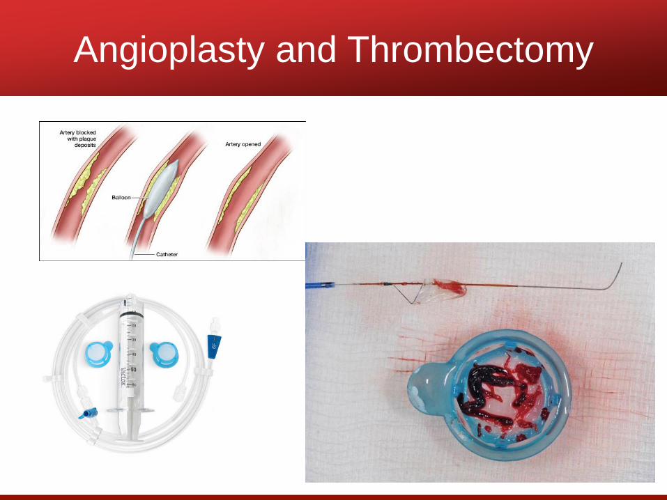
Angioplasty and Thrombectomy
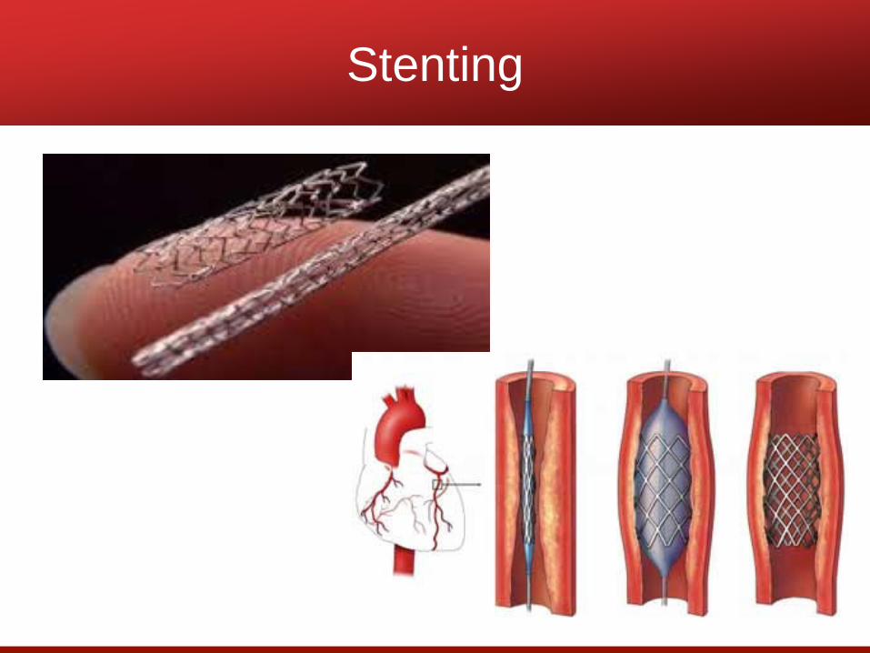
Stenting
• 100% Proximal Left Anterior Descending • 3.0mm x 38mm drug-eluting stent placed • Patient developed acute stent thrombosis
while still in the Cath Lab, so two additional drug-eluting stents needed to be placed.
• Cardiohelp (ECMO) placed • Swan-Ganz and cooling catheter inserted
Cath Lab Interventions
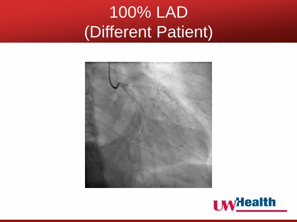
100% LAD (Different Patient)
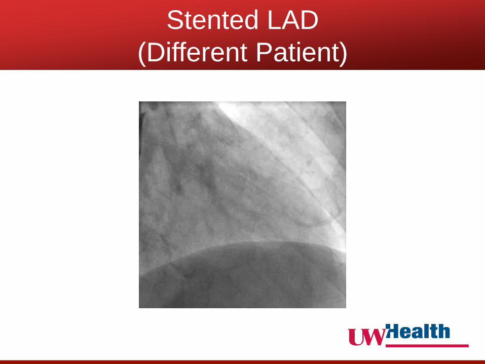
Stented LAD (Different Patient)
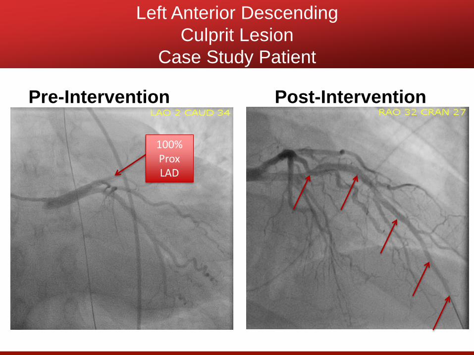
Left Anterior Descending Culprit Lesion
Case Study Patient
Pre-Intervention Post-Intervention
100% Prox LAD
• 5/15 - Patient’s right foot became ischemic due to large cannulas from ECMO. HE was taken back to the Cath Lab for revision from VA ECMO to veno-venus. IABP placed to off load the LV. Temporary bypass of SFA with arterial to arterial bypass.
• 5/19 – ECMO decannulated • 5/21 – Extubated and following commands • 5/31 – Discharged to rehab facility
Hospital Course
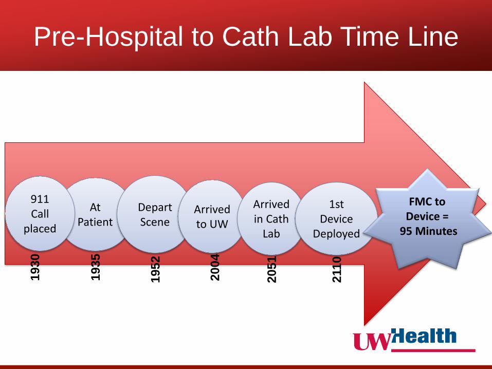
Pre-Hospital to Cath Lab Time Line
• csffefrsa At Patient
Depart Scene
Arrived to UW
911 Call
placed
1930
1935
1952
Arrived in Cath
Lab 20
04
2051
1st Device
Deployed
2110
FMC to Device =
95 Minutes
• High Quality CPR is vital to patient survival • Transmit STEMI ECG’s IMMEDIATELY to
receiving center to expedite patient care • Time is critical for STEMI and Cardiogenic
Shock patients • Detailed handoffs and providing all
documentation from the field are essential to expedite continuity of care.
Key Points
Teamwork is the key to
success!
Key Points (cont.)
• American Heart Association. (2017). Heart Disease and Stroke Statistics 2017 At-a-Glance. Retrieved from https://www.heart.org/idc/groups/ahamah-public/@wcm/@sop/@smd/documents/downloadable/ucm_491265.pdf
• American Heart Association. (2015). Hard to recognize heart attack
symptoms. Retrieved from https://www.goredforwomen.org/about-heart-disease/symptoms_of_heart_disease_in_women/hard-to-recognize-heart-attack-symptoms/
• American Heart Association. (2012). Regional systems of care demonstration project: Mission Lifeline™ STEMI systems accelerator (version 5.0). Duke University
References
• American Heart Association. (2011). STEMI Provider Manual. United States of America.
• Thiele, H., Ohman, E. M., Desch, S., Eitel, I., de Waha, S. (2015). Management of cardiogenic shock. European Heart Journal, 36, 1223-1230
References (cont.)