Embed Size (px)
Citation preview
Thematic review series: Lipid Posttranslational Modifications
Geranylgeranylation of Rab GTPases
Ka Fai Leung, Rudi Baron and Miguel C. Seabra*
Molecular and Cellular Medicine, Division of Biomedical Sciences, Imperial
College London, Exhibition Road, London SW7 2AZ
*To whom correspondence should be addressed: Prof. M.C. Seabra, Molecular and Cellular Medicine, Division of Biomedical Sciences, Sir Alexander Fleming Building,Imperial College London, Exhibition Road, London SW7 2AZ, UK. Tel: +44 20 7594 3024; Fax: +44 20 7594 3015 Email: [email protected]
by guest, on March 30, 2019
ww
w.jlr.org
Dow
nloaded from
Abstract
Rab GTPases require special machinery for protein prenylation, which include Rab Escort
Protein (REP) and Rab Geranylgeranyl Transferase (RGGT). The current model of Rab
geranylgeranylation proposes that the Rab protein must bind REP, which presents the Rab
to RGGT. Following geranylgeranylation of C-terminal cysteines in Rab, REP delivers the
prenylated protein to membranes. The REP-like protein, Rab GDP dissociation inhibitor
(RabGDI) then recycles the prenylated Rab between the membrane and the cytosol. The
recent solution of crystal structures of the Rab prenylation machinery have helped to refine
this model and provide further insights. The hydrophobic prenyl binding site of RGGT is
similar to that of geranylgeranyl transferase type-I (GGT-I) but differs from farnesyl
transferase (FT). At the bottom of the hydrophobic isoprenyl-binding pocket, a bulky Trp
residue in FT prevents binding of the longer geranylgeranyl pyrophosphate group whereas
in GGT-I and RGGT this position is occupied by the smaller residues Thr and Ser,
respectively. At the REP:RGGT interface, a highly conserved Phe in REP, which is absent
in RabGDI, plays a critical role in the formation of the REP:RGGT complex and may
explain the inability of RabGDI to complex with RGGT. A geranylgeranyl-binding site in
REP and RabGDI has been identified within domain II. However, as most Rabs undergo
double geranylgeranylation, it is still unclear how and where the second geranylgeranyl
group is stabilised. The post-prenylation events, including the specific targeting of Rabs to
target membranes and the requirement for single versus double-geranylgeranylation by
different Rabs remain obscure and should be the object of future studies.
Supplementary key words
Rab • GTPases • prenyltransferases • REP • GDI • geranylgeranyl
by guest, on March 30, 2019
ww
w.jlr.org
Dow
nloaded from
Members of the small GTPase Ras superfamily perform important regulatory functions,
from cell growth to cytoskeleton dynamics and membrane trafficking. With the exception of Ran,
Ras-like proteins undergo co- or post-translational lipid modifications, which act as hydrophobic
membrane anchors interacting with the cytoplasmic leaflet of cellular membranes and/or
participating in protein-protein interactions. The most common lipid modification affecting small
GTPases is protein prenylation, which involves the covalent addition of either farnesyl (15-
carbon) or geranylgeranyl (20-carbon) pyrophosphate to proteins via thioether linkages catalysed
by protein prenyltransferases (1). Prenylation of Ras, Rho/Rac and Rab is absolutely critical for
the proper function of the modified protein in cellular processes (also reviewed in the other
reviews in this series). The importance of protein prenylation first gained focus when it was
found that oncogenic forms of Ras proteins required prenylation for their ability to transform
cells (2, 3). Since then, the search for inhibitors of prenylation has been an active area of research
(reviewed in this series by (4)).
Three distinct protein prenyltransferases have been identified and can be classified into
two main functional classes: the CAAX prenyltransferases, consisting of farnesyltransferase (FT)
and geranylgeranyltransferase type I (GGT-I) (reviewed in this series by Beese et al (editor
please add reference, if it is too complicated then it is OK to delete the ref)), and the Rab
geranylgeranyltransferase (RGGT, also known as GGT-II) (1). Substrates for the first class
include CAAX-containing farnesylated proteins (Ras, nuclear lamins, and others) and
geranylgeranylated proteins of the (Rho/Rac families and others). The Rab protein family are
exclusive substrates of RGGT.
THE RAB PROTEIN FAMILY – STRUCTURE/FUNCTION:
The Rab proteins (ras genes from rat brain), comprising over 60 proteins, form the largest
family of the Ras superfamily of small GTPases, and are important regulators of organelle
biogenesis and vesicle transport (5). They are conserved throughout evolution, from yeast to
mammals (6). Some Rab proteins are ubiquitously expressed, while others are expressed in a
tissue-specific or developmentally-regulated manner. For example, Rab1 is found in all cell types
while others, such as Rab27a, are found in melanocytes and secretory cell types (7). Analysis of
the budding yeast S. cerevisiae genome indicates that there are 11 Rab genes called Ypt (yeast
by guest, on March 30, 2019
ww
w.jlr.org
Dow
nloaded from
protein involved in transport), some of which are redundant in their function (8). In mammals,
over 60 Rab proteins have been identified, which is not surprising considering the increase in
complexity of trafficking pathways required to carry out diverse functions in a variety of different
cell types. The evolutionary conservation of Rabs is highlighted by the fact that mouse Rab1a can
compensate for loss of Ypt1p in yeast (9).
Studies of Rab protein function suggest that they are important in vesicular membrane
transport (10, 11). Eukaryotic cells possess an elaborate internal membrane system composed of
different intracellular compartments, each serving a different function. These compartments are
highly dynamic and communicate with each other. Each Rab protein has a specific intracellular
localisation and thus regulates a specific membrane trafficking step. However, transport between
two membrane compartments may be governed by more than one Rab member and thus some
Rab proteins may exhibit redundancy in their roles.
All members of the Ras superfamily have conserved regions that are involved in binding
guanine nucleotide and phosphate/Mg2+, and have been previously referred to as G1-G3 and
PM1-PM3 respectively (12). There are two regions which undergo a significant conformational
change upon GTP binding and hydrolysis: the switch I domain, which lies in the loop 2 region,
and the switch II domain, which resides in the loop4/α2/loop5 region.. While the presence of a
di-cysteine prenylation motif at the C-terminus is generally considered a good defining feature of
a Rab protein, it is not absolute, as a few Rab proteins such as Rab8 and Rab13 contain only a
single cysteine motif. More recently, diagnostic feature distinguishing to distinguish Rabs from
other Ras-like GTPases has been proposed based on sequence alignments of Rab proteins (5).
Using this approach, five Rab family regions (RabF) were identified, which were conserved only
in Rab proteins and thus discriminated them from other Ras-like proteins (Figure 1). The RabF1
region is located in the so-called effector domain (loop2/β2) in the switch I region. The
remaining four regions, RabF2 (β3), RabF3 (loop4), RabF4 (α2/loop5) and RabF5 (β4/loop6), all
reside in and around the switch II region between β-sheets β3 and β4 (5). Since the RabF regions
cluster around the two switch domains, which undergo changes in conformation on binding GDP
or GTP, it has been suggested that these regions are involved in binding to general regulators of
Rab function, such as Rab GDP dissociation inhibitor protein (RabGDI) and Rab escort protein
(REP), as these regulatory proteins are nucleotide-sensitive (e.g associate better with the GDP
form of Rab proteins) and recognise all Rabs (13, 14).
by guest, on March 30, 2019
ww
w.jlr.org
Dow
nloaded from
In addition, four Rab subfamily regions (RabSF) were defined as regions of high
conservation within subfamilies: RabSF1 (β1), RabSF2 (α1/loop2), RabSF3 (α3/loop7) and
RabSF4 (α5) (5). RabSF1, RabSF3 and RabSF4 corresponded to three regions previously
referred to as Rab complementary-determining regions (CDRs) I, II and III, based on the crystal
structure of Rab3a complexed with its effector Rabphilin3a (15). Thus RabSF1, RabSF3 and
RabSF4 of Rab3a form a surface which mediates binding to Rabphilin3a, while RabSF2 forms
another surface on the opposite face of Rab3a and could interact with other effectors. Based on
these findings, it was proposed that effectors bind to RabF regions to discriminate between the
nucleotide-bound states, but also to RabSF regions to confer specificity.
RAB GERANYLGERANYLATION – TWO ALTERNATIVE PATHWAYS
Like the CAAX prenyltransferases, RGGT is heterodimeric and consists of distinct α-
and β-subunits. However, its mechanism of action is distinct from the other prenyltransferases.
The enzyme was first isolated from rat brain cytosol and purified as a multi-component enzyme
(components A and B) that was able to attach geranylgeranyl groups onto Rab proteins (16, 17).
Component B represents the catalytic component, now called Rab Geranylgeranyl Transferase
(RGGT). Unlike the CAAX prenyl transferases, RGGT does not recognise short peptides
containing the Rab C-terminal prenylation motif, nor does it recognise the Rab protein alone(17,
18). Instead, it binds Component A, now called Rab Escort Protein (REP), which is a Rab-
binding protein (19).
Several details concerning the mechanisms of Rab protein prenylation are yet unclear.
Biochemical assays have led to the proposal of two possible pathways (Figure 2). The classical
mechanism of Rab prenylation implicates first the association of an unprenylated Rab protein
with REP (19). The equilibrium dissociation constant was measured to be 0.2µM (although it
varies between Rabs), and the interaction relies mostly on ionic bonds and does not involve the
two C-terminal cysteine residues (18). The complex is then recognised by RGGT (Kd<1nM)
which adds two geranylgeranyl moieties to the Rab protein without prior dissociation of REP (20,
21). After the transfer of the isoprenoids onto C-terminal cysteines, the ternary complex remains
associated until the binding of a new geranylgeranyl diphosphate (GGpp) molecule, which
stimulates the release of the Rab-GG:REP complex (20). REP is then believed to escort the
prenylated Rab protein to its target membrane (22) (Figure 2).
by guest, on March 30, 2019
ww
w.jlr.org
Dow
nloaded from
An alternative pathway for Rab prenylation was also proposed (23). Solid phase
precipitation assays demonstrated that REP-1 and RGGT can form a tight complex in the
presence of GGpp (Kd~10nM) (Figure 2). This complex could associate with Rab protein, but 10
times slower than REP:Rab to RGGT:GGpp. It was proposed that in vivo the pathway followed
should depend on the concentrations of the proteins involved. At high concentrations of RGGT,
REP, Rab and GGpp, the association of Rab with RGGT:GGpp:REP complex becomes rate-
determining and is favoured, while at low concentrations the classical pathway is preferred.
RAB GERANYLGERANYL TRANSFERASE
The heterodimeric enzyme consisting of a 60kDa α-subunit and a 38kDa β-subunit
presents 30% homology with its counterparts FT and GGT-I (24). The yeast genes encoding the
α− and β-subunits of RGGT were designated BET4 and BET2, respectively (25, 26)..
Interestingly, a mutation termed bet2-1 results in lower affinity of the enzyme for GGpp. This
mutation can be suppressed by overexpression of BTS1 (25) which encodes a GGpp synthase,
suggesting that this enzyme is directly involved in GGpp accessibility by RGGT.
The crystal structure of RGGT has been solved to 2.0 Å and revealed the presence of four
distinct structural domains (27). The α-subunit is composed of three domains: a helical domain,
an immunoglobulin-like (Ig-like) domain and a leucine-rich repeat (LRR) domain. The helical
domain is structurally very similar to the α-subunit of FT despite only 22% sequence identity
between FTα and the relevant region in RGGTα. A major structural difference in RGGT is the
presence of both the Ig-like domain and the LRR domain, absent in FT or GGT-I. These domains
are also absent from lower eukaryote versions of RGGT, suggesting that the LRR and Ig-like
domains of the mammalian RGGT are not essential for the catalytic activity of the enzyme. The
function of these unique regions remains unknown.
. The β-subunit forms an α-α barrel and contains a central cavity lined with hydrophobic
residues very similar to the β-subunit of FT, which comprises the GGpp binding site. A positively
charged cluster is located near the opening of the cavity. In the FT-Fpp structure, this cluster was
shown to interact with the diphosphate head groups of Fpp (28, 29).
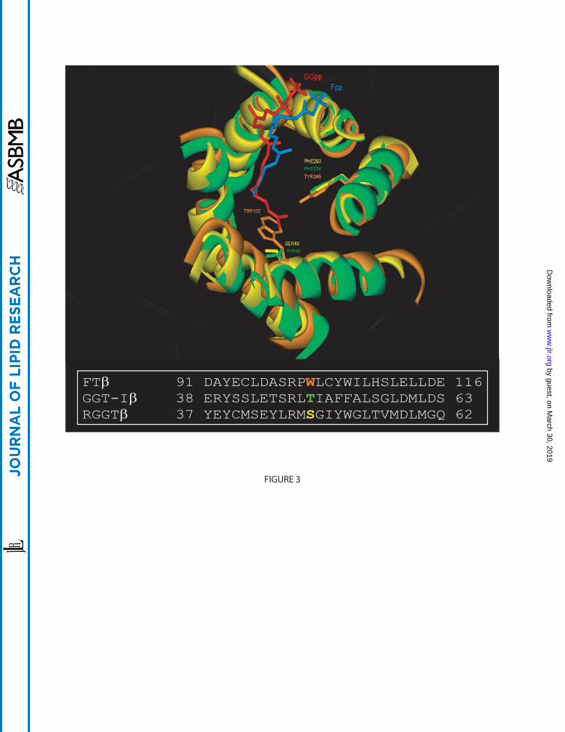
The recent structure of GGT-I (30) helped define the isoprenoid specificity of each
enzyme. In geranylgeranyl transferases, residue 49β (in GGT-I) is always a small amino acid
such as threonine, serine, valine or alanine across many species, whereas in FT it is always a
by guest, on March 30, 2019
ww
w.jlr.org
Dow
nloaded from
tryptophan. This residue fills the space in FT where the larger isoprenoid GGpp fit within GGT-I
and RGGT, thus constricting the length of the isoprenoid that fits in the binding site (Figure 3).
When the Trp residue was replaced by a Thr in FTβ, the resulting mutant preferably bound to
GGpp without any significant alteration of CAAX sequence specificity (30). In RGGT as in
GGT-I, Ser48β and Leu99β replace the more bulky Trp102β and Tyr154β of FT at the bottom of
the cavity (Figure 3). These changes significantly enlarge the binding site to accommodate GGpp.
RGGT binds GGpp with an affinity of 8±4nM, while Fpp binds less strongly (Kd=
60±8nM) and Gpp even less (Kd= 330±40nM) (31). However, these differences in affinity are
more significant when the Rab substrate is included. This may be explained by the fact that the
reaction cycle progression requires the binding of fresh isoprenoid diphosphate to displace the
product from the active site. Fpp was shown to be ineffective in displacing GGpp from the active
site of GGT-I (30).
Mutations in RGGT have been shown to cause a disease similar to Hermansky-Pudlak
syndrome (HPS) in the gunmetal (gm) mouse (32). HPS is a rare autosomal recessive, genetic
disease characterised by partial albinism, prolonged bleeding and platelet dysfunction (33). The
gm mutation was identified as a Gly to Ala substitution in a splice acceptor site within the Rggta
gene (32). While REP expression is unaffected, there is ~70% reduction in the expression of
RGGT α-subunit, with a concomitant decrease in RGGT activity. Although the reduction in
RGGT activity was observed in all tissues, defects in Rab prenylation are tissue-specific ((32, 34)
and our unpublished observations). One possible explanation for the tissue-specific phenotype is
that concentrations of Rab proteins are considerably higher in platelets and melanocytes
compared with other tissues (34). Alternatively, a subset of Rabs present lower affinity for the
prenylation machinery and are selectively affected whenever the enzyme is limiting (32).
Interestingly, Rab27 isoforms are highly expressed in tissues affected in gm mice, suggesting that
hypoprenylation of Rab27 partly contributes to the gm phenotype.
REGULATION OF RGGT ACTIVITY
Very little is known about potential enzyme regulation. One possible mechanism involves
an intramolecular interaction (27). The structural data suggests that the RGGTα N-terminus is
mobile. This region of the α-subunit binds to the β-subunit in an extended conformation by
coordinating the zinc ion with residues from both α- and β-subunits, so it was hypothesised that
by guest, on March 30, 2019
ww
w.jlr.org
Dow
nloaded from
this binding might act in an autoinhibitory manner to prevent the binding of short substrate
peptides (27). This hypothesis remains to be tested.
There is also some evidence to suggest that RGGT activity may be regulated by
phosphorylation. Stimulation of 3T3 L1 fibroblasts and adipocytes with insulin was shown to
result in the concomitant phosphorylation of the RGGT α-subunit but not the β-subunit (35). This
in turn led to a subtle increase in Rab3 and Rab4 prenylation. Therefore it is possible that Rab
prenylation can be influenced by environmental changes by responding to extracellular signals.
Interestingly, insulin was also shown to induce the phosphorylation of the FT α-subunit, leading
to increased farnesylation of Ras (36). Since FT and GGT-I share the same α-subunit, it was
proposed that insulin-induced phosphorylation may also regulate GGT-I activity, although this
has yet to be shown. While no further studies have demonstrated phosphorylation of RGGT,
future work should clarify its role in the regulation of RGGT activity.
While many specific inhibitors have been found to inhibit FT and GGT-I activity,
including FTI-277 and GGTI-298 (4), a limited number of specific inhibitors for RGGT have
been identified. For example, the monoterpene perillyl alcohol has been shown to inhibit RGGT,
but it also inhibits GGT-I. The first specific inhibitor of RGGT was a phosphonocarboxylate
called NE10790 or 3-PEHPC (37). This analogue of the nitrogen-containing bisphosphonate,
risedronate, was identified as a drug which inhibited not only prenylation of Rab proteins in
osteoclasts and J774 macrophages in vitro and bone resorption in vivo. More recently, 3-PEPC,
an analogue of 3-PEHPC, was also demonstrated to inhibit the activity of RGGT (38). The
availability of such inhibitors specific for RGGT may help in our understanding of RGGT
regulation and the mechanism of Rab prenylation.
THE REP/RABGDI PROTEIN FAMILY
Two REP isoforms are known in mammals: REP-1 and REP-2, both of which are
ubiquitously expressed, though their expression levels vary in different tissues (39). In yeast, only
a single essential gene has been identified, encoding the MRS6/MSI4 gene product (40). The REP
proteins are structurally related to RabGDI, such that they have been grouped to form the
REP/RabGDI superfamily.
REP-1 was shown to be the product of the choroideremia (CHM) gene which maps to
human locus Xq21 (41). CHM is an X-linked progressive retinal degenerative disease affecting
by guest, on March 30, 2019
ww
w.jlr.org
Dow
nloaded from
photoreceptors, retinal pigment epithelium and choroid (42). The autosomal homologue REP-2
(also known as CHML) can functionally replace REP-1, as in vitro assays suggest that it behaves
as a REP in the geranylgeranylation of Rabs (43). The substrate specificity between REPs may
differ as REP-2 may have lower affinity for Rab3 and Rab27, although the molecular basis for
this effect remains unresolved (43-45). Mutation of REP1 in CHM leads to a selective defect in
prenylation of some Rabs, including Rab27a (46). These defects may trigger the degenerative
process in CHM.
RabGDI was originally isolated from bovine brain as a protein that inhibited the
dissociation of GDP from Rab3a (13). RabGDI and REP family members all share regions of
high homology known as sequence conserved regions (SCR). Sequence alignment of the RabGDI
and REP sequences reveal five SCRs located at the N-terminal and central portions of the
molecule. The crystal structures of RabGDIα (47) and in complex with prenylated Ypt1 (48)
have been solved. They reveal that RabGDIα is composed of two main structural units, a large
multisheet domain I and a small α-helical domain II. Domain I is composed of SCR1A, SCR1B
and SCR3B, which fold back together to form a compact structure at the apex of the molecule to
form the Rab-binding platform (RBP). The less conserved SCR2 and SCR3A constitute domain
II. The SCRs form a conserved face on one side of the molecule while the opposing face is
comprised of less conserved regions.
Two crystal structures of REP-1 in complex with either RGGT or Rab7 have been
described (44, 49). As with RabGDI, REP-1 consists of two domains: a large cylindrical domain I
made up of four β-sheets and six α-helices, and a smaller domain II composed of five α-helices.
The largest region of sequence conservation with RabGDI, SCR2, covers helices D and E of
domain II and the C-terminal binding region (CBR) on domain I of REP-1. The most significant
difference between REP-1 and RabGDI is in domain I, where REP contains a large insertion
between SCR1 and SCR2. Unfortunately, the function of this insert remains unknown as well as
its structure, which was not visible in the crystal.
Since the Rab-binding platform (RBP) is a key functional element, it is highly conserved
between REP-1 and RabGDI. Out of 32 residues of REP-1 involved in the formation of the Rab
complex interface, 15 are invariant between RabGDI and REP-1, 11 are conserved and 6 are
unique for the REP-1:Rab complex (44). The contacts between the RBP and Rab7 are nearly
identical in the prenylated and unprenylated complexes, suggesting that the association of REP-1
by guest, on March 30, 2019
ww
w.jlr.org
Dow
nloaded from
with the geranylgeranyl moiety does not alter the RBP. REP-1 needs to bind unprenylated
proteins in order to promote their prenylation while this property is not required for RabGDI
function. In fact, it is widely thought that RabGDI cannot bind unprenylated Rabs effectively.
However, comparison of structural data of Rab7:REP-1 (44) and Ypt1:yGdi1p (48) complexes
indicates that they are in fact very similar in terms of hydrophobic and hydrophilic interactions
between REP/GDI and Rab proteins, therefore cannot explain why RabGDI binds preferentially
prenylated proteins.
The crystal structure of RabGDI in complex with prenylated Ypt1p revealed that the
geranylgeranyl moiety is accommodated in a hydrophobic lipid-binding site in domain II (48). A
similar binding pocket was observed in REP (44) (Figure 1). This hydrophobic pocket appears to
be blocked when REP-1 is in complex with RGGT. However, when REP is in complex with a
mono-GG Rab protein, there is a change in the conformation of helix D resulting in opening of
the binding site and allowing the accommodation of the GG moiety. The cavity is too small to
accommodate two GG groups and so it was postulated that the second lipid moiety lies in an
adjacent groove located outside the cavity. However, the structures of apo-REP-1 and di-GG-
Rab:REP-1 need first to be resolved to build a complete model of the interactions between Rab
proteins and REP.
The mobile effector loop (MEL) is a short stretch in domain II which is thought to be
another conserved functional element, although it adopts a different conformation in REP-1 than
in RabGDI (49). Mutations in the RabGDI MEL did not affect Rab binding but significantly
reduced membrane association of RabGDI and prevented the extraction of Rabs by RabGDI (50).
Therefore the MEL has been implicated in Rab recycling.
The C-terminal binding region (CBR) of REP-1 is composed of hydrophobic residues that
form a cavity above the MEL. It has been proposed that the CBR interacts with the distal part of
the C-terminus of Rab proteins. More importantly, it was demonstrated that a motif in the HVR
of Rab proteins, similar to the IKL sequence of Rab7, consisting of a polar residue flanked by
hydrophobic residues, is important for efficient Rab prenylation.
Despite the structural similarities, REP and RabGDI have unique functions. The best
understood difference involves the inability of RabGDI to bind RGGT (see more details under
REP:RGGT Complex). As discussed above, the issue of whether there are differences between
REP and RabGDI in binding affinities towards unprenylated and prenylated Rabs remains to be
by guest, on March 30, 2019
ww
w.jlr.org
Dow
nloaded from
addressed experimentally, although this is often referred as a known fact. Under steady-state,
endogenous RabGDI:Rab complexes can be isolated but not REP:Rab complexes. This may
reflect the fact that RabGDI is more abundantly expressed than REP and/or functional
differences. The most likely hypothesis at present is that REP works at the initial
prenylation/membrane association event whilst RabGDI works at a later stage in recycling Rabs
on/off membranes (22).
THE REP:RGGT COMPLEX
Recent studies described the crystal structure of REP-1 complexed with RGGT to 2.7Å
resolution and provided more insight into the interaction between the two proteins as well as why
RGGT can interact with REP but not with RabGDI (49). A previous suggestion implicated the
LRR or Ig-like domains of RGGT to interact with the insert region REP, since these are unique
sequences (27). This is in fact not the case, since this element faces the opposite direction to the
interaction surface. The REP:RGGT interface is formed by helices 8, 10, 12 of the RGGT α-
subunit, and helices D and E in domain II of REP-1 (49). Despite the high affinity with which
REP-1 can bind RGGT, the contact area is surprisingly small. An important finding was that
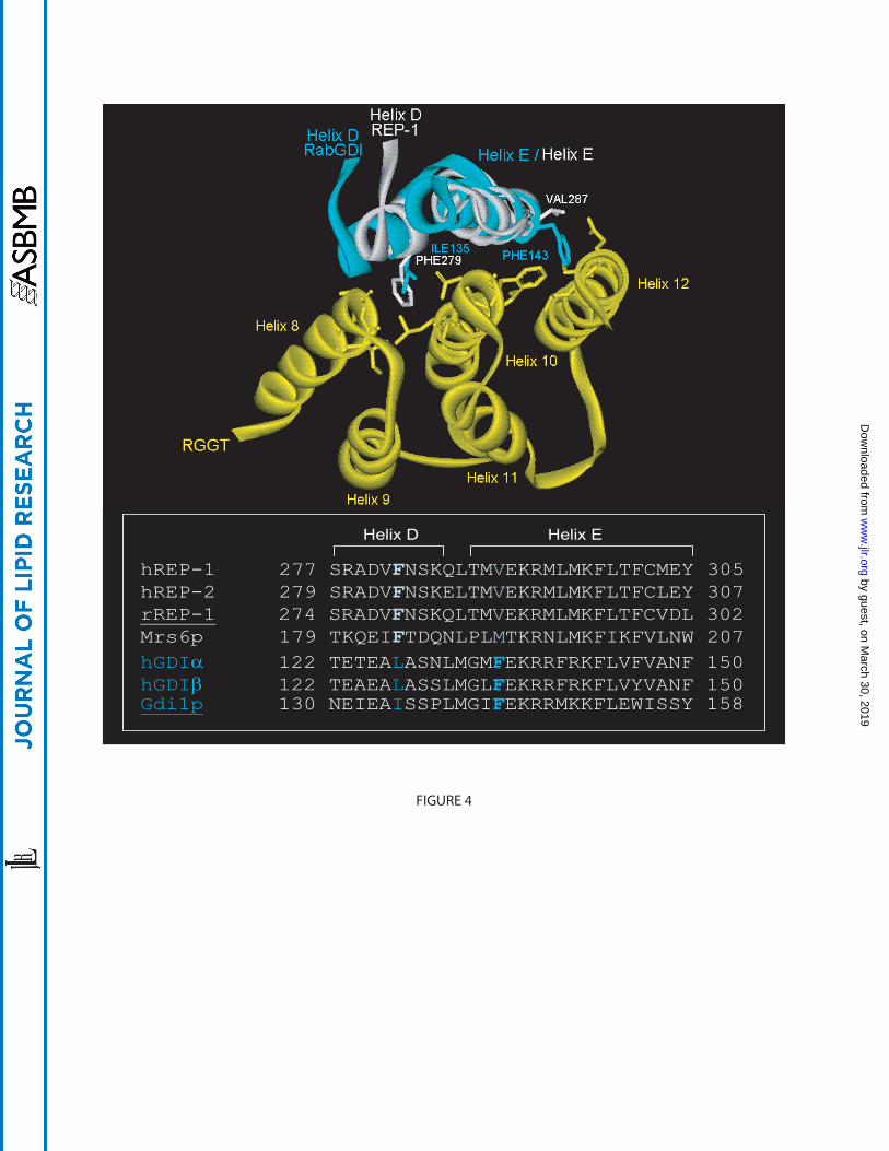
residue F279 of REP-1 plays a key role in mediating interaction with RGGT, since a F279A
substitution abolishes binding to RGGT (49). This residue protrudes deeply into the cavity
formed by α-helices 8 and 10 of RGGT and by establishing hydrophobic interactions stabilises
the REP-1:RGGT complex (Figure 4). This phenylalanine is conserved throughout the REP
family but is absent in the RabGDI family. The comparison between apo-RGGT and the REP-
1:RGGT complex suggested that complex formation leads to a dramatic decrease in flexibility of
RGGT helices 8 and 10. It was proposed that this change in conformation was to unblock a
binding site for REP helix D, which would otherwise be hindered by RGGT helix 8, allowing the
formation of a high-affinity complex. Comparison of RabGDI with REP-1 reveals another
important difference. In helix E, the conserved V287 residue in REP-1 is replaced with a
phenylalanine residue at this position in RabGDI (Figure 4). This phenylalanine is highly
conserved in the RabGDI family but is absent in the REP family. It was suggested that the bulky
aromatic side chain would clash with residues in the REP-1 binding site of RGGT. Thus despite
sharing structurally conserved domains, REP and RabGDI can be functionally segregated through
a small number of amino acid substitutions.
by guest, on March 30, 2019
ww
w.jlr.org
Dow
nloaded from
The REP:RGGT crystal structure also raised an intriguing question as to how the binding
of phosphoisoprenoid was able to stimulate the interaction of RGGT and REP, since the distance
between the REP:RGGT binding interface and the active site located on the RGGT β-subunit
exceeded 30Å. The RGGT α-subunit residue Y107 was shown to be involved in
phosphoisoprenoid-dependent interaction between REP and RGGT. Indeed, the RGGTαY107A
mutant lacks the ability to interact with REP-1 with high affinity (Kd=10nM) while the low-
affinity (Kd=2µM) isoprenoid-independent binding was unaffected (49). Thus it was proposed
that Y107α regulates a long-range conformational change that transduces phosphoisoprenoid-
binding.
A model of the sequence of events occurring during the prenylation reaction has thus
begun to emerge. Upon prenylation, the newly conjugated GGpp molecule moves from RGGT to
the REP-1 hydrophobic cavity. By invading this hydrophobic core, helices D and E of REP-1 are
displaced and the RGGT interaction with residues F279 (helix D) and R290 (helix E) are
disrupted, leading to a decrease in affinity of REP for RGGT.
FUNCTIONAL CONSEQUENCES OF RAB PRENYLATION: MONO VERSUS DI-
GERANYLGERANYLATION
The majority of Rab proteins contain two cysteine residues such as CC or CXC at the C-
terminus, and undergo two geranylgeranylation reactions, probably via consecutive independent
steps (21, 51). This double prenylation makes the Rab proteins considerably more hydrophobic
than other prenylated proteins, which may be the reason why REP is required to chaperone Rab
proteins during and after prenylation. Intriguingly, a subset of Rab proteins possess only one C-
terminal cysteine residue, usually within a CXXX motif and hence are only modified by a single
GG group (52). Interestingly, these mono-cysteine Rabs possess a CXXX motif, and in some
cases a canonic CAAX motif similar to members of the Ras and Rho family proteins. Rab8a
possesses a CVLL motif, which is potentially a substrate of GGT-I, although it is preferentially
modified by RGGT ((53) and our unpublished observations). While the presence of a single GG
group is sufficient to target mono-cysteine Rab proteins, two GG moieties are required for the
faithful targeting of di-cysteine Rabs. Recent studies have shown that when the C-terminus of
Rab proteins normally containing a di-cysteine motif, such as Rab5a and Rab27a, was replaced
by guest, on March 30, 2019
ww
w.jlr.org
Dow
nloaded from
with a mono-cysteine motif, such as CSLG or CVLL, the mutants were mistargeted to the
ER/Golgi region (54). Furthermore, Rab27a-CVLL was unable to rescue the function of wild
type Rab27a in Rab27a-/- cells (54). These findings indicate that the prenylation status is
important for the correct targeting and function of Rab proteins. Similar studies in yeast have
demonstrated that Ypt1p and Sec4p mutants with one prenylatable cysteine were similarly mis-
localised and were unable to support growth when the mutant Rab represented the sole copy in
the cell (55). This is consistent with the studies in mammalian cells and further demonstrates the
importance of di-geranylgeranylation.
The reason why some Rabs are mono-prenylated and some are di-prenylated is not clear,
but recent studies suggest that they may be targeted by different routes. The integral membrane
protein Yip1p, which has been implicated in Rab recruitment to membranes, interacts
preferentially with di-prenylated and not mono-cysteine Rabs, suggesting that different factors
may be involved in membrane recruitment (55).
Post-prenylation processing is another factor that may assist membrane recruitment of
Rabs. Rab proteins with a CXC motif, but not a CC motif, are carboxyl methylated on the C-
terminal prenylcysteine (56). Our unpublished data suggest that mono-cysteine Rabs undergo
post-prenylation processing, ie, proteolysis of the AAX tripeptide and carboxyl methylation as
observed in CAAX-containing Ras family proteins. However, the contribution of carboxyl
methylation in Rab targeting is unclear as absence of methylation does not affect the localisation
of Rab proteins ((57) and our unpublished data). The post-prenylation processing enzymes, Rce1
and Icmt, are localised in the ER (58, 59), raising the possibility that mono-cysteine Rab proteins
and CXC Rabs must transiently interact with the ER following prenylation, before delivery to
their target organelle. Rab proteins with a CC motif do not undergo methylation and therefore are
likely to be delivered directly to the target membrane.
In summary, recent biochemical and structural studies have led to an incremental advance
in our understanding of Rab geranylgeranylation. Nevertheless, much remains unknown, in
particular the molecular mechanisms underlying the exquisitely specific targeting of Rabs to their
target intracellular membranes.
by guest, on March 30, 2019
ww
w.jlr.org
Dow
nloaded from
ACKNOWLEDGEMENTS
We thank members of the lab for many insightful discussions. K.F.L and R.B. are supported by
Wellcome Trust.
by guest, on March 30, 2019
ww
w.jlr.org
Dow
nloaded from
REFERENCES
1. Casey, P.J., and Seabra, M.C. 1996. Protein prenyltransferases. J Biol Chem 271:5289-5292.
2. Hancock, J.F., Magee, A.I., Childs, J.E., and Marshall, C.J. 1989. All ras proteins are polyisoprenylated but only some are palmitoylated. Cell 57:1167-1177.
3. Willumsen, B.M., Christensen, A., Hubbert, N.L., Papageorge, A.G., and Lowy, D.R. 1984. The p21 ras C-terminus is required for transformation and membrane association. Nature 310:583-586.
4. Basso, A.D., Kirschmeier, P., and Bishop, W.R. 2005. Lipid posttranslational modifications: Farnesyl transferase inhibitors. J Lipid Res.
5. Pereira-Leal, J.B., and Seabra, M.C. 2000. The mammalian Rab family of small GTPases: definition of family and subfamily sequence motifs suggests a mechanism for functional specificity in the Ras superfamily. J Mol Biol 301:1077-1087.
6. Pereira-Leal, J.B., and Seabra, M.C. 2001. Evolution of the Rab family of small GTP-binding proteins. J Mol Biol 313:889-901.
7. Tolmachova, T., Anders, R., Stinchcombe, J.C., Bossi, G., Griffiths, G.M., Huxley, C., and Seabra, M.C. 2004. A general role for Rab27a in secretory cells. Mol Biol Cell 15:332-344.
8. Lazar, T., Gotte, M., and Gallwitz, D. 1997. Vesicular transport: how many Ypt/Rab-GTPases make a eukaryotic cell? Trends Biochem Sci 22:468-472.
9. Haubruck, H., Prange, R., Vorgias, C., and Gallwitz, D. 1989. The ras-related mouse ypt1 protein can functionally replace the YPT1 gene product in yeast. Embo J 8:1427-1432.
10. Segev, N. 2001. Ypt/rab gtpases: regulators of protein trafficking. Sci STKE 2001:RE11. 11. Zerial, M., and McBride, H. 2001. Rab proteins as membrane organizers. Nat Rev Mol
Cell Biol 2:107-117. 12. Valencia, A., Chardin, P., Wittinghofer, A., and Sander, C. 1991. The ras protein family:
evolutionary tree and role of conserved amino acids. Biochemistry 30:4637-4648. 13. Sasaki, T., Kikuchi, A., Araki, S., Hata, Y., Isomura, M., Kuroda, S., and Takai, Y. 1990.
Purification and characterization from bovine brain cytosol of a protein that inhibits the dissociation of GDP from and the subsequent binding of GTP to smg p25A, a ras p21-like GTP-binding protein. J Biol Chem 265:2333-2337.
14. Seabra, M.C. 1996. Nucleotide dependence of Rab geranylgeranylation. Rab escort protein interacts preferentially with GDP-bound Rab. J Biol Chem 271:14398-14404.
15. Ostermeier, C., and Brunger, A.T. 1999. Structural basis of Rab effector specificity: crystal structure of the small G protein Rab3A complexed with the effector domain of rabphilin-3A. Cell 96:363-374.
16. Seabra, M.C., Brown, M.S., Slaughter, C.A., Sudhof, T.C., and Goldstein, J.L. 1992. Purification of component A of Rab geranylgeranyl transferase: possible identity with the choroideremia gene product. Cell 70:1049-1057.
17. Seabra, M.C., Goldstein, J.L., Sudhof, T.C., and Brown, M.S. 1992. Rab geranylgeranyl transferase. A multisubunit enzyme that prenylates GTP-binding proteins terminating in Cys-X-Cys or Cys-Cys. J Biol Chem 267:14497-14503.
18. Anant, J.S., Desnoyers, L., Machius, M., Demeler, B., Hansen, J.C., Westover, K.D., Deisenhofer, J., and Seabra, M.C. 1998. Mechanism of Rab geranylgeranylation: formation of the catalytic ternary complex. Biochemistry 37:12559-12568.
by guest, on March 30, 2019
ww
w.jlr.org
Dow
nloaded from
19. Andres, D.A., Seabra, M.C., Brown, M.S., Armstrong, S.A., Smeland, T.E., Cremers, F.P., and Goldstein, J.L. 1993. cDNA cloning of component A of Rab geranylgeranyl transferase and demonstration of its role as a Rab escort protein. Cell 73:1091-1099.
20. Thoma, N.H., Iakovenko, A., Kalinin, A., Waldmann, H., Goody, R.S., and Alexandrov, K. 2001. Allosteric regulation of substrate binding and product release in geranylgeranyltransferase type II. Biochemistry 40:268-274.
21. Thoma, N.H., Niculae, A., Goody, R.S., and Alexandrov, K. 2001. Double prenylation by RabGGTase can proceed without dissociation of the mono-prenylated intermediate. J Biol Chem 276:48631-48636.
22. Alexandrov, K., Horiuchi, H., Steele-Mortimer, O., Seabra, M.C., and Zerial, M. 1994. Rab escort protein-1 is a multifunctional protein that accompanies newly prenylated rab proteins to their target membranes. Embo J 13:5262-5273.
23. Thoma, N.H., Iakovenko, A., Goody, R.S., and Alexandrov, K. 2001. Phosphoisoprenoids modulate association of Rab geranylgeranyltransferase with REP-1. J Biol Chem 276:48637-48643.
24. Armstrong, S.A., Seabra, M.C., Sudhof, T.C., Goldstein, J.L., and Brown, M.S. 1993. cDNA cloning and expression of the alpha and beta subunits of rat Rab geranylgeranyl transferase. J Biol Chem 268:12221-12229.
25. Jiang, Y., Rossi, G., and Ferro-Novick, S. 1993. Bet2p and Mad2p are components of a prenyltransferase that adds geranylgeranyl onto Ypt1p and Sec4p. Nature 366:84-86.
26. Rossi, G., Yu, J.A., Newman, A.P., and Ferro-Novick, S. 1991. Dependence of Ypt1 and Sec4 membrane attachment on Bet2. Nature 351:158-161.
27. Zhang, H., Seabra, M.C., and Deisenhofer, J. 2000. Crystal structure of Rab geranylgeranyltransferase at 2.0 A resolution. Structure Fold Des 8:241-251.
28. Dunten, P., Kammlott, U., Crowther, R., Weber, D., Palermo, R., and Birktoft, J. 1998. Protein farnesyltransferase: structure and implications for substrate binding. Biochemistry 37:7907-7912.
29. Long, S.B., Casey, P.J., and Beese, L.S. 1998. Cocrystal structure of protein farnesyltransferase complexed with a farnesyl diphosphate substrate. Biochemistry 37:9612-9618.
30. Taylor, J.S., Reid, T.S., Terry, K.L., Casey, P.J., and Beese, L.S. 2003. Structure of mammalian protein geranylgeranyltransferase type-I. Embo J 22:5963-5974.
31. Thoma, N.H., Iakovenko, A., Owen, D., Scheidig, A.S., Waldmann, H., Goody, R.S., and Alexandrov, K. 2000. Phosphoisoprenoid binding specificity of geranylgeranyltransferase type II. Biochemistry 39:12043-12052.
32. Detter, J.C., Zhang, Q., Mules, E.H., Novak, E.K., Mishra, V.S., Li, W., McMurtrie, E.B., Tchernev, V.T., Wallace, M.R., Seabra, M.C., et al. 2000. Rab geranylgeranyl transferase alpha mutation in the gunmetal mouse reduces Rab prenylation and platelet synthesis. Proc Natl Acad Sci U S A 97:4144-4149.
33. Huizing, M., Boissy, R.E., and Gahl, W.A. 2002. Hermansky-pudlak syndrome: vesicle formation from yeast to man. Pigment Cell Res 15:405-419.
34. Zhang, Q., Zhen, L., Li, W., Novak, E.K., Collinson, L.M., Jang, E.K., R, J.H., Elliott, R.W., and Swank, R.T. 2002. Cell-specific abnormal prenylation of Rab proteins in platelets and melanocytes of the gunmetal mouse. Br J Haematol 117:414-423.
35. Goalstone, M.L., Leitner, J.W., Berhanu, P., Sharma, P.M., Olefsky, J.M., and Draznin, B. 2001. Insulin signals to prenyltransferases via the Shc branch of intracellular signaling. J Biol Chem 276:12805-12812.
by guest, on March 30, 2019
ww
w.jlr.org
Dow
nloaded from
36. Goalstone, M.L., and Draznin, B. 1996. Effect of insulin on farnesyltransferase activity in 3T3-L1 adipocytes. J Biol Chem 271:27585-27589.
37. Coxon, F.P., Helfrich, M.H., Larijani, B., Muzylak, M., Dunford, J.E., Marshall, D., McKinnon, A.D., Nesbitt, S.A., Horton, M.A., Seabra, M.C., et al. 2001. Identification of a novel phosphonocarboxylate inhibitor of Rab geranylgeranyl transferase that specifically prevents Rab prenylation in osteoclasts and macrophages. J Biol Chem.
38. Coxon, F.P., Ebetino, F.H., Mules, E.H., Seabra, M.C., McKenna, C.E., and Rogers, M.J. 2005. Phosphonocarboxylate inhibitors of Rab geranylgeranyl transferase disrupt the prenylation and membrane localization of Rab proteins in osteoclasts in vitro and in vivo. Bone 37:349-358.
39. Desnoyers, L., Anant, J.S., and Seabra, M.C. 1996. Geranylgeranylation of Rab proteins. Biochem Soc Trans 24:699-703.
40. Waldherr, M., Ragnini, A., Schweyer, R.J., and Boguski, M.S. 1993. MRS6--yeast homologue of the choroideraemia gene. Nat Genet 3:193-194.
41. Cremers, F.P., van de Pol, D.J., van Kerkhoff, L.P., Wieringa, B., and Ropers, H.H. 1990. Cloning of a gene that is rearranged in patients with choroideraemia. Nature 347:674-677.
42. Seabra, M.C. 1996. New insights into the pathogenesis of choroideremia: a tale of two REPs. Ophthalmic Genet 17:43-46.
43. Cremers, F.P., Armstrong, S.A., Seabra, M.C., Brown, M.S., and Goldstein, J.L. 1994. REP-2, a Rab escort protein encoded by the choroideremia-like gene. J Biol Chem 269:2111-2117.
44. Rak, A., Pylypenko, O., Niculae, A., Pyatkov, K., Goody, R.S., and Alexandrov, K. 2004. Structure of the Rab7:REP-1 complex: insights into the mechanism of Rab prenylation and choroideremia disease. Cell 117:749-760.
45. Larijani, B., Hume, A.N., Tarafder, A.K., and Seabra, M.C. 2003. Multiple factors contribute to inefficient prenylation of Rab27a in Rab prenylation diseases. J Biol Chem.
46. Seabra, M.C., Ho, Y.K., and Anant, J.S. 1995. Deficient geranylgeranylation of Ram/Rab27 in choroideremia. J Biol Chem 270:24420-24427.
47. Schalk, I., Zeng, K., Wu, S.K., Stura, E.A., Matteson, J., Huang, M., Tandon, A., Wilson, I.A., and Balch, W.E. 1996. Structure and mutational analysis of Rab GDP-dissociation inhibitor. Nature 381:42-48.
48. Rak, A., Pylypenko, O., Durek, T., Watzke, A., Kushnir, S., Brunsveld, L., Waldmann, H., Goody, R.S., and Alexandrov, K. 2003. Structure of Rab GDP-dissociation inhibitor in complex with prenylated YPT1 GTPase. Science 302:646-650.
49. Pylypenko, O., Rak, A., Reents, R., Niculae, A., Sidorovitch, V., Cioaca, M.D., Bessolitsyna, E., Thoma, N.H., Waldmann, H., Schlichting, I., et al. 2003. Structure of Rab escort protein-1 in complex with Rab geranylgeranyltransferase. Mol Cell 11:483-494.
50. Luan, P., Heine, A., Zeng, K., Moyer, B., Greasely, S.E., Kuhn, P., Balch, W.E., and Wilson, I.A. 2000. A new functional domain of guanine nucleotide dissociation inhibitor (alpha-GDI) involved in Rab recycling. Traffic 1:270-281.
51. Shen, F., and Seabra, M.C. 1996. Mechanism of digeranylgeranylation of Rab proteins. Formation of a complex between monogeranylgeranyl-Rab and Rab escort protein. J Biol Chem 271:3692-3698.
52. Joberty, G., Tavitian, A., and Zahraoui, A. 1993. Isoprenylation of Rab proteins possessing a C-terminal CaaX motif. FEBS Lett 330:323-328.
by guest, on March 30, 2019
ww
w.jlr.org
Dow
nloaded from
53. Wilson, A.L., Erdman, R.A., Castellano, F., and Maltese, W.A. 1998. Prenylation of Rab8 GTPase by type I and type II geranylgeranyl transferases. Biochem J 333 ( Pt 3):497-504.
54. Gomes, A.Q., Ali, B.R., Ramalho, J.S., Godfrey, R.F., Barral, D.C., Hume, A.N., and Seabra, M.C. 2003. Membrane targeting of rab GTPases is influenced by the prenylation motif. Mol Biol Cell 14:1882-1899.
55. Calero, M., Chen, C.Z., Zhu, W., Winand, N., Havas, K.A., Gilbert, P.M., Burd, C.G., and Collins, R.N. 2003. Dual prenylation is required for rab protein localization and function. Mol Biol Cell 14:1852-1867.
56. Smeland, T.E., Seabra, M.C., Goldstein, J.L., and Brown, M.S. 1994. Geranylgeranylated Rab proteins terminating in Cys-Ala-Cys, but not Cys-Cys, are carboxyl-methylated by bovine brain membranes in vitro. Proc Natl Acad Sci U S A 91:10712-10716.
57. Bergo, M.O., Leung, G.K., Ambroziak, P., Otto, J.C., Casey, P.J., Gomes, A.Q., Seabra, M.C., and Young, S.G. 2001. Isoprenylcysteine carboxyl methyltransferase deficiency in mice. J Biol Chem 276:5841-5845.
58. Dai, Q., Choy, E., Chiu, V., Romano, J., Slivka, S.R., Steitz, S.A., Michaelis, S., and Philips, M.R. 1998. Mammalian prenylcysteine carboxyl methyltransferase is in the endoplasmic reticulum. J Biol Chem 273:15030-15034.
59. Schmidt, W.K., Tam, A., Fujimura-Kamada, K., and Michaelis, S. 1998. Endoplasmic reticulum membrane localization of Rce1p and Ste24p, yeast proteases involved in carboxyl-terminal CAAX protein processing and amino-terminal a-factor cleavage. Proc Natl Acad Sci U S A 95:11175-11180.
by guest, on March 30, 2019
ww
w.jlr.org
Dow
nloaded from
FIGURE LEGENDS
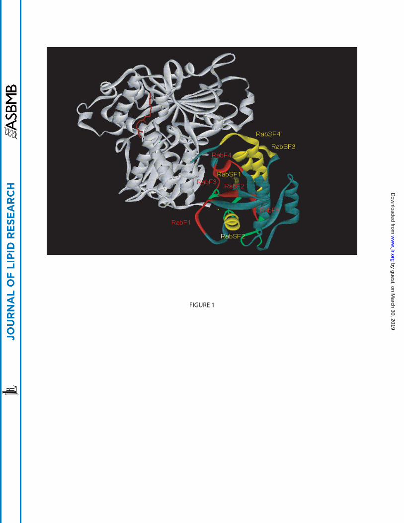
Figure 1: Crystal structure of the Rab7:REP-1 complex – regions conserved within the Rab
family. Ribbon representation of REP-1 (white) bound to Rab7 (greyish blue). The RabF regions
and RabSF regions are highlighted in red and yellow respectively. The guanine nucleotide-
binding regions are in green. GGpp (brown) located in the prenyl-binding pocket of REP-1 is
shown in ball and stick representation. All crystal structures were generated using Accelerys DS
ViewerPro 5.0. The PDB ID of Rab7GG:REP-1 is 1VG0.
Figure 2: A cartoon showing the two possible pathways for Rab protein prenylation. In the
classical pathway, newly translated Rabs bind REP and the complex is recognised by GGpp-
bound RGGT. RGGT catalyses the transfer of geranylgeranyl groups to C-terminal cysteines of
the Rab protein. Following prenylation, RGGT dissociates from REP, which remains bound to
the prenylated Rab protein and delivers it to target membranes. REP is then released into the
cytosol to take part in a new cycle of prenylation. In the alternative pathway, REP forms a
complex with RGGT in the presence of GGpp under conditions where these constituents are in
higher concentration relative to the Rab protein. The REP:RGGT:GGpp complex then binds
newly translated Rab protein and the geranylgeranylation reaction takes place. RGGT dissociates
as before while REP escorts the prenylated Rab to membranes as in the classical pathway. The Kd
values of the Rab:REP:RGGT:GGpp complex for each pathway are indicated.
Figure 3: The isoprenyl binding pocket of mammalian protein prenyltransferases. Superposition
of the isoprenyl binding pocket of Zn2+-depleted FT (orange), GGT-I (green) and RGGT
(yellow). Fpp (blue), GGpp (red) and key amino acid side chains are in ball and stick
representation. Trp102β of FT clashes with the fourth isoprene unit of GGpp and therefore
sterically hinders GGpp from binding in the pocket. The smaller residues in GGT-I and RGGT
(Thr49 and Ser48 respectively) accommodate the GGpp molecule. Sequence alignment of the β-
subunits of human FT, GGT-I and RGGT indicating the key residues, Trp102β (orange), Thr49β
(green) and Ser48β (yellow). The PDB ID of FT, GGT-I and RGGT are 1D8E, 1N4P and 1LTX,
respectively.
by guest, on March 30, 2019
ww
w.jlr.org
Dow
nloaded from
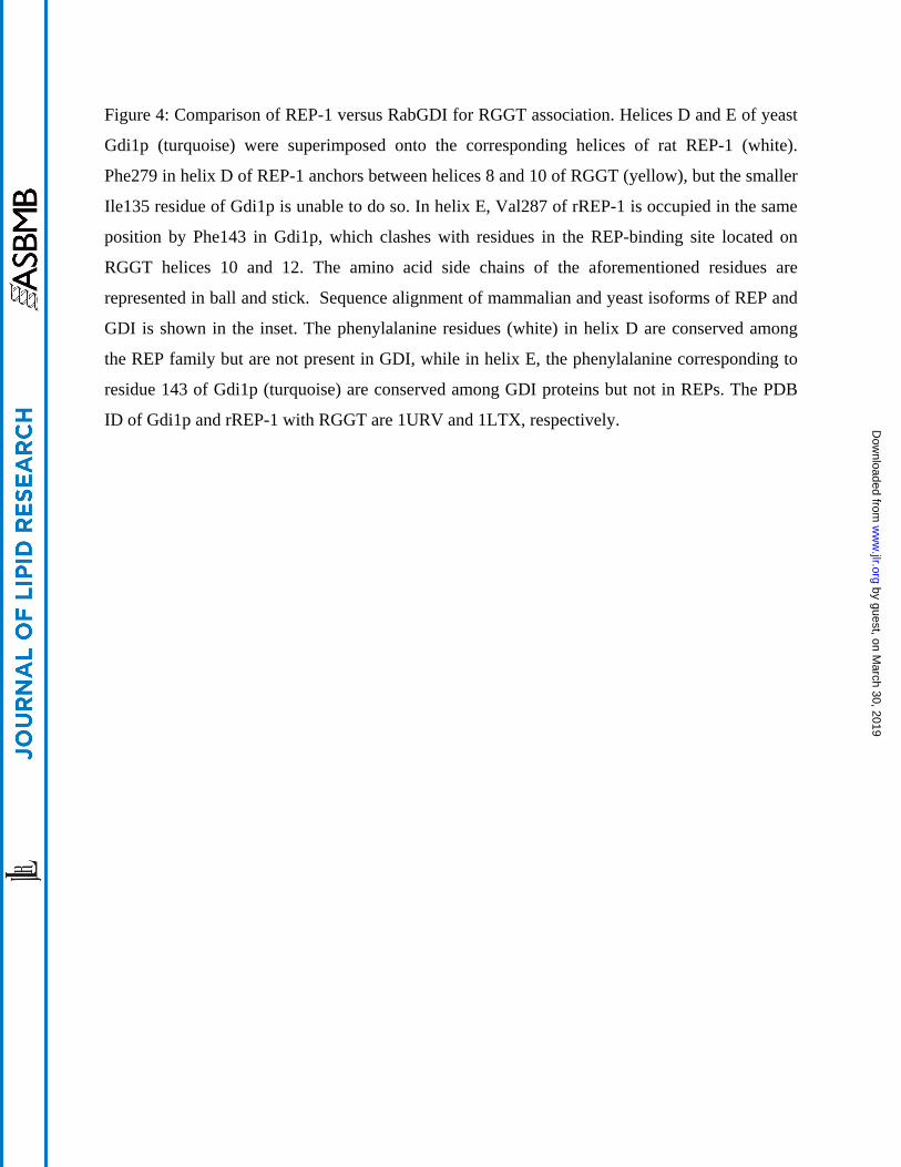
Figure 4: Comparison of REP-1 versus RabGDI for RGGT association. Helices D and E of yeast
Gdi1p (turquoise) were superimposed onto the corresponding helices of rat REP-1 (white).
Phe279 in helix D of REP-1 anchors between helices 8 and 10 of RGGT (yellow), but the smaller
Ile135 residue of Gdi1p is unable to do so. In helix E, Val287 of rREP-1 is occupied in the same
position by Phe143 in Gdi1p, which clashes with residues in the REP-binding site located on
RGGT helices 10 and 12. The amino acid side chains of the aforementioned residues are
represented in ball and stick. Sequence alignment of mammalian and yeast isoforms of REP and
GDI is shown in the inset. The phenylalanine residues (white) in helix D are conserved among
the REP family but are not present in GDI, while in helix E, the phenylalanine corresponding to
residue 143 of Gdi1p (turquoise) are conserved among GDI proteins but not in REPs. The PDB
ID of Gdi1p and rREP-1 with RGGT are 1URV and 1LTX, respectively.
by guest, on March 30, 2019
ww
w.jlr.org
Dow
nloaded from
Rab
Rab Rab
REP
REP
REP
Rab
“Classical” pathway Alternative pathway
α
βKd ~ 0.5nM Kd ~ 30nM
Target membrane
α
β
α
β
REP
REP
Rab
Rab
FIGURE 2
by guest, on March 30, 2019
ww
w.jlr.org
Dow
nloaded from
FTβ 91 DAYECLDASRPWLCYWILHSLELLDE 116GGT-Iβ 38 ERYSSLETSRLTIAFFALSGLDMLDS 63RGGTβ 37 YEYCMSEYLRMSGIYWGLTVMDLMGQ 62
FIGURE 3
by guest, on March 30, 2019
ww
w.jlr.org
Dow
nloaded from
hREP-1 277 SRADVFNSKQLTMVEKRMLMKFLTFCMEY 305hREP-2 279 SRADVFNSKELTMVEKRMLMKFLTFCLEY 307rREP-1 274 SRADVFNSKQLTMVEKRMLMKFLTFCVDL 302 Mrs6p 179 TKQEIFTDQNLPLMTKRNLMKFIKFVLNW 207hGDIα 122 TETEALASNLMGMFEKRRFRKFLVFVANF 150hGDIβ 122 TEAEALASSLMGLFEKRRFRKFLVYVANF 150Gdi1p 130 NEIEAISSPLMGIFEKRRMKKFLEWISSY 158
Helix D Helix E
FIGURE 4
by guest, on March 30, 2019
ww
w.jlr.org
Dow
nloaded from