Embed Size (px)
Citation preview

A MICROSCOPIC METHOD OF DISTINGUISHING DEADFROM LIVING BACTERIAL CELLS
GEORGES KNAYSI
Bacteriological Laboratories, New York State College of Agriculture, CornellUniversity, Ithaca, New York
Received for publication, October 10, 1934
For a long time the biologist has been attempting to discovera criterion by which a living cell can be distinguished from a deadcell by simple microscopic examination. The most popularmethod has been the application of dyes, either in the form ofthe so-called vital staining, or in the usual way on dried and fixedsmears.
1. METHODS OF "VITAL" STAINING
Dilute solutions of dyes were applied to animal or plant cellsnearly as far back as a century ago. By this means it was soughtto gain an insight into various life processes of the cells, to demon-strate certain cellular structures, or to find out whether the cellwere dead or alive. The work started by Ehrlich on nerve cellsand blood cells, and the stimulus it gave other workers, havegiven us a respectable literature on the subject, to which con-tributions are still being made from time to time.The first application of "vital" stains to bacteria was made by
Metchnikoff (1887) in his immortal study of phagocytosis. Hesays:
To convince myself of the bactericidal action of the microphages, Imade use of an old solution of vesuvin which was not in condition toalter living bacteria but stained dead ones bright brown. By adding afew drops of this solution to preparations of leucocytic exudates, Iwas able to observe that the majority of the rods (of attenuated anthraxorganism) inclosed in the protoplasm of the microphages immediately
193
on June 26, 2018 by guesthttp://jb.asm
.org/D
ownloaded from

GEORGES KNAYSI
took up the brown color, while the cells (microphages) remained color-less and continued to live, manifesting their amoeboid movements.
Among the basic anilin dyes, methylene blue and neutral redhave been the most popular, especially neutral red, introducedand warmly recommended by Ehrlich (1894). Plato (1900) usedthat dye in his extensive investigations, and Metchnikoff (1901)himself adopted it in his later work.Among "vital" staining methods may be mentioned that of
Ruziicka (1905) who recommends the use of a staining solutionmade by mixing equal volumes of 0.05 per cent neutral red and0.05 per cent methylene blue solutions in distilled water. A fewdrops of this mixture are spread over the surface of a clean slideand allowed to evaporate at 350C. This leaves a thin, dry filmof dye on which a drop of the bacterial suspension to be investi-gated is placed and examined under a cover-glass. Within ashort time, living cells take on a violet color in which the red tonepredominates. Dead cells show a predominance of blue tone.Here belong also those "vital" staining methods with acid
dyes, like that of Seiffert (1922) who used congo red. Seiffert'sstain was a dilution of 1:10 of a saturated congo red solution inphysiological salt solution. Others have used this dye in nega-tive preparations. Dead bacteria are supposed to take up thedye, living bacteria to remain colorless.2. METHODS OF APPLYING STAINS TO DRIED AND FIXED SMEARS
There is a variety of these methods described in the literature.The best known among these are Bordet's (1895) eosin-methyleneblue method, and that of Proca (1909), especially in the form ofKayser's modification.
Bordet observed in the course of his studies of phagocytosisthat phagocytosed bacteria gradually undergo a change in theirstaining reaction. At first they are cyanophilic and are coloredblue, when the smear is stained with eosin and then with methyl-ene blue. Gradually, under the action of the phagocyte theybecome eosinophilic and take up a pink color when stained in thesame way. A continuous transition in color is seen between thetwo stages.
194
on June 26, 2018 by guesthttp://jb.asm
.org/D
ownloaded from

DISTINGUISHING DEAD FROM LIVING BACTERIAL CELLS 195
Proca's method consists in staining the dried and fixed smearfor one minute with a solution made up by mixing 8 cc. of Ziehl'scarbol fuchsin with 100 cc. of Loeffler's methylene blue and 100cc. of distilled water. Dead bacteria stain red and living bac-teria blue. Applied to spores (Proca and Danila, 1909), it wasfound that viable spores remain colorless and dead spores arestained blue. Proca also observed that bacteria killed by heator disinfectants and stained with Loeffler's methylene blue, losetheir blue color and turn red if subsequently stained with diluteZiehl's carbol fuchsin (1:10). This observation was applied byKayser (1912) in his modification. Kayser recommends stain-ing of the smear three to five minutes with Loeffler's methyleneblue, rinsing, and staining from five to ten seconds with Ziehl'scarbol fuchsin, diluted 1:10. Recently this modification hasbeen warmly recommended by Gay and Clark (1934).The gram reaction has been suggested as a means of differen-
tiating dead from living cells in gram positive species, and animproved method based on that reaction has been recently de-scribed by Frazier and Boyer (1934).
SIGNIFICANCE AND LIMITATIONS OF THE PRESENT METHODS OFDISTINGUISHING DEAD FROM LIVING BACTERIA
The above brief review gives the general lines along whichmethods, based on staining reactions, have been developed inan attempt to distinguish microscopically a dead from a livingcell. In spite of some very optimistic reports, we may safely saythat none yet has proved to be dependable. The following dis-cussion shows why this is to be expected.
"Vital" staining methodsThe basis of the use of vital staining has been so far indefinite.
The general assumption is that living cells do not stain with"vital" dyes, while dead ones do. This assumption has neverbeen satisfactorily justified, and investigators hold conflictingviews on the subject. There is first the school led by Ehrlichwhich believed that the living protoplasm never stains, and thatthe appearance of stain is an evidence of death. Intracellular
on June 26, 2018 by guesthttp://jb.asm
.org/D
ownloaded from
GEORGES KNAYSI
structures which take up the stain are, according to this school,metabolic products taking no part in cell functions. In thisconnection we may mention the interesting "theory of micellarimmunity" formulated by Lumibre (1925), in which he explainsthe non-stainability of living protoplasm by its colloidal struc-ture. The opposite camp is led by Przesmycki (1897), whobelieved that both the cytoplasm and the nucleus of the livingcell may stain, and that, upon death, decoloration takes place.Similar views were held in recent years by Roskin and Semenoff(1933) and by Gavaudan (1933, 1934) and others. Betweenthese two schools we find all kinds of transitional views expressedby investigators like Prowazek (1897), Arnold (1899) and Plato(1900). This latter investigator, although denying stainabilityof the living protoplasm in blood cells and in protozoa, yet be-lieves that stained bacteria inside of the phagocytes may still bealive.The above views regarding blood cells and protozoa were
gained from microscopic observations of motility, or of the be-havior of the stained intracellular granules. In one or two cases,claims of correlation with the division of a protozoan were made.A different line of attack was followed by Fraser (1920) who
compared the number of viable yeast cells as obtained by "Vital"staining, with the number of colonies developing on the petriplate. After trying out a number of basic and acid dyes, Fraseradopted methylene blue. He found that the plate count wasconsistently less than the number of unstained cells and con-cluded that the cell loses the power of reproduction before itstains. Methylene blue was also used by Fulmer and Buchanan(1923). Rahn and Barnes (1933) used Congo red in additionto methylene blue and compared their results with the ferment-ing power and the plate count of yeast cells. Their conclusionsare, likewise, that yeast cells lose the power of reproduction first,followed, in order, by the loss of fermenting power and by theacquisition of the ability to stain with the dyes used.On going over the literature, one can but feel that much of
what is assumed in using "vital" dyes to distinguish dead fromliving cells is arbitrary and indefinite. This feeling becomes even
196
on June 26, 2018 by guesthttp://jb.asm
.org/D
ownloaded from

DISTINGUISHING DEAD FROM LIVING BACTERIAL CELLS 197
stronger when one undertakes such a study experimentally. Itseems natural to consider dead any cell that takes up a dye andliving any one that does not; for, if we suspend growing cells indilute dye solutions, they remain colorless; and, if we kill thecells by heat or chemicals, they immediately stain. But isevery stained cell a dead one or every non-stained one alive?We have seen that the authorities are not in agreement on thatpoint. And then we have the more complex question of degreeof staining. How about cells that barely show color? And cellsthat contain stained vacuoles of various kinds? Much has tobe determined before "vital" staining methods can become de-pendable.
Methods using dried and fixed smearsEven less is known of the principles behind the methods in-
volving the staining of dried and fixed smears. In general, suchmethods are evolved empirically as follows: A smear made froma living culture and stained in a certain way shows certain coloror predominance of a certain shade of color, while a culture,killed in certain ways and stained by the same method, shows adifferent color or predominance of a different shade. In attempt-ing to explain these, at best, rough reactions, some wild assump-tions have been made; for instance, the assumptions that in adried film fixed by heat, the bacteria are still alive, if the filmcame from a living culture.
In order to get an idea of the value of these methods, the readeris referred to an article by Bickert (1930) who investigated avariety of them and arrived at the conclusion that none of themis of any practical value.
I, myself, have investigated the most recommended of allthese methods, the Proca-Kayser method, and my conclusion isthat it does not show the difference between dead and living cells,but merely the difference in the degree of destruction to which thecells have been subjected. In the case of very young cells ofEscherichia coli killed by quick cooling from 450 to 50C. (Sher-man and Cameron, 1934) the method fails completely. Thereare two more objections to this method. First, the shade of
on June 26, 2018 by guesthttp://jb.asm
.org/D
ownloaded from

GEORGES KNAYST
color is rarely pure blue or pure red. It is usually violet with apredominance of blue or red shades, and all kinds of transitionalshades in between. Second, it is not suitable for quantitativework because many of the cells are washed off on rinsing.
PRESENT INVESTIGATIONS
A satisfactory solution of the present problem involves thesolution of several preliminary problems. In the first place asuitable dye must be selected. After trying out several basicand acid dyes, neutral red was chosen on account of its lowtoxicity and its low reduction potential (E' = -0.330 volts atpH 7). The concentration used was such that it had no retardingeffect on the organism studied when added to the culture medium.In the case of Escherichia coli with which most of the presentwork was done, a concentration of. 0.005 per cent was adopted,while some of the yeasts used required as little as 0.001 per centof neutral red. It was also ascertained that, when the organismswere most actively growing in a neutral-red free medium, theaddition of neutral red in the proper concentration caused noinjury.On the other hand neutral red has two disadvantages which,
however, do not interfere with the method as it is here described.The first disadvantage is the fact that neutral red acts as anacid-base indicator in the pH range of 6.8 to 8.0. The alkalinecolor is yellow and hard to see under the microscope. Themethod automatically takes care of this in the process of dilutingwith neutral-red gelatin of pH about 6. The addition of a fer-mentable sugar to the original medium will also eliminate thistrouble.The second disadvantage is encountered when a strongly re-
ducing organism like Escherichia coli is grown in a medium inwhich acid is produced. It has been known for a long time thatEscherichia coli produces discoloration in neutral-red sugar mediain the form of a yellowish-green fluorescence. Clark and Perkins(1932) have shown that this is due to an irreversible reductionof neutral red in the pH range of 4 to 6. Although the criteriaand results given in this paper have been obtained by direct
198
on June 26, 2018 by guesthttp://jb.asm
.org/D
ownloaded from

DISTINOUISHING DEAD FROM LIVING BACTERIAL CELLS 199
experimentation and are independent of aberrations in the be-havior of neutral red in the conditions described, yet numerousexperiments were performed to elucidate the circumstances ofthe appearance of fluorescence in coli cultures. The conclusionfrom these experiments is that, when coli cultures in the neutralred medium used attain their most negative potential, the pHis about 6 and only about 25 per cent of the dye is reduced, whichis not noticeable to the unaided eye. Fluorescence appears muchlater, usually in cultures twenty-four hours old or more, at 30'C.The pH of the culture is then about 5.2 and the dye should bereduced, only, to the extent of about 1 per cent. This apparentlyparadoxical fact has probably its explanation in the continuousaccumulation of small amounts of the irreversible fluorescentmaterial. As long as the bacterial population in fluorescentcultures is such that they must be diluted with neutral-red gela-tin, the objection suggested by this phenomenon disappears.The next step was to find out whether a cell can stain (in whole
or in part) and still be alive. The test organisms were Escheri-chia coli, Schizosaccharomyces pombe and another yeast isolatedfrom fermenting ale. Use was made of the technique of micro-cultures described by the author in a previous paper (Knaysi,1933). The investigated organism was grown in glucose brothcontaining neutral red far below its inhibiting concentration.Micro-cultures were made from such cultures or from neutral-red glucose broth inoculated from these cultures. The micro-cultures were incubated and observed at room temperature.Each preparation contained about 30 droplets deposited in rows,and these were mapped immediately after the preparation wascompleted, and descriptions of the cells contained in some ofthem were recorded, and this correlated with viability. And,again, after growth in certain droplets had proceeded far enoughor was completed, micro-transfers were made to fresh dropletsof the neutral-red glucose medium or, in certain cases, the mediumwas sucked out and fresh medium added, thus insuring the ob-servation of some particular cell or cells.The conclusions drawn from numerous such experiments may
be stated as follows:
on June 26, 2018 by guesthttp://jb.asm
.org/D
ownloaded from

GEORGES XNAYSI
1. Normal, healthy cells do not show the slightest evidence ofcolor either of the cytoplasm or the vacuoles and other inclusions.
2. Any cell showing evidence of staining of its cytoplasm, nomatter how faint it is, is a dead cell.
3. Cells in which the vacuoles alone are stained, may or maynot be able to grow. In the first case, the vacuole loses itscolor completely before growth takes place. In the second, thestain diffuses out into the cytoplasm, giving it a faint tinge ofcolor.
4. There is a certain number of healthy, normal-appearingcells that fail to grow, and yet they do not take up the dye for along time. These were probably alive but somehow lost thepower to reproduce. They are numerically few.
5. Before autolysis sets in, a dead cell stains deep red, but asautolysis proceeds, staining becomes gradually fainter, and onefinds a certain number of cells that look disorganized and havea low refractive index without showing evidences of staining.Such cells are not numerous and are easy to recognize. Theyprobably contain no more stainable material.
ADDITIONAL TESTS OF RELIABILITY AND SENSITIVITY OFTHE METHOD
Aside from the direct viability test described above, othermeans have been used to gain an idea about how reliable andsensitive the method is.
Cultures of Escherichia coli killed by heating to 650C., inflowing steam, or in the autoclave, take up the dye immediately,as do cultures killed by phenol and mercuric chloride. The dyemay not penetrate the cell instantaneously on account of coagu-lation of the protoplasm or the like, but the membrane of thecell stains deeply and there will be no mistake about the cellbeing stained.A most crucial test for the sensitivity of the method is to
apply it to young cultures cooled quickly from 450 to 50C. Itmay be remembered that Sherman and Cameron (1934) haverecently noted that when very young, rapidly growing culturesof Escherichia coli are quickly cooled from 450 to 10'C., over 90
200
on June 26, 2018 by guesthttp://jb.asm
.org/D
ownloaded from
DISTINGUISHING DEAD FROM LIVING BACTERIAL CELLS 201
per cent of the cells may be killed. This discovery is surprisingin view of the fact that both temperatures are included withinthe temperature range of growth of Escherichia coli. The cri-terion of death used by Sherman and Cameron having beenthe failure to grow on petri plates, it is reasonable to consider thepossibility that cooling merely interfered with the growth mech-anism of the cell instead of causing instantaneous killing. Whenthe present method was applied to such cultures, it was foundthat almost all the cells took up the dye, indicating an im-mediate death.The present method also shows that in a smear of Escherichia
coli, dried and fixed by heat, the cells are dead, contrary to astatement of Ficker (1929) adopted by various investigators, in-cluding Bickert (1930) and Gay and Clark (1934).
In addition to the above tests it was also thought desirable tocompare the numbers of viable bacteria as obtained by thepresent method with those obtained by the petri plate method.
NUMBERS OF VIABLE CELLS IN CULTURES OF ESCHERICHIA COLI,AS DETERMINED BY THE NEUTRAL RED AND BY
THE PETRI PLATE METHODS
Technique
A certain volume of the culture of Escherichia coli in which itwas intended to enumerate viable cells was diluted with 15 or20 per cent of nutrient gelatin at 350C., containing 0.005 per centneutral red. The percentage of gelatin used depended on thedegree of dilution and on the temperature of the room. Themixture was shaken thoroughly for a few minutes and replacedin the water bath until most of the foam disappeared. Then 1cc. was measured into a 99 cc. water blank previously warmed to30'C. and, immediately following this, a loopful of the bacterialsuspension was used to fill a Petroff-Hauser counting chamber.The chamber was allowed to remain one minute at 370C. whilefilling, in order to keep the viscosity of the gelatin low and preventit from raising the cover-glass, which would introduce a largeerror. During the time the chamber was at 370C., the water
on June 26, 2018 by guesthttp://jb.asm
.org/D
ownloaded from

GEORGES KNAYSI
blank which received 1 cc. of suspension was being shaken. Thechamber was then placed in a 100C. incubator to allow hardeningof the gelatin and to slow down possible growth of the organism,and it was left there until plating was completed. This pro-cedure, it was ascertained, causes no injury even to the mostsensitive cells. The value of the gelatin is to check motility ofthe organism, and to supply rigidity, as counting was made withthe oil-immersion objective.The Petroff-Hauser counting chamber has similar ruling to
ordinary haemacytometers and possesses the advantages of beingadapted for use with the oil-immersion objective and with thedark-field condenser. Its depth is 0.02 mm.; that is, it is one-fifth as deep as the chambers of ordinary haemacytometers.An average of one cell per small square is equivalent to 20 millioncells per cubic centimeter. With the possible large error in-volved, the degree of accuracy attained is remarkable, even whenthe suspension contained less than 10 million cells per cubiccentimeter. Usually about 50 small. squares were counted.
In plating, water blanks warmed to 300C. were used. Warm-ing the water blanks was for the purpose of keeping the gelatinin the liquid form. The medium used for plating was nutrientagar containing 0.5 per cent glucose. The agar was poured at450C. Before this procedure was decided upon, experimentswere performed in substituting broth blanks for water blanks,and in using different percentages of agar to make pouring pos-sible at 350C. No advantage was found for any of these modifi-cations, even with most sensitive cells. The glucose was in-corporated, however, because it made the organisms grow muchfaster and form larger colonies. Counting was usually madeafter 24-hour incubation at 370C.
ResultsThe results of comparative counts are recorded in tables 1 and
2. Table 1 includes counts of young cultures, while table 2 in-cludes the results with mature and old cultures.
In comparing any microscopic count with the plate count, itis necessary to compare the number of groups observed under
202
on June 26, 2018 by guesthttp://jb.asm
.org/D
ownloaded from

DISTINGUISHING DEAD FROM LIVING BACTERIAL CELLS 203
the microscope with the number of colonies developing on theplate. By group, is meant one cell or two or more cells that arestill united by their cell wall. This point seems too evident toneed mention, and yet, strangely enough, in the few instanceswhere a direct count of viable cells was compared with the platecount, no mention was made of groups, except in Frazier andBoyer's report. This, alone, is sufficient to magnify the dis-crepancy found between the two methods to the proportions re-corded. The argument that, upon shaking, groups (in the sensehere used) are broken up to individual cells does not hold in the
TABLE 1Number of viable cells in young cultures of Escherichia coli as determined micro-
scopically and by platingThe organism was grown in glucose broth containing 0.005 per cent of neutral
red at 30'C.
PLATE COUNTMICROSCOPIC COUNT PER CUBIC PER CUBIC
CENTIMETER OF CULTURE CENTIMETEREXPERI- AGE OF OF CULTURE PLATE COUNT X 100MENT CULTURE GROUP COUNT
Cells'X lo$ Groups Groups X 100 AverageX106Cens ~x106
hours
1 4 185.0 111.0 60.0 119.0 107.22 41 107.0 60.0 56.0 67.3 112.03 31 72.0 48.0 66.6 51.0 106.04 12 703.0 356.0 50.6 280.0 78.75 1i 2.2 1.1 50.0 1.4 127.26 31 11.2 5.8 51.8 5.9 102.0
light of the figures reported in tables 1 and 2, even for as "indi-vidualistic" an organism as Escherichia coli. The gelatin sus-pension was usually vigorously shaken for nearly five minutes,and yet the ratio of groups to individuals never reached unity,although in a few experiments with old cells it approached thatvalue. In young cultures of coli, where the cells are activelygrowing, the number of groups is nearly half of that of individuals,because most of the population consists then of pairs of sistercells. In cultures where growth is very slow, this circumstanceis non-existent, and the cells tend to be single with few pairs andan occasional short chain.
on June 26, 2018 by guesthttp://jb.asm
.org/D
ownloaded from
204 GEORGES KNAYSI
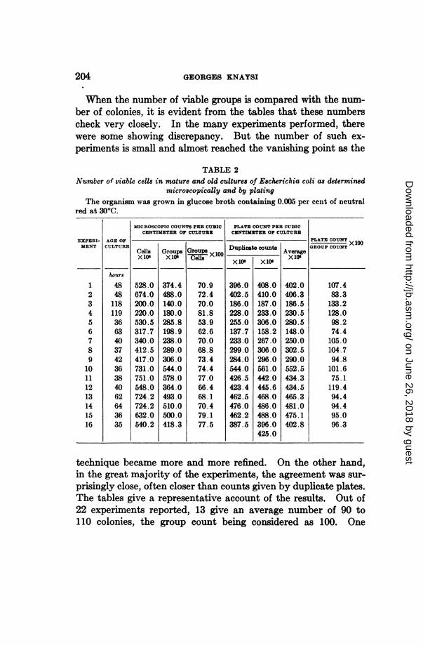
When the number of viable groups is compared with the num-ber of colonies, it is evident from the tables that these numberscheck very closely. In the many experiments performed, therewere some showing discrepancy. But the number of such ex-periments is small and almost reached the vanishing point as the
TABLE 2Number of viable cells in mature and old cultures of Escherichia coli as determined
microscopically and by platingThe organism was grown in glucose broth containing 0.005 per cent of neutral
red at 300C.
MICROSCOPIC COUNTS PER CUBIC PLATE COUNT PER CUBICCENTIMETER OF CULTURE CENTIMETER OF CULTURE
Cells Groups Group Duplicat ecounts rageX106 X10 Cells X xDupI vte rX10t
528.0674.0200.0220.0530.5317.7340.0412.5417.0731.0751.0548.0724.2724.2632.0540.2
374.4488.0140.0180.0285.8198.9238.0289.0306.0544.0578.0364.0493.0510.0500.0418.3
70.972.470.081.853.962.670.068.873.474.477.066.468.170.479.177.5
X lIU
396.0402.5186.0228.0255.0137.7233.0299.0284.0544.0426.5423.4462.5476.0462.2387.5
XA f
408.0410.0187.0233.0306.0158.2267.0306.0296.0561.0442.0445.6468.0486.0488.0396.0425.0
402.0
406.3186.5230.5280.5148.0250.0302.5290.0552.5434.3434.5465.3481.0475.1402.8
PLATE COUNT x W
GROUP COUNT
107.483.3133.2128.098.274.4105.0104.794.8101.675.1119.494.494.495.096.3
technique became more and more refined. On the other hand,in the great majority of the experiments, the agreement was sur-
prisingly close, often closer than counts given by duplicate plates.The tables give a representative account of the results. Out of22 experiments reported, 13 give an average number of 90 to110 colonies, the group count being considered as 100. One
EXPERI-MENT
1
2345678910111213141516
AGE OFCULTURE
hours
4848118119366340374236384062643635
on June 26, 2018 by guesthttp://jb.asm
.org/D
ownloaded from
DISTINGUISHING DEAD FROM LIVING BACTERIAL CELLS 205
would have no objection if his duplicate plates came consistentlyas close as this. It must be added that, in most of the cases re-ported, the gelatin suspension contained less than 25 million percubic centimeter, and in a couple of experiments with young cellsit contained less than 2 millions. A much closer agreement isobtained when the number of cells counted in the gelatin sus-pension is made larger.
SUMMARY
This paper is concerned with the use of neutral red in distin-guishing, microscopically, between dead and living cells.The technique of micro-cultures was used to study the relation
between the viability of a cell and its ability to stain in wholeor in part. It was found that cells of Escherichia coli, Schizo-saccharomyces pombe and a yeast isolated from ale are to be con-sidered dead whenever the cytoplasm proper is tinged, evenslightly, with stain. Yeast cells with stained vacuoles and un-stained cytoplasm are weakened, sick cells that may or may notrecover. In a culture, there is a certain number of cells that arenot stained and look healthy, but are unable to multiply. Theseare numerically few. Others do not stain because their autolysisis too far gone. These look disorganized, have a low refractiveindex and are easy to recognize. Their number is usually notimportant.When comparisons are made between numbers of viable cells
in cultures of Escherichia coli as obtained microscopically, usingneutral red as an indicator of viability, and numbers of viablecells as given by plating, a close agreement between the two isfound when groups, instead of single cells, are taken into con-sideration.The concentration of neutral red varies with the organisms.
In the case of Escherichia coli, a concentration of 0.005 per centwas adopted. This concentration is harmless even to the young-est and most sensitive cells, and the organism grows readily inbroth containing neutral red in that concentration.
on June 26, 2018 by guesthttp://jb.asm
.org/D
ownloaded from

206 GEORGES KNAYSI
REFERENCES
ARNOLD, J. 1899 Arch. f. path. Anat. u. Physiol. u. f. klin. Med. (Virchow's),167, 424-437.
BICKERT, F. W. 1930 Centralbl. f. Bakt. I Abt. Orig., 117, 548-551.BORDET, J. 1895 Ann. Soc. roy. des sciences medic. et nat. de Bruxelles, 4,
455-530.CLARK, W. M., AND PERKINS, M. E. 1932 Jour. Amer. Chem. Soc., 54, 1228-1248.EHRLICH, P. 1894 Allgem. med. Centralz., 63, 20.FICKER, M. 1929 In Kolle and Wassermann's Handbuch der Pathogenen Mikro-
organismen, 3rd edition, 9, 711-802.FRASER, C. G. 1920 Jour. Phys. Chem., 24, 741-748.FRAZIER, W. C., AND BOYER, A. J. 1934 Jour. Bact., 27, 31-32.FULMER, E. I., AND BtUcHANAN, R. E. 1923 Jour. Gen. Physiol., 6, 77-89.GAVAUDAN, P. 1933 Comptes Rend. a l'Acad. des Sciences, 196, 563-565.GAVAUDAN, P. 1934 Comptes Rend. a l'Acad. des Sciences, 198, 848-85.GAY, F. P., AND CLARK, A. R. 1934 Jour. Bact., 27, 175-189.KAYSER, H. 1912 Centralbl. f. Bakt. I Orig., 62, 174-176.KNAYSI, G. 1933 Jour. Bact., 26, 623-644.LtMIkRE, A. 1925 Bull. d'Histol. Apple. a la Physiol. et A la Pathol. et de Tech.
Micros., 2, 69-77.METCHNIKOFF, E. 1887 Ann. Inst. Pasteur, 1, 321-336.METCHNIKOFF, E. 1901 L'immunite. Masson et Cie, Paris publishers. Eng-
lish edition published by Cambridge University Press, 1905.PLATO 1900 Arch. f. mikros. Anat. u. Entwicklungs-gesc., 56, 868-917.PROCA, G. 1909 Comp. Rend. Soc. Biol., 67, 148-149.PROCA, G., AND DANILA, P. 1909 Comp. Rend. Soc. Biol., 67, 307-309.PROWAZEK, S. 1897 Ztschr. f. wissenschaft. Zool., 63, 187-194PRZESMYCKI, A. M. 1897 Biol. Centralbl., 17, 321-335.RAHN, O., AND BARNES, M. N. 1933 Jour. Gen. Physiol., 16, 579-592.ROSKIN, G., AND SEMENOFF, W. 1933 Ztschr. f. Zellforsch. u. mikrosk. Anat.,
19, 150-190.RtfIt}&A, V. 1905 Pfuiger's Arch. f. d. ges. Physiologie d. Mensch. u. d. Thiere.,
107, 497-534.SEIFFERT, W. 1922 Centralbl. f. Bakt. I. Abt Orig., 88, 151-158.SHERMAN, J. M., AND CAMERON, G. M. 1934 Jour. Bact., 27, 341-348.
on June 26, 2018 by guesthttp://jb.asm
.org/D
ownloaded from

![Production, Characterization and Treatment of Textile ... · and anthraquinone dyes) and lanaset dyes (Blue 5G and Bordeaux B) [5-7]. Other dyes, like dispersed dyes (Disperse yellow](https://img.pdfslide.us/doc/110x75/5e227d67c8e7e660b661c0e5/production-characterization-and-treatment-of-textile-and-anthraquinone-dyes.jpg)