Embed Size (px)
Citation preview
Genotypic Differences in the Hepatitis B Virus CorePromoter and Precore Sequences DuringSeroconversion From HBeAg to Anti-HBe
Jonas Blackberg and Karin Kidd-Ljunggren*Department of Infectious Diseases, University Hospital, Lund, Sweden
Hepatitis B virus (HBV) strains from anti-HBepositive patients often show specific mutationsin the precore gene, the core promoter region, orboth. The dynamics of seroconversion in rela-tion to the appearance of these mutations hasnot been studied and compared between de-fined HBV genotypes. Samples from patients fol-lowed during seroconversion from HBeAg toanti-HBe were amplified by polymerase chain re-action (PCR), sequenced and genotyped. Among16 sets of samples, 6 belonged to genotype A, 6to genotype D, 2 to genotype B, 1 to genotype C,and 1 to genotype E. Whereas strains from geno-types B, C and E showed changes in the corepromoter, precore codon 28 or both, genotype Aand D strains displayed a different pattern. In 4of 6 anti-HBe positive samples from genotype A,the precore had a wild-type sequence while thecore promoter sequence showed a specific TGAmutation. In another genotype A strain a precorestop mutation was preceded by a mutation incodon 15, thus conserving base-pairing at thepregenomic RNA level in this region. In contrast,all genotype D strains showed wild-type se-quences in both the core promoter and precorecodon 28 in pre- and post-seroconversionsamples. Thus, in 8 patients with a mean follow-up time of 17 months, wild-type sequences inboth the core promoter and precore codon 28were found after seroconversion to anti-HBe.This study also confirmed, for genotype D, thatHBeAg seroconversion often occurs earlier thangenomic conversion. J. Med. Virol. 60:107–112,2000. © 2000 Wiley-Liss, Inc.
KEY WORDS: HBV; wild-type; mutation; tem-poral
INTRODUCTION
During infection with hepatitis B virus (HBV), asoluble protein is found in the serum of almost allacutely and a proportion of chronically infected pa-
tients. This protein, the HBeAg [Magnius and Esp-mark, 1972], was used for many years as the maininfectivity marker of HBV [Shikata et al., 1977]. Sero-conversion from HBeAg to anti-HBe was taken origi-nally to signify a fall in viral replication and thereforeof infectivity. It is still used in some studies as amarker of successful treatment with interferon [Wonget al., 1993].
As tests for the detection of HBV DNA became avail-able more widely, it became clear that a proportion ofanti-HBe positive patients carried replicative virus andwere therefore infectious [Burk et al., 1994]. In a studyby Carman et al. [1989], a mutation leading to a trans-lational stop in precore codon 28 was seen to be asso-ciated with anti-HBe positivity in the infected patient.This mutation would prevent the production of HBeAgand has since been reported by numerous groups. Ithas been associated with more aggressive liver diseaseand with outbreaks of fulminant hepatitis [Omata etal., 1991; Liang et al., 1991].
Transcription of the precore and core genes is di-rected from the so-called core promoter region. Achange from 1762 - 1764 AGG to TGA (numbering byOkamoto et al., [1988]) in the core promoter was firstseen by Okamoto et al. (1994) to be associated with theHBeAg/anti-HBe status of the patient. In a cross-sectional study, both the core promoter mutations andthe precore codon 28 mutation were found to be highlyand equally significant for the HBe/anti-HBe pheno-type displayed [Kidd-Ljunggren et al., 1997].
The dynamics of seroconversion in relation to theappearance of the precore codon 28 mutation has beenstudied by Lai et al. [1994]. A gradual takeover of mu-tated over wild-type strains seems to occur. In a recent
Grant sponsor: Crafoord Foundation; Grant sponsor: Bristol-Myers Squibb Research Foundation; Grant Sponsor: Swedish So-ciety of Medicine; Grant sponsor: Royal Physiographic Society ofLund; Grant sponsor: Swedish Medical Research Council; Grantnumber K98-16X-11592-03A.
*Correspondence to: Karin Kidd-Ljunggren, M.D., Ph.D., De-partment of Infectious Diseases, University Hospital, S-221 85Lund, Sweden. E-mail: [email protected]
Accepted 16 July 1999
Journal of Medical Virology 60:107–112 (2000)
© 2000 WILEY-LISS, INC.
study by Chan et al. [1999], Chinese patients fromHong Kong, divided into two groups by a variation inprecore codon 15, were followed through seroconver-sion. Only 2 of 26 patients had wild-type sequences inthe core promoter and precore codon 28 after serocon-version.
A study was undertaken to determine whether thepattern that appeared in the core promoter and precorecodon 28 during seroconversion from HBeAg to anti-HBe would vary between the different HBV genotypes.In particular, the sequence pattern of well-defined ge-notype A strains was examined. Base-pairing at thepregenomic RNA level in the precore region, which isessential for replication [Junker-Niepmann et al.,1990], precludes the development of the precore stopcodon in genotype A strains [Li et al., 1993]. Thesestrains would therefore have to use other mechanismsto prevent HBeAg formation.
MATERIALS AND METHODSSerum Samples and HBV Marker Tests
Samples from 51 HBV-infected patients who hadbeen followed with routine serological testing duringseroconversion from HBeAg to anti-HBe were exam-ined retrospectively. The samples had been stored at−20°C. None of the patients had been treated with in-terferon or specific antiviral drugs. From each of the 51patients at least two samples were analyzed by poly-merase chain reaction (PCR), namely, an HBeAg posi-tive and anti-HBe negative sample, and an HBeAg nega-tive and anti-HBe positive sample. If there were morethan two samples available, the last HBeAg positiveand the first anti-HBe positive samples were chosen forthe initial PCR. In case no. 6 the first sample was posi-tive for both markers. Commercial radioimmunoassays(Abbott Laboratories, North Chicago, IL) were used forthe detection of HBsAg, HBeAg and anti-HBe.
Enzymatic Amplification and Sequencing
DNA was extracted from serum and amplified byPCR as described [Ljunggren and Kidd, 1991], usingslightly modified cycling temperatures. Oligonucleo-tide primers KL28 (58GAG ACC ACC GTG AAC GCC38) and KL6 (58GGA AAG AAG TCA GAA GGC A 38)were used to amplify the core promoter and precoreregions. Ten to 100 genome copies can be detected us-ing this protocol. The pre-S and S genes were amplifiedusing KL12 (58GGG TCA CCA TAT TCT TGG G 38) andKL33 (58ACC ACT GAA CAA ATG GCA CTA G 38). ThePCR primers were then used for direct sequencing ofthe amplified DNA by the method of Kretz et al. [1989].Standard precautions were taken to prevent carry-overof DNA [Kwok, 1990; Ljunggren and Kidd, 1991] andthere was no evidence of contamination.
Determination of Genotypes
Sequences obtained by direct sequencing werealigned with strains which genotypically were well de-fined. The genotype of each strain was determined bycomparing key nucleotide positions in the core pro-
moter, precore and S genes [Norder et al., 1993; Kidd-Ljunggren et al., 1995a,1997; Kidd and Kidd-Ljunggren, 1996].
RESULTS
In the samples from HBV-infected patients who hadbeen followed through seroconversion from HBeAg toanti-HBe, HBV DNA was detected by PCR in the first(HBeAg positive) sample from all 51 cases. In 35 cases,the second, HBeAg negative sample was negative byPCR, thus precluding further analysis. For the remain-ing 16 cases, an attempt was made to amplify all otheravailable samples. In total, 52 samples were tested(Table I). There was a mean time of 26 months betweenthe first and the last sample (range 1.5–79 months) anda mean time of 12 months between the last HBeAgpositive and the first HBeAg negative sample (range1.5–28 months).
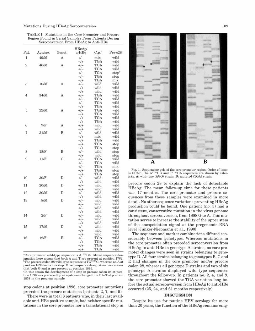
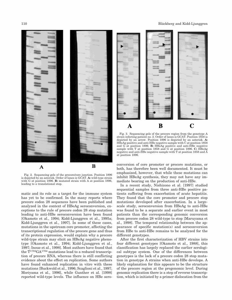
The sequences obtained for each sample, in relationto the HBeAg/anti-HBe phenotype displayed in thesample, are presented in Table I. The core promotersequence A1762GG1764 was considered to be the wild-type, whereas a mutated core promoter sequence wasT1762GA1764 (Fig. 1). The presence of A1896 instead of Gindicates a precore codon 28 stop mutation (Fig. 2). Sixsets of samples belonged to genotype A, 6 to genotypeD, 2 to genotype B and 1 each to genotypes C and E.Four of the genotype D strains showed homology to thegenotype D strains seen in Swedish drug addicts (datanot shown).
The sequence patterns seen during seroconversion,particularly at position 1762, 1764, and 1896, differedconsiderably amongst the 16 cases (Table I). In theHBeAg positive samples, 3 of 16 strains (patients 2, 8and 9) had a stop mutation in precore codon 28. In oneof these cases (patient 9) there was a mixture of wildtype and mutated strains. The stop mutation only ap-peared in one additional sample (patient 7) after sero-conversion to anti-HBe. Thus, in 12 of 16 cases, thepatient had a wild-type precore sequence after serocon-version to anti-HBe. In 4 of these 12 cases (patients 3,5, 15, and 16) there were multiple anti-HBe-positivesamples available, all displaying the wild-type precoresequence. The mean anti-HBe positive follow-up timefor these four patients was 10 months (range 21⁄2–24months). In one patient (no. 2) the genotype A straindeveloped the precore stop codon in the last availableHBeAg positive sample. This was preceded by a changefrom C at position 1858 in the first sample, to T in thesecond sample. The remaining anti-HBe positivesamples had T 1858 (Fig. 3).
In the core promoter region, the sequence patternswere similar between the first HBeAg-positive sample(10 of 16 samples had the wild-type sequence) and thelast anti-HBe positive sample (9 of 16 with wild-type).During the transition from HBeAg-positive to anti-HBepositive samples, patient no. 7 had an intermediatesample with mixed wild-type and mutated core pro-moter sequence. In 3 of the 4 patients who developed
108 Blackberg and Kidd-Ljunggren
stop codons at position 1896, core promoter mutationspreceded the precore mutations (patients 2, 7, and 9).
There were in total 8 patients who, in their last avail-able anti-HBe positive sample, had neither specific mu-tations in the core promoter nor a translational stop in
precore codon 28 to explain the lack of detectableHBeAg. The mean follow-up time for these patientswas 17 months. The core promoter and precore se-quences from these samples were examined in moredetail. No other sequence variations preventing HBeAgproduction could be found. One patient (no. 3) had aconsistent, conservative mutation in the virus genomethroughout seroconversion, from 1888 G to A. This mu-tation serves to increase the stability of the upper stemof the encapsidation signal at the pregenomic RNAlevel [Junker-Niepmann et al., 1990].
The sequence and marker combinations differed con-siderably between genotypes. Whereas mutations inthe core promoter often preceded seroconversion fromHBeAg to anti-HBe in genotype A strains, no core pro-moter changes were seen in strains belonging to geno-type D. All four strains belonging to genotypes B, C andE had changes in the core promoter and/or precorecodon 28, whereas all genotype D strains and two of sixgenotype A strains displayed wild type sequencesthroughout the follow-up. In patients no. 2, 4, and 9,the core promoter showed the TGA variation long be-fore the actual seroconversion from HBeAg to anti-HBeoccurred (25, 24, and 61 months respectively).
DISCUSSIONDespite its use for routine HBV serology for more
than 20 years, the function of the HBeAg remains enig-
TABLE I. Mutations in the Core Promoter and PrecoreRegion Found in Serial Samples From Patients During
Seroconversion From HBeAg to Anti-HBe
Pat. Age/sex Genot.HBeAg/a-HBe C.p.a Pre-c28b
1 49/M A +/− mix wild−/+ TGA wild
2 46/M A +/− TGA wild+/− TGA wild+/− TGA stopc
−/− TGA stop−/+ TGA mix
3 10/M A +/− wild wild−/+ wild wild−/+ wild wild
4 34/M A +/− TGA wild+/− TGA wild+/− TGA wild−/+ TGA wild
5 22/M A +/− TGA wild−/+ TGA wild−/+ TGA wild−/+ TGA wild
6 9/F A +/+ wild wild−/+ wild wild
7 31/M B +/− wild wild−/+ mix wild−/+ TGA wild−/+ TGA stop−/+ TGA stop
8 18/F B +/− wild stop−/+ wild stop
9 11/F C +/− TGA wild+/− TGA wild+/− TGA mix−/+ TGA stop−/+ TGA stop
10 30/F D +/− wild wild−/+ wild wild
11 20/M D +/− wild wild−/+ wild wild
12 30/M D +/− wild wild−/+ wild wild
13 8/M D +/− wild wild+/− wild wild+/− wild wild−/+ wild wild
14 2/F D +/− wild wild+/− wild wild−/+ wild wild
15 17/M D +/− wild wild−/+ wild wild−/+ wild wild
16 12/F E +/− TGA wild−/+ TGA wild−/+ TGA wild−/+ TGA wild
aCore promoter wild-type sequence is A1762GG. Mixed sequence des-ignation here means that both A and T are present at position 1762.bThe precore codon 28 wild-type sequence is TG1896G, whereas an A atposition 1896 leads to a stop. Mixed sequence designation here meansthat both G and A are present at position 1896.cIn this strain the development of a stop in precore codon 28 at posi-tion 1896 was preceded by an upstream change from C to T at position1858 in the previous sample.
Fig. 1. Sequencing gels of the core promoter region. Order of lanesis GCAT. The A1762GG and T1762GA sequences are shown by aster-isks. A: wild-type (AGG) strain. B: mutated (TGA) strain.
Mutations During HBeAg Seroconversion 109
matic and its role as a target for the immune systemhas yet to be confirmed. In the many reports whereprecore codon 28 sequences have been published andanalyzed in the context of HBeAg seroconversion, ex-ceptions to the rule of precore codon 28 stop mutationleading to anti-HBe seroconversion have been found[Okamoto et al., 1994; Kidd-Ljunggren et al., 1995a;Kidd-Ljunggren et al., 1997]. In some of these cases,mutations in the upstream core promoter, affecting thetranscriptional regulation of the precore gene and thusof its protein expression, would explain why a precorewild-type strain may elicit an HBeAg negative pheno-type [Okamoto et al., 1994; Kidd-Ljunggren et al.,1997; Inoue et al., 1998]. Most authors have found thatthe T1762GA1764 mutations lead to a reduced transcrip-tion of precore RNA, whereas there is still conflictingevidence about the effect on replication. Some authorshave found enhanced replication in vitro with thesemutations [Buckwold et al., 1996; Scaglioni et al., 1997;Moriyama et al., 1996], while Gunther et al. [1998]reported wild-type levels. The influence on HBe sero-
conversion of core promoter or precore mutations, orboth, has therefore been well documented. It must beemphasized, however, that while these mutations caninhibit HBeAg synthesis, they may not have any im-mediate bearing on the production of anti-HBe.
In a recent study, Nishizono et al. [1997] studiedsequential samples from three anti-HBe positive pa-tients suffering from exacerbation of acute hepatitis.They found that the core promoter and precore stopmutations developed after exacerbation. In a large-scale study, seroconversion from HBeAg to anti-HBewas found to be a separate and earlier event in mostpatients than the corresponding genomic conversionfrom precore codon 28 wild-type to stop [Maruyama etal., 1998]. The temporal relationship between the ap-pearance of specific mutation(s) and seroconversionfrom HBe to anti-HBe remains to be analyzed for thedifferent genotypes.
After the first characterization of HBV strains intofour different genotypes [Okamoto et al., 1988], thisclassification has largely replaced the earlier serologi-cal subtype system. One of the differences betweengenotypes is the lack of a precore codon 28 stop muta-tion in genotype A strains when anti-HBe develops. Alikely explanation for this appears to be the structureof the precore region at the pregenomic level. Duringgenomic replication there is a step of reverse transcrip-tion, which is initiated by a primer dislocation from the
Fig. 2. Sequencing gels of the precore/core junction. Position 1896is depicted by an asterisk. Order of lanes is GCAT. A: wild-type strainwith G at position 1896. B: mutated strain with A at position 1896,leading to a translational stop.
Fig. 3. Sequencing gels of the precore region from the genotype Astrain infecting patient no. 2. Order of lanes is GCAT. Position 1858 isdepicted by an arrow. Position 1896 is depicted by an asterisk. A:HBeAg positive and anti-HBe negative sample with C at position 1858and G at position 1896. B: HBeAg positive and anti-HBe negativesample with T at position 1858 and G at position 1896. C: HBeAgnegative and anti-HBe negative sample with T at position 1858 and Aat position 1896.
110 Blackberg and Kidd-Ljunggren

stable pregenomic RNA stem-loop structure of the pre-core region to the upstream DR1. In genotype Astrains, a C at position 1858 leads to disruption of thebase-pairing in the lower stem of this stem-loop struc-ture when the stop mutation (G to A) appears [Li et al.,1993; Kidd and Kidd-Ljunggren, 1996]. The levels ofreplication seem to decrease dramatically when thisdisruption occurs, and in most cases, patients infectedwith genotype A strains become PCR negative whenthey seroconvert to anti-HBe.
There seems to be a mechanism for the genotype Astrains to cope with the precore codon 28 mutation. Asseen in this study, case no. 2 was HBeAg positive andanti-HBe negative in the first sample, having a wild-type precore sequence (Fig. 3, Table I). Before the pre-core stop mutation (G to A) developed, the correspond-ing base on the opposite side of the stem (codon 15)changed from a genotype A-specific C to T, thus ensur-ing a stable base-pairing in this region. A similar ob-servation was made by Li et al. [1993]. The precore stopmutation probably happens as frequently in genotypeA strains as in the other genotypes. The only viablegenotype A strains though, will be those that also man-age to develop a corresponding mutation in precorecodon 15, preventing disruption of the stem-loop struc-ture.
In this study, the most marked difference in se-quence patterns after seroconversion was not, as re-ported in a very recent study [Chan et al., 1999], be-tween strains with C 1858 and T 1858, but betweengenotypes. All genotype D (T 1858) strains had wildtype sequences in both the core promoter and precorecodon 28, compared to two of ten strains from geno-types A (C 1858), B (T1858), C (T 1858) and E (T 1858).The comparatively high number of anti-HBe positivewild-type strains in this study may be due in part to thefollow-up time. The average observation time in thestudy by Maruyama et al. [1998] was two to threeyears, compared to 17 months for the eight wild-typecases in the present study. It is therefore possible thatseveral of the wild-type genotype D strains from anti-HBe positive patients in our study would have changedto precore stop mutants with a longer time of observa-tion.
Another difference between the patients in thisstudy and those in the report by Chan et al. [1999] isthat more than half of their patients had been treatedwith interferon, whereas none of our patients had re-ceived interferon or any other antiviral treatment. Mu-tations in precore codon 28 and 29 have been seen toappear more often during seroconversion to anti-HBein patients treated with interferon [Gunther et al.,1992].
This study shows that the pattern of core promoterand precore sequence mutations differs between HBVgenotypes during seroconversion from HBeAg to anti-HBe. We also confirmed the observation, for genotypeD (and some A) strains, that seroconversion is a tem-porally different, and often earlier event than genomicconversion in the precore [Maruyama et al, 1998] and
core promoter regions. This lends support to the viewthat these mutations may appear as an escape mecha-nism after the immunological T-cell directed clearanceof HBeAg-bearing hepatocytes.
ACKNOWLEDGMENTS
We thank Alistair Kidd for valuable discussions andSusanne Bengtsson for excellent technical help.
REFERENCES
Buckwold VE, Xu Z, Chen M, Yen TSB, Ou JH.1996. Effects of anaturally occurring mutation in the hepatitis B virus basal corepromoter on precore gene expression and viral replication. J Virol70:5845–5851.
Burk RD, Hwang LY, Ho GYF, Shafritz DA, Beasley RP.1994. Out-come of perinatal hepatitis B virus exposure is dependent on ma-ternal virus load. J Infect Dis 170:1418–1423.
Carman WF, Jacyna MR, Hadziyannis S, Karayiannis P, McGarveyMJ, Makris A, Thomas HC.1989. Mutations preventing formationof hepatitis B e antigen in patients with chronic hepatitis B infec-tion. Lancet 2:588–591.
Chan HLY, Hussain M, Lok ASF.1999. Different hepatitis B virusgenotypes are associated with different mutations in the core pro-moter and precore regions during hepatitis B e antigen serocon-version. Hepatology 29:976–984.
Gunther S, Meisel H, Reip A, Miska S, Kruger DH, Will H.1992.Frequent and rapid emergence of mutated pre-C sequences inHBV from e-antigen positive carriers who seroconvert to anti-HBeduring interferon treatment. Virology 187:271–279.
Gunther S, Piwon N, Will H.1998. Wild type levels of pregenomic RNAand replication but reduced pre-C RNA and e-antigen synthesis ofhepatitis B virus with C(1653)-T, A(1762)-T, and G(1764)-A mu-tations in the core promoter. J Gen Virol 79:375–380.
Inoue K, Yoshiba M, Sekiyama K, Okamoto H, Mayumi M.1998. Clini-cal and molecular virological differences between fulminant he-patic failures following acute and chronic infection with hepatitisB virus. J Med Virol 55:35–41.
Junker-Niepmann M, Bartenschlager R, Schaller H. 1990. A shortcis-acting sequence is required for hepatitis B virus pregenomeencapsidation and sufficient for packaging of foreign RNA. EMBOJ 9:3389–3396.
Kidd AH, Kidd-Ljunggren K.1996. A revised secondary structuremodel for the 38-end of hepatitis B virus pregenomic RNA. NucleicAcids Res 24:3295–3301.
Kidd-Ljunggren K, Oberg M, Kidd AH.1995a. The hepatitis B virus Xgene: analysis of functional domain variation and gene phylogenyusing multiple sequences. J Gen Virol 78:1469–1478.
Kidd-Ljunggren K, Ekdahl K, Oberg M, Kurathong S, LolekhaS.1995b. Hepatitis B virus strains in Thailand: genomic variantsin chronic carriers. J Med Virol 47:454–461.
Kidd-Ljunggren K, Oberg M, Kidd AH.1997. Hepatitis B virus X gene1751 to 1764 mutations: implications for HBeAg status and dis-ease. J Gen Virol 78:1469–1478.
Kretz KA, Carson GS, O’Brien JS. 1989. Direct sequencing from low-melt agarose with Sequenase. Nucleic Acids Res 17:5864.
Kwok S.1990. Procedures to minimize PCR-product carry-over. In:Innis MA. editor. PCR protocols. A guide to methods and applica-tions. San Diego:Academic Press Inc. p 142–145.
Lai ME, Solinas A, Mazzoleni AP, Deplano A, Farci P, Lisci V, PorruA, Tocco A, Balestrieri A. 1994. The role of pre-core hepatitis Bvirus mutants on the long-term outcome of chronic hepatitis Bvirus hepatitis. J Hepatol 20:773–781.
Li J-S, Tong S-P, Wen Y-M, Vitvitski L, Zhang Q, Trepo C.1993. Hepa-titis B virus genotype A rarely circulates as an HBe-minus mu-tant: possible contribution of a single nucleotide in the precoreregion. J Virol 67:5402–5410.
Liang TJ, Hasegawa K, Rimon S, Wands JR, Ben-Porath E.1991. Ahepatitis B virus mutant associated with an epidemic of fulminanthepatitis. New Engl J Med 324:1705–1709.
Ljunggren K, Kidd AH.1991. Enzymatic amplification and sequenceanalysis of precore/core DNA in HBsAg-positive patients. J MedVirol 34:179–183.
Magnius LO, Espmark LA.1972. New specificities in Australia
Mutations During HBeAg Seroconversion 111

antigen positive sera distinct from Le Bouvier determinants. JImmunol 109:1017–1021.
Maruyama T, Kuwata S, Koike K, Iino S, Yasuda K, Yotsuyanagi H,Moriya K, Maekawa H, Yamada H, Shibata Y, Milich DR. 1998.Precore wild-type DNA and immune complexes persist in chronichepatitis B after seroconversion: no association between genomeconversion and seroconversion. Hepatology 27:245–253.
Moriyama K, Okamoto H, Tsuda F, Mayumi M. 1996. Reduced pre-core transcription and enhanced core-pregenome transcription ofhepatitis B virus DNA after replacement of the precore-core pro-moter with sequences associated with e antigen-seronegative per-sistent infections. Virology 226:269–280.
Nishizono A, Kohno K, Takita-Sonoda Y, Hiraga M, Terao H, FujiokaT, Nasu M, Mifune K. 1997. Sequential analyses of the mutationsin the core upstream and precore regions of hepatitis B virus ge-nome in anti-HBe positive-carriers developing acute exacerbation.J Med Virol 53:266–272.
Norder H, Hammas B, Lee S-H, Bile K, Courouce A-M, Mushawar IK,Magnius LO.1993. Genetic relatedness of hepatitis B viral strainsof diverse geographical origin and natural variations in the pri-mary structure of the surface antigen. J Gen Virol 74:1341–1348.
Okamoto H, Tsuda F, Sakugawa H, Sastroewigjno RI, Imai M, Miya-kawa Y, Mayumi M. 1988. Typing hepatitis B virus by homology innucleotide sequence: comparison of surface antigen subtypes. JGen Virol 69:2575–2583.
Okamoto H, Tsuda F, Akahane Y, Sugai Y, Yoshiba M, Moriyama K,Tanaka T, Miyakawa Y, Mayumi M. 1994. Hepatitis B virus withmutations in the core promoter for an e antigen-negative pheno-type in carriers with antibody to e-antigen. J Virol 68:8102–8110.
Omata M, Ehata T, Yokosuka O, Hosoda K, Ohto M. 1991. Mutationsin the precore region of hepatitis B virus DNA in patients withfulminant and severe hepatitis. New Engl J Med 324:1699–1704.
Scaglioni PP, Melegari M, Wands JR.1997. Biological properties ofhepatitis B viral genomes with mutations in the precore promoterand precore open reading frame. Virology 233:374–381.
Shikata T, Karasawa T, Abe K, Uzava T, Suzuki H, Oda T, Imai M,Mayumi M, Moritsugu Y. 1977. Hepatitis B e antigen and infec-tivity of hepatitis B virus. J Infect Dis 136:571–576.
Wong DKH, Cheung AM, O’Rourke K, Naylor CD, Detsky AS, Heath-cote J.1993. Effect of alpha-interferon treatments in patients withhepatitis B e antigen-positive chronic hepatitis B. A metaanalysis.Ann Intern Med 119:312–323.
112 Blackberg and Kidd-Ljunggren


![Targeting the hepatitis B virus precore antigen with a ...adalta.com.au/wp-content/uploads/2016/04/Virology-2011.pdf · NAR, in this case the 12A-9 clone (PDB: 2COQ [47]), illustrating](https://img.pdfslide.us/doc/110x75/611f7685a1f9fb2dfd0b55b4/targeting-the-hepatitis-b-virus-precore-antigen-with-a-nar-in-this-case-the.jpg)


![HBV Treatment Guidelinessmh.mans.edu.eg/files/pdf/conf/2011/5_HBV_CASE_PRESENTATION2011.pdfPredictors of HBsAg Loss in HBeAg-Positive Patients Race: whites > nonwhites[1] Genotype[1-3]](https://img.pdfslide.us/doc/110x75/5f6b0183e56d490fbb092b32/hbv-treatment-predictors-of-hbsag-loss-in-hbeag-positive-patients-race-whites-.jpg)