Embed Size (px)
Citation preview
1
Genotype analyses of human commensal scalp fungi, Malassezia globosa, and
Malassezia restricta on the scalps of patients with dandruff and healthy subjects
Midori Hiruma1, 2, Otomi Cho2, Masataro Hiruma3, Sanae Kurakado2, Takashi Sugita2,
Shigaku Ikeda1
1Department of Dermatology and Allergology, Juntendo University Graduate School of
Medicine, 2-1-1 Hongo, Bunkyo-ku, Tokyo 113-8421 Japan 2Department of Microbiology, Meiji Pharmaceutical University, 2-522-1 Noshio,
Kiyose, Tokyo 204-8588 Japan 3Ochanomizu Institute for Medical Mycology and Allergology, Nakamura Bldg. 2F,
2-12-4 Hongo, Bunkyo-ku, Tokyo 113-0033 Japan
Correspondence
Takashi Sugita, Ph.D., Department of Microbiology, Meiji Pharmaceutical University,
2-522-1 Noshio, Kiyose, Tokyo 204-8588 Japan
Tel: +81-424-95-8762; Fax: +81-424-95-8762
E-mail: [email protected]
2
Abstract
Dandruff and seborrheic dermatitis are common afflictions of the human scalp caused
by commensal scalp fungi belonging to the genus Malassezia. Malassezia globosa and
Malassezia restricta are the predominant species found on the scalp. The intergenic
spacer regions of these species’ rRNA genes contain short sequence repeats (SSR):
(GT)n and (CT)n in M. globosa and (CT)n and (AT)n in M. restricta. In the present
study, we compared the genotypes (SSR) of M. globosa and M. restricta colonizing the
scalps of patients with dandruff and healthy individuals. For M. globosa, the genotype
(GT)10:(CT)8 (40.3%, 25/62) was predominant followed by (GT)9:(CT)8 (14.5%, 9/62)
and (GT)11:(CT)8 (14.5%, 9/62) in patients with dandruff, whereas the genotypes in
healthy subjects were diverse. For M. restricta, the genotype (CT)6:(AT)6 (59.7%,
37/62) was predominant followed by (CT)6:(AT)8 (24.2%, 15/62) in patients with
dandruff, while four genotypes, (CT)6:(AT)6 (10.5%, 6/57), (CT)6:(AT)7 (22.8%, 13/57),
(CT)6:(AT)8 (17.5%, 10/57), and (CT)6:(AT)10 (21.1%, 12/57), accounted for 71.9% of
all combinations in healthy subjects. The results of this study suggested that M. globosa
genotype (GT)10:(CT)8 and M. restricta genotype (CT)6:(AT)6 are strongly involved in
the development of dandruff.
Keywords: Dandruff, Malassezia globosa, Malassezia restricta, Genotype, IGS, rRNA
gene
3
Introduction
Dandruff and seborrheic dermatitis are common afflictions of the human scalp and are
considered to be the same basic condition differing only in magnitude. Dandruff and
seborrheic dermatitis are categorized into four sequential pathophysiological phases [1]:
1) human commensal scalp fungi (Malassezia species) interact with the epidermis; 2)
inflammation develops with clinical signs and symptoms, including erythema and
itching; 3) proliferation and differentiation in the epidermis are disrupted; and 4) the
barrier function of the skin is disrupted. Thus, Malassezia species play a significant role
in the development of dandruff and seborrheic dermatitis. As Malassezia species are
lipophilic fungi, they require fatty acids for their growth. Sebum on the human scalp
acts as a nutrient source for Malassezia growth. Lipases secreted by Malassezia species
hydrolyze sebum into triglycerides, which are further hydrolyzed into saturated and
unsaturated fatty acids. Although the former is used as a nutrient source for other
cutaneous microorganisms, the latter (e.g., oleic acid) induces inflammation of the skin
[2, 3].
The human body is covered with a great variety of microorganisms, including bacteria
and fungi [4, 5]. Malassezia species are predominant members of the cutaneous fungal
microbiome at all sites of the body and account for over 40% of the total fungal
microbiome [6]. Of the 14 species identified to date, the most clinically significant
species are Malassezia globosa and Malassezia restricta [7, 8]. These microorganisms
4
are associated with the development or exacerbation of Malassezia-related skin diseases
including dandruff, seborrheic dermatitis, pityriasis dermatitis, and atopic dermatitis
[9-11]. Both M. globosa and M. restricta are detected in patients’ skin regardless of
disease type, whereas other species are detected in <40% of cases. However, the
distribution ratio of these two species differs between skin diseases [12-18]. In
seborrheic dermatitis, M. restricta is predominant over M. globosa, while M. globosa is
predominant over M. restricta in pityriasis versicolor.
The fungal rRNA gene consists of four subunits (18S, 5.8S, 26S, and 5S) with spacer
regions (two internal transcribed spacers [ITSs] and two intergenic spacers [IGSs])
located between the subunits (Fig. 1) [7]. The 26S and ITS regions are widely used for
species taxonomy and/or identification. The IGS regions show remarkable intraspecific
diversity compared to the other subunits and spacer region; therefore, IGS analysis can
be used for strain typing or molecular epidemiology [19-23]. In addition, we found that
the IGS regions of M. globosa and M. restricta have short sequence repeats (SSR):
(GT)n and (CT)n for M. globosa, and (CT)n and (AT)n for M. restricta (Figs. 2 , 3) [20,
21].
In the present study, we analyzed the genotypes of both M. globosa and M. restricta
colonizing the scalps of patients with dandruff and healthy individuals, and we found
that specific genotypic strains selectively colonized the patients’ scalps.
5
Materials and Methods
Subjects
Samples were obtained from 62 Japanese patients with dandruff and 57 healthy subjects
control subjects (Table S1). Subjects treated with antimicrobial agents in the previous 4
weeks were excluded. This study protocol was approved by the Institutional Review
Board of our institution, and informed consent was obtained from each individual prior
to enrollment.
Collection of scale samples and Malassezia DNA extraction
Scale samples were obtained from the scalp by swabbing with Falcon™ polyester
fiber-tipped applicators (Becton, Dickinson, and Co., Sparks, MD). Briefly, a 3- × 3-cm
area of the scalp was swabbed 15 times back and forth on the x-axis and 15 times back
and forth on the y-axis.
Swabs were placed in 1.5-mL Eppendorf tubes with 1 mL of lysis solution (100 mM
Tris-HCl pH 8.0, 30 mM EDTA, and 0.5% SDS) and incubated for 15 min at 100°C.
The lysis solution was then transferred to a new tube and combined with 2 volumes of
phenol-chloroform-isoamyl alcohol (25:24:1, vol/vol/vol). The solution was then
vortexed and centrifuged at 14000 rpm. The aqueous phase was transferred to a new
6
tube, combined with chloroform-isoamyl alcohol (24:1, vol/vol), and centrifuged at
14000 rpm. DNA was precipitated with 2.5 volumes of ethanol in the presence of 3 M
sodium acetate and Ethachinmate™ (Nippon Gene, Toyama, Japan) according to the
manufacturer’s instructions. The DNA pellet was resuspended in 30 µL of TE (10 mM
Tris-HCl, pH 8.0, and 1 mM EDTA) and stored at –20°C until use.
Quantitative analysis of Malassezia DNA on the scalp by real-time PCR
The level of colonization by Malassezia was quantified by real-time PCR with TaqMan
probes according to the method of Sugita et al. [18]. DNA from M. globosa, M.
restricta, and all Malassezia species was analyzed using the ABI PRISM 7500 sequence
detection system (Applied Biosystems, Foster City, CA).
Determination of M. globosa and M. restricta genotypes
The IGS 1 regions of M. globosa and M. restricta were amplified by nested PCR using
species-specific primers. For analysis of the M. globosa genotype, the PCR conditions
were as follows: denaturation at 94°C for 1 min followed by 30 cycles of 30 s at 94°C,
30 s at 54°C, and 30 s at 72°C, with a final extension at 72°C for 10 min. The primers
used were gb-F1 (GCTTTCGAGTGCATACCACACT) and gb-R1
(GGAAATAGGATGAGAGAAACA). For nested PCR, 1 µL of the first amplification
product was added to a new reaction tube and PCR was performed as follows:
7
denaturation at 94°C for 1 min followed by 30 cycles of 30 s at 94°C, 30 s at 54°C, and
30 s at 72°C, with a final extension at 72°C for 10 min. The primers used were gb-F2
(TGCATACCACACTCGAGCGCTT) and gb-R2
(ATGTGGTAGTACGACATAGAGA). For analysis of the M. restricta genotype, the
PCR conditions were the same as those used for M. globosa. The primer sequences
were restF1 (CGACCTAGTCGACTACATCCTA), restR1
(GTGTATGTTCGGAGATACAAGC), restF2 (CTAGTCGACTACATCCTACTG),
and restR2 (GGAGATACAAGCCTCCATTCG). All products were directly sequenced
and analyzed for the number of SSR: (GT)n and (CT)n for M. globosa and (CT)n and
(AT)n for M. restricta.
Results
Level of colonization on the scalp by Malassezia species
DNA from all Malassezia species and from M. globosa and M. restricta were quantified
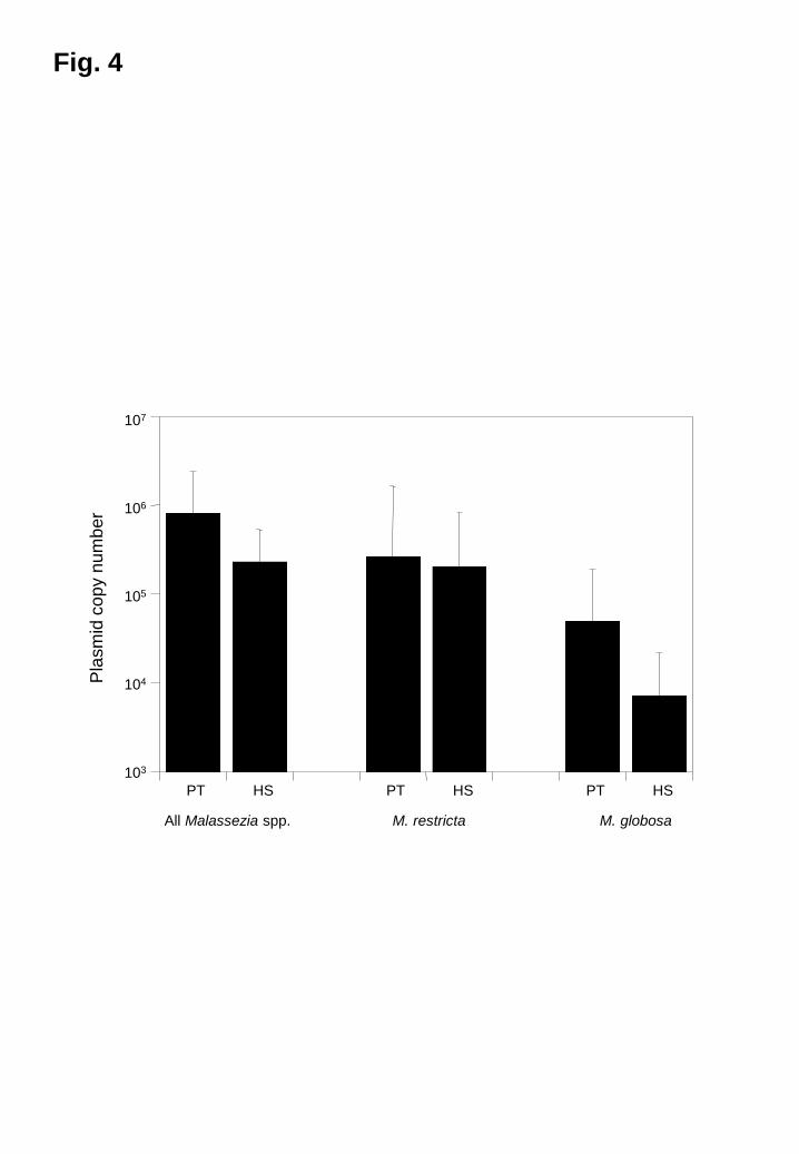
by real-time PCR. The level of colonization by all Malassezia species in patients with
dandruff was approximately three times greater than that in healthy subjects (Fig. 4).
The amounts of DNA from the two major species in the patients were also greater than
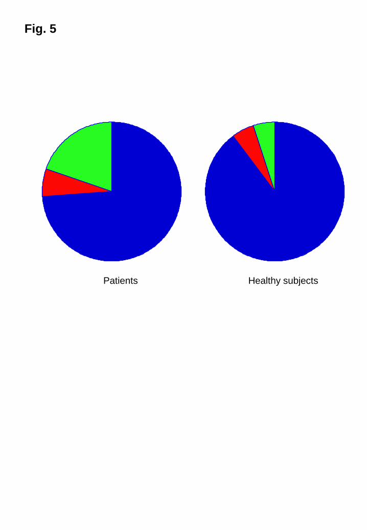
those in the healthy subjects. In dandruff patients, M. restricta and M. globosa
accounted for 74 and 6 % of all Malassezia species, respectively, and 90 and 5 % of all
Malasseia species in healthy subjects (Fig. 5). The ratio of M. restricta to all Malassezia
8
species in patients with dandruff was slightly lower than that in healthy individuals.
Analysis of Malassezia genotypes
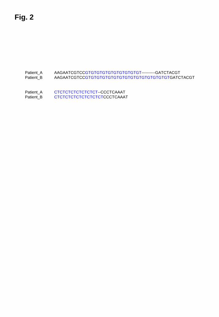
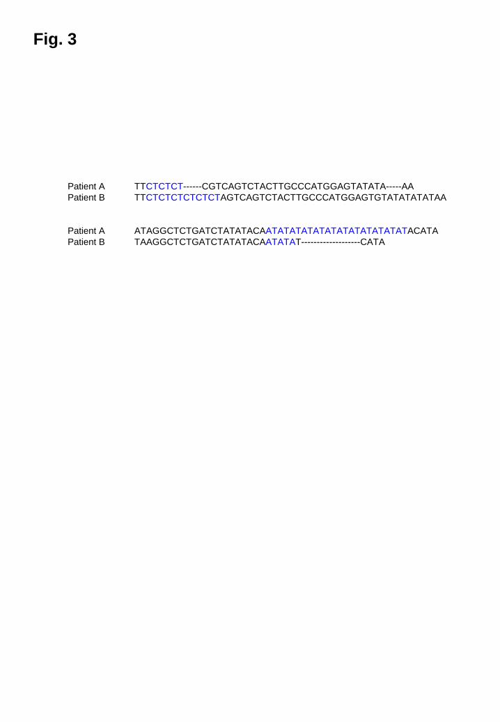
PCR products that included the GT and CT repeats of the M. globosa IGS were
approximately 300 bp in length (Fig. 2) while those that included the CT and AT
repeats of the M. restricta IGS were approximately 500 bp in length (Fig. 3).
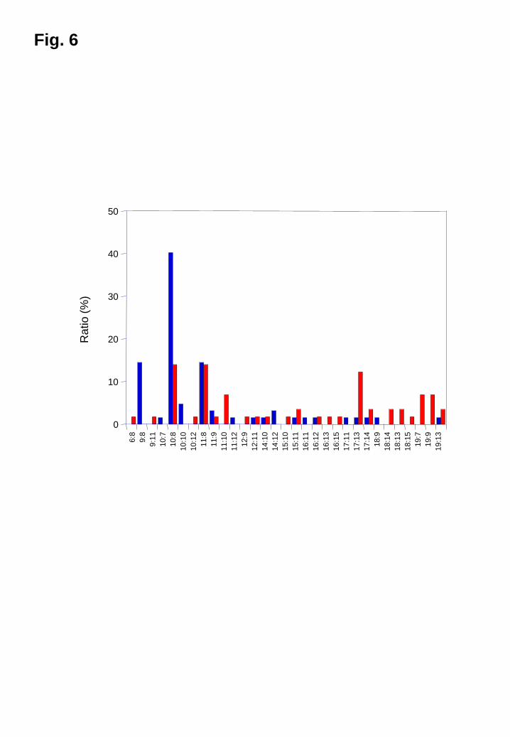
For the M. globosa IGS genotypes, 6-19 repeats were detected for GT while 6-15
repeats were detected for CT in the DNA of the patients with dandruff and healthy
individuals. A total of 31 combinations were noted. The genotype (GT)10:(CT)8 (40.3%,
25/62) was predominant followed by (GT)9:(CT)8 (14.5%, 9/62) and (GT)11:(CT)8
(14.5%, 9/62) in patients with dandruff, whereas the genotypes in the healthy subjects
were diverse. The genotypes (GT)10:(CT)8, (GT)11:(CT)8, and (GT)17:(CT)13 accounted
for 14.0% (8/57), 14.0% (8/57), and 10.5% (6/57), respectively, of the healthy subjects
(Fig. 6).
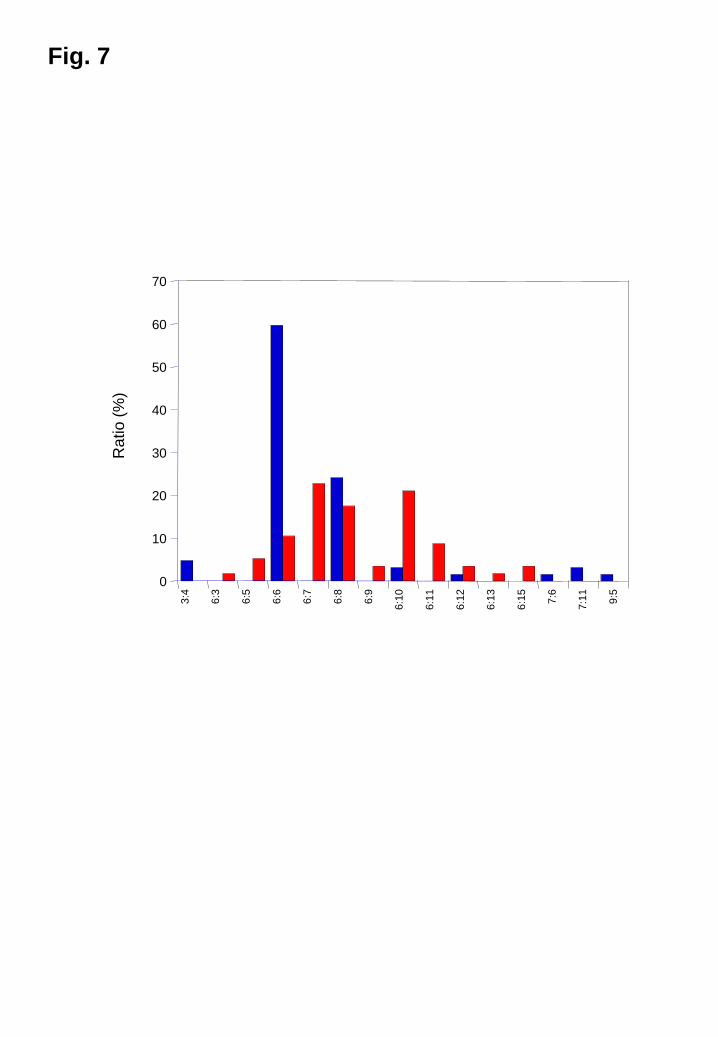
For the M. restricta IGS genotypes, 3-9 repeats were detected for CT while 3-15 repeats
were detected for AT in the DNA of the patients with dandruff and healthy individuals.
A total of 15 combinations were found. The genotype (CT)6:(AT)6 (59.7%, 37/62) was
predominant followed by (CT)6:(AT)8 (24.2%, 15/62) in patients with dandruff, while
the genotypes (CT)6:(AT)6 (10.5%, 6/57), (CT)6:(AT)7 (22.8%, 13/57), (CT)6:(AT)8
(17.5%, 10/57), and (CT)6:(AT)10 (21.1%, 12/57) accounted for 71.9% of all
9
combinations (Fig. 7).
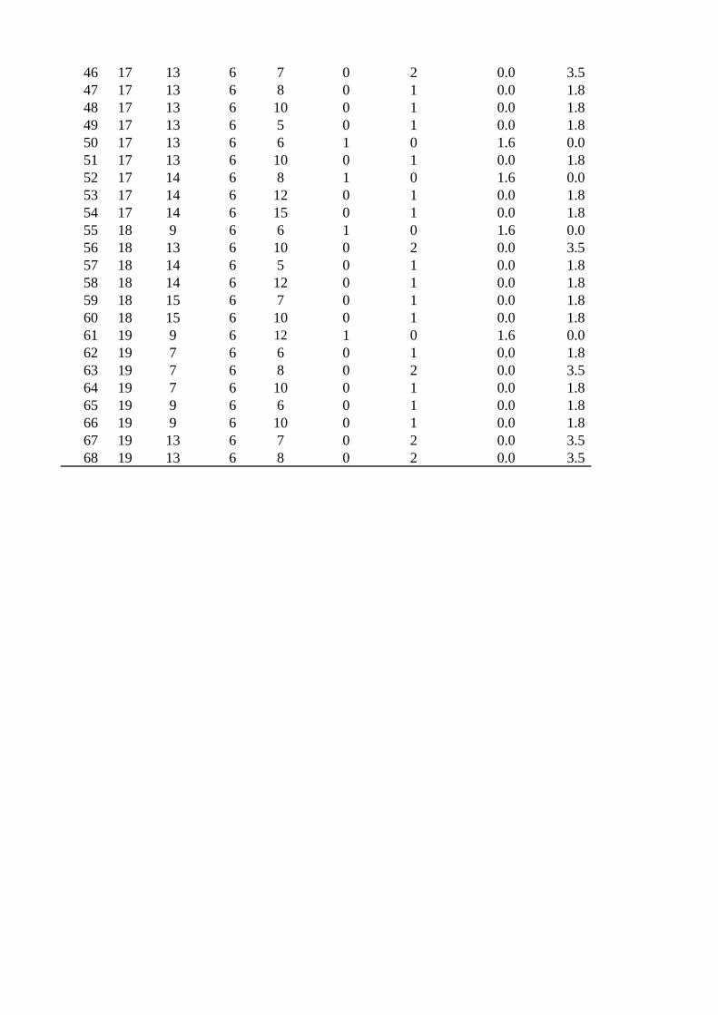
Both M. globosa and M. restricta were detected in all cases. A total of 68 combinations
of M. globosa and M. restricta genotypes were obtained from 119 cases (Table S2).
Three combinations of each genotype [30.6% (M. globosa [GT]10:[CT]8; M. restricta
[CT]6:[AT]6), 12.9% (M. globosa [GT]9:[CT]8; M. restricta [CT]6:[AT]6), and 9.7% (M.
globosa [GT]11:[CT]8; M. restricta [CT]6:[AT]8)] predominated in the dandruff patients,
while no specific genotype combination was found among the 68 combinations in
healthy subjects.
Discussion
In the present study, we found that specific genotypic strains of M. globosa and M.
restricta selectively colonized the scalps of patients with dandruff. Malassezia species
are responsible for the induction of dandruff on the scalp. In fact, scalp symptoms of
dandruff are improved by treatment with antifungal agents or pyrithione zinc shampoo
[24-26]. Gao et al. [6] determined the levels of Malassezia colonization on the forehead,
forearm, behind the ears, inner elbows, foreleg, and axillae of healthy human subjects
by qPCR, and they found that Malassezia species were predominant in the skin fungal
microbiome. In the cheek and scalp areas, the fungal microbiome consisted mainly of M.
restricta followed by M. globosa [27, 28]. In the present study, M. restricta accounted
for 98.7 ± 7.2% of the total fungal microbiome on the healthy scalp. This was also the
10
predominant microorganism over M. globosa on the patients’ scalps. The level of
Malassezia colonization on the scalps of patients should be greater than that on the
healthy scalp as patients with dandruff secrete larger amounts of sebum than healthy
subjects. In fact, the scalps of the patients showed three-fold greater levels of
Malassezia colonization compared with the scalps of the healthy individuals.
Malassezia globosa possesses 15 lipase genes in its genome [29]. This number is higher
than that in the non-lipophilic yeasts Saccharomyces cerevisiae and Cryptococcus
neoformans. The presence of multiple genes for secreted lipases suggests that this
species utilizes fatty acids from external sources as nutrients. Lee et al. [30] confirmed
the expression of several lipase genes from M. globosa and M. restricta on the scalps of
patients, and one of the M. restricta lipase genes (MRE-0242) was strongly expressed
on the patients’ scalps. Therefore, high levels of lipase production by Malassezia
species may contribute to the clinical severity of dandruff. The high level of lipase
expression may be a characteristic of this genus or a response to the chemical
composition of a patient’s sebum.
We studied the relationship between the genotypes of microorganisms and their
virulence. The rRNA gene is responsible for protein synthesis and is not directly
associated with virulence. However, the rRNA genotypes of pathogenic fungi are
correlated with their virulence factors or source of origin. Secreted aspartic protease
(SAP) is a virulence factor of the pathogenic yeast Candida albicans, and three
11
genotypes of the large subunit are correlated with SAP production ability [31]. Another
example is observed in Trichosporon asahii, which causes both deep-seated
opportunistic infections in immunocompromised hosts and summer-type
hypersensitivity pneumonitis as type III or IV allergies in healthy subjects. Of the
several T. asahii IGS genotypes, genotype I strains are infectious, while type III
strains are involved in allergic reactions [32].
With regard to Malassezia genotypes, we found that strains of specific genotypes
selectively colonized the skin of patients with atopic dermatitis [33]. That is, the
genotype of M. globosa was correlated with M. globosa-specific IgE antibody levels in
the sera of patients with atopic dermatitis. Patients were divided into two groups
according to their specific IgE antibody level: ≥ 30 IU/ml and < 30 IU/mL. In the < 30
IU/mL group, 16 genotypes were almost equivalently distributed from 2.1 to 12.5% for
each genotype, while in the > 30 IU/mL group, 92.3% of patients showed the genotype
(GT)10:(CT)8. In this study, the genotype (GT)10:(CT)8 was also predominant in patients
with dandruff (40.3%). Therefore, M. globosa genotype (GT)10:(CT)8 seems to be
common in both dandruff and atopic dermatitis. Currently, the relationship between the
M. restricta genotype and the species-specific IgE antibody level is under investigation.
Our previous study also indicated that the IGS sequences of M. restricta colonizing the
skin of seborrehic dermatitis patients consisted phylogenetically of two groups: one that
included patients with seborrehic dermatitis and another that included both patients with
12
seborrheic dermatitis and healthy subjects [21]. Although it is unclear whether the
specific genotypic strains of M. globosa and M. restricta induce or exacerbate dandruff,
or whether they selectively colonize the scalps of dandruff patients, these strains were
predominantly found on the scalps of patients when compared to healthy individuals.
This raises the question why specific genotypic strains of both microorganisms are
predominant on the scalps of patients with dandruff. This may be due to differences in
the chemical composition of scalp sebum, water content, and/or pH of the skin surface
of patients with dandruff and healthy subjects. Therapeutic agents given to dandruff
patients may also affect selective skin microbial colonization. However, none of the
patients in the present study received any antibacterial agents.
In conclusion, we found that specific genotypic strains of M. globosa and M. restricta
predominated in patients with dandruff. The virulence of these microorganisms against
the scalp is still unknown. However, the genotype of Malassezia species should be
investigated when determining the relationships between Malassezia species and
virulence against the scalp.
Conflict of interest
No conflicts of interest exist for any of the authors due to financial, commercial, or
other affiliations.
13
Legends to Figures
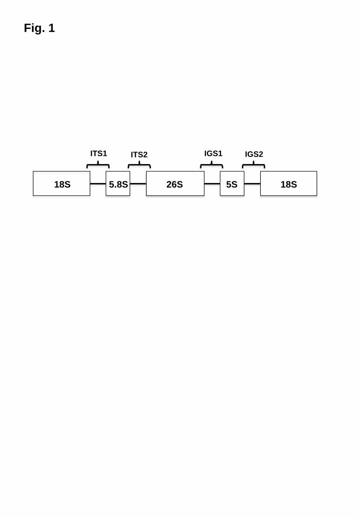
Fig. 1
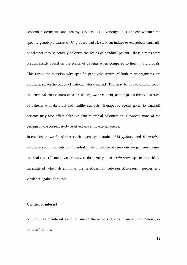
Primary structure of the fungal rRNA gene
The fungal rRNA gene consists of four subunits (18S, 5.8S, 26S, and 5S) and two
spacer regions (ITS and IGS). Approximately 100 copies are present in the genome.
ITS, internal transcribed spacer; IGS, intergenic spacer.
Fig. 2
(GT)n and (CT)n repeats in the IGS region of M. globosa
Two representative examples are shown.
Fig. 3
(CT)n and (AT)n repeats in the IGS region of M. restricta
Two representative examples are shown.
Fig. 4
The level of Malassezia colonization as determined by qPCR
The levels of all Malassezia species, M. restricta, and M. globosa were determined.
PT, patients; HS, healthy subjects.
14
Fig. 5
M. restricta and M. globosa colonization
The ratios of the levels of M. restricta and M. globosa colonization relative to all
Malassezia species are shown.
Blue, M. restricta; red, M. globosa; green; other Malassezia species
Fig. 6
Distribution of M. globosa genotypes colonizing the scalps of patients with dandruff
and healthy subjects
Blue, patients with dandruff; red, healthy subjects.
n:n = the numbers of (GT)n and (CT)n.
Fig. 7
Distribution of M. restricta genotypes colonizing the scalps of patients with dandruff
and healthy subjects
Blue, patients with dandruff; red, healthy subjects.
n:n = the numbers of (CT)n and (AT)n.
Supporting Information
15
Table S1
Subjects included in this study
Table S2
Genotypes of M. globosa and M. restricta found in this study
References
1. Schwartz JR, Messenger AG, Tosti A, Todd G, Hordinsky M, Hay RJ, Wang X,
Zachariae C, Kerr KM, Henry JP, Rust RC, Robinson MK. A comprehensive
pathophysiology of dandruff and seborrheic dermatitis - towards a more precise
definition of scalp health. Acta Derm Venereol. 2013;93:131-7.
2. DeAngelis YM, Gemmer CM, Kaczvinsky JR, Kenneally DC, Schwartz JR,
Dawson TL Jr. Three etiologic facets of dandruff and seborrheic dermatitis:
Malassezia fungi, sebaceous lipids, and individual sensitivity. J Investig Dermatol
Symp Proc. 2005;10:295-7.
3. Dawson TL Jr. Malassezia globosa and restricta: breakthrough understanding of the
etiology and treatment of dandruff and seborrheic dermatitis through whole-genome
analysis. J Investig Dermatol Symp Proc. 2007;12:15-9.
4. Grice EA, Segre JA. The skin microbiome. Nat Rev Microbiol. 2011;9:244-53.
5. Findley K, Oh J, Yang J, Conlan S, Deming C, Meyer JA, Schoenfeld D, Nomicos
16
E, Park M, Kong HH, Segre JA. Topographic diversity of fungal and bacterial
communities in human skin. Nature 2013;498:367-70.
6. Gao Z, Perez-Perez GI, Chen Y, Blaser MJ. Quantitation of major human cutaneous
bacterial and fungal populations. J Clin Microbiol . 2013;48:3575-81.
7. Sugita T, Boekhout T, Velegraki A. Epidemiology of Malassezia-related skin
diseases. In: Boekhout T, Mayser P, Guého-Kellermann E, Velegraki A, editors.
Malassezia and the Skin, New York: Springer Berlin Heidelberg; 2010. pp. 65-119.
8. Sugita T, Zhang E, Tanaka T, Tajima M, Tsuboi R, Ishibashi Y, Nishikawa A.
Atopic dermatitis and skin fungal microorganisms. In: Esparza-Gordillo J, Dkio I,
editors. Atopic dermatitis–disease etiology and clinical management. Rijeka:
InTech Open Access Company; 2012. pp. 123–140.
9. Erchiga VC, Hay RJ. Pityriasis versicolor and other Malassezia skin diseases. In:
Boekhout T, Mayser P, Guého-Kellermann E, Velegraki A, editors. Malassezia and
the Skin, New York: Springer Berlin Heidelberg; 2010. pp. 175-99.
10. Gaitanis G, Mayser P, Scheynius A, Crameri R. Malassezia yeasts in seborrheic and
atopic eczemas. In: Boekhout T, Mayser P, Guého-Kellermann E, Velegraki A,
editors. Malassezia and the Skin, New York: Springer Berlin Heidelberg; 2010. pp.
201-18.
11. Gaitanis G, Velegraki A, Mayser P, Bassukas ID. Skin diseases associated with
Malassezia yeasts: facts and controversies. Clin Dermatol . 2013;31:455-63.
17
12. Zhang E, Tanaka T, Tajima M, Tsuboi R, Nishikawa A, Sugita T. Characterization
of the skin fungal microbiota in patients with atopic dermatitis and in healthy
subjects. Microbiol Immunol. 2011;55:625-32.
13. Kaga M, Sugita T, Nishikawa A, Wada Y, Hiruma M, Ikeda S. Molecular analysis
of the cutaneous Malassezia microbiota from the skin of patients with atopic
dermatitis of different severities. Mycoses 2011;54:e24-8.
14. Takahata Y, Sugita T, Kato H, Nishikawa A, Hiruma M, Muto M. Cutaneous
Malassezia flora in atopic dermatitis differs between adults and children. Br J
Dermatol. 2007;157:1178-82.
15. Amaya M, Tajima M, Okubo Y, Sugita T, Nishikawa A, Tsuboi R. Molecular
analysis of Malassezia microflora in the lesional skin of psoriasis patients. J
Dermatol 2007;34:619-24.
16. Tajima M, Sugita T, Nishikawa A, Tsuboi R. Molecular analysis of Malassezia
microflora in seborrheic dermatitis patients: comparison with other diseases and
healthy subjects. J Invest Dermatol 2008;128:345-51.
17. Takahata Y, Sugita T, Hiruma M, Muto M. Quantitative analysis of Malassezia in
the scale of patients with psoriasis using a real-time polymerase chain reaction assay.
Br J Dermatol. 2007;157:670-3.
18. Sugita T, Tajima M, Tsubuku H, Tsuboi R, Nishikawa A. Quantitative analysis of
cutaneous Malassezia in atopic dermatitis patients using real-time PCR. Microbiol
18
Immunol. 2006;50:549-52.
19. Sugita T, Nakajima M, Ikeda R, Matsushima T, Shinoda T. Sequence analysis of the
ribosomal DNA intergenic spacer 1 regions of Trichosporon species. J Clin
Microbiol. 2002;40:1826-30.
20. Sugita T, Kodama M, Saito M, Ito T, Kato Y, Tsuboi R, Nishikawa A. Sequence
diversity of the intergenic spacer region of the rRNA gene of Malassezia globosa
colonizing the skin of patients with atopic dermatitis and healthy individuals. J Clin
Microbiol. 2003;41:3022-7.
21. Sugita T, Tajima M, Amaya M, Tsuboi R, Nishikawa A. Genotype analysis of
Malassezia restricta as the major cutaneous flora in patients with atopic dermatitis
and healthy subjects. Microbiol Immunol. 2004;48:755-9.
22. Sugita T, Takeo K, Hama K, Virtudazo E, Takashima M, Nishikawa A, Kucsera J,
Dorogi J, Komori S, Nakagaki K, Vollekova A, Slavikova E, Farkas V. DNA
sequence diversity of intergenic spacer I region in the non-lipid-dependent species
Malassezia pachydermatis isolated from animals. Med Mycol. 2005;43:21-6.
23. Diaz MR, Boekhout T, Kiesling T, Fell JW. Comparative analysis of the intergenic
spacer regions and population structure of the species complex of the pathogenic
yeast Cryptococcus neoformans. FEMS Yeast Res. 2005;5:1129-40.
24. Bulmer AC, Bulmer GS. The antifungal action of dandruff shampoos.
Mycopathologia 1999;147:63-5.
19
25. Piérard-Franchimont C, Piérard GE, Arrese JE, De Doncker P. Effect of
ketoconazole 1% and 2% shampoos on severe dandruff and seborrhoeic dermatitis:
clinical, squamometric and mycological assessments. Dermatology 2001;202:171-6.
26. Schmidt-Rose T, Braren S, Fölster H, Hillemann T, Oltrogge B, Philipp P, Weets G,
Fey S. Efficacy of a piroctone olamine/climbazol shampoo in comparison with a
zinc pyrithione shampoo in subjects with moderate to severe dandruff. Int J Cosmet
Sci. 2011;33:276-82.
27. Zhang E, Tanaka T, Tsuboi R, Makimura K, Nishikawa A, Sugita T.
Characterization of Malassezia microbiota in the human external auditory canal and
on the sole of the foot. Microbiol Immunol. 2012;56:238-44.
28. Clavaud C, Jourdain R, Bar-Hen A, Tichit M, Bouchier C, Pouradier F, El Rawadi
C, Guillot J, Ménard-Szczebara F, Breton L, Latgé JP, Mouyna I. Dandruff is
associated with disequilibrium in the proportion of the major bacterial and fungal
populations colonizing the scalp. PLoS One 2013; 8:e58203.
29. Xu J, Saunders CW, Hu P, Grant RA, Boekhout T, Kuramae EE, Kronstad JW,
Deangelis YM, Reeder NL, Johnstone KR, Leland M, Fieno AM, Begley WM, Sun
Y, Lacey MP, Chaudhary T, Keough T, Chu L, Sears R, Yuan B, Dawson TL Jr.
Dandruff-associated Malassezia genomes reveal convergent and divergent virulence
traits shared with plant and human fungal pathogens. Proc Natl Acad Sci. USA
2007;104:18730-5.
20
30. Lee YW, Lee SY, Lee Y, Jung WH. Evaluation of expression of lipases and
phospholipases of Malassezia restricta in patients with seborrheic dermatitis. Ann
Dermatol. 2013;25:310-4.
31. Sugita T, Kurosaka S, Yajitate M, Sato H, Nishikawa A. Extracellular proteinase
and phospholipase activity of three genotypic strains of a human pathogenic yeast,
Candida albicans. Microbiol Immunol. 2002;46:881-3.
32. Sugita T, Ikeda R, Nishikawa A. Analysis of Trichosporon isolates obtained from
the houses of patients with summer-type hypersensitivity pneumonitis. J Clin
Microbiol. 2004;42:5467-71.
33. Cho O, Saito M, Tsuboi R, Kato H, Nishikawa A, Nakajima S, Sugita T.
Relationships among the genotypes of Malassezia globosa colonizing patients with
atopic dermatitis, the clinical severity of the disease, and the level of specific IgE
antibodies. J Clin Experimental Dermatol Res. 2013;4:197.
5.8S 18S 26S 5S 18S
ITS1 ITS2 IGS1 IGS2
Fig. 1
Patient_A AAGAATCGTCCGTGTGTGTGTGTGTGTGTGT----------GATCTACGT Patient_B AAGAATCGTCCGTGTGTGTGTGTGTGTGTGTGTGTGTGTGTGATCTACGT Patient_A CTCTCTCTCTCTCTCT--CCCTCAAAT Patient_B CTCTCTCTCTCTCTCTCTCCCTCAAAT
Fig. 2
Patient A TTCTCTCT------CGTCAGTCTACTTGCCCATGGAGTATATA-----AA Patient B TTCTCTCTCTCTCTAGTCAGTCTACTTGCCCATGGAGTGTATATATATAA Patient A ATAGGCTCTGATCTATATACAATATATATATATATATATATATATACATA Patient B TAAGGCTCTGATCTATATACAATATAT-------------------CATA
Fig. 3
103 PT HS PT HS PT HS
Plas
mid
cop
y nu
mbe
r
104
105
106
107
All Malassezia spp. M. restricta M. globosa
Fig. 4
Patients Healthy subjects
Fig. 5
6:8
9:8
9:11
10
:7
10:8
10
:10
10:1
2 11
:8
11:9
11
:10
11:1
2 12
:9
12:1
1 14
:10
14:1
2 15
:10
15:1
1 16
:11
16:1
2 16
:13
16:1
5 17
:11
17:1
3 17
:14
18:9
18
:14
18:1
3 18
:15
19:7
19
:9
19:1
3
0
10
20
30
40
50
Fig. 6
Rat
io (%
)
3:4
6:3
6:5
6:6
6:7
6:8
6:9
6:10
6:11
6:12
6:13
6:15
7:6
7:11
9:5
0
10
20
30
40
50
60
70
Fig. 7
Rat
io (%
)
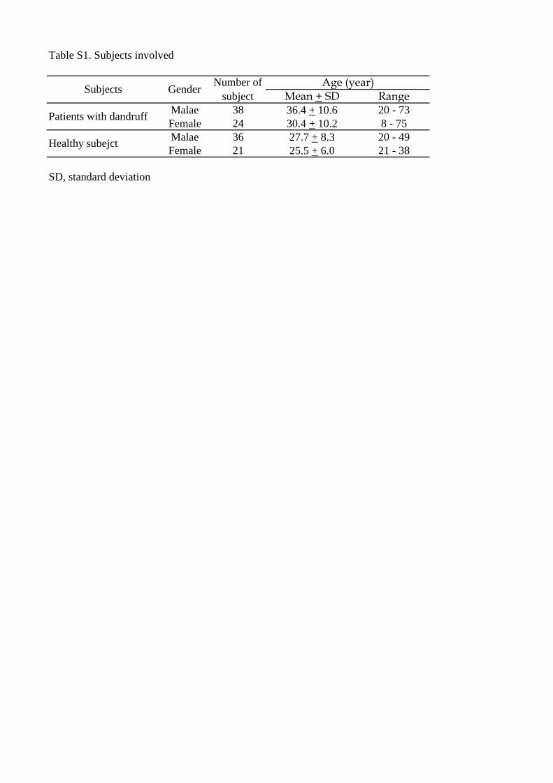
Table S1. Subjects involved
Mean + SD RangeMalae 38 36.4 + 10.6 20 - 73Female 24 30.4 + 10.2 8 - 75Malae 36 27.7 + 8.3 20 - 49Female 21 25.5 + 6.0 21 - 38
SD, standard deviation
Healthy subejct
Subjects Gender Number ofsubject
Age (year)
Patients with dandruff
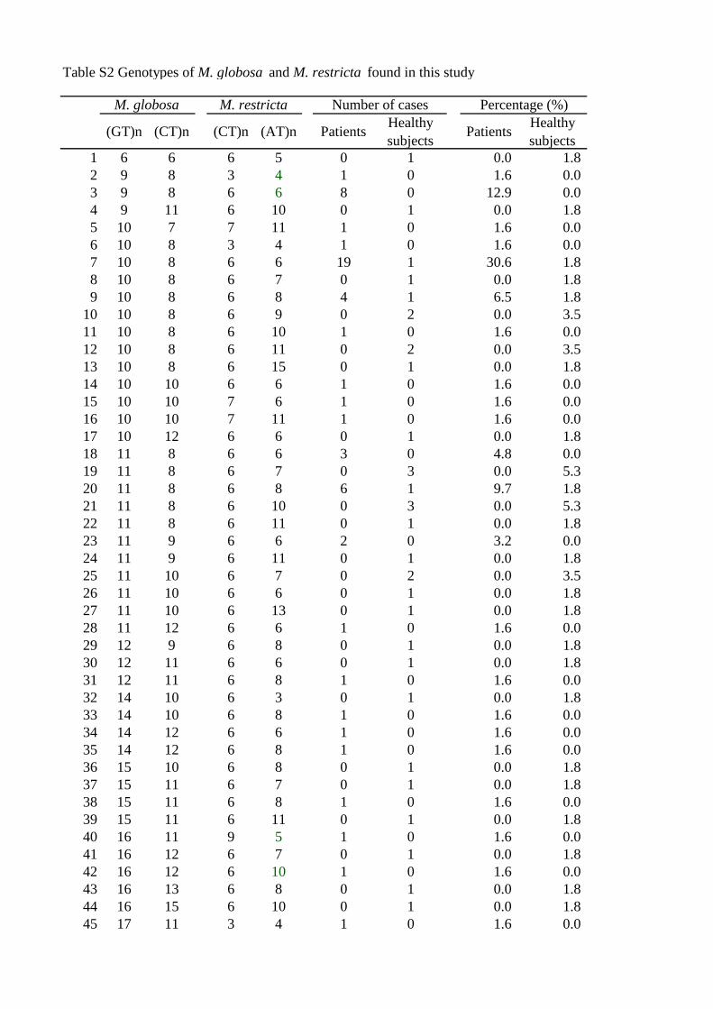
Table S2 Genotypes of M. globosa and M. restricta found in this study
(GT)n (CT)n (CT)n (AT)n Patients Healthysubjects Patients Healthy
subjects1 6 6 6 5 0 1 0.0 1.82 9 8 3 4 1 0 1.6 0.03 9 8 6 6 8 0 12.9 0.04 9 11 6 10 0 1 0.0 1.85 10 7 7 11 1 0 1.6 0.06 10 8 3 4 1 0 1.6 0.07 10 8 6 6 19 1 30.6 1.88 10 8 6 7 0 1 0.0 1.89 10 8 6 8 4 1 6.5 1.8
10 10 8 6 9 0 2 0.0 3.511 10 8 6 10 1 0 1.6 0.012 10 8 6 11 0 2 0.0 3.513 10 8 6 15 0 1 0.0 1.814 10 10 6 6 1 0 1.6 0.015 10 10 7 6 1 0 1.6 0.016 10 10 7 11 1 0 1.6 0.017 10 12 6 6 0 1 0.0 1.818 11 8 6 6 3 0 4.8 0.019 11 8 6 7 0 3 0.0 5.320 11 8 6 8 6 1 9.7 1.821 11 8 6 10 0 3 0.0 5.322 11 8 6 11 0 1 0.0 1.823 11 9 6 6 2 0 3.2 0.024 11 9 6 11 0 1 0.0 1.825 11 10 6 7 0 2 0.0 3.526 11 10 6 6 0 1 0.0 1.827 11 10 6 13 0 1 0.0 1.828 11 12 6 6 1 0 1.6 0.029 12 9 6 8 0 1 0.0 1.830 12 11 6 6 0 1 0.0 1.831 12 11 6 8 1 0 1.6 0.032 14 10 6 3 0 1 0.0 1.833 14 10 6 8 1 0 1.6 0.034 14 12 6 6 1 0 1.6 0.035 14 12 6 8 1 0 1.6 0.036 15 10 6 8 0 1 0.0 1.837 15 11 6 7 0 1 0.0 1.838 15 11 6 8 1 0 1.6 0.039 15 11 6 11 0 1 0.0 1.840 16 11 9 5 1 0 1.6 0.041 16 12 6 7 0 1 0.0 1.842 16 12 6 10 1 0 1.6 0.043 16 13 6 8 0 1 0.0 1.844 16 15 6 10 0 1 0.0 1.845 17 11 3 4 1 0 1.6 0.0
M. globosa M. restricta Number of cases Percentage (%)
46 17 13 6 7 0 2 0.0 3.547 17 13 6 8 0 1 0.0 1.848 17 13 6 10 0 1 0.0 1.849 17 13 6 5 0 1 0.0 1.850 17 13 6 6 1 0 1.6 0.051 17 13 6 10 0 1 0.0 1.852 17 14 6 8 1 0 1.6 0.053 17 14 6 12 0 1 0.0 1.854 17 14 6 15 0 1 0.0 1.855 18 9 6 6 1 0 1.6 0.056 18 13 6 10 0 2 0.0 3.557 18 14 6 5 0 1 0.0 1.858 18 14 6 12 0 1 0.0 1.859 18 15 6 7 0 1 0.0 1.860 18 15 6 10 0 1 0.0 1.861 19 9 6 12 1 0 1.6 0.062 19 7 6 6 0 1 0.0 1.863 19 7 6 8 0 2 0.0 3.564 19 7 6 10 0 1 0.0 1.865 19 9 6 6 0 1 0.0 1.866 19 9 6 10 0 1 0.0 1.867 19 13 6 7 0 2 0.0 3.568 19 13 6 8 0 2 0.0 3.5