Embed Size (px)
Citation preview
molecules
Article
Genotoxic and Cytotoxic Properties of Zinc OxideNanoparticles Phyto-Fabricated from the Obscure MorningGlory Plant Ipomoea obscura (L.) Ker Gawl
Mahadevamurthy Murali 1, Satish Anandan 2,3, Mohammad Azam Ansari 4 , Mohammad A. Alzohairy 5,Mohammad N. Alomary 6 , Sarah Mousa Maadi Asiri 7, Ahmad Almatroudi 5,* , M. C. Thriveni 8,Sudarshana Brijesh Singh 9, Hittanahallikoppal Gajendramurthy Gowtham 9, Mohammed Aiyaz 9,Chandrashekar Srinivasa 10, Asna Urooj 2 and Kestur Nagaraj Amruthesh 1,*
�����������������
Citation: Murali, M.; Anandan, S.;
Ansari, M.A.; Alzohairy, M.A.;
Alomary, M.N.; Asiri, S.M.M.;
Almatroudi, A.; Thriveni, M.C.; Singh,
S.B.; Gowtham, H.G.; et al. Genotoxic
and Cytotoxic Properties of Zinc
Oxide Nanoparticles
Phyto-Fabricated from the Obscure
Morning Glory Plant Ipomoea obscura
(L.) Ker Gawl. Molecules 2021, 26, 891.
https://doi.org/10.3390/
molecules26040891
Academic Editor: Nicola Micale
Received: 29 December 2020
Accepted: 31 January 2021
Published: 8 February 2021
Publisher’s Note: MDPI stays neutral
with regard to jurisdictional claims in
published maps and institutional affil-
iations.
Copyright: © 2021 by the authors.
Licensee MDPI, Basel, Switzerland.
This article is an open access article
distributed under the terms and
conditions of the Creative Commons
Attribution (CC BY) license (https://
creativecommons.org/licenses/by/
4.0/).
1 Applied Plant Pathology Laboratory, Department of Studies in Botany, University of Mysore,Manasagangotri, Mysore 570006, Karnataka, India; [email protected]
2 Department of Studies in Food Science and Nutrition, University of Mysore,Manasagangotri, Mysore 570006, Karnataka, India; [email protected] (S.A.);[email protected] (A.U.)
3 Department of Clinical Nutrition and Dietetics, Sri Devaraj Urs Academy of Higher Education and Research,Kolar 563101, Karnataka, India
4 Department of Epidemic Disease Research, Institutes for Research and Medical Consultations (IRMC),Imam Abdulrahman Bin Faisal University, Dammam 31441, Saudi Arabia; [email protected]
5 Department of Medical Laboratories, College of Applied Medical Sciences, Qassim University,Qassim 51431, Saudi Arabia; [email protected]
6 National Center for Biotechnology, Life Science and Environmental Research Institute, King Abdulaziz Cityfor Science and Technology, Riyadh P.O. Box 6086, Riyadh 11442, Saudi Arabia; [email protected]
7 Department of Biophysics, Institute for Research and Medical Consultations (IRMC), Imam Abdulrahman BinFaisal University, Dammam 31441, Saudi Arabia; [email protected]
8 Central Sericultural Germplasm Resources Centre, Central Silk Board, Ministry of Textiles, Thally Road,TVS Nagar, Hosur 635109, Tamil Nadu, India; [email protected]
9 Department of Studies in Biotechnology, University of Mysore, Manasagangotri,Mysore 570006, Karnataka, India; [email protected] (S.B.S.);[email protected] (H.G.G.); [email protected] (M.A.)
10 Department of Studies in Biotechnology, Davangere University, Davangere 577007, Karnataka, India;[email protected]
* Correspondence: [email protected] (A.A.); [email protected] (K.N.A.)
Abstract: The study was undertaken to investigate the antioxidant, genotoxic, and cytotoxic poten-tialities of phyto-fabricated zinc oxide nanoparticles (ZnO-NPs) from Ipomoea obscura (L.) Ker Gawl.aqueous leaf extract. The UV-visible spectral analysis of the ZnO-NPs showed an absorption peak at304 nm with a bandgap energy of 3.54 eV, which are characteristics of zinc nanoparticles. Moreover,the particles were of nano-size (~24.26 nm) with 88.11% purity and were agglomerated as observedthrough Scanning Electron Microscopy (SEM). The phyto-fabricated ZnO-NPs offered radical scav-enging activity (RSA) in a dose-dependent manner with an IC50 of 0.45 mg mL−1. In addition, thegenotoxicity studies of ZnO-NPs carried out on onion root tips revealed that the particles were ableto significantly inhibit the cell division at the mitotic stage with a mitotic index of 39.49%. Further,the cytotoxic studies on HT-29 cells showed that the phyto-fabricated ZnO-NPs could arrest the celldivision as early as in the G0/G1 phase (with 92.14%) with 73.14% cells showing early apoptoticsymptoms after 24 h of incubation. The results of the study affirm the ability of phyto-fabricatedZnO-NPs from aqueous leaf extract of I. obscura is beneficial in the cytotoxic application.
Keywords: cytotoxicity; genotoxicity; cell cycle analysis by flow cytometry; ZnO-NPs; HT-29 cells;Allium cepa; Ipomoea obscura
Molecules 2021, 26, 891. https://doi.org/10.3390/molecules26040891 https://www.mdpi.com/journal/molecules

Molecules 2021, 26, 891 2 of 16
1. Introduction
Nanotechnology is a multidimensional discipline that has the potential to transformall the fields of science. Nanoparticles have gained importance due to their surface area tovolume ratio and the behaviors that result from that [1,2]. In light of the environmental andbiological risks due to the toxicity of used chemicals in synthetic metal–oxide nanoparticles,biologically synthesized nanoparticles have gained considerable importance as they areconsidered to be stable, eco-friendly, and also possess biological properties [3–6]. Differentmethods have been employed for the synthesis of nanoparticles, of which the hydrothermalmethod is one of the alternatives to the synthetic methods, as it is simple, cost-effective,eco-friendly, and less hazardous [7–9]. Zinc oxide nanoparticles (ZnO-NPs) have gainedmore attention compared to other metal–oxide nanoparticles because of their broaderapplications in scientific fields, including biological applications [8,10,11]. Apart frommicroorganisms, plant extracts have been used as reducing or capping agents as these playa pivotal and versatile role during the synthesis of nanoparticles, which are essential fortheir functions and applications in various fields [7,12–14].
It has been well documented in the literature that the chemical synthesis of nanoparti-cles yields toxic byproducts that are not eco-friendly and cost-effective, and researchers areconcentrating on alternative methods [4–6]. Biosynthesis of nanoparticles using plant ex-tract has gained interest due to its abundance of sources available in the environment [8,9].Plants have a rich source of secondary metabolites present in them, which act as reduc-ing/capping agents during the synthesis of nanoparticles [6,12,13]. In the recent past,ZnO-NPs have been synthesized from every part of the plant, such as leaves [8], stem [15],tuber [16], root [14], flower [7], fruit [17], and seed [18]. They have also shown to possessmany biological properties such as antioxidant, antimicrobial, genotoxicity, cytotoxicity,etc., thereby indicating the efficacy toward their application in the field of pharmaceuti-cals [5,8,9,14,15]. The toxicity of chemically synthesized nanoparticles has paved the wayfor plant-mediated biosynthesis of nanoparticles due to their stable and eco-friendly naturein addition to their enhanced biological properties compared to chemically synthesizednanoparticles [2,5,8,9].
Ipomea spp. have been in continuous use in nutrition, medicine, rituals, and agricul-ture [19]. These plant species possess antimicrobial, spasmolytic, analgesic, psychometric,hypotensive, and anticancer activities. The plant is also used to cure inflammation, con-stipation, kidney ailments, digestive disorders, etc. [19,20]. From the literature, it may benoted that a few studies have been reported on the synthesis of metal nanoparticles fromIpomea spp. which have been evaluated for antioxidant and cytotoxic effects [21]; however,to date, no report on the phyto-fabrication of ZnO-NPs from Ipomea obscura has been made.Hence, a study on antioxidant, genotoxic, and cytotoxic potentialities of phyto-fabricatedzinc oxide nanoparticles (ZnO-NPs) from I. obscura aqueous leaf extract was evaluated.
2. Materials and Methods2.1. Collection and Phyto-Fabrication of Zinc Oxide Nanoparticles from I. obscura
Healthy leaves of Ipomea obscura (L.) Ker Gawl. were collected from Manasagangotri,Mysuru, Karnataka, India and authenticated at the Dept. of Studies in Botany, Universityof Mysore, Mysuru. Phyto-fabrication of ZnO-NPs from I. obscura leaves was carried outaccording to our previous studies with modifications [8]. In brief, about 25 g of freshleaves of the plant was collected, washed, and blot dried. The collected sample was thenblended in a blender with 250 mL of distilled water and filtered (Whatman No. 1 filterpaper). Further, 25 mL of the filtrate was heated to 80 ◦C (on a magnetic stirrer) and2.5 g of zinc nitrate hexahydrate was added with constant stirring, and the reaction mixturewas stirred until the mixture became paste. The obtained sample was then transferred to asilica furnace and calcinated at 300 ◦C for 2 h, and the obtained product (ZnO-NPs) wasstored in air-tight vials until further use.
Molecules 2021, 26, 891 3 of 16
2.2. Physico-Chemical Characterization of ZnO-NPs
The UV-Vis Spectrophotometer (DU730, Beckman Coulter, Krefeld, Germany) wasused to determine the optical density of the phyto-fabricated ZnO-NPs. X-Ray Diffraction(XRD) study was performed out on an X-Ray Diffractometer (Rigaku SmartLab, Tokyo,Japan), and Scherrer’s formula was applied to determine the particle size. The DynamicLight Scattering (DLS) analysis was also carried out to learn the particle size distributionof ZnO-NPs using the Nanotrack Wave particle size analyzer (Microtrack, PA, USA).The morphology of the nanoparticles was evaluated by Scanning Electron Microscopy(HITACHI-S-3400N, Tokyo, Japan) at 5 kV acceleration. The phyto-fabricated ZnO-NPswere placed on a carbon-coated copper in a tiny amount, allowed to air-dry, and imageswere taken. The elemental analysis (qualitative as well as quantitative) was carried out byEnergy Dispersive Spectroscopy (EDS) (Noran System 7, Thermoscientific, WI, USA). Thebinding properties of the ZnO-NPs and aqueous extract of I. obscura was investigated by FT-IR with a resolution of 4 cm−1 between 4000 to 400 cm−1 spectral range on a PerkinElmerSpectrum ATR2000 (Singapore).
2.3. Evaluation of Phyto-Fabricated ZnO-NPs for Biological Potentialities2.3.1. 2,2-diphenyl-1-picrylhydrazyl (DPPH) Radical Scavenging Activity
The radical scavenging activity (RSA) of phyto-fabricated ZnO-NPs was performedby following the method of Hemanth Kumar et al. [5]. In brief, 3.5 mL of 0.1 mM DPPHcontaining 0.2, 0.4, 0.6, 0.8, and 1 mg mL−1 concentrations of ZnO-NPs were sonicatedbefore incubating for 30 min at 37 ± 2 ◦C (under dark conditions) and the absorbance wasmeasured at 517 nm in a spectrophotometer. The percentage of RSA of the nanoparticleswas determined.
Radical Scavenging Activity (%) =A − bsorbance of Control − Absorbance of Test sample
A − bsorbance of Control× 100 (1)
2.3.2. Genotoxic Analysis of Phyto-Fabricated ZnO-NPs by the Allium cepa Method
The genotoxicity of ZnO-NPs was determined in the root tips of healthy onion bulbs [5].Onion bulbs with fresh roots (2–3 cm long) grown on glass vials were transferred to glassvials containing phyto-fabricated ZnO-NPs (0.2, 0.4, 0.6, 0.8, and 1 mg mL−1) and weresubjected to incubation for 24 h at room temperature. The ZnO-NPs treated onion root tipswere watchfully cut out and fixed in Carnoy’s Fixative II (for 24 h) and later relocated in70% ethanol. Later, the root tips were taken and squashed, and the cells were observedfor cell division and, also, to record the mitotic index. Sterile Distilled Water (SDW)and Methotrexate treated root tips were designated as the negative and positive control,respectively. A total of ten microscopic fields per root sample were observed with fourroots per treatment.
Mitotic Index (%) =Number of dividing cells
Total number of cells× 100 (2)
2.3.3. Cytotoxic Analysis of Phyto-Fabricated ZnO-NPs3-(4,5-dimethylthiazol-2-yl)-2,5-diphenyl tetrazolium bromide (MTT) Assay
MTT assay was carried out to identify the anticancer properties of phyto-fabricatedZnO-NPs according to the method of Selvakumaran et al. [22]. Cell lines (HT-29) (procuredfrom NCCS, Pune, India) of 80% confluent cells were trypsinized. Each well was seededwith 100 µL of cell suspension (5 × 10⁵) in Dulbecco’s modified Eagle’s medium (DMEM)and further incubated for 24 h at 37 ◦C (humidified atmosphere containing 5% CO2). Afterincubation, the media were discarded, and each well was loaded with 150 µL of freshDMEM media containing ZnO-NPs (0.2, 0.4, 0.6, 0.8, and 1 mg) with fetal bovine serum(10%) and incubated for 24 h. The incubated samples were aspirated and 100 µL of MTTsolution (0.5 mg mL−1 in 1× PBS filtered through 0.2 µM filter) was added and subjectedto incubation for 4 h. From the incubated samples, MTT reagent was removed and DMSO

Molecules 2021, 26, 891 4 of 16
(100 µL) was added to solubilize formazan rapidly. SDW and colchicine treated cells weredesignated as a negative and positive control, respectively. Each sample was subjected toUV-Vis spectroscopy at 570 nm, and the percentage of cell viability was calculated.
Cell Viability (%) =Absorbance of sampleAbsorbance of control
× 100 (3)
Cell Cycle Analysis
About 3 mL well−1 of HT-29 cell suspension (5 × 10⁵) was loaded into each well of a6-well plate and subjected to incubation at 37 ◦C for 24 h (5% CO2). After incubation, thegrowth media were discarded by aspiration and reloaded with 1 mL of phyto-fabricatedZnO-NPs (1 mg) and colchicine (320 µg) before subjecting to incubation in humified condi-tions. The treated cells were detached by trypsinization and subjected to centrifugationfor 10 min at 2000 rpm and repeatedly washed with PBS. Later, the cell pellet was fixedwith ethanol (700 µL) and further incubated (for 1 h at −20 ◦C). Subsequently, the cellswere washed twice with PBS (ice cold) by cold centrifugation for 10 min at 4000 rpm.The resultant cell pellet was resuspended in PBS (1 mL) containing propidium iodide(50 mg mL−1), RNase A (50 mg mL−1), and Triton X-100 (0.1%) and incubated for 30 minunder dark conditions before analyzing using a flow cytometer (Cell Lab Quanta™, SC,Beckman Coulter, CA, USA).
Annexin V-FITC Staining Assay
The phyto-fabricated ZnO-NPs treated (1 mg) and respective control cells, as men-tioned above, were subjected to AnnexinV-FITC staining by using Annexin V-FITC Apop-tosis Detection Kit (Invitrogen, USA) following the manufacturer’s protocol. In brief, theZnO-NP treated and untreated cells were resuspended in binding buffer (100 µL) contain-ing PI (10 µL) and subjected to incubation for 5 min (dark conditions). The stained cellsamples were further subjected to analysis by a flow cytometer (Cell Lab Quanta™, SC,Beckman Coulter, CA, USA).
Analysis of Cell Morphology
Morphological changes in HT-29 cells upon treatment with phyto-fabricated ZnO-NPs were performed on a 6 well-plate. Each well was seeded with 1 mL of DMEMmedia containing HT-29 cells (1 × 10⁵ cells mL−1) and incubated for 24 h. The HT-29 cellsuspension was discarded after incubation by aspiration and loaded with 1 mL of freshDMEM media containing ZnO-NPs (1 mg mL−1). The treated samples were incubated atroom temperature for 24 h aseptically. After incubation, the cellular morphology changeswere observed using phase-contrast inverted microscopy at 20X magnification (Zeiss AxioVert. A1, Jena, Germany).
2.4. Statistical Analysis
The experimental studies were carried out with four replicates for each experiment.They were statistically analyzed by subjecting to arcsine transformation and analysis ofvariance (ANOVA) using SPSS, version 17 (SPSS Inc., Chicago, IL, USA). The significant dif-ferences between the treatment mean values were determined by the Honestly SignificantDifference (HSD) obtained by Tukey’s test at p ≤ 0.05 levels.
3. Results and Discussion3.1. Physico-Chemical Characterization of Phyto-Fabricated ZnO-NPs
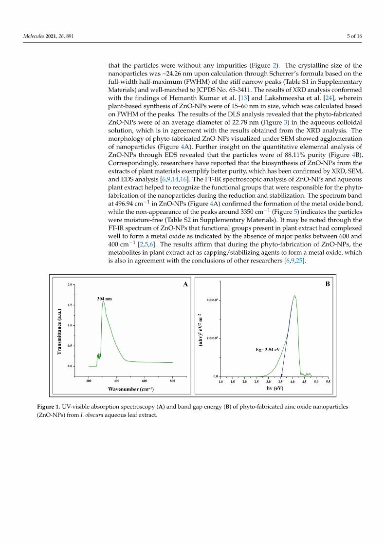
The phyto-fabricated ZnO-NPs offered an absorption peak of 304 nm (Figure 1A) withan energy band gap of 3.54 eV (Figure 1B). The spectral analysis results agreed with theresults of many other studies in which the absorption peak and bandgap energy of phyto-fabricated ZnO nanoparticles were between 280 to 400 nm and 3.2 eV to 3.5 eV, respectively,which is crucial for their applications in biological and pharmaceutical fields [23]. TheXRD analysis of nanoparticles revealed stiff narrow diffraction peaks, thereby confirming
Molecules 2021, 26, 891 5 of 16
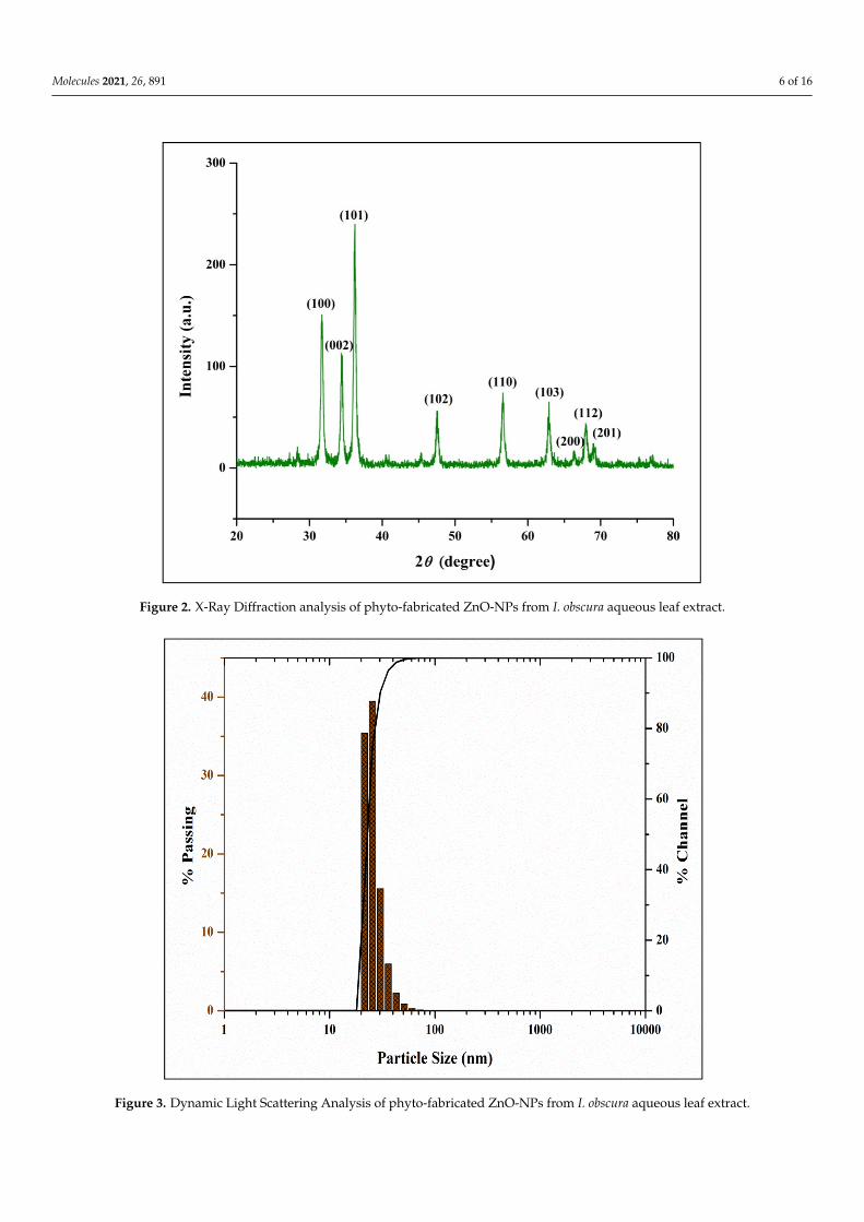
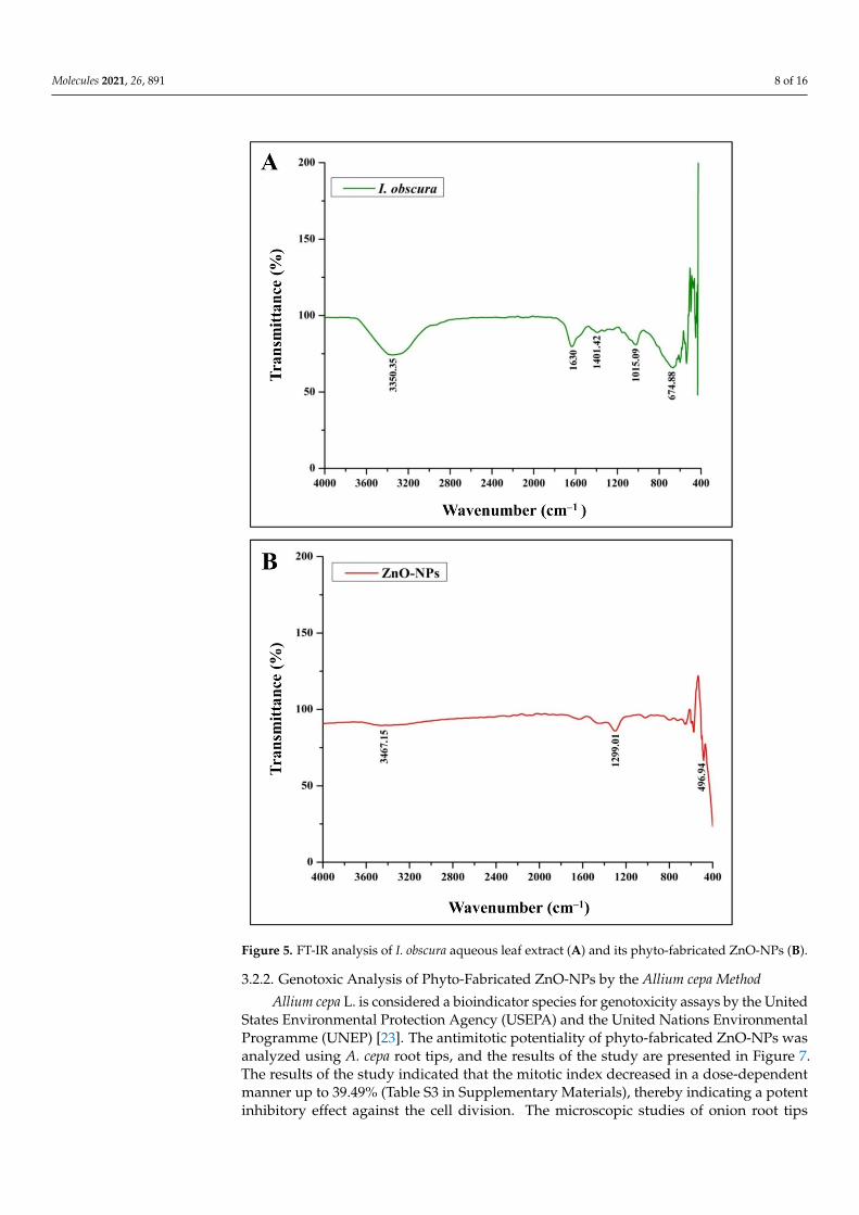
that the particles were without any impurities (Figure 2). The crystalline size of thenanoparticles was ~24.26 nm upon calculation through Scherrer’s formula based on thefull-width half-maximum (FWHM) of the stiff narrow peaks (Table S1 in SupplementaryMaterials) and well-matched to JCPDS No. 65-3411. The results of XRD analysis conformedwith the findings of Hemanth Kumar et al. [13] and Lakshmeesha et al. [24], whereinplant-based synthesis of ZnO-NPs were of 15–60 nm in size, which was calculated basedon FWHM of the peaks. The results of the DLS analysis revealed that the phyto-fabricatedZnO-NPs were of an average diameter of 22.78 nm (Figure 3) in the aqueous colloidalsolution, which is in agreement with the results obtained from the XRD analysis. Themorphology of phyto-fabricated ZnO-NPs visualized under SEM showed agglomerationof nanoparticles (Figure 4A). Further insight on the quantitative elemental analysis ofZnO-NPs through EDS revealed that the particles were of 88.11% purity (Figure 4B).Correspondingly, researchers have reported that the biosynthesis of ZnO-NPs from theextracts of plant materials exemplify better purity, which has been confirmed by XRD, SEM,and EDS analysis [6,9,14,16]. The FT-IR spectroscopic analysis of ZnO-NPs and aqueousplant extract helped to recognize the functional groups that were responsible for the phyto-fabrication of the nanoparticles during the reduction and stabilization. The spectrum bandat 496.94 cm−1 in ZnO-NPs (Figure 4A) confirmed the formation of the metal oxide bond,while the non-appearance of the peaks around 3350 cm−1 (Figure 5) indicates the particleswere moisture-free (Table S2 in Supplementary Materials). It may be noted through theFT-IR spectrum of ZnO-NPs that functional groups present in plant extract had complexedwell to form a metal oxide as indicated by the absence of major peaks between 600 and400 cm−1 [2,5,6]. The results affirm that during the phyto-fabrication of ZnO-NPs, themetabolites in plant extract act as capping/stabilizing agents to form a metal oxide, whichis also in agreement with the conclusions of other researchers [6,9,25].
Molecules 2020, 25, x FOR PEER REVIEW 5 of 16
phyto-fabricated ZnO nanoparticles were between 280 to 400 nm and 3.2 eV to 3.5 eV, respectively, which is crucial for their applications in biological and pharmaceutical fields [23]. The XRD analysis of nanoparticles revealed stiff narrow diffraction peaks, thereby confirming that the particles were without any impurities (Figure 2). The crys-talline size of the nanoparticles was ~24.26 nm upon calculation through Scherrer’s for-mula based on the full-width half-maximum (FWHM) of the stiff narrow peaks (Table S1 in Supplementary Materials) and well-matched to JCPDS No. 65-3411. The results of XRD analysis conformed with the findings of Hemanth Kumar et al. [13] and Lakshmeesha et al. [24], wherein plant-based synthesis of ZnO-NPs were of 15–60 nm in size, which was calculated based on FWHM of the peaks. The results of the DLS analysis revealed that the phyto-fabricated ZnO-NPs were of an average diameter of 22.78 nm (Figure 3) in the aqueous colloidal solution, which is in agreement with the results obtained from the XRD analysis. The morphology of phyto-fabricated ZnO-NPs visualized under SEM showed agglomeration of nanoparticles (Figure 4A). Further insight on the quantitative elemental analysis of ZnO-NPs through EDS revealed that the particles were of 88.11% purity (Figure 4B). Correspondingly, researchers have reported that the biosynthesis of ZnO-NPs from the extracts of plant materials exemplify better purity, which has been confirmed by XRD, SEM, and EDS analysis [6,9,14,16]. The FT-IR spectroscopic analysis of ZnO-NPs and aqueous plant extract helped to recognize the functional groups that were responsible for the phyto-fabrication of the nanoparticles during the reduction and stabilization. The spectrum band at 496.94 cm−1 in ZnO-NPs (Figure 4A) confirmed the formation of the metal oxide bond, while the non-appearance of the peaks around 3350 cm−1 (Figure 5) indicates the particles were moisture-free (Table S2 in Supplementary Materials). It may be noted through the FT-IR spectrum of ZnO-NPs that functional groups present in plant extract had complexed well to form a metal oxide as indicated by the absence of major peaks between 600 and 400 cm−1 [2,5,6]. The results affirm that dur-ing the phyto-fabrication of ZnO-NPs, the metabolites in plant extract act as cap-ping/stabilizing agents to form a metal oxide, which is also in agreement with the con-clusions of other researchers [6,9,25].
Figure 1. UV-visible absorption spectroscopy (A) and band gap energy (B) of phyto-fabricated zinc oxide nanoparticles (ZnO-NPs) from I. obscura aqueous leaf extract. Figure 1. UV-visible absorption spectroscopy (A) and band gap energy (B) of phyto-fabricated zinc oxide nanoparticles(ZnO-NPs) from I. obscura aqueous leaf extract.
Molecules 2021, 26, 891 6 of 16
Molecules 2020, 25, x FOR PEER REVIEW 6 of 16
Figure 2. X-Ray Diffraction analysis of phyto-fabricated ZnO-NPs from I. obscura aqueous leaf ex-tract.
Figure 3. Dynamic Light Scattering Analysis of phyto-fabricated ZnO-NPs from I. obscura aqueous leaf extract.
Figure 2. X-Ray Diffraction analysis of phyto-fabricated ZnO-NPs from I. obscura aqueous leaf extract.
Molecules 2020, 25, x FOR PEER REVIEW 6 of 16
Figure 2. X-Ray Diffraction analysis of phyto-fabricated ZnO-NPs from I. obscura aqueous leaf ex-tract.
Figure 3. Dynamic Light Scattering Analysis of phyto-fabricated ZnO-NPs from I. obscura aqueous leaf extract.
Figure 3. Dynamic Light Scattering Analysis of phyto-fabricated ZnO-NPs from I. obscura aqueous leaf extract.

Molecules 2021, 26, 891 7 of 16
Molecules 2020, 25, x FOR PEER REVIEW 7 of 16
Figure 4. Scanning Electron Micrograph (A) and Energy Dispersive Spectra (B) of phyto-fabricated ZnO-NPs.
Figure 4. Scanning Electron Micrograph (A) and Energy Dispersive Spectra (B) of phyto-fabricated ZnO-NPs.
3.2. Evaluation of Phyto-Fabricated ZnO-NPs for Biological Potentialities3.2.1. DPPH Radical Scavenging Activity
The percent RSA of ZnO-NPs from I. obscura increases with an increase in the particles’dose with a half-maximal inhibitory concentration (IC50) of 0.45 mg mL−1 (Figure 6). Incomparison, ascorbic acid (positive control) possessed 75% inhibition at 50 µg mL−1. Ithas been well-documented that the hydroxyl free radicals result in DNA damage andlipid peroxidation, which is directly correlated to cancer-related complications [5]. Theresults obtained from the study are in corroboration with the findings of other researchersin which ZnO-NPs synthesized using biological route possessed effective antioxidantactivity [5,26]. The antioxidant nature of the green synthesized ZnO-NPs may be correlatedto the secondary metabolites that act as agents of capping/reducing/stabilizing duringnanoparticle synthesis [10,21].
Molecules 2021, 26, 891 8 of 16
Molecules 2020, 25, x FOR PEER REVIEW 8 of 16
Figure 5. FT-IR analysis of I. obscura aqueous leaf extract (A) and its phyto-fabricated ZnO-NPs (B).
3.2. Evaluation of Phyto-Fabricated ZnO-NPs for Biological Potentialities 3.2.1. DPPH Radical Scavenging Activity
The percent RSA of ZnO-NPs from I. obscura increases with an increase in the parti-cles' dose with a half-maximal inhibitory concentration (IC50) of 0.45 mg mL−1 (Figure 6). In comparison, ascorbic acid (positive control) possessed 75% inhibition at 50 µg mL−1. It has been well-documented that the hydroxyl free radicals result in DNA damage and li-pid peroxidation, which is directly correlated to cancer-related complications [5]. The results obtained from the study are in corroboration with the findings of other research-ers in which ZnO-NPs synthesized using biological route possessed effective antioxidant
Figure 5. FT-IR analysis of I. obscura aqueous leaf extract (A) and its phyto-fabricated ZnO-NPs (B).
3.2.2. Genotoxic Analysis of Phyto-Fabricated ZnO-NPs by the Allium cepa Method
Allium cepa L. is considered a bioindicator species for genotoxicity assays by the UnitedStates Environmental Protection Agency (USEPA) and the United Nations EnvironmentalProgramme (UNEP) [23]. The antimitotic potentiality of phyto-fabricated ZnO-NPs wasanalyzed using A. cepa root tips, and the results of the study are presented in Figure 7.The results of the study indicated that the mitotic index decreased in a dose-dependentmanner up to 39.49% (Table S3 in Supplementary Materials), thereby indicating a potentinhibitory effect against the cell division. The microscopic studies of onion root tips

Molecules 2021, 26, 891 9 of 16
exposed to ZnO-NPs showed chromosomal abnormalities in more cells than untreatedcontrol root tips, where typical cell divisions were observed (Figure 8). The inhibition ofcell division upon exposure to ZnO-NPs affirms the genotoxic nature of the nanoparticles(cell arrest in mitosis eventually leading to apoptosis) and conforms with the findingsof other researchers wherein ZnO-NPs synthesized from plant extracts offered effectiveantimitotic properties [13,23,27]. Further, in our previous studies, we had evaluated theeffect of commercially available zinc oxide for their genotoxicity and found that there wasno significant genotoxic effect of the treatment, but normal cell divisions were noticed evenafter treating at 2.5 mg mL−1 concentration [12]. It has been widely reported that ZnO-NPssynthesized using plant extracts with genotoxic nature will also have potential anticancerproperties [28,29].
Molecules 2020, 25, x FOR PEER REVIEW 9 of 16
activity [5,26]. The antioxidant nature of the green synthesized ZnO-NPs may be corre-lated to the secondary metabolites that act as agents of capping/reducing/stabilizing during nanoparticle synthesis [10,21].
Figure 6. DPPH radical scavenging activity of phyto-fabricated ZnO-NPs from I. obscura aqueous leaf extract. Each value is the mean for three replicates (n = 3), and bars sharing the same letters are not significantly different (p ≤ 0.05) according to Tukey’s Honestly Significant Difference (HSD). The vertical bar indicates the standard error.
3.2.2. Genotoxic Analysis of Phyto-Fabricated ZnO-NPs by the Allium Cepa Method Allium cepa L. is considered a bioindicator species for genotoxicity assays by the
United States Environmental Protection Agency (USEPA) and the United Nations Envi-ronmental Programme (UNEP) [23]. The antimitotic potentiality of phyto-fabricated ZnO-NPs was analyzed using A. cepa root tips, and the results of the study are presented in Figure 7. The results of the study indicated that the mitotic index decreased in a dose-dependent manner up to 39.49% (Table S3 in Supplementary Materials), thereby indicating a potent inhibitory effect against the cell division. The microscopic studies of onion root tips exposed to ZnO-NPs showed chromosomal abnormalities in more cells than untreated control root tips, where typical cell divisions were observed (Figure 8). The inhibition of cell division upon exposure to ZnO-NPs affirms the genotoxic nature of the nanoparticles (cell arrest in mitosis eventually leading to apoptosis) and conforms with the findings of other researchers wherein ZnO-NPs synthesized from plant extracts offered effective antimitotic properties [13,23,27]. Further, in our previous studies, we had evaluated the effect of commercially available zinc oxide for their genotoxicity and found that there was no significant genotoxic effect of the treatment, but normal cell di-visions were noticed even after treating at 2.5 mg mL−1 concentration [12]. It has been widely reported that ZnO-NPs synthesized using plant extracts with genotoxic nature will also have potential anticancer properties [28,29].
Figure 6. DPPH radical scavenging activity of phyto-fabricated ZnO-NPs from I. obscura aqueous leaf extract. Each value isthe mean for three replicates (n = 3), and bars sharing the same letters are not significantly different (p ≤ 0.05) according toTukey’s Honestly Significant Difference (HSD). The vertical bar indicates the standard error.
3.2.3. Cytotoxic Analysis of Phyto-Fabricated ZnO-NPsMTT Assay
The cytotoxic effect of ZnO-NPs from I. obscura was evaluated by MTT assay againstHT-29 cells. The assay studied is considered to be one of the most reliable methods to pri-marily assess the effect of compounds on cell lines [30]. The phyto-fabricated ZnO-NPs gavedose-dependent cytotoxicity wherein the cell viability decreased from 88.32% to 43.17%(0.2 to 1 mg mL−1) with an IC50 value of 0.91 mg mL−1. The standard drug colchicineoffered around 70% cytotoxicity at 100 µg mL−1. A significant rise in the percent cellinhibition was observed in a dose-dependent manner compared to control (Figure 9). Like-wise, biosynthesized ZnO-NPs inhibited human breast cancer cell lines (MDA-MB 231 andMCF-7) evaluated through MTT assay and correlated to its antioxidant potentiality [31].
Cell Cycle Analysis
The cell cycle includes G1, S, G2, and mitosis (M); in brief, in the G1 phase, RNAand proteins are produced for the DNA replication, the S phase includes DNA synthesis,and in the G2 phase, new proteins are synthesized for cell division, whereas in the Mstage, nuclear and cytoplasmic divisions take place [32]. It is well noted that anticancer

Molecules 2021, 26, 891 10 of 16
agents can arrest the cell cycle before inducing apoptosis [33]. Similarly, ZnO-NP treatmentwas able to arrest the cell cycle at the G0/ G1 phase along with a significant reductionin the accumulation of cells at the S phase after 24 h of treatment in the present study(Figure 10). A total of 92.14% (G0/G1 phase), 5.82% (S phase), and 0.12% (G2 phase)HT-29 cells were observed after 24 h treatment with ZnO-NPs (Figure 10B). Further, itwas also noted that the results on cell division upon treatment with ZnO-NPs were com-parable to the effects of the standard drug (Figure 10D). In accordance with the results,Kaminskyy et al. [34] reported that molecules/drugs have the ability to arrest the cell cyclein the G2/ M or S phase, which was directly correlated to their sensitivity of the cell linestoward the anticancer agents.Molecules 2020, 25, x FOR PEER REVIEW 10 of 16
Figure 7. Genotoxic potential of phyto-fabricated ZnO-NPs from I. obscura aqueous leaf extract. Each value is the mean for three replicates (n = 3) and bars sharing the same letters are not signifi-cantly different (p ≤ 0.05) according to Tukey’s HSD. The vertical bar indicates the standard error.
Figure 8. Representative images of chromosomal aberrations observed in onion root meristem cells upon treatment with ZnO-NPs.
Figure 7. Genotoxic potential of phyto-fabricated ZnO-NPs from I. obscura aqueous leaf extract. Each value is the mean forthree replicates (n = 3) and bars sharing the same letters are not significantly different (p ≤ 0.05) according to Tukey’s HSD.The vertical bar indicates the standard error.
Annexin V-FITC Staining Assay
The biochemical hallmarks of cancer include cleavage of intracellular substrates, DNAfragmentation, and phosphatidylserine (PS), which is actively localized on the inner leafletof the plasma membrane in healthy cells [35]. The flipping of its distribution is generallyaccepted as one of the apoptotic biomarkers [36]. The loss of PS asymmetry can be detectedwith the staining of Annexin V, which specifically binds PS; the latter can be detectedby flow cytometry when fluorescently conjugated [37]. Likewise, Annexin V/ PI doublestaining was used to determine the effect of ZnO-NPs upon treatment of HT-29 cells todetect the stages of apoptosis (Figure 11). ZnO-NPs induced early apoptotic (73.14%) at24 h, whereas upon colchicine treatment, there was abigger increase in late apoptotic cells(91.61%) than in early apoptotic cells (5.24%). The results of the study affirm that theZnO-NPs may have generated ROS and oxidative stress, which leads to apoptosis in theHT-29 cell line. Consistent with our results, ZnO-NPs were able to induce cytotoxicityin LTEP-a-2 cells [38]. In agreement with the results of the present study, Bai et al. [39]reported the induction of significant cytotoxicity in human ovarian cells upon treatmentwith ZnO-NPS and correlated the cytotoxic effect of the nanoparticles to the generation ofROS and oxidative stress.

Molecules 2021, 26, 891 11 of 16
Molecules 2020, 25, x FOR PEER REVIEW 10 of 16
Figure 7. Genotoxic potential of phyto-fabricated ZnO-NPs from I. obscura aqueous leaf extract. Each value is the mean for three replicates (n = 3) and bars sharing the same letters are not signifi-cantly different (p ≤ 0.05) according to Tukey’s HSD. The vertical bar indicates the standard error.
Figure 8. Representative images of chromosomal aberrations observed in onion root meristem cells upon treatment with ZnO-NPs.
Figure 8. Representative images of chromosomal aberrations observed in onion root meristem cells upon treatmentwith ZnO-NPs.
Molecules 2020, 25, x FOR PEER REVIEW 11 of 16
3.2.3. Cytotoxic Analysis of Phyto-Fabricated ZnO-NPs
MTT Assay The cytotoxic effect of ZnO-NPs from I. obscura was evaluated by MTT assay against
HT-29 cells. The assay studied is considered to be one of the most reliable methods to primarily assess the effect of compounds on cell lines [30]. The phyto-fabricated ZnO-NPs gave dose-dependent cytotoxicity wherein the cell viability decreased from 88.32% to 43.17% (0.2 to 1 mg mL−1) with an IC50 value of 0.91 mg mL−1. The standard drug colchicine offered around 70% cytotoxicity at 100 µg mL−1. A significant rise in the percent cell inhibition was observed in a dose-dependent manner compared to control (Figure 9). Likewise, biosynthesized ZnO-NPs inhibited human breast cancer cell lines (MDA-MB 231 and MCF-7) evaluated through MTT assay and correlated to its antioxi-dant potentiality [31].
Figure 9. In vitro cytotoxicity study of phyto-fabricated ZnO-NPs from I. obscura aqueous leaf ex-tract against HT-29 cell lines by MTT assay. Each value is the mean for three replicates (n = 3) and bars sharing the same letters are not significantly different (p ≤ 0.05) according to Tukey’s HSD. The vertical bar indicates the standard error.
Cell Cycle Analysis The cell cycle includes G1, S, G2, and mitosis (M); in brief, in the G1 phase, RNA and
proteins are produced for the DNA replication, the S phase includes DNA synthesis, and in the G2 phase, new proteins are synthesized for cell division, whereas in the M stage, nuclear and cytoplasmic divisions take place [32]. It is well noted that anticancer agents can arrest the cell cycle before inducing apoptosis [33]. Similarly, ZnO-NP treatment was able to arrest the cell cycle at the G0/ G1 phase along with a significant reduction in the accumulation of cells at the S phase after 24 h of treatment in the present study (Figure 10). A total of 92.14% (G0/G1 phase), 5.82% (S phase), and 0.12% (G2 phase) HT-29 cells were observed after 24 h treatment with ZnO-NPs (Figure 10B). Further, it was also noted that the results on cell division upon treatment with ZnO-NPs were comparable to the effects of the standard drug (Figure 10D). In accordance with the results, Kaminskyy et al. [34] reported that molecules/drugs have the ability to arrest the cell cycle in the G2/ M or S phase, which was directly correlated to their sensitivity of the cell lines toward the an-ticancer agents.
Figure 9. In vitro cytotoxicity study of phyto-fabricated ZnO-NPs from I. obscura aqueous leaf extract against HT-29 celllines by MTT assay. Each value is the mean for three replicates (n = 3) and bars sharing the same letters are not significantlydifferent (p ≤ 0.05) according to Tukey’s HSD. The vertical bar indicates the standard error.

Molecules 2021, 26, 891 12 of 16
Molecules 2020, 25, x FOR PEER REVIEW 12 of 16
Figure 10. Representative graph of flow cytometry of HT-29 cell cycle analysis. (A): Control; (B): ZnO-NPs; (C): Colchicine; (D): Percentage of HT-29 cells. Each value is the mean of three replicates (n = 3) and bars sharing the same letters are not significantly different (p ≤ 0.05) according to Tuk-ey's HSD. The vertical bar indicates the standard error.
Annexin V-FITC Staining Assay The biochemical hallmarks of cancer include cleavage of intracellular substrates,
DNA fragmentation, and phosphatidylserine (PS), which is actively localized on the in-ner leaflet of the plasma membrane in healthy cells [35]. The flipping of its distribution is generally accepted as one of the apoptotic biomarkers [36]. The loss of PS asymmetry can be detected with the staining of Annexin V, which specifically binds PS; the latter can be detected by flow cytometry when fluorescently conjugated [37]. Likewise, Annexin V/ PI double staining was used to determine the effect of ZnO-NPs upon treatment of HT-29 cells to detect the stages of apoptosis (Figure 11). ZnO-NPs induced early apoptotic (73.14%) at 24 h, whereas upon colchicine treatment, there was abigger increase in late apoptotic cells (91.61%) than in early apoptotic cells (5.24%). The results of the study af-firm that the ZnO-NPs may have generated ROS and oxidative stress, which leads to apoptosis in the HT-29 cell line. Consistent with our results, ZnO-NPs were able to in-duce cytotoxicity in LTEP-a-2 cells [38]. In agreement with the results of the present study, Bai et al. [39] reported the induction of significant cytotoxicity in human ovarian cells upon treatment with ZnO-NPS and correlated the cytotoxic effect of the nanoparti-cles to the generation of ROS and oxidative stress.
Figure 10. Representative graph of flow cytometry of HT-29 cell cycle analysis. (A): Control; (B): ZnO-NPs; (C): Colchicine;(D): Percentage of HT-29 cells. Each value is the mean of three replicates (n = 3) and bars sharing the same letters are notsignificantly different (p ≤ 0.05) according to Tukey’s HSD. The vertical bar indicates the standard error.
Molecules 2020, 25, x FOR PEER REVIEW 13 of 16
Figure 11. Representative graph of flow cytometry analysis of HT-29 cells with Annexin-V fluo-rescein isothiocyanate (FITC). (A): Control; (B): ZnO-NPs; (C): Colchicine; (D): Percentage of HT-29 cells. Each value is the mean of three replicates (n = 3) and bars sharing the same letters are not significantly different (p ≤ 0.05) according to Tukey's HSD. The vertical bar indicates the standard error.
Morphological Evaluation HT-29 Cells by Phase-Contrast Microscopy The morphological changes within the cells were evaluated to authenticate the re-
sults obtained from the cytotoxic studies. It was observed that ZnO-NPs and colchicine treated HT-29 cells showed morphological changes such as blebbing in the cell mem-brane, shrinkage of cells, the formation of apoptotic bodies, nuclear fragmentation, and loss of membrane stability (Figure 12E,F). In contrast, no morphological changes were observed in control HT-29 cells even after 24 h incubation (Figure 12C). Similar to the observations of the present study, Vijaykumar et al. [40] also observed morphological changes in A549 cells upon treatment with ZnO-NPs green synthesized using Laurus no-bilis. These changes in the morphology in cell lines upon treatment with ZnO-NPs are correlated to the intracellular ROS generation, dysfunction of mitochondria, and leakage of plasma membrane leading to cell death [41]. From the studies, we presume that the ZnO-NP exposure to HT-29 cells will induce the overproduction of ROS, which leads to the over expression of the caspase-12 protein in HT-29 cells, thus triggering the ER stress resulting in HT-29 cell damage and finally inducing apoptosis/necrosis in HT-29 cells [38]. The results of the study confirm that the phyto-fabricated ZnO-NPs possess effective cytotoxic properties against HT-29 cells.
Figure 11. Representative graph of flow cytometry analysis of HT-29 cells with Annexin-V fluorescein isothiocyanate (FITC).(A): Control; (B): ZnO-NPs; (C): Colchicine; (D): Percentage of HT-29 cells. Each value is the mean of three replicates(n = 3) and bars sharing the same letters are not significantly different (p ≤ 0.05) according to Tukey’s HSD. The vertical barindicates the standard error.

Molecules 2021, 26, 891 13 of 16
Morphological Evaluation HT-29 Cells by Phase-Contrast Microscopy
The morphological changes within the cells were evaluated to authenticate the re-sults obtained from the cytotoxic studies. It was observed that ZnO-NPs and colchicinetreated HT-29 cells showed morphological changes such as blebbing in the cell membrane,shrinkage of cells, the formation of apoptotic bodies, nuclear fragmentation, and loss ofmembrane stability (Figure 12E,F). In contrast, no morphological changes were observed incontrol HT-29 cells even after 24 h incubation (Figure 12C). Similar to the observations ofthe present study, Vijaykumar et al. [40] also observed morphological changes in A549 cellsupon treatment with ZnO-NPs green synthesized using Laurus nobilis. These changes in themorphology in cell lines upon treatment with ZnO-NPs are correlated to the intracellularROS generation, dysfunction of mitochondria, and leakage of plasma membrane leading tocell death [41]. From the studies, we presume that the ZnO-NP exposure to HT-29 cells willinduce the overproduction of ROS, which leads to the over expression of the caspase-12protein in HT-29 cells, thus triggering the ER stress resulting in HT-29 cell damage andfinally inducing apoptosis/necrosis in HT-29 cells [38]. The results of the study confirm thatthe phyto-fabricated ZnO-NPs possess effective cytotoxic properties against HT-29 cells.Molecules 2020, 25, x FOR PEER REVIEW 14 of 16
Figure 12. Representative phase-contrast inverted microscopic images showing the morphological changes observed in HT-29 cells upon treatment with phyto-fabricated ZnO-NPs. (A,D): Control; (B,E): ZnO-NPs treated; (C,F): Colchicine treated; 1: Membrane blebbing; 2: Nuclear fragmentation; 3: Cell shrinkage; 4: Apoptotic bodies.
4. Conclusions In conclusion, the process of phyto-fabrication of ZnO-NPs from I. obscura leaf ex-
tract was carried out for the first time in the present study. The physico-chemical char-acterization of the nanoparticles possessed bandgap energy of 3.54 eV, ~24.26 nm size with 88.11% purity. The obtained nanoparticles were free from moisture, as evidenced through FT-IR analysis. The antioxidant study revealed dose-dependent RSA with the particles showing IC50 of 0.45 mg mL−1. Further, the phyto-fabricated ZnO-NPs showed genotoxicity and cytotoxicity against A. cepa meristem and HT-29 cells, respectively, which were comparable results to that of standard drugs used. The results affirm that the phyto-fabricated ZnO-NPs could be a potent substitute for the synthetic drug used presently for cytotoxicity.
Supplementary Materials: The following are available online, Table S1: Estimated crystallite size of phyto-fabricated ZnO-NPs from I. obscura leaf extract; Table S2: FT-IR spectra with possible assignments of phyto-fabricated ZnO-NPs and I. obscura leaf extract; Table S3: Genotoxicity of phyto-fabricated ZnO-NPs from I. obscura leaf extract.
Author Contributions: M.M., S.A., and M.A.A. (Mohammad Azam Ansari) conceptualized and performed the experiments. M.M., H.G.G., M.A., S.B.S., S.M.M.A., and M.A.A. (Mohammad A. Alzohairy) analyzed the experimental data. M.M., M.N.A., C.S., A.A., and M.C.T. performed the statistical analysis and validated the results. K.N.A. and A.U., as research supervisors, were in-volved in planning, execution, and contributed reagents/materials to carry out the experiments. All the authors prepared the original draft, reviewed, edited, and approved the final manuscript. All authors have read and agreed to the published version of the manuscript.
Funding: The first author (Murali, M.) would like to acknowledge the University Grants Commis-sion (UGC)-New Delhi, India for providing the financial support under UGC Post-Doctoral Fel-lowship for SC/ST Candidates (No. F/PDFSS-2015-17-KAR-11846).
Institutional Review Board Statement: Not Applicable.
Informed Consent Statement: Not Applicable.
Data Availability Statement: The data presented in this study are available in this manuscript.
Figure 12. Representative phase-contrast inverted microscopic images showing the morphological changes observed inHT-29 cells upon treatment with phyto-fabricated ZnO-NPs. (A,D): Control; (B,E): ZnO-NPs treated; (C,F): Colchicinetreated; 1: Membrane blebbing; 2: Nuclear fragmentation; 3: Cell shrinkage; 4: Apoptotic bodies.
4. Conclusions
In conclusion, the process of phyto-fabrication of ZnO-NPs from I. obscura leaf extractwas carried out for the first time in the present study. The physico-chemical characterizationof the nanoparticles possessed bandgap energy of 3.54 eV, ~24.26 nm size with 88.11%purity. The obtained nanoparticles were free from moisture, as evidenced through FT-IRanalysis. The antioxidant study revealed dose-dependent RSA with the particles showingIC50 of 0.45 mg mL−1. Further, the phyto-fabricated ZnO-NPs showed genotoxicity andcytotoxicity against A. cepa meristem and HT-29 cells, respectively, which were comparableresults to that of standard drugs used. The results affirm that the phyto-fabricated ZnO-NPscould be a potent substitute for the synthetic drug used presently for cytotoxicity.
Supplementary Materials: The following are available online, Table S1: Estimated crystallite sizeof phyto-fabricated ZnO-NPs from I. obscura leaf extract; Table S2: FT-IR spectra with possible

Molecules 2021, 26, 891 14 of 16
assignments of phyto-fabricated ZnO-NPs and I. obscura leaf extract; Table S3: Genotoxicity ofphyto-fabricated ZnO-NPs from I. obscura leaf extract.
Author Contributions: M.M., S.A., and M.A.A. (Mohammad Azam Ansari) conceptualized andperformed the experiments. M.M., H.G.G., M.A., S.B.S., S.M.M.A., and M.A.A. (Mohammad A.Alzohairy) analyzed the experimental data. M.M., M.N.A., C.S., A.A., and M.C.T. performed thestatistical analysis and validated the results. K.N.A. and A.U., as research supervisors, were involvedin planning, execution, and contributed reagents/materials to carry out the experiments. All theauthors prepared the original draft, reviewed, edited, and approved the final manuscript. All authorshave read and agreed to the published version of the manuscript.
Funding: The first author (Murali, M.) would like to acknowledge the University Grants Commission(UGC)-New Delhi, India for providing the financial support under UGC Post-Doctoral Fellowshipfor SC/ST Candidates (No. F/PDFSS-2015-17-KAR-11846).
Institutional Review Board Statement: Not Applicable.
Informed Consent Statement: Not Applicable.
Data Availability Statement: The data presented in this study are available in this manuscript.
Acknowledgments: The authors thank the Department of Studies in Botany and the Department ofStudies in Biotechnology, University of Mysore, Mysuru, for providing facilities to carry out research.The authors would also like to thank the Institute of Excellence (IOE) and University with Potentialof Excellence (UPE) Authorities University of Mysore, Mysuru, for instrumentation facilities.
Conflicts of Interest: The authors declare no competing interests.
Sample Availability: Samples of the compounds are available from the authors.
References1. Nagajyothi, P.; Cha, S.J.; Yang, I.J.; Sreekanth, T.; Kim, K.J.; Shin, H.-M. Antioxidant and anti-inflammatory activities of zinc oxide
nanoparticles synthesized using Polygala tenuifolia root extract. J. Photochem. Photobiol. B Biol. 2015, 146, 10–17. [CrossRef]2. Mahendra, C.; Chandra, M.N.; Murali, M.; Abhilash, M.; Singh, S.B.; Satish, S.; Sudarshana, M. Phyto-fabricated ZnO nanoparticles
from Canthiumdicoccum (L.) for antimicrobial, anti-tuberculosis and antioxidant activity. Process. Biochem. 2020, 89, 220–226.[CrossRef]
3. Gunalan, S.; Sivaraj, R.; Rajendran, V. Green synthesized ZnO nanoparticles against bacterial and fungal pathogens. Prog. Nat.Sci. 2012, 22, 693–700. [CrossRef]
4. Annu, A.A.; Ahmed, S. Green Synthesis of Metal, Metal Oxide Nanoparticles, and Their Various Applications. In Handbook ofEcomaterials; Martínez, L., Kharissova, O., Kharisov, B., Eds.; Springer International Publishing AG: Cham, Switzerland, 2018;pp. 1–45. [CrossRef]
5. Kumar, N.K.H.; Murali, M.; Satish, A.; Singh, S.B.; Gowtham, H.G.; Mahesh, H.M.; Lakshmeesha, T.R.; Amruthesh, K.N.;Jagannath, S. Bioactive and Biocompatible Nature of Green Synthesized Zinc Oxide Nanoparticles from Simaroubaglauca DC.: AnEndemic Plant to Western Ghats, India. J. Clust. Sci. 2020, 31, 523–534. [CrossRef]
6. Ansari, M.A.; Murali, M.; Nagabhushana, H.; Alzohairy, M.A.; Almatroudi, A.; Alomary, M.N.; Udayashankar, A.C.; Singh, S.B.;Asiri, S.M.M.; Ashwini, B.S.; et al. Cinnamomumverum Bark Extract Mediated Green Synthesis of ZnO Nanoparticles and TheirAntibacterial Potentiality. Biomolecules 2020, 10, 336. [CrossRef] [PubMed]
7. Sharma, D.; Sabela, M.I.; Kanchi, S.; Mdluli, P.S.; Singh, G.; Stenström, T.A.; Bisetty, K. Biosynthesis of ZnO nanoparticles usingJacaranda mimosifolia flowers extract: Synergistic antibacterial activity and molecular simulated facet specific adsorption studies. J.Photochem. Photobiol. B Biol. 2016, 162, 199–207. [CrossRef] [PubMed]
8. Murali, M.; Mahendra, C.; Rajashekar, N.; Sudarshana, M.S.; Raveesha, K.A.; Amruthesh, K.N. Antibacterial and antioxidantproperties of biosynthesized zinc oxide nanoparticles from Ceropegia candelabrum L.—An endemic species. Spectrochim. Acta PartA Mol. Biomol. Spectrosc. 2017, 179, 104–109. [CrossRef]
9. Anandan, S.; Murali, M.; Ansari, M.A.; Alzohairy, M.A.; Alomary, M.N.; FarhaSiraj, S.; HaluguddeNagaraja, S.; Chikkamada-iah, M.; ThimappaRamachandrappa, L.; Krishnappa, N.K.H.; et al. Biosynthesized ZnO-NPs from Morusindica AttenuatesMethylglyoxal-Induced Protein Glycation and RBC Damage: In-Vitro, In-Vivo and Molecular Docking Study. Biomolecules 2019,9, 882. [CrossRef]
10. Suresh, D.; Nethravathi, P.C.; Rajanaika, H.; Nagabhushana, H.; Sharma, S.C. Green synthesis of multifunctional zinc oxide (ZnO)nanoparticles using Cassia fistula plant extract and their photodegradative, antioxidant and antibacterial activities. Mater. Sci.Semicond. Process. 2015, 31, 446–454. [CrossRef]
11. Žukiene, R.; Snitka, V. Zinc oxide nanoparticle and bovine serum albumin interaction and nanoparticles influence on cytotoxicityin vitro. Colloids Surf. B Biointerfaces 2015, 135, 316–323. [CrossRef] [PubMed]

Molecules 2021, 26, 891 15 of 16
12. Mahendra, C.; Murali, M.; Manasa, G.; Ponnamma, P.; Abhilash, M.R.; Lakshmeesha, T.R.; Satish, A.; Amruthesh, K.N.;Sudarshana, M.S. Antibacterial and antimitotic potential of bio-fabricated zinc oxide nanoparticles of Cochlospermumreligiosum(L.). Microb. Pathog. 2017, 110, 620–629. [CrossRef]
13. Krishnappa, H.K.N.; Andia, J.D.; Manjunatha, S.; Murali, M.; Amruthesh, K.; Jagannath, S.; Andia, D. Antimitotic and DNA-binding potential of biosynthesized ZnO-NPs from leaf extract of Justiciawynaadensis (Nees) Heyne—A medicinal herb. Biocatal.Agric. Biotechnol. 2019, 18, 101024. [CrossRef]
14. Liu, D.; Liu, L.; Yao, L.; Peng, X.; Li, Y.; Jiang, T.; Kuang, H. Synthesis of ZnO nanoparticles using radish root extract for effectivewound dressing agents for diabetic foot ulcers in nursing care. J. Drug Deliv. Sci. Technol. 2020, 55, 101364. [CrossRef]
15. Joel, C.; Badhusha, M.S.M. Green synthesis of ZnO nanoparticles using Phyllanthusembilica stem extract and their antibacterialactivity. Pharm. Lett. 2016, 8, 218–223.
16. Satpathy, S.; Patra, A.; Ahirwar, B.; Hussain, M.D. Antioxidant and anticancer activities of green synthesized silver nanoparticlesusing aqueous extract of tubers of Puerariatuberosa. Artif. Cells Nanomed. Biotechnol. 2018, 46, S71–S85. [CrossRef]
17. Rana, N.; Chand, S.; Gathania, A.K. Green synthesis of zinc oxide nano-sized spherical particles using Terminaliachebula fruitsextract for their photocatalytic applications. Int. Nano Lett. 2016, 6, 91–98. [CrossRef]
18. Sundaraselvan, S.; Quine, S.D. Green Synthesis of Zinc Oxide Nanoparticles using Seed Extract of Murrayakoenigii and theirantimicrobial activity against Some Human Pathogens. J. Nanosci. Tech. 2017, 3, 289–292.
19. Pereda-Miranda, R.; Bah, M. Biodynamic constituents in the Mexican morning glories: Purgative remedies transcendingboundaries. Curr. Top. Med. Chem. 2003, 3, 111–131. [CrossRef]
20. Wills, R.; Rangga, A. Determination of carotenoids in Chinese vegetables. Food Chem. 1996, 56, 451–455. [CrossRef]21. Venkateasan, A.; Prabakaran, R.; Sujatha, V. Phytoextract-mediated synthesis of zinc oxide nanoparticles using aqueous leaves
extract of Ipomoea pes-caprae (L).R. br revealing its biological properties and photocatalytic activity. Nanotechnol. Environ. Eng.2017, 2, 8. [CrossRef]
22. Selvakumaran, M.; Pisarcik, D.A.; Bao, R.; Yeung, A.T.; Hamilton, T.C. Enhanced cisplatin cytotoxicity by disturbing the nucleotideexcision repair pathway in ovarian cancer cell lines. Cancer Res. 2003, 63, 1311–1316.
23. Sirelkhatim, A.; Mahmud, S.; Seeni, A.; Kaus, N.H.M.; Ann, L.C.; Bakhori, S.K.M.; Hasan, H.; Mohamad, D. Review on ZincOxide Nanoparticles: Antibacterial Activity and Toxicity Mechanism. Nano-Micro Lett. 2015, 7, 219–242. [CrossRef]
24. Lakshmeesha, T.R.; Sateesh, M.K.; Prasad, B.D.; Sharma, S.C.; Kavyashree, D.; Chandrasekhar, M.; Nagabhushana, H. Reactivityof Crystalline ZnO Superstructures against Fungi and Bacterial Pathogens: Synthesized Using Nerium oleander Leaf Extract. Cryst.Growth Des. 2014, 14, 4068–4079. [CrossRef]
25. Yuvakkumar, R.; Suresh, J.; Saravanakumar, B.; Nathanael, A.J.; Hong, S.I.; Venkatachalam, R. Rambutan peels promotedbiomimetic synthesis of bioinspired zinc oxide nanochains for biomedical applications. Spectrochim. Acta Part A Mol. Biomol.Spectrosc. 2015, 137, 250–258. [CrossRef] [PubMed]
26. Stan, M.; Popa, A.; Toloman, D.; Silipas, T.-D.; Vodnar, D.C. Antibacterial and Antioxidant Activities of ZnO NanoparticlesSynthesized Using Extracts of Allium sativum, Rosmarinusofficinalis and Ocimumbasilicum. Acta Met. Sin. Engl. Lett. 2016, 29,228–236. [CrossRef]
27. Sabir, S.; Arshad, M.; Chaudhari, S.K. Zinc Oxide Nanoparticles for Revolutionizing Agriculture: Synthesis and Applications. Sci.World J. 2014, 2014, 1–8. [CrossRef]
28. Tapadiya, G.G.; Lamale, J.J.; Khadabadi, S.S.; Saboo, S.S. Phytochemical screening and antioxidant, antimitotic, and antiprolifera-tive activities of Trichodesmaindicum shoot. Anc. Sci. Life 2014, 34, 113–118. [CrossRef]
29. Tettey, C.; Shin, H.-M. Evaluation of the antioxidant and cytotoxic activities of zinc oxide nanoparticles synthesized usingscutellariabaicalensis root. Sci. Afr. 2019, 6, e00157. [CrossRef]
30. Price, P.; McMillan, T.J. Use of the tetrazolium assay in measuring the response of human tumor cells to ionizing radiation. CancerRes. 1990, 50, 1392–1396.
31. Umar, H.; Kavaz, D.; Rizaner, N. Biosynthesis of zinc oxide nanoparticles using Albizialebbeck stem bark, and evaluation of itsantimicrobial, antioxidant, and cytotoxic activities on human breast cancer cell lines. Int. J. Nanomed. 2018, 14, 87–100. [CrossRef]
32. Vaja, F.; Guran, C.; Ficai, D.; Ficai, A.; Oprea, O. Cytotoxic effects of ZnO nanoparticles incorporated in mesoporous silica. UPBSci. Bull. 2014, 76, 55–66.
33. Shapiro, G.I.; Harper, J.W. Anticancer drug targets: Cell cycle and checkpoint control. J. Clin. Investig. 1999, 104, 1645–1653.[CrossRef]
34. Kaminskyy, V.; Lootsik, M.D.; Stoika, R. Correlation of the cytotoxic activity of four different alkaloids, from Chelidoniummajus(greater celandine), with their DNA intercalating properties and ability to induce breaks in the DNA of NK/Ly murine lymphomacells. Open Life Sci. 2006, 1, 2–15. [CrossRef]
35. Saraste, A. Morphologic and biochemical hallmarks of apoptosis. Cardiovasc. Res. 2000, 45, 528–537. [CrossRef]36. Fadok, V.; Voelker, D.R.; Campbell, P.A.; Cohen, J.J.; Bratton, D.L.; Henson, P.M. Exposure of phosphatidylserine on the surface of
apoptotic lymphocytes triggers specific recognition and removal by macrophages. J. Immunol. 1992, 148, 2207.37. Van Engeland, M.; Nieland, L.J.; Ramaekers, F.C.; Schutte, B.; Reutelingsperger, C.P. Annexin V-affinity assay: A review on an
apoptosis detection system based on phosphatidylserine exposure. Cytom. J. Int. Soc. Anal. Cytol. 1988, 31, 1–9. [CrossRef]38. Wang, C.; Hu, X.; Gao, Y.; Ji, Y. ZnO Nanoparticles Treatment Induces Apoptosis by Increasing Intracellular ROS Levels in
LTEP-a-2 Cells. BioMed Res. Int. 2015, 2015, 423287. [CrossRef]

Molecules 2021, 26, 891 16 of 16
39. Bai, D.-P.; Zhang, X.-F.; Zhang, G.-L.; Huang, Y.-F.; Gurunathan, S. Zinc oxide nanoparticles induce apoptosis and autophagy inhuman ovarian cancer cells. Int. J. Nanomed. 2017, 12, 6521–6535. [CrossRef] [PubMed]
40. Vijayakumar, S.; Vaseeharan, B.; Malaikozhundan, B.; Shobiya, M. Laurusnobilis leaf extract mediated green synthesis of ZnOnanoparticles: Characterization and biomedical applications. Biomed. Pharmacother. 2016, 84, 1213–1222. [CrossRef] [PubMed]
41. Song, W.; Zhang, J.; Guo, J.; Zhang, J.; Ding, F.H.; Li, L.; Sun, Z. Role of the dissolved zinc ion and reactive oxygen species incytotoxicity of ZnO nanoparticles. Toxicol. Lett. 2010, 199, 389–397. [CrossRef] [PubMed]

![Cytotoxic and genotoxic investigation on barbatimão ... · Cytotoxic and genotoxic investigation on barbatimão [Stryphnodendron adstringens (Mart.) Coville, 1910] extract Juliana](https://img.pdfslide.us/doc/110x75/5c4e860393f3c3245e2a46d1/cytotoxic-and-genotoxic-investigation-on-barbatimao-cytotoxic-and-genotoxic.jpg)