Embed Size (px)
Citation preview
a SpringerOpen Journal
Jiang et al. SpringerPlus 2013, 2:458http://www.springerplus.com/content/2/1/458
SHORT REPORT Open Access
Genomic and expression analysis of a solutecarrier protein (CcSLC25a5) gene from Cyprinuscarpio LinnaeusLi Jiang1*†, Anda Cheng1,2†, Yangyang Wang1,3 and Baoyong Zhang1,3
Abstract
Using the Genefishing method, we identified seven potential regulatory genes involved in the process of scalemorphogenesis in fishes. We further characterized a novel solute carrier protein gene (CcSLC), from the commoncarp which is differentially expressed in mirror carp and Jianli. The ORF encodes a peptide of 298 amino acids witha molecular mass of 31.5 kDa and a theoretical isoelectric point of 7.49. ScanProsite analysis indicated that it is aputative solute carrier protein that contains a substrate binding site. CcSLC was detected in carp embryos by in situhybridization in the 70%-epiboly, 6-somite, and 14-somite embryonic stages. Gene expression stopped at the longpec stage. However, CcSLC25a5 was re-expressed during the initiation of scale formation in the regions that werescale covered. These findings provide novel insights into the features of early carp embryo and scale development.
Keywords: Cyprinus carpio Linnaeus; Scale development; Solute carrier protein; Scale initiation; Embryodevelopment
IntroductionMembrane transporters are the gatekeepers for all cellsand organelles, controlling uptake and efflux of crucialcompounds such as sugars, nucleotides, inorganic ions,and drugs (Hediger et al. 2004). They are responsible forsubstrate movement across both cytoplasmic mem-branes of cells and internal membranes of organelles(Sreedharan et al. 2011). Transporters can be dividedinto ABC transporters, pumps, ion channels, waterchannels, and solute carriers. Membrane bound proteinsrepresent about 27% of the entire human proteome.Among the membrane bound proteins, the SLC trans-porters are the second largest group after G proteincoupled receptors (Lagerstrom 2008, Almen et al. 2009).Transporters can be divided into two families, passiveand active transporters. The active transporters usediverse energy-coupling mechanisms to allow the move-ment of molecules across a membrane against a concen-tration gradient. The passive transporters, also known as
* Correspondence: [email protected]†Equal contributors1The Center for Applied Aquatic Genomics, Chinese Academy of FisherySciences, Beijing 100141, ChinaFull list of author information is available at the end of the article
© 2013 Jiang et al.; licensee Springer. This is anAttribution License (http://creativecommons.orin any medium, provided the original work is p
facilitated transporters, allow passage of solutes (e.g.,glucose, amino acids, urea) across membranes downtheir electrochemical gradients (Hediger et al. 2004).Appreciation of the role that transport proteins play in
the absorption, distribution, and elimination of a widevariety of drugs in clinical use is increasing. As the lar-gest group of secondary transporters, SLC transportersare becoming the focus of an increasing number of stud-ies because they control transmembrane movement ofmany types of important substrates. The human genomecontains approximately 360 unique SLC protein genesgrouped into 48 families (Ren et al. 2007; Fredrikssonet al. 2008). Approximately 19 of the SLC gene familieshave been reported to transport xenobiotics including:organic anion polypeptides (SLCO), oligopeptides (SLC15)(Russel et al. 2002; Brandsch et al. 2008; Dobson and Kell2008; Rubio and Daniel 2008), organic anion/cations(SLC22) (Koepsell et al. 2007; Ciariboli 2008), and organiccations (SLC47) (Tanihara et al. 2007; Moriyama et al.2008; Matsushima et al. 2009).The SLC25 gene encodes mitochondrial carriers (MCs),
which are membrane-integrated proteins that localize tothe inner membranes of mitochondria and catalyze thetranslocation of solutes across the membranes (Plamieri,
Open Access article distributed under the terms of the Creative Commonsg/licenses/by/2.0), which permits unrestricted use, distribution, and reproductionroperly cited.
Jiang et al. SpringerPlus 2013, 2:458 Page 2 of 7http://www.springerplus.com/content/2/1/458
2004). The MCs provide a critical link between the mito-chondria and the cytosol by facilitating the flux of solutesthrough the permeable barrier of the inner mitochondrialmembrane. The substrates transported by the MCs rangefrom the smallest H+ to the largest ATP molecule, imply-ing that they have a broad array of functions in diversemetabolic processes. Defects in MC genes lead to severaldiseases such as type II citrullinaemia (SLC25A13; OMIM215700), hyperornithine-hyperammone- homocitrulline-mia (HHH) syndrome (SLC25A15; OMIM 238970), Stanleysyndrome (SLC25A20; OMIM 212138), Amish microceph-aly (SLC25A20; OMIM 607196), and autosomal dominantprogressive external ophthalmoplegia (adPEO) (SLC25A4;OMIM 157640). The complete amino acid sequence ofthe ATP/ADP carrier was identified in beef heart mito-chondria (Aquila et al. 1982; Aquila et al. 1985).Post-genomic era studies have enabled us to identify
many more mitochondria carrier families (MCFs) simul-taneously without laborious cloning or purification proce-dures. Although much is known about the characteristicsand functions of MCFs in human and plants, their bio-logical roles in fish remain unknown. In our studies, wecloned the CcSLC25a5 (Cyprinus Carpio SLC25a5) geneusing Genefishing kits from the skins of the mirror carp,which has interspersed scales, and the Jianli, that has fullscales. The expression pattern of SLC25a5 during differentdevelopmental stages was determined by whole-mount insitu hybridization.
Materials and methodsAnimalsMirror carp and Jianli (Cyprinus carpio Linnaeus) werecultivated at Experimental Station of the Wuxi Fresh-water Center, Jiangsu, China. The mirror carp was de-rived by domesticating the common carp and selectingfor a scale-reduced mutation fgfr1a (Rohner et al. 2009).The skin tissues from mirror carp and Jianli were har-vested with forceps and immediately homogenized in 1 mlTrizol (Invitrogen).
First-strand cDNA synthesisTotal RNA extracted from the skin tissues using Trizol re-agent (Invitrogen) was used to synthesis the first-strandcDNA. Subsequent reverse transcription was performedaccording to the manufacture’s protocol (Seegene, Seoul,South Korea). The final reaction volume was 20 uL andcontained: 3 ug of purified total RNA, 4 uL of 5× reactionbuffer, 5 uL of dNTPs (2 mM each), 2 uL of 10 uM dT-ACP1 (5′-GTCTACCAGGCATTCGCTTCATXXXXXGCCATCGACC-3′), 0.5 uL RNase inhibitor (40 U/uL;Invitrogen, USA), and 1 uL of reverse transcriptase (200U/uL,Invitrogen). First-strand cDNAs were diluted using 80 uLof DNase-free water for GenefishingTM PCR, and storedat −20°C.
ACP (Annealing Control Primer)-based Genefishing PCRDEGs (Differential Expressed Genes) were screened byACP-based PCR methodology using the GenefishingDEG Kits (Seegene). Briefly, second-strand cDNA wassynthesized at 50°C during in the first-stage PCR reac-tion. The final reaction was conducted in a 20 uL vol-ume containing: 3–5 uL of diluted first-strand cDNA, 1uL of dT-ACP2 (10 uM), 1 uL of 10 uM arbitrary ACP(Hwang et al. 2005), and 10 uL of 2× Master Mix(Seegene). The PCR protocol for second-strand synthesiswas: one cycle at 94°C for 5 minutes, followed by 50°Cfor 3 minutes, and 72°C for 1 minute. Once the second-strand DNA synthesis was completed, a second-stagePCR amplification protocol was conducted that consistedof: 40 cycles of 94°C for 40 seconds, 65°C for 40 seconds,and a 5 minute final extension at 72°C. The amplifiedPCR products were separated in a 2% agarose gel andstained with ethidium bromide.
Cloning and sequencingPCR bands indicating genes with differential expressionwere extracted from the gel using a DNA extraction kit(Zomanbio, China). The bands were directly cloned intoa pEASY-T vector (Trans, China) according to the man-ufacturer’s instructions. The cloned plasmids weresequenced.
Whole-mount in situ hybridizationRNA probes were prepared from a 206 bp CDS (CodingSequence) region of the gene SLC25a5 in common carpand labeled with digoxigenin-UTP using T3 or T7 RNApolymerase (T3 for production of the antisense probe,T7 for the sense probe). The embryonic and develop-mental stages of the embryos used for whole-mount insitu hybridization were assessed using haf (hours afterfertilization) and various morphological criteria (Kaneand Kimmel 1993) as described by Westerfield (1993).The RNA probes were hybridized to the tissue overnightat 65°C. The embryos and juvenile fish from each devel-opmental stage were imaged using an Olympus BH-2microscope (Olympus Optical, Tokyo, Japan). The pri-mers used to create the probes were: Forward: 5′-TGGGTAACTGCTTGGTGAAGATCTCC-3′, and Reverse: 5′-ACCAGCAACAGCAGTCACAGTCTGA-3′.
ResultsThe mirror carp and Jianli have differential geneexpression in skin tissuesTo identify the differentially expressed genes that are as-sociated with development of the skin and appendages,skin samples from mirror carp and Jianli were assayedby Genefishing. The Genefishing assay used an anchoredprimer in combination with 20 arbitrary primers (ArbitraryACP, Annealing Control Primer) (Table 1). We obtained
Table 2 Seven candidate GOIS (gene of interest) bygenefishing
GOI number Protein
1 Solute carrier family 25 alpha, member 5 (slc25a5)
2 Hairless protein
3 Cyprinus carpio translationally-controlledtumor protein mRNA, complete cds
4 Danio rerio myosin heavy chain, fast skeletalmuscle-like, 85 bp (89%)
5 Actin, alpha, cardiac muscle 1 b [Danio rerio],367/389 (94%)
6 Dictyostelium discoideum AX4 hypothetical protein,84/87 (97%)
7 Fibroblast growth factor 4
Jiang et al. SpringerPlus 2013, 2:458 Page 3 of 7http://www.springerplus.com/content/2/1/458
seven DEGs from all ACP primers (Table 2), among theseDEGs, two PCR products from ACP28 and ACP29 primerswere identified that had significantly different expressionlevels (Figure 1). The differentially expressed bands weresubcloned into pEasy-T3 vector and sequenced. The se-quences were compared using the blast program in theNCBI sequence database (http://wei.sohu.com/20121011/n354628269.shtml). The blast results showed that the dif-ferentially expressed genes identified using the primersACP28 and ACP29 were homologues of the zebrafishSLC25a5 (Solute carrier) and TPT1 (Tumor protein,translationaly-controlled1) genes, respectively. We havenamed them CcSLC25a5 (The Common carp SLC25a5)and CcTPT1 (The Common carp TPT1).
CcSLC25a5 gene structure and its isoformsOne of the identified genes, CcSLC25a5, was selected forfurther analysis. We designed the primers located in theconserved region of the gene using the partial referencesequence of CcSLC25a5 isoform 1. Nine different isoformsof the CcSLC25a5 gene were identified in the commoncarp and Jianli. The genes encode six different SLC25a5protein isoforms due to codon degeneracy. Four isoformswere found in the common carp and two isoforms were
Table 1 Primers used in genefishing for amplifying thedifferential expressed genes in skin of jianli and mirrorcarp
Primers Primer sequence
ACP1 GTCTACCAGGCATTCGCTTCATXXXXXGCCATCGACC
ACP2 GTCTACCAGGCATTCGCTTCATXXXXXAGGCGATGCC
ACP3 GTCTACCAGGCATTCGCTTCATXXXXXCCGGAGGATG
ACP4 GTCTACCAGGCATTCGCTTCATXXXXXGCTGCTCGCG
ACP5 GTCTACCAGGCATTCGCTTCATXXXXXAGTGCGCTCG
ACP6 GTCTACCAGGCATTCGCTTCATXXXXXGGCCACATCG
ACP7 GTCTACCAGGCATTCGCTTCATXXXXXCTGCGGATCG
ACP8 GTCTACCAGGCATTCGCTTCATXXXXXGGTCACGGAG
AC P9 GTCTACCAGGCATTCGCTTCATXXXXXGATGCCGCTG
ACP10 GTCTACCAGGCATTCGCTTCATXXXXXTGGTCGTGCC
ACP11 GTCTACCAGGCATTCGCTTCATXXXXXCTGCAGGACC
ACP12 GTCTACCAGGCATTCGCTTCATXXXXXACCGTGGACG
ACP13 GTCTACCAGGCATTCGCTTCATXXXXXGCTTCACCGC
ACP14 GTCTACCAGGCATTCGCTTCATXXXXXGCAAGTCGGC
ACP15 GTCTACCAGGCATTCGCTTCATXXXXXCCACCGTGTG
ACP16 GTCTACCAGGCATTCGCTTCATXXXXXGTCGACGGTG
ACP17 GTCTACCAGGCATTCGCTTCATXXXXXCAAGCCCACG
ACPI18 GTCTACCAGGCATTCGCTTCATXXXXXCGGAGCATCC
ACP19 GTCTACCAGGCATTCGCTTCATXXXXXCTCTGCGAGC
ACP2D GTCTACCAGGCATTCGCTTCATXXXXXGACGTTGGCG
found in the mirror carp (Figure 2). The full-length CcSLCcDNA consists of 1697 nucleotides with an 897 base pairopen reading frame (ORF). The ORF encodes a peptide of298 amino acids with a molecular mass of 31.5 kDa and atheoretical isoelectric point of 7.49. There are seven aminoacid positions that are different between the isoforms(Figure 2). SLC25a5 has six putative transmembrane do-mains, which are composed of three repeats of mitochon-drial carrier protein features by searching the Pfamdatabase (Finn et al. 2010). SLC25a5 also contains onesubstrate binding site (Figure 2) and encodes an adeninenucleotide translocator (ANT2). ANT2 (also named asSLC25a5) is the most abundant mitochondrial protein(Itoi et al. 2005).Using the CcSLC25a5 mRNA sequence as a query, we
searched our assembled genome database of Cyprinuscarpio (not published), two copies of CcSLC25a5 exist onone scaffold (scaffold ID: 000000476), they are adjacentto each other and have the converse gene directions.By comparing the genomic and transcript sequences, theCcSLC25a5 has four exons and three introns (Figure 3).We then compared the carp gene to known zebrafish andhuman isoforms. Zebrafish have ANT1 (SLC25a4) andANT2 (SLC25a5) genes, while humans have three ANTgenes: ANT1, ANT2, and ANT3. Similar to the carp, thezebrafish ANT1 and the human ANT1, ANT2, andANT3 genes contain four exons (Figure 3). The excep-tion is the zebrafish ANT2 gene which contains threeexons (Figure 3).
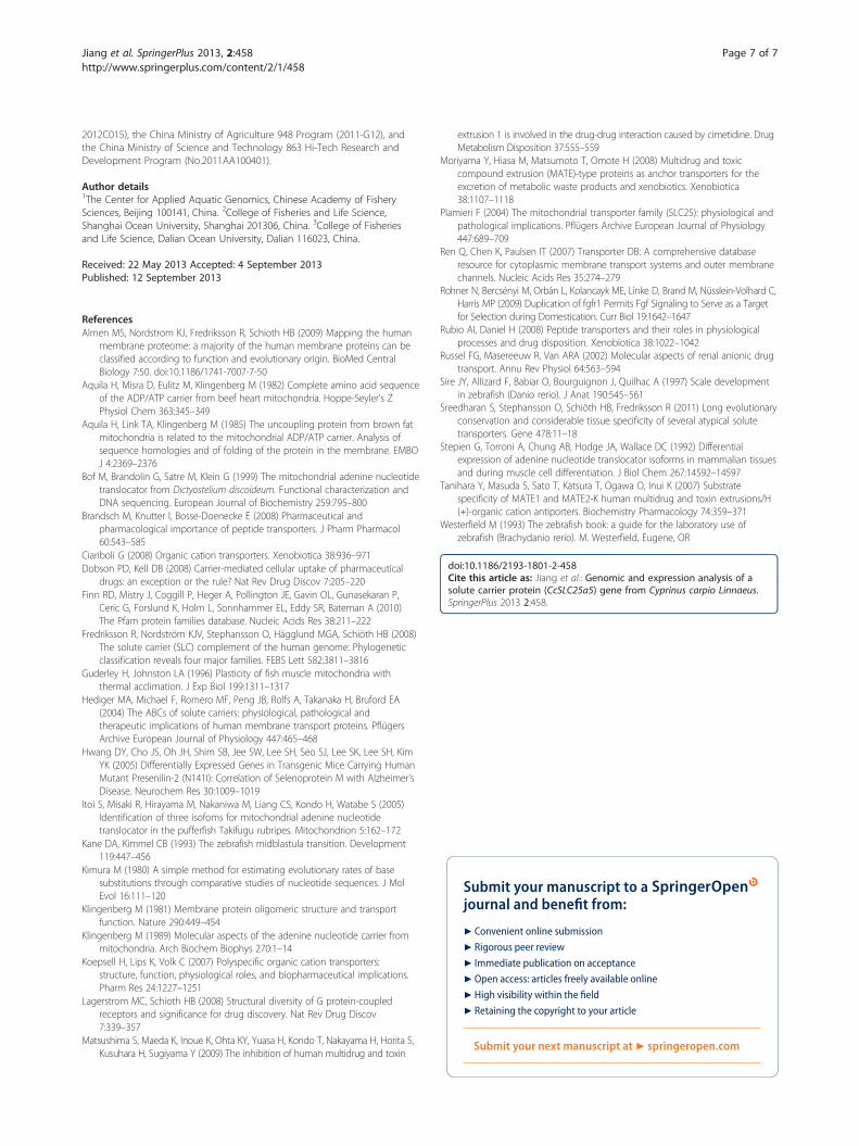
Phylogenetic analysis of cyprinid fishes and mammalianSLC25a5 isoformsTo analyze the phylogenetic relationship of ANTs incyprinid fishes, a phylogenetic tree was constructedusing neighbor-joining from deduced amino acid se-quences (Figure 4). The six carp isoforms were clusteredinto one branch. They are more closely related to humanand bovine ANT3, and more distantly to mammalian
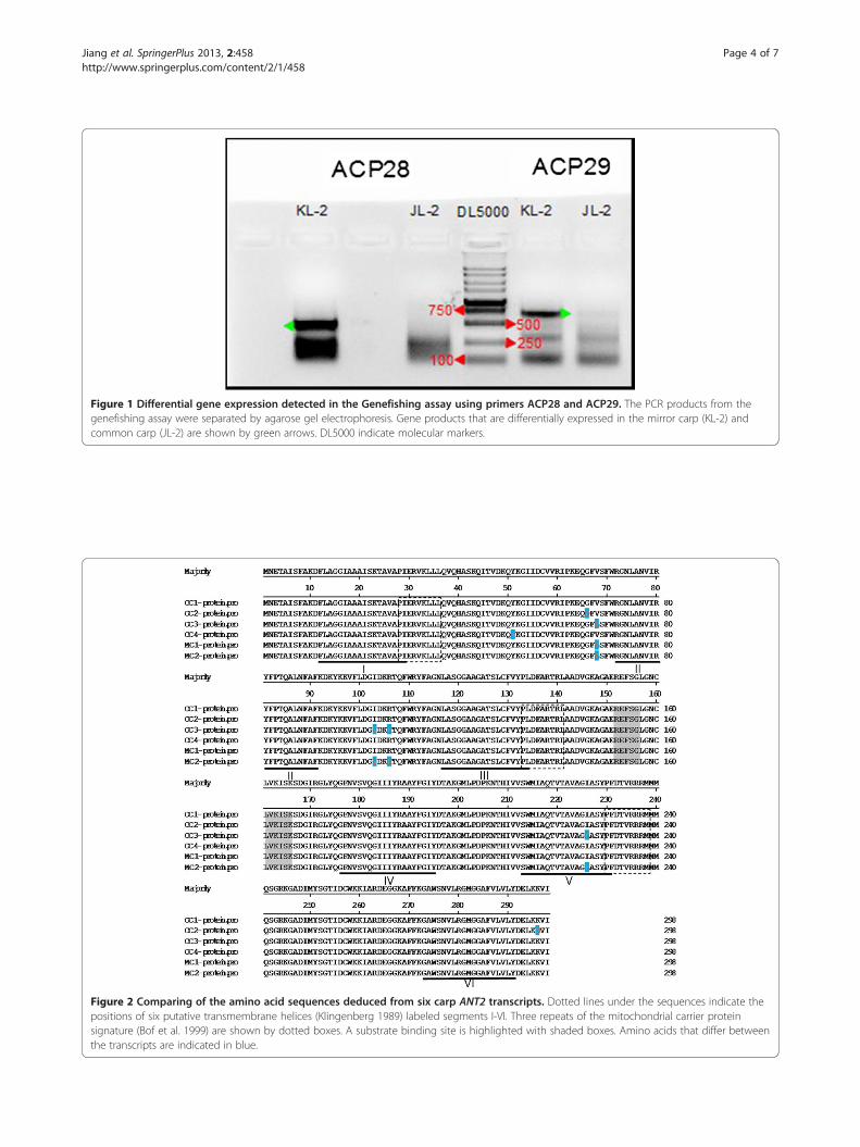
Figure 1 Differential gene expression detected in the Genefishing assay using primers ACP28 and ACP29. The PCR products from thegenefishing assay were separated by agarose gel electrophoresis. Gene products that are differentially expressed in the mirror carp (KL-2) andcommon carp (JL-2) are shown by green arrows. DL5000 indicate molecular markers.
Figure 2 Comparing of the amino acid sequences deduced from six carp ANT2 transcripts. Dotted lines under the sequences indicate thepositions of six putative transmembrane helices (Klingenberg 1989) labeled segments I-VI. Three repeats of the mitochondrial carrier proteinsignature (Bof et al. 1999) are shown by dotted boxes. A substrate binding site is highlighted with shaded boxes. Amino acids that differ betweenthe transcripts are indicated in blue.
Jiang et al. SpringerPlus 2013, 2:458 Page 4 of 7http://www.springerplus.com/content/2/1/458
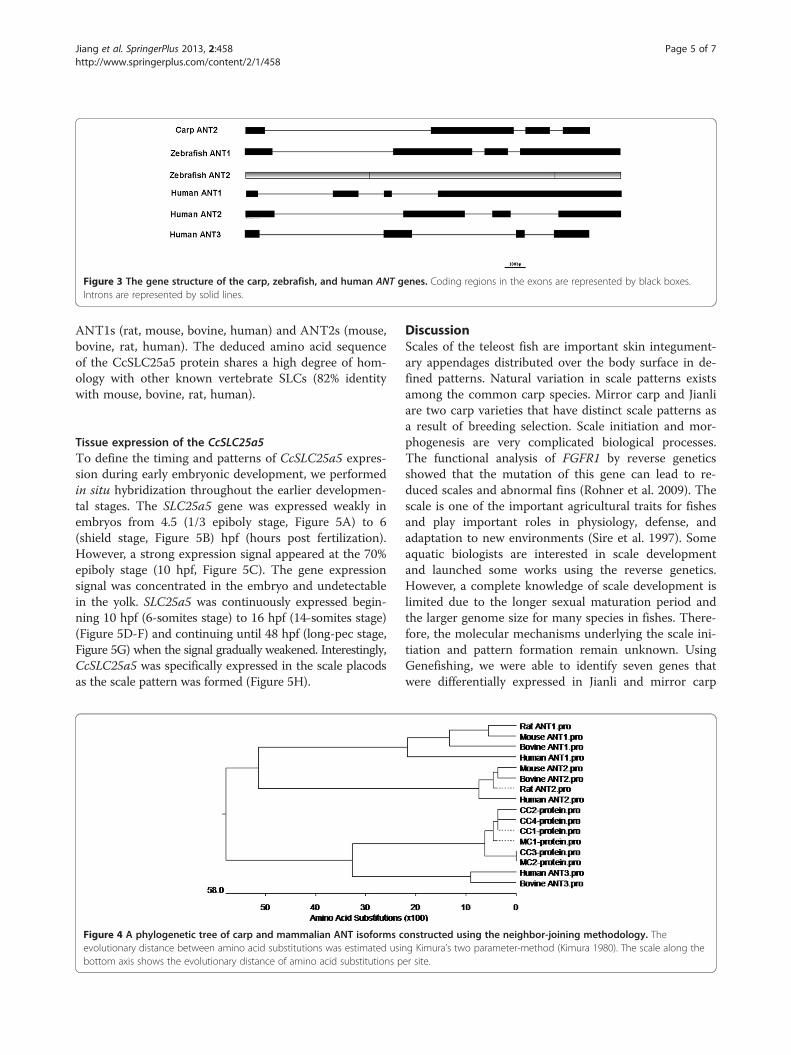
Figure 3 The gene structure of the carp, zebrafish, and human ANT genes. Coding regions in the exons are represented by black boxes.Introns are represented by solid lines.
Jiang et al. SpringerPlus 2013, 2:458 Page 5 of 7http://www.springerplus.com/content/2/1/458
ANT1s (rat, mouse, bovine, human) and ANT2s (mouse,bovine, rat, human). The deduced amino acid sequenceof the CcSLC25a5 protein shares a high degree of hom-ology with other known vertebrate SLCs (82% identitywith mouse, bovine, rat, human).
Tissue expression of the CcSLC25a5To define the timing and patterns of CcSLC25a5 expres-sion during early embryonic development, we performedin situ hybridization throughout the earlier developmen-tal stages. The SLC25a5 gene was expressed weakly inembryos from 4.5 (1/3 epiboly stage, Figure 5A) to 6(shield stage, Figure 5B) hpf (hours post fertilization).However, a strong expression signal appeared at the 70%epiboly stage (10 hpf, Figure 5C). The gene expressionsignal was concentrated in the embryo and undetectablein the yolk. SLC25a5 was continuously expressed begin-ning 10 hpf (6-somites stage) to 16 hpf (14-somites stage)(Figure 5D-F) and continuing until 48 hpf (long-pec stage,Figure 5G) when the signal gradually weakened. Interestingly,CcSLC25a5 was specifically expressed in the scale placodsas the scale pattern was formed (Figure 5H).
Figure 4 A phylogenetic tree of carp and mammalian ANT isoforms cevolutionary distance between amino acid substitutions was estimated usinbottom axis shows the evolutionary distance of amino acid substitutions p
DiscussionScales of the teleost fish are important skin integument-ary appendages distributed over the body surface in de-fined patterns. Natural variation in scale patterns existsamong the common carp species. Mirror carp and Jianliare two carp varieties that have distinct scale patterns asa result of breeding selection. Scale initiation and mor-phogenesis are very complicated biological processes.The functional analysis of FGFR1 by reverse geneticsshowed that the mutation of this gene can lead to re-duced scales and abnormal fins (Rohner et al. 2009). Thescale is one of the important agricultural traits for fishesand play important roles in physiology, defense, andadaptation to new environments (Sire et al. 1997). Someaquatic biologists are interested in scale developmentand launched some works using the reverse genetics.However, a complete knowledge of scale development islimited due to the longer sexual maturation period andthe larger genome size for many species in fishes. There-fore, the molecular mechanisms underlying the scale ini-tiation and pattern formation remain unknown. UsingGenefishing, we were able to identify seven genes thatwere differentially expressed in Jianli and mirror carp
onstructed using the neighbor-joining methodology. Theg Kimura’s two parameter-method (Kimura 1980). The scale along theer site.
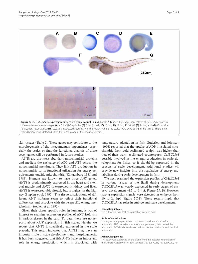
Figure 5 The CcSLC25a5 expression pattern by whole-mount in situ. Panels A-G show the expression pattern of CcSLC25a5 genes indifferent developmental stages. (A) 4.5 haf (1/3 epiboly), (B) 6 haf (shield), (C) 10 haf, (D) 12 haf, (E) 16 haf, (F) 24 haf, and (G) 48 haf afterfertilization, respectively. (H) SLC25a5 is expressed specifically in the regions where the scales were developing in the skin. (I) There is nohybridization signal detected using the sense probe as the negative control.
Jiang et al. SpringerPlus 2013, 2:458 Page 6 of 7http://www.springerplus.com/content/2/1/458
skin tissues (Table 2). These genes may contribute to themorphogenesis of the integumentary appendages, espe-cially the scales or fins, the functional analysis of theseseven genes will be performed in future studies.ANTs are the most abundant mitochondrial proteins
and mediate the exchange of ADP and ATP across themitochondrial membrane. They link ATP production inmitochondria to its functional utilization for energy re-quirements outside mitochondria (Klingenberg 1981 and1989). Humans are known to have three ANT genes.ANT1 is predominantly expressed in the heart and skel-etal muscle and ANT2 is expressed in kidney and liver.ANT3 is expressed ubiquitously but is highest in the kid-ney (Stepien et al. 1992). The tissue distributions of dif-ferent ANT isoforms seem to reflect their functionaldifferences and associate with tissue-specific energy me-tabolism (Stepien et al. 1992).Given their tissue specific roles in humans, it was of
interest to examine expression profiles of ANT isoformsin various tissues in the carp. To date, there are no re-ports about ANT expression in fish scales. Herein, wereport that ANT2 is specifically expressed in the scaleplacods. This result indicates that ANT2 may have animportant role in scale development and morphogenesis.It has been suggested that fish ANTs have an importantrole in energy production, which is associated with
temperature adaptation in fish. Guderley and Johnston(1996) reported that the uptake of ADP in isolated mito-chondria from cold-acclimated sculpin was higher thanthat of their warm-acclimated counterparts. CcSLC25a5possibly involved in the energy production in scale de-velopment for fishes, so it should be expressed in theprocess of scale development. Additional studies willprovide new insights into the regulation of energy me-tabolism during scale development in fish.We next examined the expression profiles of CcSLC25a5
in various tissues of the Jianli during development.CcSLC25a5 was weakly expressed in early stages of em-bryo development (4.5 to 6 hpf, Figure 5A-B). However,strong expression signals were detected in embryos from10 to 24 hpf (Figure 5C-F). These results imply thatCcSLC25a5 has roles in embryo and scale development.
Competing interestThe authors declare that no competing interests exist.
Authors’ contributionsLJ designed the project, carried out research and made the draftedmanuscript. ADC carried out most of the experiments, YYW revised themanuscript, BYZ did data collection. All authors read and approved the finalmanuscript.
AcknowledgementsThis study was supported by the grants from the Research Foundation ofthe Chinese Academy of Fishery Sciences (No. 2011C015, No. 2010C017, No
Jiang et al. SpringerPlus 2013, 2:458 Page 7 of 7http://www.springerplus.com/content/2/1/458
2012C015), the China Ministry of Agriculture 948 Program (2011-G12), andthe China Ministry of Science and Technology 863 Hi-Tech Research andDevelopment Program (No.2011AA100401).
Author details1The Center for Applied Aquatic Genomics, Chinese Academy of FisherySciences, Beijing 100141, China. 2College of Fisheries and Life Science,Shanghai Ocean University, Shanghai 201306, China. 3College of Fisheriesand Life Science, Dalian Ocean University, Dalian 116023, China.
Received: 22 May 2013 Accepted: 4 September 2013Published: 12 September 2013
ReferencesAlmen MS, Nordstrom KJ, Fredriksson R, Schioth HB (2009) Mapping the human
membrane proteome: a majority of the human membrane proteins can beclassified according to function and evolutionary origin. BioMed CentralBiology 7:50. doi:10.1186/1741-7007-7-50
Aquila H, Misra D, Eulitz M, Klingenberg M (1982) Complete amino acid sequenceof the ADP/ATP carrier from beef heart mitochondria. Hoppe-Seyler’s ZPhysiol Chem 363:345–349
Aquila H, Link TA, Klingenberg M (1985) The uncoupling protein from brown fatmitochondria is related to the mitochondrial ADP/ATP carrier. Analysis ofsequence homologies and of folding of the protein in the membrane. EMBOJ 4:2369–2376
Bof M, Brandolin G, Satre M, Klein G (1999) The mitochondrial adenine nucleotidetranslocator from Dictyostelium discoideum. Functional characterization andDNA sequencing. European Journal of Biochemistry 259:795–800
Brandsch M, Knutter I, Bosse-Doenecke E (2008) Pharmaceutical andpharmacological importance of peptide transporters. J Pharm Pharmacol60:543–585
Ciariboli G (2008) Organic cation transporters. Xenobiotica 38:936–971Dobson PD, Kell DB (2008) Carrier-mediated cellular uptake of pharmaceutical
drugs: an exception or the rule? Nat Rev Drug Discov 7:205–220Finn RD, Mistry J, Coggill P, Heger A, Pollington JE, Gavin OL, Gunasekaran P,
Ceric G, Forslund K, Holm L, Sonnhammer EL, Eddy SR, Bateman A (2010)The Pfam protein families database. Nucleic Acids Res 38:211–222
Fredriksson R, Nordström KJV, Stephansson O, Hägglund MGA, Schiöth HB (2008)The solute carrier (SLC) complement of the human genome: Phylogeneticclassification reveals four major families. FEBS Lett 582:3811–3816
Guderley H, Johnston LA (1996) Plasticity of fish muscle mitochondria withthermal acclimation. J Exp Biol 199:1311–1317
Hediger MA, Michael F, Romero MF, Peng JB, Rolfs A, Takanaka H, Bruford EA(2004) The ABCs of solute carriers: physiological, pathological andtherapeutic implications of human membrane transport proteins. PflügersArchive European Journal of Physiology 447:465–468
Hwang DY, Cho JS, Oh JH, Shim SB, Jee SW, Lee SH, Seo SJ, Lee SK, Lee SH, KimYK (2005) Differentially Expressed Genes in Transgenic Mice Carrying HumanMutant Presenilin-2 (N141I): Correlation of Selenoprotein M with Alzheimer’sDisease. Neurochem Res 30:1009–1019
Itoi S, Misaki R, Hirayama M, Nakaniwa M, Liang CS, Kondo H, Watabe S (2005)Identification of three isofoms for mitochondrial adenine nucleotidetranslocator in the pufferfish Takifugu rubripes. Mitochondrion 5:162–172
Kane DA, Kimmel CB (1993) The zebrafish midblastula transition. Development119:447–456
Kimura M (1980) A simple method for estimating evolutionary rates of basesubstitutions through comparative studies of nucleotide sequences. J MolEvol 16:111–120
Klingenberg M (1981) Membrane protein oligomeric structure and transportfunction. Nature 290:449–454
Klingenberg M (1989) Molecular aspects of the adenine nucleotide carrier frommitochondria. Arch Biochem Biophys 270:1–14
Koepsell H, Lips K, Volk C (2007) Polyspecific organic cation transporters:structure, function, physiological roles, and biopharmaceutical implications.Pharm Res 24:1227–1251
Lagerstrom MC, Schioth HB (2008) Structural diversity of G protein-coupledreceptors and significance for drug discovery. Nat Rev Drug Discov7:339–357
Matsushima S, Maeda K, Inoue K, Ohta KY, Yuasa H, Kondo T, Nakayama H, Horita S,Kusuhara H, Sugiyama Y (2009) The inhibition of human multidrug and toxin
extrusion 1 is involved in the drug-drug interaction caused by cimetidine. DrugMetabolism Disposition 37:555–559
Moriyama Y, Hiasa M, Matsumoto T, Omote H (2008) Multidrug and toxiccompound extrusion (MATE)-type proteins as anchor transporters for theexcretion of metabolic waste products and xenobiotics. Xenobiotica38:1107–1118
Plamieri F (2004) The mitochondrial transporter family (SLC25): physiological andpathological implications. Pflügers Archive European Journal of Physiology447:689–709
Ren Q, Chen K, Paulsen IT (2007) Transporter DB: A comprehensive databaseresource for cytoplasmic membrane transport systems and outer membranechannels. Nucleic Acids Res 35:274–279
Rohner N, Bercsényi M, Orbán L, Kolancayk ME, Linke D, Brand M, Nüsslein-Volhard C,Harris MP (2009) Duplication of fgfr1 Permits Fgf Signaling to Serve as a Targetfor Selection during Domestication. Curr Biol 19:1642–1647
Rubio AI, Daniel H (2008) Peptide transporters and their roles in physiologicalprocesses and drug disposition. Xenobiotica 38:1022–1042
Russel FG, Masereeuw R, Van ARA (2002) Molecular aspects of renal anionic drugtransport. Annu Rev Physiol 64:563–594
Sire JY, Allizard F, Babiar O, Bourguignon J, Quilhac A (1997) Scale developmentin zebrafish (Danio rerio). J Anat 190:545–561
Sreedharan S, Stephansson O, Schiöth HB, Fredriksson R (2011) Long evolutionaryconservation and considerable tissue specificity of several atypical solutetransporters. Gene 478:11–18
Stepien G, Torroni A, Chung AB, Hodge JA, Wallace DC (1992) Differentialexpression of adenine nucleotide translocator isoforms in mammalian tissuesand during muscle cell differentiation. J Biol Chem 267:14592–14597
Tanihara Y, Masuda S, Sato T, Katsura T, Ogawa O, Inui K (2007) Substratespecificity of MATE1 and MATE2-K human multidrug and toxin extrusions/H(+)-organic cation antiporters. Biochemistry Pharmacology 74:359–371
Westerfield M (1993) The zebrafish book: a guide for the laboratory use ofzebrafish (Brachydanio rerio). M. Westerfield, Eugene, OR
doi:10.1186/2193-1801-2-458Cite this article as: Jiang et al.: Genomic and expression analysis of asolute carrier protein (CcSLC25a5) gene from Cyprinus carpio Linnaeus.SpringerPlus 2013 2:458.
Submit your manuscript to a journal and benefi t from:
7 Convenient online submission
7 Rigorous peer review
7 Immediate publication on acceptance
7 Open access: articles freely available online
7 High visibility within the fi eld
7 Retaining the copyright to your article
Submit your next manuscript at 7 springeropen.com