Embed Size (px)
Citation preview

Developmental Biology 299 (2006) 594–608www.elsevier.com/locate/ydbio
Genomes & Developmental Control
Myogenic regulatory factors Myf5 and Myod function distinctly duringcraniofacial myogenesis of zebrafish
Cheng-Yung Lin, Rong-Feng Yung, Hung-Chieh Lee, Wei-Ta Chen,Yau-Hung Chen 1, Huai-Jen Tsai ⁎
Institute of Molecular and Cellular Biology, National Taiwan University, Room 307, Fisheries Science Building,No. 1, Section 4, Roosevelt Road, Taipei 106, Taiwan
Received for publication 30 January 2006; revised 21 July 2006; accepted 19 August 2006Available online 24 August 2006
Abstract
The functions of Myf5 and Myod are well known in trunk myogenesis. However, the roles that Myf5 and Myod play during craniofacialmyogenesis are far from well known. We observed that zebrafish myf5 was detected in the primordia of the obliques, lateral rectus,sternohyoideus, and pharyngeal mesoderm cores. In contrast, myod transcripts were expressed in all head muscle precursors at later stages.Knockdown of myf5 revealed that Myf5 was required for the development of the obliques, lateral rectus, sternohyoideus, and all pharyngealmuscles, whereas knockdown of myod proved that Myod was required for the development of superior rectus, medial rectus, inferior rectus, lateralrectus, and the ventral pharyngeal muscles. myod mRNA did not rescue the loss of the cranial muscle caused by injecting myf5-morpholino, orvice versa, suggesting that the functions of Myf5 and Myod were not redundant in head paraxial mesoderm, a finding different from theirfunctions in trunk myogenesis. Myf5, but not Myod, was required for the forward migration of myf5-positive oblique precursors. All evidencesreveal that Myf5 and Myod function independently during cranial myogenesis. On the basis of the expression patterns of myf5 and myod, wepropose a model to present how Myf5 and Myod are involved in head myogenesis of zebrafish.© 2006 Elsevier Inc. All rights reserved.
Keywords: Myf5; Myod; Cranial myogenesis; Zebrafish
Introduction
The paraxial mesoderm comprises the anterior (head orcephalic) mesoderm and the posterior (trunk somites and tail)mesoderm of vertebrates (Pownall et al., 2002). Unlike trunkmuscle, which originates from somites, head muscle is derivedfrom the unsegmented, nonepithelial paraxial mesodermflanking the hindbrain and midbrain and from the prechordalmesoderm regions. Head muscle develops in two regions:branchiomeric and nonbranchiomeric (Noden, 1983; Couly etal., 1992; Trainor et al., 1994; Hacker and Guthrie, 1998;Mackenzie et al., 1998). Branchiomeric muscle includes themuscles of mastication, derived from the first or mandibular
⁎ Corresponding author. Fax: +886 2 2363 8483.E-mail address: [email protected] (H.-J. Tsai).
1 Present address: Graduate Institute of Life Sciences, Tamkang University,Taipei, Taiwan.
0012-1606/$ - see front matter © 2006 Elsevier Inc. All rights reserved.doi:10.1016/j.ydbio.2006.08.042
arch; the muscles of facial expression, derived from the secondor hyoid arch; and the muscles of the pharynx and larynx,derived from more caudal arches (Kaufman and Bard, 1999).Non-branchiomeric head muscle includes extraocular muscles,derived from the anterior-most paraxial and prechordalmesoderm; and tongue muscles, derived from the hypoglossalcord and originating in the anterior somites (Mackenzie et al.,1998; Kaufman and Bard, 1999).
The basic helix–loop–helix myogenic regulatory factors(MRFs) play crucial functions that trigger the expression ofmuscle structural proteins and permit the assembly of functionalmyofibers (Molkentin and Olson, 1996). Myf5 and Myod directthe myogenic lineage, evidenced by the finding that double-mutant mice do not form skeletal muscle, a result of the absenceof precursor myoblasts (Rudnicki et al., 1993; Kaul et al., 2000;Kablar et al., 2003). In the absence of these factors, progenitorcells remain multipotent and their cell fates can change(Tajbakhsh et al., 1996; Kablar and Rudnicki, 1999). However,

595C.-Y. Lin et al. / Developmental Biology 299 (2006) 594–608
in Myf5−/− mutants, muscles occur normally in limb andbranchial arch progenitors (Kablar et al., 1997); in Myod−/−
embryos. Skeletal muscles in trunk develop normally and Myf5is significantly up-regulated (Rudnicki et al., 1992). AlthoughMyod mutant embryos exhibit the delayed development of limbmusculature, the limb myogenesis still keeps on processing(Kablar et al., 1997), indicating that Myod and Myf5, havefunctional redundancy during somitogenesis (Pownall et al.,2002). In zebrafish, embryos that received myf5- or myod-morpholino oligonucleotide (MO) alone developed somitesnormally (Lee et al., 2006), suggesting that myf5 and myodperform complementary functions during somitogenesis. How-ever, Kassar-Duchossory et al. (2004) state an epistaticrelationship among MRFs, that is, Mrf4 acts upstream ofmyod and directs embryonic multipotent cells into the myogeniclineage. This finding contradicts the theory that myogenicidentity is conferred only by Myf5 and Myod.
Several transcription factors and signaling modulators playimportant roles in mediating the response of signals fromsurrounding tissues to induce expression of MRFs duringskeletal myogenesis (Borycki and Emerson, 2000; Sabourin andRudnicki, 2000; Tajbakhsh and Buckingham, 2000). However,the finding of distinct regulatory networks of MRFs in head andin trunk myogenesis has been reported by many investigators(Patapoutian et al., 1993; Tajbakhsh et al., 1997; Hacker andGuthrie, 1998; Mootoosamy and Dietrich, 2002). Mice lackingMyf5 and the paired homeodomain transcription factor Pax3 donot develop skeletal muscle in the trunk or limb, yet headmuscle forms normally (Tajbakhsh et al., 1997). Furthermore,mice lacking Capsulin and MyoR fail to express Myf5 in thefirst arch and lost a subset of mandibular arch-derived muscle,but trunk muscle development was not affected (Lu et al.,2002). In chick, Lbx1/Pax7/Paraxis have distinct regulatorycascades in head and in trunk myogenesis (Mootoosamy andDietrich, 2002) and the Wnt signals, which promote trunkmyogenesis, have been proven to block head myogenesis(Tzahor et al., 2003). In zebrafish cranial muscle development,myod expression is found only in branchiomeric muscle andextraocular muscle (Schilling and Kimmel, 1997).
Throughout vertebrate evolution, head muscle developmenthas shown tremendous diversification (Goodrich, 1958).However, little is known about head muscle development inlower vertebrates. The epistatic relationship and the comple-mentary effects among MRFs in muscle systems not derivedfrom somites, such as craniofacial muscle development, havenever been reported. Moreover, the role of the MRFs that areinvolved in head muscle development has not been elucidated.In this report, we highlight the functions of zebrafish Myf5 andMyod during cranial muscle development. Myf5 gene tran-scripts are detected transiently in branchial arch mesodermcores and extraocular muscle, and inferior oblique and superioroblique primordial cells. However, when myf5 translation isinhibited, almost all head muscle, including that derived fromtrunk paraxial mesoderm, is lost. On the other hand, whenMyod translation is inhibited, major cranial muscles are stillpresent, such as adductor hyomandibulae, adductor mandibulae,adductor opercula, dilator operculi, inferior oblique, levator
arcus palatine, sternohyoideus, and superior oblique. We alsoprove that Myf5 and Myod do not have redundant functions inhead paraxial mesoderm. Knockdown of Myf5 abolished themigration of cranial muscle primordia. Therefore, on the basisof the expression pattern of myf5 and myod, we propose threeputative pathways of how Myf5 and Myod regulate thedevelopment of craniofacial muscles.
Materials and methods
Zebrafish transgenic lines
Two transgenic lines of zebrafish, Tg(α-actin:RFP) and Tg(myf5:EGFP),were used in this study. The enhanced green fluorescent protein (EGFP)reporter cDNA in the plasmid pZα-EGFP-ITR (Hsiao et al., 2001), whichcontains an upstream 4-kb segment of zebrafish fast muscle α-actin fused withthe EGFP reporter and flanked by an internal repeat sequence of an adeno-associated virus, was replaced by red fluorescent protein (RFP) cDNA fromcoral (DsRed, Clontech). The pDsRed 2-1 (Clontech) was cut first with NotIthen with BamHI after the NotI site was blunted. It was ligated into the pZα-EGFP-ITR vector that was cut with XhoI, blunted, then cut with BamHI. Theresultant plasmid, namely pZα-DsRed-ITR, was linearized by NotI andresuspended at a concentration of 25 ng/μl in double-distilled water mixedwith 0.1% (v/v) phenol red prior for microinjection to generate the transgenicline Tg(α-actin:RFP). Parental pairs that produced RFP-positive embryos wereseparated and mated with wild-type individuals to confirm the putative germ-line transmitting parent. After screening, RFP-positive F1 embryos were raisedto adulthood and crossed with wild-type zebrafish to generate a heterozygoticF2 generation. RFP-positive F2 individuals were then crossed with each otherto generate homozygotic F3 fish, which were used to produce 100% RFP-positive F4 offspring. A similar strategy was used to generate the transgenicline Tg(myf5:EGFP), except that the microinjected plasmid was pZMYP-BAC80E, in which an upstream region of zebrafish myf5 was fused with theEGFP reporter.
Knockdown of Myf5 and Myod in zebrafish embryos
Antisense MOs were designed specifically for translation inhibition of myf5-MO, TACGTCCATGATTGGTTTGGTGTTG, which was complementary tonucleotides (nt) 28-52, respectively, of zebrafish myf5 cDNA (GenBankaccession no. NM131576). The myod-MO, GTTTTTTCTACCTCAACAGCC-TATA, was complementary to nt 180–204 of zebrafish myod cDNA (GenBankaccession no. NM131262). All oligonucleotides were prepared at a stockconcentration of 1 mM and were diluted to the desired concentrations, that is,either 4, 2, or 1 ng, for microinjection into each embryo.
Fish embryos and whole-mount in situ hybridization
The procedures for zebrafish culture, embryo collection, fluorescentobservation, and whole-mount in situ hybridization have been describedpreviously (Lee et al., 2006), except that myf5 (GenBank accession no.NM131576), myod (GenBank accession no. NM131262), myogenin (GenBankaccession no. NM131006), mrf4 (GenBank accession no. NM001003982), α-actin (GenBank accession no. AF180887), and met (GenBank accession no.NM001007124) were used as probes. They were digoxigenin-labeled, after wecloned their partial DNA fragments. The designation of head muscle and thedevelopmental stage of zebrafish were following those of Schilling and Kimmel(1997).
mRNA preparation for the rescue experiment
Capped mRNAs of myf5 and myod were synthesized according to theprotocol of the manufacturer (Epicentre). The resultant mRNAs were diluted to44 ng/μl and 22 ng/μl for myf5 mRNA and myod mRNA, respectively, withdistilled water. Approximately 2.3 nl was used in injection into one-cell stageembryos.

596 C.-Y. Lin et al. / Developmental Biology 299 (2006) 594–608
Time-lapse and imaging analyses
Embryos derived from the transgenic line Tg(myf5:EGFP) were dechor-ionized and anesthetized with buffered ethyl m-aminoboenzoate (Tricaine;Sigma). Then, embryos were transferred to 0.5% agar containing an embryomedium with 10 mM HEPES and Tricaine. Axiovert 200 M InvertedMicroscope (Zeiss) was used to capture the image approximately every20 min from 40- to 48-hpf period. Embryos were always kept at 28.5°C on theheated microscope stage during time-lapse analysis. Image analysis wasprocessed by using MetaMorph 7.0 software (Molecular Derice).
Results
All cranial muscles are tagged with RFP in the transgeniczebrafish line Tg(α-actin:RFP)
To characterize the functions of Myf5 and Myod during headdevelopment,we generated a zebrafish transgenic lineTg(α-actin:RFP) that carried a DNA construct in which the RFP reporter wasdriven by a zebrafish fast-muscle α-actin promoter. Whole-mountin situ hybridization revealed that the RFP reporter gene started totranscribe at 14 h postfertilization (hpf) in the somites of embryosderived from Tg(α-actin:RFP), indicating that the transcription oftransgenic and endogenous α-actin genes was initiated at thesame stage (Figs. 1A, B). Red fluorescent signal was observedfirst in somites at 20 hpf (data not shown), and this signal appearedweaker in the newly formed somites than in the old ones (Figs. 1C,D). Similarly, α-actin transcripts were detected first in the headregion at 36 hpf, as were RFP transcripts. However, RFP signalswere observed first in all cranial muscles at 55 hpf (data notshown). After 55 hpf, all cranial skeletalmuscles of this transgenicTg(α-actin:RFP) fish were tagged clearly by RFP (Figs. 1E, F, G,H). We also noticed that the expression of α-actin in the head wassimilar to that of myod.
Expression patterns of myf5 and myod in zebrafish headmuscle development
We detected the spatiotemporal expression patterns of myf5and myod from 24 through 48 hpf. At 24 hpf, myf5 was detectedin the posterior region near the eye (Fig. 2A). At 30 hpf, thesemyf5-positive cells distributed at the first branchial archmesoderm core (Figs. 2A, B, C). In addition, myf5 started toexpress in the inferior oblique (io) and lateral rectus (lr) muscleprimordial cells (Fig. 2B). At 32 hpf, myf5 was expressed notonly in io and lr, but also in the muscle primordia of superioroblique (so) muscle and of the first, the second, and the thirdbranchial arches (Fig. 2C). After 32 hpf,myf5 transcripts rapidlywere absent in the first arch and lr, but expression increased inthe third branchial arch mesoderm core (Figs. 2C, E). At 36 hpf,myf5 was expressed predominately in sternohyoideus (sh),which migrates from the anterior somites (Fig. 2E). At 36through 48 hpf, myf5 continued to be expressed in the muscleprimordia of so, io, sh, and themesoderm cores of the second andthe third branchial arches (Figs. 2G, I). After 48 hfp, myf5transcripts gradually decreased in the head region.
Unlike the expression pattern of myf5, myod transcripts weredetected first in the head muscle primordia of the superior rectus
(sr), medial rectus (mr), and inferior rectus (ir), lr, and in the firstbranchial arch mesoderm core at 32 hfp (Fig. 2D). At 36 hpf,myod was detected in the first (masticatory plate, MP;intermandibularis, IM) and the second arch mesoderm cores(constrictor hyoideus dorsalis, CHD; constrictor hyoideusventralis, CHV). Thereafter, these mesoderm cores were cleavedindividually into dorsal (MP in the first arch; CHD in the secondarch) and ventral (IM in the first arch; CHV in the second arch)areas (Figs. 2F, H, J) (Schilling and Kimmel, 1994). At 42 to48 hpf, all the cranial muscle were myod-positive (Figs. 2H, J).
By comparing the expression patterns of myf5 and myod inhead, we found that almost all the cranial muscle expressedmyf5 in the early stages and then expressed myod afterward.At 32 hpf, the first arch mesoderm core was myf5-positive.However, when the first arch subdivided into MP and IM, themyf5 transcripts started to decrease at 36 hpf, as in the secondand third arch. The expression of myf5 was decreased greatlyafter 42 hpf. Instead, myod became positive in these muscles at36 hpf. A group ofmyf5-negative muscle primordia, such as mr/ir/sr, became myod-positive after 32 hfp (Figs. 2F, H). Inaddition, after we compared the spatiotemporal expressions ofmyf5, myod, myogenin, and α-actin in the wild-type embryosand the RFP reporter signaling in the transgenic line Tg(α-actin:RFP) (please see Supplemental data 1), we hypothesize thatMyf5 andMyod are involved in cranial muscle development butthat they play roles differently.
Functions of Myf5 and Myod in zebrafish cranial muscledevelopment
To determine whether myf5 and myod play roles duringcraniofacial muscle development, we microinjected MOs toknock down Myf5 or Myod specifically. When myf5-MO wasinjected into zygotes from Tg(α-actin:RFP) fish, only the muscleprimordia of sr, mr, and irwereRFP-positive at 72 hpf (Fig. 3Avs.B), even until 7 days postfertilization (dpf; data not show); theremainder of the cranial muscle was lost inmyf5morphants, evenat 7 dpf. The transcripts of myod and myogenin were onlydetected in the primordia of sr, mr, and ir at 36 hpf (Fig. 3D) and58 hpf (Figs. 3H, J) in myf5 morphants. The MO-inducedphenotypes were dose-dependent (Table 1). Next, we detected et1expression inmyf5morphants to reveal whether the loss of ventralcranial muscle in myf5 morphants was due to the abnormaldevelopment of pharyngeal ventral mesoderm cores. Resultsshowed that the pharyngeal ventral mesoderm cores of myf5morphants developed normally (Fig. 3E vs. F). These resultsindicate that Myf5 knockdown did not affect cranial mesodermcores development. Myf5 expression in the arch 1 and 2mesoderm cores is necessary for initiating the further myogenesisof ventral mesoderm cores. In addition, although myod andmyogeninwere expressed in all cranial muscle (Figs. 3G, I), Myf5knockdown resulted in restrictingmyod andmyogenin expressionin the primordia of sr, mr, and ir (Figs. 3H, J). Thus, we proposethat Myf5 is necessary for the development of the extraocularmuscles so, io, and lr, and all pharyngeal muscles.
When myod-MO was microinjected to specifically inhibit thetranslation of myod in the embryos derived from both wild-type

Fig. 1. Tagging all head muscles by using the transgenic zebrafish line Tg(α-actin:RFP). Endogenous α-actin transcripts (A) and red fluorescent protein (RFP) reportertranscripts (B) were detected by whole-mount in situ hybridization of zebrafish embryos at 14 hpf (lateral view: A, B). RFP expressions in the embryos derived fromthe transgenic line (α-actin:RFP) at 22.5 hpf (C, D), 72 hpf (E, F), and 120 hpf (G, H) were observed from a lateral view (E, G) and from a ventral view (F, H). Panel Dis a magnification of panel C. RFP appeared in the formed somites (D, arrowhead) but was absent in the newly forming somite (D, arrow) at 22.5 hpf. Meanwhile, allcranial muscles were labeled by red fluorescent signal in the embryos both at 72 and at 120 hpf. The muscles are designated following the scheme of Schilling andKimmel (1997): ah, adductor hyoideus; am, adductor mandibulae; ao, adductor operculi; do, dilator operculi; dpw1–5, dorsal pharyngeal wall 1–5; hh, hyohyoideus;ih, interhyoideus; ima, intermandibularis anterior; imp, intermandibularis posterior; io, inferior oblique; ir, inferior rectus; lap, levator arcus palatini; lr, lateral rectus;mr, medial rectus; sh, sternohyoideus; so, superior oblique; sr, superior rectus; and tv 1–5, transvs. ventralis 1–5.
597C.-Y. Lin et al. / Developmental Biology 299 (2006) 594–608
and Tg(α-actin:RFP) fish, the RFP signals were present in theextraocular muscles so and io, and in the dorsal pharyngealmuscles, such as lap, do, ah, ao, and sh at 72 hpf (Figs. 4Avs. B, Cvs. D), although the RFP signals were reduced slightly. Never-theless, the RFP signals were lost in the extraocular muscles sr,mr, ir, and lr and in the ventral pharyngeal muscles ima, imp, ih,and hh. Moreover, the myf5 transcripts appeared normal in myodmorphants both at 30 hpf (Fig. 4F) and at 36 hpf (Fig. 4H),whereas the myogenin transcripts were expressed slightly in so,io, lap/do, am, ah, ao, and sh (Fig. 4I vs. J). Like RFP signals,neither myf5 nor myogenin was expressed in the extraocularmuscles sr, mr, ir, and lr and in the ventral pharyngeal musclesima, imp, ih, and hh in myod morphants. The red fluorescent
signal was too weak to be observed in the primordia of am inmyod morphants before 72 hpf, but myogenin was detected byusing whole-mount in situ hybridization, supporting the theorythat myogenin was expressed in the promordia of am. However,neither the RFP signal nor in situhybridization was detected in sr,mr, ir, lr, ima, imp, ih, and hh, even until 7 dpf (data not shown).The phenotypes induced by MO treatment were dose-dependent(Table 2). Again, these results suggest that the absence of cranialmuscle in the myod morphants was myod-specific, not due to thedelay of development in the MO-treated embryos. Althoughmyod was expressed in all cranial muscles, myod knockdown didnot affect the expression of myf5 in so, io, dorsal arch, and sh(Figs. 4F, H), butmyogenin transcript and the RFP-labeledmuscle

Fig. 2. The temporal expressions of myf5 and myod during cranial muscle development. The temporal expressions of myf5 (A–C, E, G, I) and myod (D, F, H, J)transcripts were analyzed by whole-mount in situ hybridization in zebrafish embryos. The transcript of myf5 was detected in the craniofacial region at 24 hpf (A,arrow); in the io, the precursor of lr, and 1st arch (lr/1st arch) at 30 hpf (B); in the so, io, lr, 1st, 2nd, and 3rd arches at 32 hpf (C); in the so, io, sh, 2nd, and 3rd arches at36 hpf (E); and in the so, io, and sh at 42–48 hpf (G, I). myf5 was not expressed in the lr and 1st arch at 36 hpf (E). Meanwhile, myod was expressed in the craniofacialmuscles mr/ir/sr, lr, and 1st arch at 32 hpf (D); in the mr/ir, sr, lr, MP, IM, CHD, and CHV at 36 hpf (F); and in all the cranial muscle at 42–48 hpf (H, J). CD: theconstrictor dorsalis, which differentiates to lap and do; CHD: the constrictor hyoideus dorsalis, which differentiates to ah and ao; CHV: the constrictor hyoideusventralis, which differentiates to ih and hh; IM: the intermandibularis, which differentiates to ima and imp; MP: the masticatory plate, which differentiates to CD andam. lr, lateral rectus; for other abbreviations, see the legend of Fig. 1.
598 C.-Y. Lin et al. / Developmental Biology 299 (2006) 594–608
fibers were reduced, indicating that Myod helped to enhance themyogenesis of the headmuscles so, io, lap, do, am, ah, ao, and sh.Myod is required for the development of the extraocular musclessr, mr, ir, and lr and the ventral pharyngeal muscles ima, imp, ih,and hh. Myf5 and Myod play their own distinct roles duringcranial myogenesis of zebrafish.
Embryos that received either myf5- or myod-MO did not loseall the cranial muscle. However, when both myf5- and myod-MOs were injected into embryos derived from the transgenicline Tg(α-actin:RFP), all cranial muscle labeled with RFP wasabsent in the head region (Figs. 5A, B). We also detected theexpression of myogenin and myf4 in this myf5/myod morphant

Fig. 3. Myf5 is required for cranial muscle development, except the primordia of mr/ir/sr. Embryos derived from the transgenic line Tg(α-actin:RFP; A, B) and fromthe wild-type strain (C–J) were used. All the embryos were lateral views except panels E and F, which were dorsal–lateral view. The embryos injected with 4 ng ofmyf5-morpholino oligonucleotide (MO) to inhibit myf5 translation specifically, were studied to observe the development of cranial muscle (B) and the expression ofmyod, et1, andmyogenin (D, F, H, J) at the stages indicated. Red fluorescent protein (RFP) signal was detected only in the mr/ir and sr primordia in themyf5 transgenicmorphant (Avs. B).myodwas expressed only in the mr/ir/sr primordia in the myf5 wild-type morphant at 36 hpf (C vs. D) and at 58 hpf (G vs. H). Similarly,myogeninwas detected only in the mr/ir/sr primordia in myf5 morphant (I vs. J). However, the et1 transcript was changed little in the ventral arch mesoderm core in the myf5morphant (E vs. F, arrows), indicating that the development of the ventral arch mesoderm core in myf5 morphant was normal.
599C.-Y. Lin et al. / Developmental Biology 299 (2006) 594–608
and found that myogenin and myf4 were not expressed in anycranial muscle (Figs. 5C vs. D, E vs. F). Furthermore,Myogenin and MRF4 did not initiate cranial myogenesis ofzebrafish when both Myf5 and Myod lost their function.
To determine whether there is a redundant function betweenMyf5 and Myod during cranial muscle development, we co-
injected myf5-MO with myod mRNAs in embryos. Interest-ingly, myod mRNA did not rescue the loss of the cranial musclecaused by myf5-MO: only the mr, ir, and sr were observed (Fig.5G). When myf5 mRNA and myf5-MO were co-injected, all thecranial muscles displayed a normal phenotype (Table 1).Similarly, the number of embryos displaying the normal
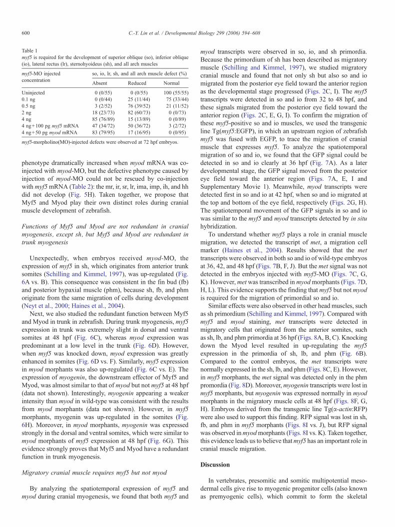
Table 1myf5 is required for the development of superior oblique (so), inferior oblique(io), lateral rectus (lr), sternohyoideus (sh), and all arch muscles
myf5-MO injectedconcentration
so, io, lr, sh, and all arch muscle defect (%)
Absent Reduced Normal
Uninjected 0 (0/55) 0 (0/55) 100 (55/55)0.1 ng 0 (0/44) 25 (11/44) 75 (33/44)0.5 ng 3 (2/52) 76 (39/52) 21 (11/52)2 ng 18 (23/73) 82 (60/73) 0 (0/73)4 ng 85 (76/89) 15 (13/89) 0 (0/89)4 ng+100 pg myf5 mRNA 47 (34/72) 50 (36/72) 3 (2/72)4 ng+50 pg myod mRNA 83 (79/95) 17 (16/95) 0 (0/95)
myf5-morpholino(MO)-injected defects were observed at 72 hpf embryos.
600 C.-Y. Lin et al. / Developmental Biology 299 (2006) 594–608
phenotype dramatically increased when myod mRNA was co-injected with myod-MO, but the defective phenotype caused byinjection of myod-MO could not be rescued by co-injectionwith myf5mRNA (Table 2): the mr, ir, sr, lr, ima, imp, ih, and hhdid not develop (Fig. 5H). Taken together, we propose thatMyf5 and Myod play their own distinct roles during cranialmuscle development of zebrafish.
Functions of Myf5 and Myod are not redundant in cranialmyogenesis, except sh, but Myf5 and Myod are redundant intrunk myogenesis
Unexpectedly, when embryos received myod-MO, theexpression of myf5 in sh, which originates from anterior trunksomites (Schilling and Kimmel, 1997), was up-regulated (Fig.6A vs. B). This consequence was consistent in the fin bud (fb)and posterior hypaxial muscle (phm), because sh, fb, and phmoriginate from the same migration of cells during development(Neyt et al., 2000; Haines et al., 2004).
Next, we also studied the redundant function between Myf5and Myod in trunk in zebrafish. During trunk myogenesis, myf5expression in trunk was extremely slight in dorsal and ventralsomites at 48 hpf (Fig. 6C), whereas myod expression waspredominant at a low level in the trunk (Fig. 6D). However,when myf5 was knocked down, myod expression was greatlyenhanced in somites (Fig. 6D vs. F). Similarly, myf5 expressionin myod morphants was also up-regulated (Fig. 6C vs. E). Theexpression of myogenin, the downstream effector of Myf5 andMyod, was almost similar to that of myod but not myf5 at 48 hpf(data not shown). Interestingly, myogenin appearing a weakerintensity than myod in wild-type was consistent with the resultsfrom myod morphants (data not shown). However, in myf5morphants, myogenin was up-regulated in the somites (Fig.6H). Moreover, in myod morphants, myogenin was expressedstrongly in the dorsal and ventral somites, which were similar tomyod morphants of myf5 expression at 48 hpf (Fig. 6G). Thisevidence strongly proves that Myf5 and Myod have a redundantfunction in trunk myogenesis.
Migratory cranial muscle requires myf5 but not myod
By analyzing the spatiotemporal expression of myf5 andmyod during cranial myogenesis, we found that both myf5 and
myod transcripts were observed in so, io, and sh primordia.Because the primordium of sh has been described as migratorymuscle (Schilling and Kimmel, 1997), we studied migratorycranial muscle and found that not only sh but also so and iomigrated from the posterior eye field toward the anterior regionas the developmental stage progressed (Figs. 2C, I). The myf5transcripts were detected in so and io from 32 to 48 hpf, andthese signals migrated from the posterior eye field toward theanterior region (Figs. 2C, E, G, I). To confirm the migration ofthese myf5-positive so and io muscles, we used the transgenicline Tg(myf5:EGFP), in which an upstream region of zebrafishmyf5 was fused with EGFP, to trace the migration of cranialmuscle that expresses myf5. To analyze the spatiotemporalmigration of so and io, we found that the GFP signal could bedetected in so and io clearly at 36 hpf (Fig. 7A). As a laterdevelopmental stage, the GFP signal moved from the posterioreye field toward the anterior region (Figs. 7A, E, I andSupplementary Movie 1). Meanwhile, myod transcripts weredetected first in so and io at 42 hpf, when so and io migrated atthe top and bottom of the eye field, respectively (Figs. 2G, H).The spatiotemporal movement of the GFP signals in so and iowas similar to the myf5 and myod transcripts detected by in situhybridization.
To understand whether myf5 plays a role in cranial musclemigration, we detected the transcript of met, a migration cellmarker (Haines et al., 2004). Results showed that the mettranscripts were observed in both so and io of wild-type embryosat 36, 42, and 48 hpf (Figs. 7B, F, J). But the met signal was notdetected in the embryos injected with myf5-MO (Figs. 7C, G,K). However,metwas transcribed inmyodmorphants (Figs. 7D,H, L). This evidence supports the finding thatmyf5 but notmyodis required for the migration of primordial so and io.
Similar effects were also observed in other head muscles, suchas sh primordium (Schilling and Kimmel, 1997). Compared withmyf5 and myod staining, met transcripts were detected inmigratory cells that originated from the anterior somites, suchas sh, lb, and phm primordia at 36 hpf (Figs. 8A, B, C). Knockingdown the Myod level resulted in up-regulating the myf5expression in the primordia of sh, lb, and phm (Fig. 6B).Compared to the control embryos, the met transcripts werenormally expressed in the sh, lb, and phm (Figs. 8C, E). However,in myf5 morphants, the met signal was detected only in the phmpromordia (Fig. 8D). Moreover,myogenin transcripts were lost inmyf5 morphants, but myogenin was expressed normally in myodmorphants in the migratory muscle cells at 48 hpf (Figs. 8F, G,H). Embryos derived from the transgenic line Tg(α-actin:RFP)were also used to support this finding. RFP signal was lost in sh,fb, and phm in myf5 morphants (Figs. 8I vs. J), but RFP signalwas observed inmyodmorphants (Figs. 8I vs. K). Taken together,this evidence leads us to believe thatmyf5 has an important role incranial muscle migration.
Discussion
In vertebrates, presomitic and somitic multipotential meso-dermal cells give rise to myogenic progenitor cells (also knownas premyogenic cells), which commit to form the skeletal

Fig. 4. Myod is required for the development of posterior extraocular recti and ventral branchial muscles. Embryos derived from the transgenic line Tg(α-actin:RFP)(A–D) and from the wild-type strain (E–J) were used. Embryos injected with 4 ng of myod-morpholino oligonucleotide (MO) to inhibit specifically myod translationwere followed to observe the development of cranial muscle (B, D) and the expression of myf5 (F, H) andmyogenin (J; lateral views: A, B, E–J; ventral views: C, D) atthe stages indicated. The anterior extraocular recti (io and so), dorsal branchial muscle (ah, ao, do, and lap), and sh developed normally in the myod transgenicmorphant (A vs. B; C vs. D). The posterior extraocular (sr, mr, ir, and lr) and the ventral branchial muscle (ima, imp, ih, and hh) were totally lost. When wild-typeembryos were injected with myod-MO, myf5 was expressed normally in the myod morphants at 30 hpf (E vs. F) and at 36 hpf (G vs. H). Whereas, the expression ofmyogenin was decreased in myod morphants at 58 hpf (I vs. J), indicating a reduction in myogenin-positive muscle fibers.
601C.-Y. Lin et al. / Developmental Biology 299 (2006) 594–608
muscles of the trunk, limbs, and head. Meanwhile, the moreanterior nonsomitic paraxial and prechordal head mesoderm isthe source of some head muscles (reviewed by Wachtler andJacob, 1986; Christ and Ordahl, 1995). The migration ofparaxial mesoderm to the branchial arches contributes pre-cursors that develop into facial muscles in chicks (Hacker and
Guthrie, 1998; Noden et al., 1999). Like other vertebrates,zebrafish cranial muscles originate from the paraxial mesoderm(Kimmel et al., 1990; Noden, 1983). Muscles that originatefrom the paraxial mesoderm of the first and second pharyngealarches develop the dorsal (lap, do, am, ah, ao) and the ventral(ima, imp, ih, hh) portions of cranial muscles.

Table 2myod is required for the development of superior rectus (sr), medial rectus (mr),inferior rectus (ir), lateral rectus (lr), and ventral arch muscles
myod-MO injectedconcentration
sr, mr, ir, lr, and ventral arch muscle defect (%)
Absent Reduced Normal
Uninjected 0 (0/117) 0 (0/117) 100 (117/117)0.1 ng 0 (0/93) 2 (2/93) 98 (91/93)0.5 ng 5 (4/74) 81 (60/74) 14 (10/74)2 ng 61 (61/100) 36 (36/100) 0 (0/100)4 ng 86 (51/59) 14 (8/59) 0 (0/59)4 ng+100 pg myod mRNA 41 (39/96) 55 (53/96) 4 (4/96)4 ng+50 pg myf5 mRNA 85 (60/71) 15 (11/71) 0 (0/71)
myod-morpholino(MO)-injected defects were observed at 72 hpf embryos.
602 C.-Y. Lin et al. / Developmental Biology 299 (2006) 594–608
Myf5 and Myod play crucial functions to trigger theexpression of muscle structural proteins and finally to permitthe assembly of myofibers (Molkentin and Olson, 1996;
Fig. 5. Myf5 andMyod function independently to activate progenitor lineages inmuscles o(MO) with 4 ng of myod-MO to inhibit specifically both myf5 and myod translation, reexpression ofmyogenin andmrf4 (D, F) at 72 hpf. Panel Bwasmagnified from the head arein themyf5 andmyod double-knockdownmorphants derived from the transgenic line Tg(αwere not detected in the myf5 and myod double-knockdown morphants (I vs. J). The mymorphants (G). Similarly, the myf5 mRNA did not rescue the formation of RFP-labeled p
Buckingham, 2001). Tajbakhsh et al. (1997) reported thatPax3 and Myf5 of mice follow two distinct myogenicpathways and that Myod acts genetically downstream ofthese genes for myogenesis in trunk muscle development;Myf5 and Myod regulate the head muscle formationindependently. The regulation of myf5 is markedly differentfrom that of myod. Obviously, more detailed knowledge aboutthe functions of Myf5 and Myod on cranial muscledevelopment is needed.
In this study, we provide strong evidence to show the distinctroles that Myf5 and Myod play during craniofacial muscledevelopment of zebrafish. In the myf5-knockdown morphants,the development of all the cranial muscles, except sr, mr, and ir,was impeded severely. The primordia of sr, mr, and ir developednormally in the myf5 morphants. Furthermore, in situhybridization also proved that myf5 was not transcribed in sr,
f the head region. Embryos co-injectedwith 4 ng ofmyf5-morpholino oligonucleotidespectively, were used to observe the development of cranial muscle (A, B) and thea of panelA.No red fluorescent protein (RFP) signalwas detected inmuscle primordia-actin:RFP; A, B). Similarly, the transcripts ofmyogenin (C vs. D) andmrf4 (E vs. F)od mRNA did not rescue the formation of RFP-labeled primordia muscles in myf5rimordia muscle in myodmorphants (H). For abbreviations, see the legend of Fig. 1.

Fig. 6. Loss of Myod up-regulated Myf5 expression, and vice versa, in the cranial muscle that migrated from the anterior somites. The expression of myf5 wasincreased in sternohyoideus (sh) primordia, fin bud (fb), and posterior hypaxial muscle (phm) that migrated from the anterior somites, when embryos received myod-morpholino oligonucleotide (MO; vs. B). This consequence was consistent in trunk myogenesis. There was weakmyf5 expression in the trunk of wild-type embryos at48 hpf (C). However,myf5 appeared predominantly in the dorsal (ds) and ventral (vs) parts of somites in the myodmorphants (E), indicating that myod knockdown up-regulated the expression of myf5 both in sh, which originated from trunk somite, and in trunk muscle. Similarly, the expression of myodwas also enhanced in the trunkof myf5 morphants, compared to wild-type embryos (D vs. F). The myogenin expression was similar to that of myf5 in myod morphants (E, G) and to that of myod inmyf5 morphants (F, H).
603C.-Y. Lin et al. / Developmental Biology 299 (2006) 594–608
mr, and ir. On contrast, few cranial muscles, such as sr, mr, andir, and the ventral set of muscles (ima, imp, ih, and hh) weretotally lost in the myod-knockdown morphants, suggesting thatMyf5 and Myod may function independently and distinctly insome cranial muscles during zebrafish embryogenesis. Theembryos injected with either myf5- or myod-MO in this studycould survive more than 8 days. Thus, we conclude that theMO-induced phenotypes are specific.
Regulatory networks of Myf5 and Myod during cranialmyogenesis are intricate
Myf5 and Myod are indispensable for cranial muscledevelopment of zebrafish, because Myf5 or Myod have itsown role without being redundant. Based on the expression
patterns of myf5 and myod, there are three different regulatorymechanisms of all craniofacial myogenesis (Fig. 9).
During cranial muscle development, the arch I and IImesoderm cores are subdivided into dorsal and ventralmesoderm cores. The dorsal mesoderm cores are the precursorof lap, do, am, ah, and ao, whereas the ventral mesoderm coresare the precursors of ima, imp, ih, and hh. We find that bothmyf5 and myod are detected in the dorsal mesoderm cores, butonly myod is expressed in the ventral groups. On the basis ofdata shown in this study, we propose two regulatory pathways(Fig. 9C): in pathway I, Myf5 per se is capable of initiatingmyogenesis. Once Myf5 is expressed, the expression of myodstarts to increase, and then myogenesis proceeds further. Thisfinding is illustrated by the expression of the muscledifferentiation marker myogenin: myod and downstream

Fig. 7. Myf5 is required for the forward migration of the superior oblique (so) and inferior oblique (io) primordia toward the anterior eye region. Embryos derived from the transgenic line Tg(myf5:EGFP), in which anupstream region of zebrafish myf5 was fused with enhanced green fluorescent protein (EGFP), were used to trace the migration of cranial muscle that express myf5. The primordia of io and so labeled with EGFP wereclearly visible in the embryos derived from the transgenic line Tg(myf5:EGFP) at 36–48 hpf under fluorescent microscopy (A, E, I; arrows). Whole-mount in situ hybridization of wild-type zebrafish embryos showed thatthe expression of met, a cell marker of migration, was positive in the io and so at 36–48 hpf (B, F, J; arrows). Embryos injected with myf5-morpholino oligonucleotide (MO) and myod-MO to inhibit myf5 and myodtranslations, respectively, expressed met at the stages indicated. The met transcript was not expressed in the io and so of myf5morphants (C, G, K), but met was expressed normally in the io and so of myodmorphants (D,H, L; arrows). For abbreviations, see the legend of Fig. 1.
604C.-Y.
Lin
etal./
Developm
entalBiology
299(2006)
594–608

Fig. 8. Myf5 is required for the migration of the sternohyoideus (sh) primordia from anterior somites. The transcripts of myf5 (A), myod (B), and met (C) were detectedin sternohyoideus (sh) premordia, fin bud (fb), and posterior hypaxial muscle (phm), that were derived from anterior somites at 36 hpf. Embryos injected with myf5-morpholino oligonucleotide (MO) and myod-MO to inhibit specifically myf5 and myod translations, respectively, expressed met at the stages indicated. Theexpression of met was almost lost in myf5 morphants; only slight signals were detected in the phm (D; arrow). However, met was expressed normally in myodmorphants (E). At 48 hpf, myogenin was expressed in the sh, fb, and phm of wild-type (F) and myod morphants (H), but myogenin was not expressed in the sh, fb, orphm of myf5 morphants (G). Instead, the expression of myogenin was up-regulated in the trunk of myf5 morphants. Embryos derived from the transgenic line Tg(α-actin:RFP; I–K) were used. At 72 hpf, the sh, fb, and phm showing red fluorescent signal were clearly observed both in wild-type (I; arrow) and myod morphants (K;arrow) but not in myf5 morphants (J). For abbreviations, see the legend to Fig. 1.
605C.-Y. Lin et al. / Developmental Biology 299 (2006) 594–608
myogenin were not expressed in the dorsal mesoderm cores ofthe myf5 morphants (Fig. 3). In contrast, myogenin transcriptsare detected in the primordia of myod morphants (Fig. 4),suggesting that dorsal arch muscle development of cranialmyogenesis does not initiate in the absence of Myf5. However,knockdown of myod does not impair myogenesis, because thedownstream differentiation marker myogenin is still expressedin these muscles, although myogenesis proceeds less efficiently.The alternative pathway for regulating the arch mesoderm coreis Pathway II: muscle primordia subdivided from the myf5-positive core are initiated to undergo myogenesis by Myod. Wefind that ima, imp, ih, and hh are lost in both myf5 morphantsand myod morphants. These results indicate that both myf5 and
myod are necessary for ventral core mesoderm to undergomyogenesis.
Unlike in the arch mesoderm core, the development ofextraocular muscles is regulated by three different pathways(Fig. 9C). Although both myf5 and myod transcripts weredetected in io and so, the primordia of io and so are lost in themyf5 morphants but not in the myod morphants, indicating thatMyf5 and Myod modulate the development of the extraocularmuscles io and so through Pathway I. On the other hand, lr islost in the myf5 and myod morphants, suggesting that Myf5 andMyod modulate the development of the extraocular muscles lrthrough Pathway II. In the development of sr, mr, and ir;however, myod transcript is expressed but not myf5 transcript.

Fig. 9. A plausible model to present the distinct modulation of Myf5 and Myod during craniofacial myogenesis of zebrafish. (A) Schematic illustration of the dynamicexpression of myf5 and myod in the cranial muscle of zebrafish embryos at 32 and 42 hpf. The myf5- or myod-positive muscle fibers are labeled in red; myf5- andmyod-negative ones are labeled in gray. (B) Schematic illustration of the presence (red) or absence (gray) of cranial muscle fibers in the embryos injected with eithermyf5-morpholino oligonucleotide (MO; left) or myod-MO (right) at 72 hpf. (C) Schematic diagram of all cranial muscles that are categorized into three groups(represented in red, blue, and green) on the basis of three regulatory pathways of myf5 and myod during development: (I) for so, io, sh, and dorsal arch, not only doesmyf5 modulate myogenesis directly to generate myogenesis at the basal level but myf5 also triggers myod expression to enhance myogenesis at a high level; (II) for lrand ventral arch, myf5 defines the cell fate of muscle and myod is the major factor of myogenesis; and (III) for sr, mr, and ir, myod modulates myogenesis directly.
606 C.-Y. Lin et al. / Developmental Biology 299 (2006) 594–608
This result is also supported by tracing the GFP signal from thetransgenic line that carries the upstream region of zebrafishmyf5 fused with GFP reporter (Fig. 7). Thus, it is Myod butnot Myf5 that modulates the development of the muscleprimordia of sr, mr, and ir through Pathway III.
On the other hand, the primordia of sh, which originates fromthe anterior somites, was lost in the myf5 morphants but not inmyod morphants, suggesting that sh is modulated throughPathway I. Thus, it is worthwhile to study whether other factorsare involved for controlling the development of all cranialmuscles.
Myf5 and Myod function differently for craniofacial and fortrunk myogenesis
Myf5−/− and Myod−/− mutant mice are viable and fertile(Kaul et al., 2000, Rudnicki et al., 1992). However, Myf5/Myodnull embryos do not form skeletal muscles and die at birthbecause of respiratory failure (Rudnicki et al., 1993). Thus,Myf5 and Myod function redundantly for skeletal myogenesis.However, unexpectedly, we found that this redundancy presentfor trunk myogenesis is not the case for cranial myogenesis inzebrafish, as evidenced by co-injection of myf5-MO with myodmRNA not rescuing the myf5-MO-induced phenotype (Fig.5G). Similarly, co-injected myod-MO and myf5 mRNA did notrescue the defects caused by myod-MO (Fig. 5H). Therefore,we reason that the functions of Myf5 and Myod are not
redundant in cranial myogenesis. Nevertheless, the cranialmuscle sh, which originates from trunk paraxial mesoderm, isan exception to this rule. The expression of myf5 in sh is up-regulated in the myod morphants (Fig. 6B), suggesting that theprimordium of sh progresses as does trunk myogenesis, evenwhen they migrate into the head region. This finding alsoindicates that Myf5 and Myod indeed have different regulatorymechanisms between head and trunk paraxial mesoderm,suggesting that cranial myogenesis is governed by a head-specific regulatory cascade, which is fundamentally distinctfrom the regulatory cascade in the trunk.
Myf5 and Myod function distinctly during development of thedorsal and ventral cranial muscles
In this study, MO-knockdown of Myf5 level results in loss ofall cranial muscles, except sr, mr, and ir, suggesting that Myf5 isa key modulator during cranial muscle development. AlthoughSchilling and Kimmel (1997) reported that myod is expressed inall cranial muscles of embryos in zebrafish, loss of Myodfunction actually does not impede the development of certaincranial muscle fibers, such as ah, am, ao, do, io, lap, sh, and so(Fig. 4). It is highly likely that Myod is required for thespecification of the ventral cranial muscles but definitively notfor the dorsal ones. On the other hand, Barrallo-Gimeno et al.(2004) reported that, in the mutant mobm610/tfap2a, am ispresent but lap and do are absent and suggested that tfap2a is

607C.-Y. Lin et al. / Developmental Biology 299 (2006) 594–608
necessary for the specification of the dorsal cranial muscles butnot for the ventral ones. Thus, we propose that Myf5 may be akey modulator to control the development of major cranialmuscles, whereas Myod and Tfap2a may be key modulators tocontrol the development of the ventral and dorsal cranialmuscles, respectively.
Epistatic relationship of the zebrafish MRFs in cranialmyogenesis
Recently, the epistatic relationship of mouse MRFs has beenproposed that both Myf5 and Mrf4 act upstream of Myod todirect embryonic multipotent cells into the myogenic lineage(Kassar-Duchossory et al., 2004). However, the epistaticrelationship of the MRFs in cranial myogenesis is still unclear.Our study shows that embryos co-injected with myf5- andmyod-MOs lost all the cranial muscles in the head region (Figs.5A, B). In addition, in situ hybridization revealed that myo-genin and mrf4 were not expressed in myf5/myod morphants(Figs. 5D, F). This evidence indicates that Myogenin and Mrf4are not capable of initiating cranial myogenesis. We speculatethat myogenin and mrf4 may serve as differentiation genes.
Myf5 is required for the migration of myogenic precursor cells
In rodent, met null mice lost the hypaxial mesoderm derivedfrom the migratory muscles, such as the diaphragm, tongue,limb, and associated shoulder musculature. Also, met served asa migratory marker (Bladt et al., 1995; Dietrich et al., 1999). Inzebrafish, the primordium of the cranial muscle sh, whichoriginates from the anterior somites, has been defined (Schillingand Kimmel, 1997). In this report, we show that the primordiaof io and so migrate during cranial myogenesis (Fig. 7). Whole-mount in situ hybridization showed that met is expressed in so,io, and sh primordia (Figs. 7, 8). Interestingly, met transcript islost in the myf5 morphants, but met displays normally in themyod morphants, suggesting that myf5 is necessary for cranialmuscle primordia migration.
In the development of mouse limb, myf5 and myodtranscripts are not detected in the migratory muscle primordiauntil they migrate into the limb buds (Tajbakhsh andBuckingham, 1994; Birchmeier and Brohmann, 2000). Unlikethe mouse limb, in zebrafish cranial muscle development, themigratory cells are myf5- and myod-positive, even when theyundergo migration. In Xenopus, p38 mitogen-activated proteinkinase (MAPK) regulates the expression of Xmyf5 and affectsdistinct myogenic programs (Keren et al., 2005). Inhibition ofp38 MAPK prevents the expression of Xmyf5 but not ofXmyod. The ventral body wall muscles, whose migratoryprecursors originate in the ventral part of somites, are reducedgreatly when p38 MAPK and Xmyf5 are knocked down.Together, the early activation of Myf5 and Myod in frog andfish may suggest the rapidity of their early developments toproduce functional muscles for swimming.
In summary, we address the functions of Myf5 and Myodduring cranial muscle development in zebrafish. Myf5 andMyod play distinct roles in cranial myogenesis. A putative
model that demonstrates three pathways for Myf5 and Myodfunction in craniofacial muscle development is proposed. Inaddition, the expression of met in morphants supports thefinding that Myf5, a well-known MRF that is involved in trunkmyogenesis, also controls the cranial cell migration. This articleis the first report to reveal the functions of Myf5 and Myodduring cranial myogenesis in zebrafish.
Acknowledgments
This work was supported by the National Science Council,Republic of China, under grand no. NSC-95-2313-B-002-067-MY2. We are also grateful to Dr. Chang-Hao Yang and MissShu-Chen Shen, Department of Medical Research, NationalTaiwan University Hospital, for helping in time-lapse analysisand molecular imaging.
Appendix A. Supplementary data
Supplementary data associated with this article can be found,in the online version, at doi:10.1016/j.ydbio.2006.08.042.
References
Bladt, F., Riethmacher, D., Isenmann, S., Birchmeier, C., 1995. Essential role forthe c-met receptor in the migration of myogenic precursor cells into the limbbud. Nature 376, 768–771.
Barrallo-Gimeno, A., Holzschuh, J., Driever, W., Knapik, E.W., 2004. Neuralcrest survival and differentiation in zebrafish depends on mont blanc/tfap2agene function. Development 131, 1463–1477.
Birchmeier, C., Brohmann, H., 2000. Genes that control the development ofmigrating muscle precursor cells. Curr. Opin. Cell Biol. 12, 725–730.
Borycki, A.G., Emerson Jr., C.P., 2000. Multiple tissue interactions and signaltransduction pathways control somite myogenesis. Curr. Top. Dev. Biol. 48,165–224.
Buckingham, M., 2001. Skeletal muscle formation in vertebrates. Curr. Opin.Genet. Dev. 11, 440–448.
Christ, B., Ordahl, C.P., 1995. Early stages of chick somite development. Anat.Embryol. 191, 381–396.
Couly, G.F., Coltey, P.M., Le Douarin, N.M., 1992. The developmental fateof the cephalic mesoderm in quail–chick chimeras. Development 114,1–15.
Dietrich, S., Abou-Rebyeh, F., Brohmann, H., Bladt, F., Sonnenberg-Riethmacher,E., Yamaai, T., Lumsden, A., Brand-Saberi, B., Birchmeier, C., 1999. The roleof SF/HGF and c-Met in the development of skeletal muscle. Development126, 1621–1629.
Goodrich, E.S., 1958. Studies on the Structure and Development of Vertebrates.Dover Publications, New York.
Hacker, A., Guthrie, S., 1998. A distinct developmental programme for thecranial paraxial mesoderm in the chick embryo. Development 125,3461–3472.
Haines, L., Neyt, C., Gautier, P., Keenan, D.G., Bryson-Richardson, R.J.,Hollway, G.E., Cole, N.J., Currie, P.D., 2004. Met and Hgf signalingcontrols hypaxial muscle and lateral line development in the zebrafish.Development 131, 3869–4857.
Hsiao, C.D., Hsieh, F.J., Tsai, H.J., 2001. Enhanced expression and stabletransmission of transgenes flanked by inverted terminal repeats from adeno-associated virus in zebrafish. Dev. Dyn. 220, 323–336.
Kablar, B., Rudnicki, M.A., 1999. Development in the absence of skeletalmuscle results in the sequential ablation of motor neurons from the spinalcord to the brain. Dev. Biol. 208, 93–109.
Kablar, B., Krastel, K., Ying, C., Asakura, A., Tapscott, S.J., Rudnicki, M.A.,

608 C.-Y. Lin et al. / Developmental Biology 299 (2006) 594–608
1997. MyoD and Myf-5 differentially regulate the development of limbversus trunk skeletal muscle. Development 124, 4729–4738.
Kablar, B., Krastel, K., Tajbakhsh, S., Rudnicki, M.A., 2003. Myf5 and MyoDactivation define independent myogenic compartments during embryonicdevelopment. Dev. Biol. 2, 307–318.
Kassar-Duchossory, L., Gayraud-Morel, B., Gomes, D., Rocancourt, D.,Buckingham, M., Shinin, V., Tajbakhsh, S., 2004. Mrf4 determines skeletalmuscle identity in Myf5:Myod double-mutant mice. Nature 431, 466–471.
Kaufman, M.H., Bard, J.B.L., 1999. The Anatomical Basis of MouseDevelopment. Academic Press, San Diego, pp. 60–76.
Kaul, A., Koster, M., Neuhaus, H., Braun, T., 2000. Myf-5 revisited: loss ofearly myotome formation does not lead to a rib phenotype in homozygousMyf-5 mutant mice. Cell 102, 17–19.
Keren, A., Bengal, E., Frank, D., 2005. p38 MAP kinase regulates theexpression of XMyf5 and affects distinct myogenic programs during Xe-nopus development. Dev. Biol. 288, 73–86.
Kimmel, C.B., Warga, R.M., Schilling, T.F., 1990. Origin and organization ofthe zebrafish fate map. Development 108, 581–594.
Lee, H.C., Huang, H.Y., Lin, C.Y., Chen, Y.H., Tsai, H.J., 2006. Foxd3 mediateszebrafish myf5 expression during early somitogenesis. Dev. Biol. 290,359–372.
Lu, J.R., Bassel-Duby, R., Hawkins, A., Chang, P., Valdez, R., Wu, H., Gan, L.,Shelton, J.M., Richardson, J.A., Olson, E.N., 2002. Control of facial muscledevelopment by MyoR and capsulin. Science 298, 2378–2381.
Mackenzie, S., Walsh, F.S., Graham, A., 1998. Migration of hypoglossalmyoblast precursors. Dev. Dyn. 213, 349–358.
Molkentin, J.D., Olson, E.N., 1996. Defining the regulatory networks for muscledevelopment. Curr. Opin. Genet. Dev. 4, 445–453.
Mootoosamy, R.C., Dietrich, S., 2002. Distinct regulatory cascades for head andtrunk myogenesis. Development 129, 573–583.
Noden, D.M., 1983. The embryonic origins of avian cephalic and cervicalmuscles and associated connective tissues. Am. J. Anat. 168, 257–276.
Noden, D.M., Marcucio, R., Borycki, A.G., Emerson Jr., C.P., 1999.Differentiation of avian craniofacial muscles: I. Patterns of earlyregulatory gene expression and myosin heavy chain synthesis. Dev.Dyn. 216, 92–116.
Neyt, C., Jagla, K., Thisse, C., Thisse, B., Haines, L., Currie, P.D., 2000.Evolutionary origins of vertebrate appendicular muscle. Nature 408,82–85.
Patapoutian, A., Miner, J.H., Lyons, G.E., Wold, B., 1993. Isolated sequences
from the linked Myf-5 and MRF4 genes drive distinct patterns of muscle-specific expression in transgenic mice. Development 118, 61–69.
Pownall, M.E., Gustafsson, M.K., Emerson Jr., C.P., 2002. Myogenic regulatoryfactors and the specification of muscle progenitors in vertebrate embryos.Annu. Rev. Cell Dev. Biol. 18, 747–783.
Rudnicki, M.A., Braun, T., Hinuma, S., Jaenisch, R., 1992. Inactivation ofMyoD in mice leads to up-regulation of the myogenic HLH geneMyf-5 andresults in apparently normal muscle development. Cell 71, 383–390.
Rudnicki, M.A., Schnegelsberg, P.N., Stead, R.H., Braun, T., Arnold, H.H.,Jaenisch, R., 1993. MyoD or Myf-5 is required for the formation of skeletalmuscle. Cell 75, 1351–1359.
Sabourin, L.A., Rudnicki, M.A., 2000. The molecular regulation of myogenesis.Clin. Genet. 57, 16–25.
Schilling, T.F., Kimmel, C.B., 1994. Segment and cell type lineage restrictionsduring pharyngeal arch development in the zebrafish embryo. Development120, 483–494.
Schilling, T.F., Kimmel, C.B., 1997. Musculoskeletal patterning in the pharyn-geal segments of the zebrafish embryo. Development 124, 2945–2960.
Tajbakhsh, S., Buckingham, M., 1994. Mouse limb muscle is determined in theabsence of the earliest myogenic factor myf-5. Proc. Natl. Acad. Sci. U. S. A.91, 747–751.
Tajbakhsh, S., Buckingham, M., 2000. The birth of muscle progenitor cellsin the mouse: spatiotemporal considerations. Curr. Top. Dev. Biol. 48,225–268.
Tajbakhsh, S., Bober, E., Babinet, C., Pournin, S., Arnold, H., Buckingham, M.,1996. Gene targeting the myf-5 locus with nlacZ reveals expression of thismyogenic factor in mature skeletal muscle fibres as well as early embryonicmuscle. Dev. Dyn. 206, 291–300.
Tajbakhsh, S., Rocancourt, D., Cossu, G., Buckingham, M., 1997. Redefiningthe genetic hierarchies controlling skeletal myogenesis: Pax-3 and Myf-5 actupstream of MyoD. Cell 89, 127–138.
Trainor, P.A., Tan, S.S., Tam, P.P., 1994. Cranial paraxial mesoderm:regionalisation of cell fate and impact on craniofacial development inmouse embryos. Development 120, 2397–2408.
Tzahor, E., Kempf, H., Mootoosamy, R.C., Poon, A.C., Abzhanov, A., Tabin,C.J., Dietrich, S., Lassar, A.B., 2003. Antagonists of Wnt and BMPsignaling promote the formation of vertebrate head muscle. Genes Dev. 17,3087–3099.
Wachtler, F., Jacob, M., 1986. Origin and development of the cranial skeletalmuscles. Biol. Anat. 29, 24–46.