Embed Size (px)
Citation preview

GeNeViSTA
Non-immune Fetal Hydrops: An UpdateGayatri N 1, Ashwani Tandon 2 and Prajnya Ranganath 1
1Department of Medical Genetics, Nizam’s Institute of Medical Sciences, Hyderabad2Department of Pathology, Nizam’s Institute of Medical Sciences, Hyderabad
Email: [email protected]
Introduction
Hydrops fetalis is a Greek term which refers tothe pathological accumulation of uid in fetal softtissues and serous cavities. Non-immune fetal hy-drops (NIFH) is de ned as uid accumulation in atleast 2 fetal body compartments in the absence ofred cell isoimmunisation (Moise, 2008). Abnormal
uid collection may be ascites, pleural effusion,pericardial effusion or generalised skin edema(skin thickness >5mm) (Figure 1). Other frequentsonographic ndings associated with fetal hydropsinclude placental thickening and polyhydramnios.The placental thickness (in mm) is normally equalto the gestational age (in weeks) +/- 10 mm; if theplacental thickness exceeds this range, it is con-sidered as increased placental thickening (Moise,2008). With the widespread use of routine antiDprophylaxis in Rh-negative mothers, prevalence ofRhD alloimmunisation and associated hydrops hasdramatically decreased and especially in developedcountries, NIFH now accounts for almost 90% ofcases of hydrops fetalis. The progressive fallin the incidence of immunologic hydrops fetalishas fostered growing interest in non-immune fetalhydrops. The world-wide prevalence of NIFH isestimated to range from 1 in 1500 to 1 in 3800births (Bellini et al., 2015).
The identi cation of fetal hydrops by antena-tal ultrasound requires extensive search for theetiology which includes a wide range of diseasesincluding several genetic disorders. Even afterundergoing numerous investigations, in a goodnumber of cases the etiology remains unknown.In addition, the prognosis is usually poor with aperinatal loss of 70–90%, except in rare cases ofspontaneous resolution of parvovirus B19 infec-tion.
Etiology of Non-immune Fetal Hydrops
Non-immune fetal hydrops is a nonspeci c ndingand can be the manifestation of a wide varietyof disorders (Bellini et al., 2009). The cause canbe found in nearly 60% of cases prenatally andin around 85% of cases when postnatal tests areincluded. Identi cation of the exact etiology helpsin accurate prognostication of the recurrence riskfor subsequent pregnancies of the couple andde nite prenatal testing can be offered in theirfuture pregnancies (Moreno et al., 2013). The mostcommon etiologies include cardiovascular causes,chromosomal anomalies and hematological abnor-malities. Other conditions associated with NIFHinclude fetal infections, fetal malformations, inbornerrors of metabolism, lethal skeletal dysplasias, nu-merous other single gene disorders, fetal tumoursand placental abnormalities.
The important etiological associations of NIFHare listed in Table 1 (Moise, 2008).
Pathophysiology of non-immune fetalhydrops
The basic pathophysiological mechanism of fetalhydrops is imbalance in the regulation of uid be-tween vascular and interstitial spaces. Fluid move-ments between vascular and interstitial spaces areregulated by ltration of uid across the capillarywall as described by the Starling equation whichstates that the uid movement due to ltrationacross the wall of a capillary is dependent on thebalance between the hydrostatic pressure gradientand the oncotic pressure gradient across the capil-lary. When these pressure gradients are disturbeddue to various pathophysiological mechanisms,there is an increased uid accumulation in the
Genetic Clinics 2017 | July - September | Vol 10 | Issue 3 10
GeNeViSTA
interstitial spaces, which leads to fetal hydrops.Increased knowledge and understanding of theunderlying mechanisms that disturb the uid equi-librium would therefore be of great importancein identifying potential therapeutic interventions
(Bellini et al., 2012).
The pathophysiology underlying the variouscauses of nonimmune fetal hydrops has beendepicted in ow charts 1 and 2 (Bellini et al., 2012).
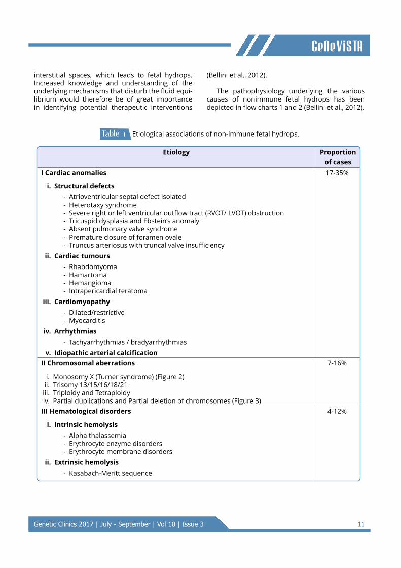
Table 1 Etiological associations of non-immune fetal hydrops.
Etiology Proportionof cases
I Cardiac anomalies
i. Structural defects- Atrioventricular septal defect isolated- Heterotaxy syndrome- Severe right or left ventricular out ow tract (RVOT/ LVOT) obstruction- Tricuspid dysplasia and Ebsteins anomaly- Absent pulmonary valve syndrome- Premature closure of foramen ovale- Truncus arteriosus with truncal valve insufficiency
ii. Cardiac tumours- Rhabdomyoma- Hamartoma- Hemangioma- Intrapericardial teratoma
iii. Cardiomyopathy- Dilated/restrictive- Myocarditis
iv. Arrhythmias- Tachyarrhythmias / bradyarrhythmias
v. Idiopathic arterial calcification
17-35%
II Chromosomal aberrations
i. Monosomy X (Turner syndrome) (Figure 2)ii. Trisomy 13/15/16/18/21iii. Triploidy and Tetraploidyiv. Partial duplications and Partial deletion of chromosomes (Figure 3)
7-16%
III Hematological disorders
i. Intrinsic hemolysis- Alpha thalassemia- Erythrocyte enzyme disorders- Erythrocyte membrane disorders
ii. Extrinsic hemolysis- Kasabach-Meritt sequence
4-12%
Genetic Clinics 2017 | July - September | Vol 10 | Issue 3 11

GeNeViSTA
iii. Red cell aplasia- Parvovirus B19 infection- Diamond-Blackfan syndrome- Dyserythropoietic anemia- Congenital leukemia
iv. Fetomaternal hemorrhageIV Twin–Twin transfusion 3-10%V InfectionsTORCHES CLAP (Toxoplasma, Rubella, Herpes simplex, Echovirus, Syphilis, Cy-tomegalovirus, Coxsackie virus, Leptospirosis, AIDS, Adenovirus, Parvovirus)
5-7%
VI Syndromes
i. Autosomal dominant disorders- Cornelia de Lange syndrome- Congenital myotonic dystrophy- Noonan syndrome- Tuberous sclerosis
ii. Autosomal recessive disorders- Lethal multiple pterygium syndrome (Figure 4)- Neu Laxova syndrome- Cumming syndrome- Elejalde syndrome
3-4%
VII Skeletal dysplasias
- Asphyxiating thoracic dysplasia- Short rib thoracic dysplasia with/ without polydactyly (Figure 5)- Achondrogenesis- Osteogenesis imperfecta type 2- Lethal osteopetrosis- Lethal Kneist- like dysplasia- Chondrodysplasia punctate (Conradi-Hunermann variant)- Greenberg chondrodystrophy- Caffey syndrome
3-4%
VIII Gastrointestinal disorders
i. Intestinal haemorrhage and meconium peritonitis due to bowel perforationii. Hepatic disorders
- Cholestasis /congenital portal hypertension- Hepatitis/hepatic brosis- Hepatic cirrhosis with portal hypertension- Polycystic liver disease
0.5-4%
IX Renal anomalies
- Congenital nephrosis (Finnish type)- Polycystic kidney disease- Renal vein thrombosis
2-3%
Genetic Clinics 2017 | July - September | Vol 10 | Issue 3 12

GeNeViSTA
X Inborn errors of metabolismi. Lysosomal storage disorders (Figure 6)
- Mucopolysaccharidosis types 1, 4, 7- Sphingolipidoses (GM1 gangliosidosis, Galactosialidosis, Farber disease,
Gaucher disease, Niemann-Pick disease type A)- Mucolipidosis type 1 (Sialidosis) and type 2 (I cell disease)- Transport defects (Niemann-Pick disease type 3 and Sialic acid storage
disease)ii. Non-lysosomal disease
- Glycogen storage disease type 2- Long- chain hydroxyl- acyl CoA dehydrogenase de ciency- Carnitine de ciency- Congenital disorder of glycosylation type I/IX
1-2%
XI Placental causes
- Chorioangioma of placenta/ Subchorial placental hematoma- Umbilical cord abnormalities (true knots of cord, umbilical cord torsion,
angiomyxoma of umbilical cord, umbilical vein thrombosis)
1%
XII Miscellaneous 3-15%XIII Unknown 15-25%
Flow chart 1:
Chromosomal/
syndromes
Cardiovascular
disorders
Twin-twin
transfusion
syndrome
Hematologic
disorders
Infections
Anemia
Hypoxia Liver failure
Receptor Donor
Polycythemia
Hyperviscosity
Cardiac
malformations,
disturbed
neurological
function,
lymphatic
dysplasia
Decreased
cardiac
output
Increased
oxygen
extraction and
cardiac output
Congestive
heart failure
Obstructive
venous
flow/impaired
venous return
Increased central
venous pressure
Increased interstitial fluid
accumulation
NIFH
Genetic Clinics 2017 | July - September | Vol 10 | Issue 3 13

GeNeViSTA
Flow chart 2:
Renal
disorders
Placental
disorders/
Tumours
Inborn errors
of metabolism
Thoracic
causes
Liver failure Protein
losing
enteropathy
Volume
overload
Imbalance of
interstitial fluid
production
Fluid
accumulation
Low plasma
oncotic
pressure
Congestive
heart failure
Increased
central venous
pressure
Lymphatic
dysplasia
Reduced
lymph
flow
Increased
interstitial fluid
accumulation
NIFH
Impaired
venous return
Gastrointestinal
disorders
Increased
intrathoracic
pressure
Skeletal
dysplasia
Hepatomegaly
A B C
Figure 1 Antenatal ultrasound ndings suggestive of fetal hydrops. A: Fetal scalp edema, B: Fetal ascites,C: Fetal pleural effusion.
Genetic Clinics 2017 | July - September | Vol 10 | Issue 3 14

GeNeViSTA
A B
Figure 2 Fetus with Turner syndrome. A. Au-topsy ndings of webbing of neck,subcutaneous edema and joint con-tractures. B. Karyotype showing 45,X.
A B
Figure 3 Fetus with unbalanced chromosomaltranslocation. A. Autopsy ndings offacial dysmorphism and generalisedsubcutaneous edema. B. Karyotypeshowing 46, SC, der 5, t(5;10) (p15.3;q24.3) mat.
A B
Figure 4 Fetus with autopsy ndings suggestiveof lethal multiple pterygium syndrome.A & B: Cystic hygroma, webbing ofneck, contractures of joints and ptery-gia across joints.
A CB
Figure 5 Fetus with short rib thoracic dyspla-sia. A. Autopsy ndings of gener-alised subcutaneous edema, rhizomeliclimb shortening and narrow and shortthorax. B. Skeletal radiograph show-ing narrow thorax with short ribs andshortening of humerus and femur.
A B
C
Figure 6 Fetus with Gaucher disease. A & B:Autopsy ndings of generalised subcu-taneous edema and enlarged liver andspleen. C: Sequence chromatogram ofthe GBA gene showing the homozygouspathogenic mutation.
Detection of Non-immune fetal hydrops
Detailed antenatal ultrasonography (USG) is theinitial diagnostic modality for any case with non-immune fetal hydrops, and apart from detectingthe hydrops per se also helps to assess the severityof hydrops and to detect associated malformations(Figure 1). Sonography can even provide importantclues to the underlying cause of the hydrops inmany cases. Increased nuchal translucency is often
Genetic Clinics 2017 | July - September | Vol 10 | Issue 3 15

GeNeViSTA
the rst sign of NIFH due to chromosomal abnor-malities. Cases secondary to cardiac abnormalityusually show signi cant cardiomegaly (Skoll et al.,1991). A fetus with anemia-related hydrops is likelyto demonstrate the presence of pleural uid andskin edema (Skoll et al., 1991). The middle cerebralartery peak systolic velocity (MCA PSV) >1.5 MoM(multiples of median) indicates fetal anemia infetuses of more than 16 weeks of gestation. Fetalhydrops associated with metabolic disorders isusually severe with massive ascites and signi cantthickening of the skin. Additional USG ndingsin various fetal infections associated with fetalhydrops include intrauterine growth retardation,polyhydramnios/ oligohydramnios, microcephaly,ventriculomegaly, intracranial calci cation, cardiacanomalies, liver calci cations and echogenic bowel(SOGC clinical practice guidelines, 2013). However,in most cases, further investigations are requiredto clearly diagnose the etiology.
Stepwise evaluation for non-immunefetal hydrops
As NIFH is an etiologically heterogeneous condi-tion, each case of NIFH would require stepwiseevaluation for all the known causes, in order toascertain the exact etiological diagnosis. As a sig-ni cant proportion of cases have a genetic etiology,identi cation of the exact cause in each case isvery important for accurate counseling regardingthe recurrence risk and prenatal diagnostic testingfor future pregnancies.
Step 1: Fetal imaging
i. Detailed obstetrical ultrasound which shouldinclude a detailed survey for anomalies of thefetus, placenta, umbilical cord and amniotic
uid and assessment of the fetal Doppler (Mid-dle cerebral artery) and fetal echocardiogram.
Step 2: Tests in the mother
i. VDRL test for syphilis and TORCH serology.Maternal TORCH serology should be done in allcases of NIFH occuring for the rst time in thefamily.
ii. SS-A and SS-B antibodies to be tested in themother in cases of fetal bradyarrhythmia.
Step 3: Invasive testing
i. Amniocentesis: For fetal karyotyping or chro-mosomal microarray analysis; PCR for Cy-
tomegalo virus/PCR for parvovirus-B19/toxo-plasmosis in selected cases; DNA extractionfor further molecular genetic studies; enzymeassay for lysosomal storage disorders.
ii. Fetal blood sampling: Complete blood picturewith red blood cell count, white blood cellcount and platelet count; TORCH serology/PCR for viral infections and viral and bacterialcultures; liver function tests including serumtotal protein and albumin in some cases.
In case of antenatal doppler evidence of fetalanemia, PCR for Parvovirus B19 and moleculargenetic testing for alpha thalassemia should bedone in the fetal sample.
Figure 7 Histopathology showing abnormal cal-ci c deposits in various fetal organs ina fetus with Idiopathic infantile arterialcalci cation. A & B: Haematoxylin-eosin& Vonkossa stain for pulmonary vesselcalci cation, C: Myocardial calci cation,D: Cerebral calci cation, E: Renal cortexcalci cation, F: Placental villi calci ca-tion, G: Aortic calci cation, H: Renalarterial calci cation.
Step 4: Postnatal evaluation
i. If the fetal samples have not been procuredantenatally or the antenatal samples are inad-equate, fetal cord blood/ skin biopsy/ umbilicalcord sample should be collected after deliveryfor karyotyping/ enzyme assays/ DNA extrac-tion. If fetal cord blood is being collected,about 2-3 ml of fetal cord blood should becollected in a heparinized vacutainer and about5ml in an EDTA vacutainer. If fetal autopsy is
Genetic Clinics 2017 | July - September | Vol 10 | Issue 3 16

GeNeViSTA
planned, the fetal body and placenta should bepreserved in 10% formalin.
ii. Whole body skeletal radiographs of the fetus-anteroposterior and lateral views
iii. Head to foot external dysmorphology evalua-tion
iv. Internal dissection of fetal organsv. Histopathology of fetal organs, placenta and
umbilical cord (Figure 7)vi. Immunohistopathology, as required, for detec-
tion of lymphodysplasia; lymphodysplasia maybe the underlying pathophysiological mecha-nism in a number of cases including chromo-somal abnormalities. Immunohistochemicalstudies with CD31 and CD34 are helpful es-pecially if the lymphodysplasia lesional areasare small and not visible on gross examination(Bellini et al., 2010).
Whole body fetal radiographs, detailed fetaldysmorphology evaluation, internal organ dissec-tion and histopathological examination of the fetalorgans and the placenta should done in everycase. If a speci c etiology is identi ed withthis rst-tier evaluation, speci c cytogenetic ormolecular genetic testing can be done in the fetalsample for con rmation of the same. As per var-ious literature reports, perinatal autopsy providesimportant additional information or changes theultrasonography-based diagnosis in 22-76% cases.
In cases where autopsy evaluation does notreveal a speci c etiology, karyotype and enzymeassays for common NIFH-associated lysosomalstorage disorders can be done. In cases wherethe above evaluation is inconclusive and the causeremains unknown, higher resolution genetic test-ing techniques i.e. chromosomal microarray andexome sequencing can be done in the fetal DNAsample, for copy number variations and singlegene etiologies respectively. Both parents can betested further, as relevant, for the genetic etiologyidenti ed in the fetus.
Prognosis
Prognosis depends upon the etiology, the gesta-tional age at onset and whether pleural effusionsare present. In general, the earlier the hydropsoccurs, the poorer the prognosis. In particular,pleural effusions and polyhydramnios prior to 20weeks of gestation are poor prognostic signs be-cause of increased risks of pulmonary hypoplasiaand preterm labour/ premature rupture of mem-branes, respectively. On the other hand, absence
of aneuploidy and absence of major structuralabnormalities confer a better prognosis. Despitecontinued advances in perinatal care NIFH con-tinues to be associated with signi cant mortality(Simpson et al., 2006).
Therapeutic options
Fetal treatment for NIHF depends on the etiologyand gestational age. Some of the therapies forselected etiologies are listed in Table 2 (SOGCclinical practice guidelines, 2013).
NIFH related to fetal toxoplasmosis treated withmaternal administration of pyrimethamine and sul-fadiazine and NIFH related to fetal syphilis treatedwith penicillin resolves but the overall prognosisdue to cerebral complications remains high. Fetalcytomegalovirus infection has been treated withmaternal and direct fetal administration of hyper-immune globulin. However there are only a fewreported cases where this therapy was attemptedand they did not resolve with this therapy.
Genetic counseling
Genetic counseling is an integral component of themanagement of any family with non-immune fetalhydrops. If the cause of hydrops is identi ed,the nature of abnormality, pattern of inheritanceand recurrence risk in future pregnancies can bedetermined. In cases of hydrops due to cardio-vascular anomalies, the recurrence risk dependson the type of anomaly and varies from 3-50%.Hydrops due to infections is less likely to recur.Hydrops due to chromosomal abnormalities usu-ally have a recurrence risk of around 1%, unlessthey are associated with a familial chromosomalrearrangment, in which case the recurrence riskwould be higher, depending on the nature of thechromosomal anomaly. If the fetal hydrops is dueto autosomal recessive disorders there is 25% riskof recurrence in the subsequent pregnancies of thecouple. If NIFH is due to an autosomal dominantcondition, most often it would be due to a de novomutation, but there would be a small but signi cantrisk of recurrence in subequent pregnancies dueto the possibility of gonadal mosaicism for thepathogenic mutation in either parent. IdiopathicNIFH generally has a low recurrence risk. Prenataltesting, as required, can be offered for subsequentpregnancies of the couple through targeted cyto-genetic/molecular genetic testing, based on theetiology identi ed in the affected fetus.
Genetic Clinics 2017 | July - September | Vol 10 | Issue 3 17

GeNeViSTA
Table 2 Therapeutic modalities for some causes of non-immune fetal hydrops.
Etiology TherapyTwin to twin transfusion syndrome Laser ablation of placental anastomoses
or selective terminationTwin-reversal arterial perfusion Percutaneous radiofrequency ablationCardiac arrhythmias Maternal transplacental administration of
antiarrhythmic medicationsFetal anemia Fetal blood sampling followed by
intrauterine transfusionFetal hydrothorax/ pleural effusion associated withbronchopulmonary sequestration
Placement of thoracoamniotic shunt/needle drianage of effusion
Fetal CPAM - (Congenital pulmonary airway malformation)Macrocystic Needle drainage/ Thoracoamniotic shuntMicrocystic Corticosteroid therapyLarge bronchopulmonary sequestration NdYAG Laser of the feeding vesselFetal thyrotoxicosis Antithyroid drugs
Conclusion
Non-immune fetal hydrops is a signi cant causeof prenatal and perinatal morbidity and mortal-ity. With the use of advanced genetic testingtechnologies such as chromosomal microarray andwhole exome/ whole genome sequencing, we arelikely to identify the underlying genetic basis ina greater proportion of cases with NIFH. This inturn would help to provide a greater insight intothe etiopathogenesis of NIFH and help to identifypotential therapeutic targets for this condition.
References
1. Bellini C, et al. Etiology of nonimmune hydropsfetalis: a systematic review. Am J Med Genet A2009; 149A: 844-851.
2. Bellini C, et al. A diagnostic ow chart for nonimmune hydrops fetalis. Am J Med Genet A2009; 149A: 852-853.
3. Bellini C, et al. Immunohistochemistry in nonimmune hydrops fetalis: A single center expe-rience in 79 fetuses. Am J Med Genet A 2010;152A: 1189-1196.
4. Bellini C, Hennekam RC. Non-immune hydropsfetalis: a short review of etiology and patho-
physiology. Am J Med Genet A 2012; 8A:597-605.
5. Bellini C, et al. Etiology of non immune fe-tal hydrops: An update. Am J Med Genet A2015;167A: 1082-1088.
6. Moise Jr K. Ultrasound evaluation of hydropsfetalis. In: Ultrasonography in Obstetrics andGynecology. Fifth edition. Ed. Peter W Callen.Elsevier Saunders, Pennsylvania, USA; 2008: p676- 697.
7. Moreno CA, et al. Non–immune hydrops fe-talis: A prospective study of 53 cases. Am J MedGenet A 2013;161A: 3078-3086.
8. Puri RD, et al. Utility of fetal autopsy to corrob-orate antenatal ultrasound ndings. Am J MedGenet A 2016; 170 A: 2119-2126.
9. Skoll MA, et al. Is the ultrasound de nitionof uid collections in non- immune hydropsfetalis helpful in de ning the underlying causeor predicting outcome? Ultrasound ObstetGynecol 1991; 1: 309-312.
10. Simpson JH, et al. Severity of non-immunehydrops fetalis at birth continues to predictsurvival despite advances in perinatal care.Fetal Diagn Ther 2006; 21:380-382.
11. Society of Obstetricians and Gynaecologists ofCanada (SOGC) clinical practice guidelines. JObstet Gynaecol 2013; 35: e1-e14.
Genetic Clinics 2017 | July - September | Vol 10 | Issue 3 18