Embed Size (px)
Citation preview

Genetic variant at coronary artery disease and ischemicstroke locus 1p32.2 regulates endothelial responsesto hemodynamicsMatthew D. Krausea, Ru-Ting Huanga, David Wua, Tzu-Pin Shentua, Devin L. Harrisona, Michael B. Whalenb,Lindsey K. Stolzeb, Anna Di Rienzoc, Ivan P. Moskowitzc,d,e, Mete Civelekf, Casey E. Romanoskib, and Yun Fanga,1
aDepartment of Medicine, The University of Chicago, Chicago, IL 60637; bDepartment of Cellular and Molecular Medicine, The University of Arizona,Tucson, AZ 85721; cDepartment of Human Genetics, The University of Chicago, Chicago, IL 60637; dDepartment of Pediatrics, The University of Chicago,Chicago, IL 60637; eDepartment of Pathology, The University of Chicago, Chicago, IL 60637; and fDepartment of Biomedical Engineering, The University ofVirginia, Charlottesville, VA 22908
Edited by Shu Chien, University of California, San Diego, La Jolla, CA, and approved October 19, 2018 (received for review June 25, 2018)
Biomechanical cues dynamically control major cellular processes,but whether genetic variants actively participate in mechanosens-ing mechanisms remains unexplored. Vascular homeostasis istightly regulated by hemodynamics. Exposure to disturbed bloodflow at arterial sites of branching and bifurcation causes constitu-tive activation of vascular endothelium contributing to athero-sclerosis, the major cause of coronary artery disease (CAD) andischemic stroke (IS). Conversely, unidirectional flow promotesquiescent endothelium. Genome-wide association studies (GWAS)have identified chromosome 1p32.2 as strongly associated withCAD/IS; however, the causal mechanism related to this locusremains unknown. Using statistical analyses, assay of transposaseaccessible chromatin with whole-genome sequencing (ATAC-seq),H3K27ac/H3K4me2 ChIP with whole-genome sequencing (ChIP-seq),and CRISPR interference in human aortic endothelial cells (HAECs), ourresults demonstrate that rs17114036, a common noncoding poly-morphism at 1p32.2, is located in an endothelial enhancer dynamicallyregulated by hemodynamics. CRISPR-Cas9–based genome editingshows that rs17114036-containing region promotes endothelial qui-escence under unidirectional shear stress by regulating phospholipidphosphatase 3 (PLPP3). Chromatin accessibility quantitative trait locus(caQTL) mapping using HAECs from 56 donors, allelic imbalanceassay from 7 donors, and luciferase assays demonstrate that CAD/IS-protective allele at rs17114036 in PLPP3 intron 5 confers increasedendothelial enhancer activity. ChIP-PCR and luciferase assays showthat CAD/IS-protective allele at rs17114036 creates a binding site fortranscription factor Krüppel-like factor 2 (KLF2), which increases theenhancer activity under unidirectional flow. These results demon-strate that a human SNP contributes to critical endothelial mechano-transduction mechanisms and suggest that human haplotypes andrelated cis-regulatory elements provide a previously unappreciatedlayer of regulatory control in cellular mechanosensing mechanisms.
GWAS | mechanotransduction | coronary artery disease | endothelial cells |shear stress
Mechanical stimuli regulate major cellular functions and playcritical roles in the pathogenesis of diverse human diseases
(1). This is especially important in the vasculature, where endo-thelial cells are activated by local disturbed flow in arterial regionsprone to atherosclerosis (2–5), the major cause of coronary arterydisease (CAD) and ischemic stroke (IS). The role of biomechanicalforces on the noncoding and regulatory regions of the human ge-nome is unexplored. Recent studies demonstrated that the non-coding, nontranscribed human genome is enriched in cis-regulatoryelements (6). In particular, enhancers are distinct genomic regionsthat contain binding sites for sequence-specific transcription factors(7). Enhancers spatially and temporally control gene expressionwith cell type- and cell state-specific patterns (8). Notably, top-associated human disease-associated SNPs are frequently locatedwithin enhancers that explicitly activate genes in disease-relevant
cell types (9). The nature of mechanosensitive enhancers andtheir biological roles in vascular functions have not been identified.Atherosclerotic disease is the leading cause of morbidity and
mortality worldwide. Genome-wide association studies (GWAS)identified chromosome 1p32.2 as one of the most strongly as-sociated loci with susceptibility to CAD and IS (10–12). Onecandidate gene in this locus is phospholipid phosphatase 3(PLPP3; also known as phosphatidic acid phosphatase-type 2B),which inhibits endothelial inflammation and promotes mono-layer integrity by hydrolyzing lysophosphatidic acid (LPA) thatactivates endothelium (13, 14). Our recent study demonstratedthat PLPP3 expression is significantly increased in vascular en-dothelium by unidirectional flow in vitro and in vivo (13).Moreover, expression quantitative trait locus (eQTL) mappingshowed that CAD-protective allele at 1p32.2 is associated withincreased PLPP3 expression in an endothelium-specific manner
Significance
Biomechanical stimuli control major cellular functions and playcritical roles in human diseases. Although studies have implicatedgenetic variation in regulating key biological functions, whetherhuman genetic variants participate in the processes by which cellssense and respond to biomechanical cues remains unclear. Thisstudy provides a line of evidence supporting an underappreciatedrole of genetic predisposition in cellular mechanotransduction.Using genetics approaches and genome editing, our data dem-onstrate that rs17114036, a common noncoding polymorphismimplicated in coronary artery disease and ischemic stroke bygenome-wide association studies, dynamically regulates endothelialresponses to atherosclerosis-related blood flow (hemodynamics) viaa noncoding DNA region important for transcription activation (en-hancer). These results provide molecular insights linking disease-associated genetic variants to cellular mechanobiology.
Author contributions: M.D.K., R.-T.H., D.W., T.-P.S., D.L.H., M.B.W., L.K.S., A.D.R., I.P.M.,M.C., C.E.R., and Y.F. designed research; M.D.K., R.-T.H., D.W., T.-P.S., D.L.H., M.B.W.,L.K.S., A.D.R., I.P.M., M.C., C.E.R., and Y.F. performed research; M.D.K., R.-T.H., T.-P.S.,D.L.H., M.B.W., L.K.S., A.D.R., I.P.M., M.C., C.E.R., and Y.F. analyzed data; and M.D.K.,R.-T.H., D.W., T.-P.S., M.C., C.E.R., and Y.F. wrote the paper.
The authors declare no conflict of interest.
This article is a PNAS Direct Submission.
This open access article is distributed under Creative Commons Attribution-NonCommercial-NoDerivatives License 4.0 (CC BY-NC-ND).
Data deposition: The sequencing data reported in this paper have been deposited in theGene Expression Omnibus (GEO) database, https://www.ncbi.nlm.nih.gov/geo (accessionno. GSE112340).1To whom correspondence should be addressed. Email: [email protected].
This article contains supporting information online at www.pnas.org/lookup/suppl/doi:10.1073/pnas.1810568115/-/DCSupplemental.
Published online November 14, 2018.
www.pnas.org/cgi/doi/10.1073/pnas.1810568115 PNAS | vol. 115 | no. 48 | E11349–E11358
MED
ICALSC
IENCE
S
Dow
nloa
ded
by g
uest
on
Apr
il 15
, 202
0
(13). However, whether genetic variants and mechanosensingmechanisms converge on PLPP3 expression is unclear. In addi-tion, causal SNPs at locus 1p32.2 remain unknown.Using statistical analyses, assay of transposase accessible
chromatin with whole-genome sequencing (ATAC-seq), H3K27acChIP with whole-genome sequencing (ChIP-seq), H3K4me2ChIP-seq, luciferase assays, and CRISPR-based approaches, wereport that rs17114036-containing genomic region at 1p32.2 caus-atively promotes endothelial expression of PLPP3 and governs theatheroresistant endothelial phenotype under unidirectional shearstress by functioning as a mechanosensitive endothelial enhancer.Using human aortic endothelial cells (HAECs) isolated from acohort of human subjects, we performed transcriptome analyses andchromatin accessibility quantitative trait locus (caQTL) mappingshowing nucleotide-specific epigenetic and transcriptomic effects ofrs17114036 in humans. Allelic imbalance (AI) assays, ChIP-PCR,and luciferase assays collectively demonstrate that, due to a singlebase pair change, the CAD/IS-protective allele at rs17114036 con-fers increased activity of an endothelial intronic enhancer that isdynamically activated by unidirectional blood flow and transcriptionfactor Krüppel-like factor 2 (KLF2). This report elucidates un-derlying molecular mechanisms related to CAD/IS locus 1p32.2and linking human disease-associated genetic variants to criticalmechanotransduction mechanisms. The molecular insights suggestthat human genetic variants provide a layer of molecular control bywhich cells convert physical stimuli into biological signaling viatissue-specific enhancers.
ResultsBayesian Refinement and Conditional and Joint Multiple-SNPAnalyses Predict That rs17114036 and rs2184104 Are Possible CausalSNPs Located in CAD/IS Locus 1p32.2. rs17114036 is the tag SNPused in most CAD/IS GWAS (10–12) and in our eQTL mapping(13); however, there are 44 common SNPs in high linkage dis-equilibrium (LD; r2 > 0.8) with rs17114036, and any of theseSNPs could conceivably be a causal variant. To predict possiblecausal SNPs at the 1p32.2 locus, we conducted two statisticalanalyses. First, we used a Bayesian statistical approach to assignposterior probabilities and credible sets of SNPs that refine theassociation signals of GWAS-detected loci (15). Second, we appliedconditional and joint association analyses using summary-level sta-tistics of GWAS data to predict causal variants (16). Using Bayes’theorem in the cohort of 45 SNPs at 1p32.2, we identified 15 SNPswith >95% posterior probability to be causal (Fig. 1A and SI Ap-pendix, Table S1). Using the approximate conditional and joint as-sociation analyses, we identified seven 1p32.2-associated SNPs to bepossible causal (Fig. 1A and SI Appendix, Table S1). Only two SNPs,rs17114036 and rs2184104, were predicted to be causal by bothmethods.
CAD/IS-Associated SNP rs17114036 Is Located in an Enhancer Element(chr1:56962213–56963412) in HAECs. Both rs17114036 and rs2184104 arelocated in noncoding regions. To probe the regulatory functions ofthese two SNPs in vascular endothelium, we performed ATAC-seq aswell as H3K27ac and H3K4me2 ChIP-seq in HAECs. ATAC-seq is ahigh-throughput, genome-wide method to define chromatin accessi-bility that correlates with precise measures of transcription factorbinding (17). The combination of H3K27ac and H3K4me2 ChIP-seqmarks was used to identify active enhancers. Since the humanPLPP3 gene is expressed from the minus strand in the annotatedhuman genome, we use alleles in the minus strand at rs17114036 andrs2184104 in this manuscript. It is important to note that, becauseCAD/IS risk alleles at rs17114036 (T) and rs2184104 (A) are majoralleles in all ethnic groups (70–99% frequency) (18), our experiments,unless specified otherwise, were conducted in HAEC lines fromdonors who carry major alleles at rs17114036 and rs2184104. Asdemonstrated in Fig. 1B, rs17114036 in the intron 5 of the PLPP3resides in an enhancer-like element [chr1:56962213–56963412, Uni-
versity of California, Santa Cruz (UCSC) version hg19] identified byATAC-seq and H3K27ac/H3K4me2 ChIP-seq in HAECs. Encyclo-pedia of DNA Elements (ENCODE) also reported a DNase hyper-sensitive site and an H3K27ac/H3K4me1 peak in an ∼1-kb regionenclosing rs17114036 in human umbilical vein endothelial cells(HUVECs) (6) (SI Appendix, Fig. S1). Notably, this region does notexhibit enhancer-like marks in other ENCODE cell types, such asK562, GM12878, and NHEK cells (SI Appendix, Fig. S1). In contrast,the other putative causal SNP, rs2184104, is located ∼120 kb down-stream of the PLPP3 transcription start site at a location that lacksenhancer-like features (Fig. 1B). ENCODE data also signify an inactivechromatin domain surrounding rs2184104 (SI Appendix, Fig. S1). SIAppendix, Fig. S2 shows the ATAC-seq andH3K27ac/H3K4me2 tracksin HAECs at 1p32.2 locus. The enhancer activity of chr1:56962213–56963412 was experimentally demonstrated by a luciferase reporterassay (Fig. 1C). Plasmid transfection was first validated in HAECsusing electroporation of pmax green fluorescent protein (pmaxGFP)-expressing constructs (SI Appendix, Fig. S3). A 1,200-bp DNA se-quence corresponding to human chr1:56962213–56963412 was clonedupstream of firefly luciferase that was driven by a minimal promoter.Reporter assays demonstrated that insertion of this putative enhancerregion with major allele T at rs17114036 significantly increased theluciferase activity in HAECs (Fig. 1C). No significant enhancer activitywas implicated when rs2184104-containing region (chr1:56911623–56912823) was cloned into the same reporter vector compared with thers17114036-containing region (SI Appendix, Fig. S4). We further clonedthe rs17114036-containing region into a luciferase vector that containsthe human PLPP3 promoter. Endogenous human PLPP3 promoterled to a 7.9-fold higher luciferase activity in HAECs compared with thevector with minimal promoter (Fig. 1C). Moreover, insertion ofchr1:56962213–56963412 resulted in a 2.14-fold increase in luciferaseactivity compared with the vector with only PLPP3 promoter (Fig. 1C).Minimal enhancer activities chr1:56962213–56963412 were detectedwhen the constructs were expressed in the nonendothelium cell lineHEK 293 (SI Appendix, Fig. S5). ATAC-seq, H3K27ac/H3K4me2ChIP-seq, and luciferase assays altogether demonstrate thatchr1:56962213–56963412 functions as an enhancer in HAECs.
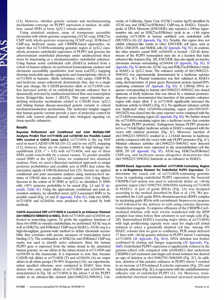
CRISPR-Based Approaches Identified rs17114036-Containing Regionas a cis-Regulatory Element for Endothelial PLPP3 Expression. Todetermine the causal role of rs17114036-containing genomiclocus in regulating endothelial PLPP3 expression, the bacterialCRISPR-Cas9 system was used to selectively delete an ∼66-bpgenomic region (chr1:56962783–56962849) enclosing rs17114036in HAECs. A pair of guide RNAs (Fig. 2A) was designedaccording to the method described by Ran et al. (19). We pre-assembled the Cas9–guide RNA ribonucleoprotein (RNP) complexby incubating guide RNAs with recombinant Streptococcus pyogenesCas9 followed by the delivery to cells using cationic liposometransfection reagents. To improve efficiency of the CRISPR-Cas9–mediated deletion, cells were reverse transfected with the RNPcomplex four times before flow cytometry to sort single cells (Fig.2B). Immortalized HAECs (carrying major alleles at rs17114036)with high proliferating capacity were used for single-cell clonalisolation to select a genetically identical cell line. Among 459HAEC colonies that we grew to confluency, PCR assays detected17 lines with ∼66-bp genetic deletion in PLPP3 intron 5 enclosingrs17114036 (SI Appendix, Fig. S6A). DNA deletion was furtherconfirmed by cloning and Sanger sequencing (SI Appendix, Fig.S6B). Endothelial PLPP3 expression is significantly reduced in thegenome-edited cells compared with teloHAECs that underwentCRISPR-Cas9 treatment and single-cell clonal isolation but showedno sign of deletion at chr1:56962783–56962849 (Fig. 2C). In addi-tion, deletion of this putative enhancer in PLPP3 intron 5 resultedin an increase of LPA-induced E-selectin expression (Fig. 2D) andleukocyte adhesion (Fig. 2E), in agreement with the antiinflammatory/adhesive role of endothelial PLPP3 (13, 14). Moreover, trans-endothelial electrical resistance detected increased monolayer
E11350 | www.pnas.org/cgi/doi/10.1073/pnas.1810568115 Krause et al.
Dow
nloa
ded
by g
uest
on
Apr
il 15
, 202
0
permeability in rs17114036-deleted HAECs (Fig. 2F), consistentwith PLPP3’s role in maintaining endothelial monolayer integrity(13, 14). CRISPR interference was recently developed to suppressthe activity of cis-regulatory elements (20). Here, we showed thatrs17114036-targeted (Fig. 2G) but not rs2184104-targeted (SIAppendix, Fig. S7) guide RNAs significantly reduce PLPP3 mRNAexpression in HAECs. These results demonstrate that rs17114036-containing region causatively regulates PLPP3 expression andendothelial functions.
Unidirectional Flow Increases the Enhancer Activity at chr1:56962213–56963412 in Vascular Endothelium. Given the critical role of he-modynamics in controlling endothelial PLPP3 transcription (13),
we tested whether shear stresses regulate the enhancer activity ofchr1:56962213–56963412. ATAC-seq and H3K27ac ChIP-seqwere conducted in HAECs subjected to “atheroprotective” uni-directional flow representing wall shear stress in human distalinternal carotid artery or “atherosusceptible” flow mimickinghemodynamics in human carotid sinus (21). ATAC-seq capturedincreased open chromatin at chr1:56962213–56963412 in HAECsunder unidirectional flow compared with cells under disturbedflow (Fig. 3A). H3K27ac ChIP-seq indicated an increased en-hancer activity of chr1:56962213–56963412 in HAECs underunidirectional flow (Fig. 3A). In addition, genetic deletion ofthe rs17114036-containing region by CRISPR-Cas9 significantly
Fig. 1. CAD-associated SNP rs17114036 is located in an enhancer element (chr1:56962213–56963412) in HAECs. (A) In silico prediction of causal SNPs in the CAD locus1p32.2. Diagrams demonstrate the association (−log10P) and LD pattern of a total of 45 common SNPs in the 1p32.2 locus. Green circles indicate possible causal SNPspredicted by the Bayesian Statistical Approach. Red circles indicate possible causal SNPs predicted by the Approximate Conditional and Joint Association Analysis.Purple circles indicate putative causal SNPs predicted by both statistical analyses. (B) Chromatin accessibility and canonical enhancer marks in chr1:56962213–56963412 region enclosing rs17114036 in HAEC. ATAC-seq and H3K27ac/H3K4me2 ChIP-seq collectively identified an enhancer-like region in chr1:56962213–56963412 in HAECs. All sequencing experiments were performed in duplicate, and the merged tracks are shown. (C) Enhancer activity of chr1:56962213–56963412 invascular endothelium. DNA sequences of chr1:56962213–56963412 were cloned into luciferase reporters (firefly luciferase construct pGL4) that contain a minimalpromoter or human PLPP3 promoter. The red asterisks denote the relative position of rs17114036 in the luciferase construct. Dual luciferase reporter assays wereconducted in HAECs 24 h after the electroporation-based transfection (using pRL-TK plasmid carrying Renilla luciferase as transfection controls) in HAECs, detectingincreased firefly luciferase as the result of insertion of chr1:56962213–56963412. Data represent mean ± SEM. ***P < 0.0005 as determined by Student’s t test.
Krause et al. PNAS | vol. 115 | no. 48 | E11351
MED
ICALSC
IENCE
S
Dow
nloa
ded
by g
uest
on
Apr
il 15
, 202
0

impaired unidirectional flow-induced PLPP3 expression in HAECs(Fig. 3B). Moreover, enhancer activity at chr1:56962213–56963412,measured by H3K27ac ChIP-PCR, was increased in control HAECbut not in rs17114036-(biallelic) deleted cells when subjected to 24 hof unidirectional flow (Fig. 3C). These results collectively demon-strate that enhancer activity of chr1:56962213–56963412 is dynam-ically activated by the atheroprotective unidirectional flow toregulate endothelial PLPP3.
CAD/IS-Protective Allele C at rs1711403 Confers a Higher EnhancerActivity of chr1:56962213–56963412. GWAS have linked the minorallele C at rs17114036 at 1p32.2 to reduced CAD/IS susceptibility(10–12), and our eQTL mapping described increased PLPP3 ex-pression in HAECs with minor allele C (13). Here, we investigated
the genotype-dependent effect of rs17114036 on the enhanceractivity of chr1:56962213–56963412 by ATAC-seq and luciferaseassays. In addition to HAEC lines carrying major (risk) allele T atrs17114036, we conducted ATAC-seq in HAECs isolated from do-nors who are heterozygous (T/C; ∼20% of Europeans) at rs17114036,allowing us to perform caQTL mapping. caQTL was recently de-veloped to detect between-individual signaling in cis-regulatory ele-ments as a function of genetic variants (22). ATAC-seq detectedsignificantly increased numbers of reads corresponding to rs17114036-containing region in HAEC lines that contain one CAD-protectiveallele (T/C) compared with HAECs from donors homozygous ofCAD risk allele (T/T) (Fig. 4A), supporting increased chromatin ac-cessibility associated with C allele at rs17114036. In addition, weconducted RNA-seq analysis in these cells, demonstrating that there
A
B
C D
Non-DeletionClones
DeletionClones
0.0
0.5
1.0
1.5
RelativePLPP3mRNA
***
Non-DeletionClones
DeletionClones
0.0
0.5
1.0
1.5
2.0
2.5
RelativeSELEmRNA
*
Non-DeletionClones
DeletionClones
0
1
2
3
4
RelativeCellAdhesion ***
Non-DeletionClones
DeletionClones
0.7
0.8
0.9
1.0
1.1
1.2
RelativeResistance
**
Non-Targeting
rs17114036Targeting
0.0
0.5
1.0
1.5
RelativePLPP3mRNA
*
dCas9-KRAB
E
F
Adhesion MoleculesEndothelial CellsMonocytes
Endothelial CellsIon CurrentElectrode G
70 bp offset
5'3'
Purified Cas9 sgRNA RNP Complexrs17114036
Cells insuspension
dCas9
PLPP3Transcription
rs17114036
5'3'
KRAB
KRAB
+
Treatment 1 Treatment 2 Treatment 3 Treatment 4 PCR ScreeningSingle Cell
Sorting
576 Clones
Fig. 2. CRISPR-based approaches identify rs17114036-containing genomic locus as a cis-regulatory element regulating endothelial PLPP3 expression. (A) RNARNP complex that contains recombinant S. pyogenes Cas9 and two sgRNAs flanking rs17114036. (B) Experimental overview of CRISPR-Cas9–mediated genomicdeletion in HAECs. TeloHAECs were treated with RNP complex four times before single-cell sorting and isogenic clone selection. (C) Reduced PLPP3 expression, (D)elevated E-selectin expression, (E) increased leukocyte adhesion, and (F) higher monolayer permeability in teloHAECs with genomic deletion at rs17114036-containing region. (G) Reduced PLPP3 expression in HAECs treated with CRISPR interference targeting rs17114036-containing region. n = 3–8. Data representmean ± SEM. *P < 0.05 as determined by Student’s t test; **P < 0.005 as determined by Student’s t test; ***P < 0.0005 as determined by Student’s t test.
E11352 | www.pnas.org/cgi/doi/10.1073/pnas.1810568115 Krause et al.
Dow
nloa
ded
by g
uest
on
Apr
il 15
, 202
0

Fig. 3. Unidirectional flow (UF) increases enhancer activity at chr1:56962213–56963412 that transcriptionally activates PLPP3 in human aortic endothelium.(A) Increased chromatin accessibility and H3K27ac mark at chr1:56962213–56963412 in HAECs subjected to 24-h atheroprotective UF compared with cellsunder 24-h atherosusceptible disturbed flow. The PLPP3 locus is shown and zoomed in to demonstrate details around the enhancer region of interest, withrs17114036 highlighted with a vertical blue line. (B) Reduced UF-induced PLPP3 expression in HAECs with genomic deletion at rs17114036-containing genomiclocus. Control and genome-edited (∼66-bp deletion) HAECs were subjected to 24-h UF. The y axis represents the fold change of PLPP3 mRNA quantitiesbetween the static conditions and UF for each individual clone. Nondeletion clones, n = 3; deletion clones, n = 4. (C) H3K27ac ChIP-PCR performed in twoCRISPR clones; one nondeletion clone and one biallelic deletion clone harboring the 66-bp deletion near rs17114036. The cells were subjected to static or UFconditions before cross-linking and ChIP. PCR primers were designed to amplify a region within the enhancer but not overlapping the deleted region. Dataare shown as fold change of the UF-treated samples compared with static conditions. Data represent mean ± SEM. *P < 0.05 as determined by Student’s t test;***P < 0.0005 as determined by Student’s t test.
Krause et al. PNAS | vol. 115 | no. 48 | E11353
MED
ICALSC
IENCE
S
Dow
nloa
ded
by g
uest
on
Apr
il 15
, 202
0

is a strong correlation between enhanced chromatin accessibility inrs17114036-containing region and increased mRNA levels of PLPP3in HAECs (Fig. 4B) and further suggesting that chr1:56962213–56963412 functions as an enhancer in promoting endothelialPLPP3 transcription. Moreover, ATAC-seq experiments in HAEClines heterozygous at rs17114036 further allow us to determinewhether the chromosome with C at rs17114036 exhibits higherchromatin accessibility at chr1:56962213–56963412 compared withthe chromosome with T allele. This is achieved by the AI analysis,which assigns next generation sequencing reads overlapping het-erozygous sites to one chromosome or the other for allele-specificsignals (23). ATAC-seq detected reads enriched from the C-containing chromosome compared with that from T allele inHAECs heterozygous at rs1711403 (Fig. 4C), further supporting the
increased chromosome accessibility associated with C allele atrs17114036. Lastly, luciferase assays were conducted to supportthe genotype-dependent enhancer activity of chr1:56962213–56963412. Replacement of T allele with C allele led to a muchhigher luciferase activity (∼5.2-fold, C vs. T) in endothelium (Fig.4D). Polymorphisms (A or G) at rs2184104 had no effect of thechr1:56911623–56912823 in the luciferase assay (SI Appendix, Fig.S8). Taken together, these results demonstrate that CAD-protective allele C at rs17114036 confers a higher enhancer ac-tivity of chr1:56962213–56963412 to promote PLPP3 expression invascular endothelium.
CAD/IS-Protective C Allele at rs1711403 Promotes Flow-Induced, KLF2-Mediated Enhancer Activity of chr1:56962213–56963412. We furtherexamined whether the genetic variants at rs17114036 modulate
Fig. 4. CAD-protective allele C at rs17114036 confers higher enhancer activity of chr1:56962213–56963412. (A) Increased ATAC-seq reads in chr1:56962213–56963412 region from HAECs isolated from people heterogeneous (T/C) at rs17114036 compared with HAECs from people heterozygous (T/T) at rs17114036.(B) A positive correlation (R = 0.6, P value = 6.23e-06) between ATAC-seq reads at chr1:56962213–56963412 and PLPP3 mRNA detected by RNA-seq in 56 HAEClines. (C) Increased ATAC-seq reads at rs17114036-containing genomic locus from C (rs17114036)-containing chromosome compared with T-containingchromosome in HAEC lines heterozygous (T/C) at rs17114036. (D) Increased enhancer activity of chr1:56962213–56963412 with C allele at rs17114036 com-pared with T allele. Dual luciferase reporter assays were conducted in teloHAEC. The red and green asterisks denote the relative position of rs17114036polymorphisms in the luciferase construct. n = 4–6. Data represent mean ± SEM. ***P < 0.0005 as determined by Student’s t test.
E11354 | www.pnas.org/cgi/doi/10.1073/pnas.1810568115 Krause et al.
Dow
nloa
ded
by g
uest
on
Apr
il 15
, 202
0

the flow-induced enhancer activity of chr1:56962213–56963412.Luciferase assays detected an increase of the enhancer activity ofchr1:56962213–56963412 (with protective C allele at rs17114036)in cells under 18-h unidirectional flow compared with disturbedflow (Fig. 5A), while no significant increase of the enhanceractivity by unidirectional flow was detected with the risk T allele(SI Appendix, Fig. S9). In addition to the ATAC-seq experimentsin HAECs homozygous at rs17114036 under flow (Fig. 3), weperformed ATAC-seq analysis in four HAEC lines heterozygousat rs17114036 under 24-h unidirectional flow to perform openchromatin AI analysis. SI Appendix, Fig. S10 demonstrates that,in all four selected HAEC lines heterozygous at rs17114036,unidirectional flow increases ATAC-seq peaks in the proposedenhancer region in PLPP3 intron 5, in agreement with increasedATAC-seq reads in rs17114036-containing region (Fig. 5B).Moreover, AI analysis showed an enrichment of ATAC readsfrom the chromosome harboring the protective C allele (Fig.5B). In contrast, ATAC-seq detected no AI at rs6421497, acommon SNP in high LD with rs17114036 (SI Appendix, Fig.S11). Indeed, the protective allele C at rs17114036 creates aCACC box that is a binding site for KLF2, which mediates theflow sensitivity of a cohort of endothelial genes, including PLPP3(13, 24–27). We then tested whether KLF2 dynamically activatesthis rs17114036-containing enhancer and if rs17114036 allelesimpact KLF2-mediated enhancer activity. The affinity ofKLF2 to the rs17114036-containing locus was determined byKLF2 ChIP-PCR assays in HAECs carrying a protective allele atrs17114036 (T/C), showing a physical binding of KLF2 to thers17114036-containing region and the CACC site in the PLPP3promoter (Fig. 5C). Enhancer activities of chr1:56962213–56963412 were further determined in HAECs as a function ofKLF2 expression. Constructs of enhancer (chr1:56962213–56963412) and PLPP3 promoter were cotransfected with KLF2-overexpressing plasmids. Luciferase assays detected a 2.9-foldincrease of luciferase activity in the T allele-containing con-struct as the result of KLF2 overexpression (Fig. 5D). Moreover,KLF2 overexpression led to a 4.7-fold increase of luciferaseactivity when T allele was substituted by the protective allele C atrs17114036 (Fig. 5D). Collectively, KLF2 ChIP-PCR and lucif-erase assays demonstrate that CAD/IS-protective allele C atrs17114036 confers a higher KLF2-dependent enhancer activityof chr1:56962213–56963412 in vascular endothelium.
DiscussionAlthough it is proposed that genetic and environmental factorsjointly influence the risk of most common human diseases, theinterplay between genetic predisposition and biomechanical cuesat the molecular level is poorly understood. The biology un-derlying the majority of CAD and IS GWAS loci remains to beelucidated (28). Most of the CAD and IS SNPs reside in thenoncoding genome. Gupta et al. (29) recently reported that thenoncoding common variant at rs9349379, implicated in CAD byGWAS, regulates endothelin 1 expression in endothelium.Atherosclerotic lesions preferentially develop in elastic ar-teries where vascular endothelial cells are activated by localdisturbed flow (2–5). As of now, it remains unknown whetherdisease-associated genetic variants contribute to mechanosensingmechanisms by which cells sense and convert biomechanicalstimuli to biological signaling. Our results here elucidate theconvergence of CAD/IS genetic predisposition and mechano-transduction mechanisms in endothelial PLPP3 expression. Sta-tistical analyses, whole-genome chromatin accessibility/enhancermarks, CRISPR interference (CRISPRi), genome editing, enhancerassays, caQTL mapping, and AI assay collectively demonstratethat CAD/IS locus 1p32.2 harbors a mechanosensitive endothe-lial enhancer that regulates PLPP3 expression. Moreover, CAD/IS-protective allele at rs17114036 confers an increased enhancer
activity that is dynamically regulated by unidirectional flow andtranscription factor KLF2 (Fig. 5E).Dysregulation of mechanosensing mechanisms contributes to
the etiology of a wide range of human diseases in cardiovascular,pulmonary, orthopedic, muscular, and reproductive systems (1).The genetic basis of these complex human diseases has beenstrongly suggested by GWAS, but the interplay between geneticvariants and mechanosensing mechanisms has not been in-vestigated. Our data provide a line of evidence supporting thegenetic regulation of mechanotransduction mechanisms incomplex human diseases and suggest an underappreciated roleof genetic predisposition in cellular mechanosensing processes.Transcriptional enhancers orchestrate the majority of cell
type-specific patterns of gene expression and play key roles indevelopment, evolution, and disease (7), which are tightly reg-ulated by mechanical cues (1). Our data provide molecular evi-dence that the noncoding genome actively participates in cellularmechanotransduction mechanisms that are influenced by humangenetic variances. In addition to the flow regulation of the spe-cific locus 1p32.2, our results provide a dataset to systematicallydetermine the mechanosensitive chromatin accessibility andputative enhancer regions at the whole-genome scale in vascularendothelium. It is important to note that most of the epigenomestudies including ENCODE were conducted in cells withoutphysiological or pathophysiological mechanical stimuli, such asHUVECs under static (no flow) conditions (6). Since majorendothelial functions are tightly and dynamically regulated byhemodynamics flow, our whole-genome epigenome profiling inHAECs under atherorelevant flows may benefit future studies toinvestigate mechanical regulation of the noncoding genome invascular cells. Indeed, we have applied model-based analysis ofChIP-seq (30) and HOMER differential analysis (31), whichunbiasedly identified rs17114036-containing locus as 1 of36,965 open chromatin sites that are activated by unidirectionalflow (SI Appendix, Fig. S12).Mechanosensitive transcription factors have been proposed as
major regulators to determine endothelial functions relevant toatherogenesis. For instance, nuclear factor-κB and HIF-1α me-diate gene sets associated with proinflammatory, procoagulant,and glycolytic endothelial phenotypes under disturbed flow (32–35), while KLFs and nuclear factor erythroid 2-like 2 regulategene networks promoting the quiescent endothelial phenotypeunder unidirectional flow (25–27, 36–39). However, the in-teraction between flow-sensitive transcription factors anddisease-associated genetic predisposition in vascular functionshas not been suggested. Our results demonstrate that a geneticvariant can influence important endothelial functions via anoncoding enhancer region recognized by the mechanosensitivetranscription factor KLF2. These results are consistent withemerging evidence showing that top-scoring disease-associatedSNPs are frequently located within enhancers explicitly activein disease-relevant cell types (9, 40). Moreover, the data suggestthat disease-associated genetic variants, via modulation of tran-scription factor binding, may regulate the enhancer activitiesdynamically responding to biomechanical cues that are instrumentalto key cellular processes.GWAS related to atherosclerotic diseases have suggested
previously unsuspected loci, genes, and biology involved in li-poprotein metabolism (28), resulting in the development of newcholesterol-lowering therapies (41). Despite that dyslipidemia isa major systemic risk factor of CAD and IS, atherosclerotic le-sions largely initiate and develop at arterial regions of atypicalvascular geometry associated with disturbed flow. Previousstudies demonstrated that cellular mechanotransduction mech-anisms, particularly endothelial responses to hemodynamics,causatively contribute to the focal nature of atherosclerotic le-sions (2–5, 42). Our studies here demonstrate that genetic vari-ants contribute to not only interindividual variation in plasma
Krause et al. PNAS | vol. 115 | no. 48 | E11355
MED
ICALSC
IENCE
S
Dow
nloa
ded
by g
uest
on
Apr
il 15
, 202
0

Fig. 5. CAD-protective C allele at rs17114036 promotes flow-induced, KLF2-mediated enhancer activity of chr1:56962213–56963412. (A) Increased enhanceractivity of chr1:56962213–56963412 (with C allele at rs17114036) under unidirectional flow (UF) but not disturbed flow (DF). Experiment was performed inbiological triplicate and technical triplicate. Data represent mean ± SEM. *P < 0.05 as determined by two-way ANOVA; **P < 0.005 as determined by two-wayANOVA. The green asterisk denotes the relative position of rs17114036 in the luciferase construct. (B) Increased ATAC-seq reads in rs17114036-containing regionin HAECs under UF compared with DF. ATAC-seq was conducted in four HAEC lines heterozygous at rs17114036 under 24-h atherorelevant flows, detectingincreased ATAC reads in cells under UF and higher reads from the C allele-containing chromosome compared with T allele. (C) KLF2 affinity to CACC sites inhuman PLPP3 promoter and intron 5. ChIP-qPCR was performed with either a control IgG antibody or the antibody against HA followed by qPCR using primersdetecting CACC sites in PLPP3 promoter or rs17114036-enclosing region from control HAECs (Ctrl) or HAECs transfected with KLF2 transcripts with HA tag. Primersthat detect a site ∼200 bp from the CACC at rs17114036 were used as a negative control. n = 4. (D) Increased enhancer activity of chr1:56962213–56963412 byKLF2 overexpression. Dual luciferase reporter assays were conducted in HAECx transfected with luciferase constructs containing the PLPP3 promoter and enhancerwith either the major (T) or minor (C) allele at s17114036 along with KLF2-overexpressing or control plasmids. KLF2 overexpression resulted in a 2.9-fold increasedluciferase activity in HAECs transfected with T allele-containing construct and a 4.7-fold increase in cells transfected with C allele-containing construct. n = 3. Datarepresent mean ± SEM. *P < 0.05 as determined by Student’s t test. (E) The interplay between hemodynamic forces, chromatin landscapes at PLPP3 intron 5, andrs17114036 at the molecular level in regulating endothelial PLPP3 expression and vascular functions.
E11356 | www.pnas.org/cgi/doi/10.1073/pnas.1810568115 Krause et al.
Dow
nloa
ded
by g
uest
on
Apr
il 15
, 202
0
lipid concentrations (43) but also, endothelial responses to bloodflow. Indeed, genetic variants at rs17114036 predict CAD sus-ceptibility independent of traditional systemic risk factors, suchas cholesterol and diabetes mellitus (10, 11). Recent GWASidentified 15 new CAD risk loci near genes of key functions inendothelial, smooth muscle, and white blood cells (44), furtherhighlighting the potential importance of genetic contribution tothe arterial wall-specific mechanisms in atherogenesis. Our re-sults indicate that CAD genetic predisposition and disturbedflow converge to inhibit endothelial PLPP3 expression and thatrestoration of endothelial PLPP3 in atherosusceptible regionsmay provide an attractive approach for future arterial wall-basedatherosclerosis therapy complementary to current pharmaco-logical treatments targeting systemic risk factors.Our studies demonstrate that the latest human genetics ap-
proaches, such as caQTL mapping (22), AI assay (23), CRISPRi(20), and CRISPR-based assays (19), are powerful tools to in-vestigate possible genetic contributions to cellular mechano-transduction. Miao et al. (45) recently applied CRISPR-Cas9 toachieve high efficiency of a 10-kb deletion of an enhancer re-gion in bulk HUVECs. Here, we expanded the applications ofCRISPR-based techniques to investigate key vascular functions.Isogenic adult aortic endothelial lines subjected to CRISPR-based deletion were successfully selected to determine thecausal role of ∼66-bp genomic region in regulating endothelialPLPP3. Nevertheless, one limitation here is that we are stillunable to replace this human SNP at rs17114036 in adult aorticendothelium, although we have tried various methods to pro-mote homology-directed repair. This will be the subject of afuture study. Nevertheless, caQTL mapping (22) and AI assay(23) provide complementary approaches detecting at the single-nucleotide resolution that CAD/IS-protective allele at rs17114036confers a higher enhancer activity at the PLPP3 intron 5.Cellular mechanotransduction is required for physiological
control of tissue homeostasis, while abnormal cell response tomechanical forces promotes pathologies of numerous humandiseases. Although investigations, such as GWAS, have sug-gested the genetic basis of complex human diseases, the interplaybetween genetic predispositions of mechanosensing mechanismsremains virtually unknown. Our results identified that CAD-associated genetic variant at rs17114036 interacts with hemody-namics in concert to regulate endothelial PLPP3 expression andconsequently, key vascular functions. Moreover, our experiments
provide evidence supporting the regulatory role of the non-coding, nontranscribed genome in mechanotransduction mecha-nisms. In summary, this study demonstrates that human haplotypesand related cis-regulatory elements provide an important layer ofmolecular control by which cells convert physical stimuli intobiological signaling.
MethodsATAC-Seq. ATAC-seq was performed as previously described (17) usingTn5 transposase (Illumina). Libraries were sequenced on an Illumina HiSeq4000 according to the manufacturer’s specifications by the Genomics CoreFacility at the University of Chicago. The reads were aligned to the UCSChg19 genome using Bowtie2 (46). ATAC-seq was conducted in HAECs understatic conditions or subjected to 24-h unidirectional flow or disturbed flow.
caQTL Mapping and AI. caQTL mapping was performed to test for associationbetween genotype at rs17114036 and chromatin accessibility measuredby ATAC-seq. We pulled genotypes for HAEC donors from our previousstudy (47) and imputed linked alleles using IMPUTE2 and SHAPEIT as wepublished previously (40). Association testing between ATAC-seq tags at thers17114036 enhancer and genotype was performed using the CombinedHaplotype Test in WASP (23).
To perform AI analysis that assigns next generation sequencing readsoverlapping heterozygous sites to one chromosome or the other, wequantified ATAC-seq tags at the rs17114036 enhancer using HOMER’sannotatePeaks function to express the log2-normalized tags in this region.
CRISPR-Cas9–Mediated Deletion of Enhancer in TeloHAECs. The CRISPR re-agents were adapted from the Alt-R system from IDT. The guide RNAs weredesigned using an online tool at crispr.mit.edu/ to minimize off-targetingeffects using two guides to create an ∼66-bp deletion. The guide RNAs weremade by annealing the transactivating CRISPR RNA (tracrRNA) to the singleguide RNA (sgRNA). The RNP complex of S. Pyogenes Cas9 and sgRNA wasformed by placing the components together at room temperature and thenimmediately transfected into cells using Lipofectamine RNAiMAX (Thermo). Foreach successive treatment, the reagent amounts were scaled relative to the sizeof the destination vessel to compensate for the number of cells in the reaction.The volumes for each part of the reaction were increased 4×when treating cellsfrom the 96-well to the 6-well and 16× when moving from the 6-well to the T-75 flask.
Detailed methods are available in SI Appendix.
ACKNOWLEDGMENTS. This work was funded by NIH Grants T32 HL007381(to M.D.K.), F32 HL134288 (to D.W.), T32 EB009412 (to D.L.H.), R00 HL121172(to M.C.), R00 HL123485 (to C.E.R.), R01 HL136765 (to Y.F.), and R01HL138223 (to Y.F.) as well as American Heart Association Grant BGIA7080012(to Y.F.)
1. Jaalouk DE, Lammerding J (2009) Mechanotransduction gone awry. Nat Rev Mol Cell
Biol 10:63–73.2. Davies PF, Civelek M, Fang Y, Fleming I (2013) The atherosusceptible endothelium:
Endothelial phenotypes in complex haemodynamic shear stress regions in vivo.
Cardiovasc Res 99:315–327.3. Hahn C, Schwartz MA (2009) Mechanotransduction in vascular physiology and ath-
erogenesis. Nat Rev Mol Cell Biol 10:53–62.4. Zhou J, Li YS, Chien S (2014) Shear stress-initiated signaling and its regulation of
endothelial function. Arterioscler Thromb Vasc Biol 34:2191–2198.5. Gimbrone MA, Jr, García-Cardeña G (2016) Endothelial cell dysfunction and the
pathobiology of atherosclerosis. Circ Res 118:620–636.6. Consortium EP; ENCODE Project Consortium (2012) An integrated encyclopedia of
DNA elements in the human genome. Nature 489:57–74.7. Ong CT, Corces VG (2011) Enhancer function: New insights into the regulation of
tissue-specific gene expression. Nat Rev Genet 12:283–293.8. Heinz S, Romanoski CE, Benner C, Glass CK (2015) The selection and function of cell
type-specific enhancers. Nat Rev Mol Cell Biol 16:144–154.9. Ernst J, et al. (2011) Mapping and analysis of chromatin state dynamics in nine human
cell types. Nature 473:43–49.10. Schunkert H, et al.; Cardiogenics; CARDIoGRAM Consortium (2011) Large-scale asso-
ciation analysis identifies 13 new susceptibility loci for coronary artery disease. Nat
Genet 43:333–338.11. Deloukas P, et al.; CARDIoGRAMplusC4D Consortium; DIAGRAM Consortium;
CARDIOGENICS Consortium; MuTHER Consortium; Wellcome Trust Case Control
Consortium (2013) Large-scale association analysis identifies new risk loci for
coronary artery disease. Nat Genet 45:25–33.12. Dichgans M, et al.; METASTROKE Consortium; CARDIoGRAM Consortium; C4D Con-
sortium; International Stroke Genetics Consortium (2014) Shared genetic suscepti-
bility to ischemic stroke and coronary artery disease: A genome-wide analysis of
common variants. Stroke 45:24–36.13. Wu C, et al. (2015) Mechanosensitive PPAP2B regulates endothelial responses to
atherorelevant hemodynamic forces. Circ Res 117:e41–e53.14. Panchatcharam M, et al. (2014) Mice with targeted inactivation of ppap2b in endo-
thelial and hematopoietic cells display enhanced vascular inflammation and perme-
ability. Arterioscler Thromb Vasc Biol 34:837–845.15. Maller JB, et al.; Wellcome Trust Case Control Consortium (2012) Bayesian refinement
of association signals for 14 loci in 3 common diseases. Nat Genet 44:1294–1301.16. Yang J, et al. (2012) Conditional and joint multiple-SNP analysis of GWAS summary
statistics identifies additional variants influencing complex traits. Nat Genet 44:
369–375.17. Buenrostro JD, Giresi PG, Zaba LC, Chang HY, Greenleaf WJ (2013) Transposition of
native chromatin for fast and sensitive epigenomic profiling of open chromatin, DNA-
binding proteins and nucleosome position. Nat Methods 10:1213–1218.18. Abecasis GR, et al.; 1000 Genomes Project Consortium (2012) An integrated map of
genetic variation from 1,092 human genomes. Nature 491:56–65.19. Ran FA, et al. (2013) Genome engineering using the CRISPR-Cas9 system. Nat Protoc 8:
2281–2308.20. Qi LS, et al. (2013) Repurposing CRISPR as an RNA-guided platform for sequence-
specific control of gene expression. Cell 152:1173–1183.21. Dai G, et al. (2004) Distinct endothelial phenotypes evoked by arterial waveforms
derived from atherosclerosis-susceptible and -resistant regions of human vasculature.
Proc Natl Acad Sci USA 101:14871–14876.22. Kumasaka N, Knights AJ, Gaffney DJ (2016) Fine-mapping cellular QTLs with RASQUAL
and ATAC-seq. Nat Genet 48:206–213.23. van de Geijn B, McVicker G, Gilad Y, Pritchard JK (2015) WASP: Allele-specific software
for robust molecular quantitative trait locus discovery. Nat Methods 12:1061–1063.
Krause et al. PNAS | vol. 115 | no. 48 | E11357
MED
ICALSC
IENCE
S
Dow
nloa
ded
by g
uest
on
Apr
il 15
, 202
0
24. Dekker RJ, et al. (2006) KLF2 provokes a gene expression pattern that establishesfunctional quiescent differentiation of the endothelium. Blood 107:4354–4363.
25. SenBanerjee S, et al. (2004) KLF2 is a novel transcriptional regulator of endothelialproinflammatory activation. J Exp Med 199:1305–1315.
26. Parmar KM, et al. (2006) Integration of flow-dependent endothelial phenotypes byKruppel-like factor 2. J Clin Invest 116:49–58.
27. Dekker RJ, et al. (2005) Endothelial KLF2 links local arterial shear stress levels to theexpression of vascular tone-regulating genes. Am J Pathol 167:609–618.
28. Nurnberg ST, et al. (2016) From loci to biology: Functional genomics of genome-wideassociation for coronary disease. Circ Res 118:586–606.
29. Gupta RM, et al. (2017) A genetic variant associated with five vascular diseases is adistal regulator of endothelin-1 gene expression. Cell 170:522–533.e15.
30. Zhang Y, et al. (2008) Model-based analysis of ChIP-seq (MACS). Genome Biol 9:R137.31. Heinz S, et al. (2010) Simple combinations of lineage-determining transcription fac-
tors prime cis-regulatory elements required for macrophage and B cell identities. MolCell 38:576–589.
32. Lan Q, Mercurius KO, Davies PF (1994) Stimulation of transcription factors NF kappa Band AP1 in endothelial cells subjected to shear stress. Biochem Biophys Res Commun201:950–956.
33. Khachigian LM, Resnick N, Gimbrone MA, Jr, Collins T (1995) Nuclear factor-kappa Binteracts functionally with the platelet-derived growth factor B-chain shear-stressresponse element in vascular endothelial cells exposed to fluid shear stress. J ClinInvest 96:1169–1175.
34. Wu D, et al. (2017) HIF-1α is required for disturbed flow-induced metabolic re-programming in human and porcine vascular endothelium. eLife 6:e25217.
35. Feng S, et al. (2017) Mechanical activation of hypoxia-inducible factor 1α drives en-dothelial dysfunction at atheroprone sites. Arterioscler Thromb Vasc Biol 37:2087–2101.
36. Huang RT, et al. (2017) Experimental lung injury reduces Krüppel-like factor 2 to in-crease endothelial permeability via regulation of RAPGEF3-Rac1 signaling. Am JRespir Crit Care Med 195:639–651.
37. Fang Y, Davies PF (2012) Site-specific microRNA-92a regulation of Kruppel-like factors4 and 2 in atherosusceptible endothelium. Arterioscler Thromb Vasc Biol 32:979–987.
38. Li Z, et al. (2017) Krüppel-like factor 4 regulation of cholesterol-25-hydroxylase andliver X receptor mitigates atherosclerosis susceptibility. Circulation 136:1315–1330.
39. Zhou G, et al. (2012) Endothelial Kruppel-like factor 4 protects against athero-thrombosis in mice. J Clin Invest 122:4727–4731.
40. Hogan NT, et al. (2017) Transcriptional networks specifying homeostatic and in-flammatory programs of gene expression in human aortic endothelial cells. eLife 6:e22536.
41. Sabatine MS, et al.; FOURIER Steering Committee and Investigators (2017) Evolocumaband clinical outcomes in patients with cardiovascular disease. N Engl J Med 376:1713–1722.
42. Nam D, et al. (2009) Partial carotid ligation is a model of acutely induced disturbedflow, leading to rapid endothelial dysfunction and atherosclerosis. Am J Physiol HeartCirc Physiol 297:H1535–H1543.
43. Willer CJ, et al.; Global Lipids Genetics Consortium (2013) Discovery and refinement ofloci associated with lipid levels. Nat Genet 45:1274–1283.
44. Howson JMM, et al.; CARDIoGRAMplusC4D; EPIC-CVD (2017) Fifteen new risk loci forcoronary artery disease highlight arterial-wall-specific mechanisms. Nat Genet 49:1113–1119.
45. Miao Y, et al. (2018) Enhancer-associated long non-coding RNA LEENE regulatesendothelial nitric oxide synthase and endothelial function. Nat Commun 9:292.
46. Langmead B, Salzberg SL (2012) Fast gapped-read alignment with Bowtie 2. NatMethods 9:357–359.
47. Romanoski CE, et al. (2010) Systems genetics analysis of gene-by-environment inter-actions in human cells. Am J Hum Genet 86:399–410.
E11358 | www.pnas.org/cgi/doi/10.1073/pnas.1810568115 Krause et al.
Dow
nloa
ded
by g
uest
on
Apr
il 15
, 202
0