Embed Size (px)
Citation preview
www.phgfoundation.org
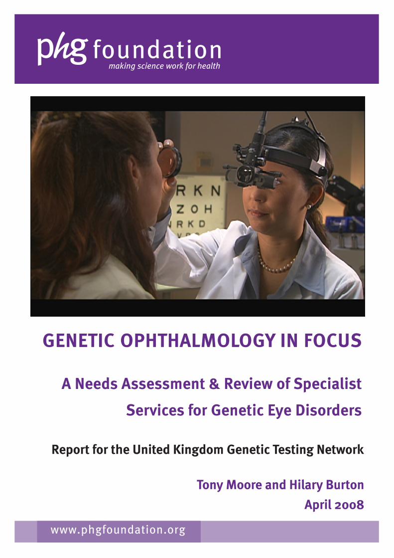
GENETIC OPHTHALMOLOGY IN FOCUS
Report for the United Kingdom Genetic Testing Network
Tony Moore and Hilary Burton
April 2008
A Needs Assessment & Review of Specialist
Services for Genetic Eye Disorders
Authors
Tony Moore Professor of Ophthalmology and Chairman of the Group
Institute of OphthalmologyUCL, London
Hilary BurtonConsultant in Public Health Medicine and Programme Director
PHG Foundation, Cambridge
Project coordinator
Corinna AlbergPHG Foundation, Cambridge
For additional copies of the Report please contact:
The full report can be downloaded from the PHG Foundation website: http://www.phgfoundation.org/pages/projectlist.htm
Acknowledgements The Project team would like to thank all members of the Working Group for sharing so freely their expertise and in particular Clive Fisher for organising the contribution of the voluntary organisations. We would also like to acknowledge the support of Catey Bunce of Moorfields Eye Hospital and Jugnoo Rahi from the Institute of Child Health, for assistance with the epidemiological chapter. Thanks are due to the Department of Health for funding the meetings and to Jo Melhuish and the Institute of Ophthalmology for arranging and hosting the Working Group meetings. The PHG Foundation is concerned with the vital task of preparing and enabling health systems to evaluate and prioritise new scientific and medical knowledge and technologies, and integrate them effectively into their service and practice.
It works at the forefront of biomedical innovation and public health with a current focus on public health genomics, and the translation of genome-based knowledge and technologies for the benefit of population health.
�www.phgfoundation.org
Summary of policy pointsAll patients with genetic eye disorders should have access to specialist care from teams with particular knowledge and experience in the diagnosis and management of these rare conditions. Such teams must combine specialist ophthalmology, genetics, genetic counselling, laboratory molecular genetics and electrophysiology.
A UK needs assessment and review of services was undertaken at the request of the UKGTN Sub-Committee on Clinical Appropriateness. Chaired by Professor Tony Moore and coordinated by the PHG Foundation, the review had a particular emphasis on the availability and utility of genetic testing in eye disorders both now and in the near future.
It is estimated that each year around 150 children and 250 adults of working age are newly diagnosed as blind or partially sighted, as a result of a genetic disorder. Others require expert help as potentially affected family members.
There is a wide disparity across the UK in provision of specialist services with some areas having little or no service.
Genetic testing in ophthalmology has demonstrable clinical utility in the main areas of increased information from better diagnosis and prognosis, decreased morbidity and mortality through preventive care and informing treatment options, improved process of care and provision of information to assist reproductive choice. Molecular diagnosis will become increasingly important with the development of novel treatments that are genotype specific, as new technologies such as microarray become available, and as our knowledge of genes associated with susceptibility to complex disorders increases. There are, however, many barriers to testing including overall lack of capacity, complexity of the underlying genetics in these disorders, technological aspects, cost, lack of formal information on test evaluation and methods of funding.
Specialist provision needs to be expanded and developed across the UK. The prime strategic elements to achieve this are:
Developing and supporting commissioning by Primary Care Trust commissioners for specialist genetics ophthalmology with urgent review in areas with no provision
• Developing a service specification for specialist genetic ophthalmology servicesDeveloping integrated service models to ensure comprehensive services now and future ability to respond effectively to new technologies. This is likely to be based on a limited number of regional or supraregional centres where ophthalmologists, ideally with a sub-specialty interest in genetics, either work alongside clinical geneticists in joint clinics or liaise closely with the regional genetics service to ensure that families receive a high quality and comprehensive package of care. Ensuring access to specialist services and new technologies through development and implementation of care pathways, referral criteria, systems for shared care, and appropriate information systems.Increasing clinical capacity including, medical, surgical, nursing, genetic counselling, electrophysiology and other specialist support services.Keeping genetic test provision under review as needs and technologies develop. Strategic work should continue to ensure test evaluation, coordination and efficiency of test provision, test accessibility and ‘gate-keeper’ functions, funding of genetic tests, the appropriateness of prioritisation tools and consider the use of commercial providers.Promoting the development of ophthalmic genetics as a sub-speciality within ophthalmology through the Specialised Services National Definition Set. This will also require that sub-speciality training in inherited eye disease is provided within Higher Surgical Training programmes in OphthalmologyPromoting special interest training in genetic ophthalmology for geneticists and genetic counsellors through links to the specialist genetic ophthalmology centres.
• Increasing knowledge and awareness about genetics in mainstream ophthalmology
Finally, it is recommended that an Implementation Board be set up with appropriate and representative membership in order to maintain momentum and oversee the next steps.
•
•
•
•
•
•
•
�www.phgfoundation.org
Foreword for commissioners and policy-makers
This detailed examination of genetics in ophthalmology was undertaken to provide a concrete example through which generic questions about genetics in mainstream medicine might be addressed. It provides a practical illustration of the opportunities offered by genetic and genomic science both now and in the near future, the ways in which health services need to adapt and develop in order to take advantage of the new science, the opportunities and barriers they face and some of the policy options that will be important in shaping future services.
We believe such a detailed examination, undertaken with the many stakeholders involved in such a service, is a necessary step in translating science into health services. Although the body of the report is specific to ophthalmology, many general concepts, such as those of clinical utility in genetic testing, problems in developing genetic tests and the work on new technologies are applicable in most clinical areas.
With parallel developments of genetics in many other mainstream specialities, the findings of service inequity and the implications for service development evident from examination of this one clinical area will be multiplied. There is an opportunity now for health services to be shaped in a way that can best capitalise on genomic advances, but this will only happen through firm involvement of policy makers and commissioners in partnership with service providers and users. The issues that will need to be addressed are drawn together in the final chapter, which is, therefore, of direct relevance to commissioners and policy-makers concerned with realising the potential of genomics throughout the health service.
5www.phgfoundation.org
Contents
1 Introduction and background
7
2 Epidemiology
11
� The patient viewpoint
20
� Genetic ophthalmology services
26
5 Evaluating genetic tests in ophthalmology: exploring clinical utility
29
6 Laboratory services
�9
7 Survey of clinical genetic ophthalmology services
50
8 Horizon scanning
71
9 Discussion and recommendations
77
10 Lessons for mainstream medicine
87
Appendices
1 Stakeholder group participant list
92
2 References
9�
� List of abbreviations used
96

6www.phgfoundation.org
7www.phgfoundation.org
Chapter 1 Introduction and background
1.1 Introduction
Ophthalmology is an area of mainstream medicine where molecular genetic testing for inherited eye disease is becoming an important aspect of the service. There are a large number of single gene disorders causing disease of various structures of the eye (such as the retina, lens and cornea), which are associated with visual impairment. Genetic mutations may result in conditions affecting the eye alone or may be associated with other systemic abnormalities such as hearing impairment, progressive neurological deficits, learning disability and physical abnormalities. Genetic factors are also important in common conditions causing visual impairment, including age-related macular degeneration (AMD), glaucoma and cataract.
This needs assessment and review was undertaken as a detailed example of genetics within an important mainstream clinical area. Throughout, the opportunity was taken to identify lessons that would be applicable in other areas of mainstream medicine. These lessons are presented in the final chapter.
1.2 Aims of the working group
The aims and objectives of the Working Group were as follows:
Aim
To undertake a needs assessment for specialist genetic ophthalmology services and molecular genetic testing in monogenic and complex eye disease.
Scope
To include disorders of the eye causing bilateral visual impairment, excluding other disorders such as strabismus and refractive errors, and excluding most syndromic conditions.To include disorders of children and adults.To include England, Wales, Scotland and Northern Ireland.
Objectives
A To undertake a needs assessment covering the following main aspects:
1. To briefly review the epidemiology of single gene disorders of the eye and the contribution of these disorders to visual loss.2. To list the genetic tests currently available in UK genetics laboratories. To describe the current clinical services and gaps in service provision.�. To obtain participants' views on current important gaps in the provision of genetic tests and clinical services.�. To review current knowledge of the genetic factors involved in the pathogenesis of complex eye disease (age-related macular degeneration and glaucoma). To consider the implications of this knowledge for clinical practice.5. To collect and summarise available information on the evaluation of currently available tests (eg submitted gene dossiers on these conditions).6. To engage with patient and voluntary groups to obtain their views on available
8www.phgfoundation.org
services and the need for genetic tests.7. To develop an understanding of the ACCE Framework (Analytic validity, Clinical validity, Clinical utility, Ethical, legal and social issues) for genetic test evaluation in the context of ophthalmology.8. To describe the main parameters of clinical utility in ophthalmology including economic considerations where possible. 9. To develop a mechanism for prioritisation of current and future genetic tests in
ophthalmology and consider the generalisability of this for genetic testing in other specialities.
10. To assess the likely impact of new genomic technologies on the diagnosis and management of genetic eye disease ('horizon scanning').
11. To make recommendations on generalisability to other specialities.
B To report to the UK Genetic Testing Network (UKGTN) Sub-Committee on Clinical Appropriateness by September 2007
1.3 Method
The working group was led by Professor Moore and the project supported by Hilary Burton, Rajalakshmi Lakshman and Corinna Alberg at the PHG Foundation, Cambridge1. The working group included experts on laboratory, clinical and genetic aspects of ophthalmology genetics. Patient and voluntary organisation viewpoint was provided by Clive Fisher from the British Retinitis Pigmentosa Society.
Participants were invited, through discussion between the Chairman and the project team, and the full Working Group was as follows:
Professor Tony Moore, Professor of Ophthalmology, Institute of Ophthalmology (Chairman)Dr Hilary Burton, Programme Director, PHG Foundation, Cambridge (Project Manager)Ms Corinna Alberg, PHG Foundation, Cambridge (Project Coordinator)Professor Graeme Black, Clinical Geneticist, University of ManchesterMs Sue Carless, Genetic Nurse Counsellor, Birmingham Hospital, Edgbaston, BirminghamMs Susan Downes, Consultant Ophthalmologist, Oxford Eye HospitalMr Clive Fisher, Board Member, British Retinitis Pigmentosa SocietyDr Rajalakshmi Lakshman, Specialist Registrar, PHG Foundation, CambridgeMs Sue Lydeard, Research Manager, Moorfields Eye Hospital, LondonDr Simon Ramsden, Molecular Geneticist, St Mary’s Hospital, ManchesterMr Ananth Viswanathan, Consultant Ophthalmologist, Moorfields Eye HospitalProfessor John Yates, Professor of Medical Genetics, University of Cambridge
The Working Group met four times between June 2006 and June 2007. The meetings were used to provide an expert viewpoint in order to:
gain agreement on the key issuesdesign and provide advice on the further detailed review workconsider and comment on the emerging findingsdecide on the main recommendationscomment and assist in the writing of the final report
1 Note: in April 2007 the PHG Foundation was founded as the successor organisation to the Public Health Genetics Unit. Although the work was begun by the PHGU, all references in this document will be to the PHG Foundation
•••••
9www.phgfoundation.org
Working sub-groups
Detailed working for the various chapters was undertaken by agreed individuals supported by informal sub-groups. In particular the epidemiology chapter was led by Rajalakshmi Lakshman supported by Catey Bunce of Moorfields Eye Hospital and Jugnoo Rahi from the Institute of Child Health. The input of voluntary organisations was obtained through focus groups organised by Clive Fisher and supported by members of the PHG Foundation, the chapter on laboratory services was led by Graeme Black and Simon Ramsden and the service review was coordinated by Hilary Burton and Corinna Alberg from the PHG Foundation. In the horizon scanning chapter, the section on glaucoma was written by Gurdeep Sagoo of the PHG Foundation, and that on age-related macular degeneration by Professor John Yates. The chapter on clinical utility was developed by Hilary Burton, building on emerging concepts of genetic test evaluation and in collaboration with clinical experts. Work on prioritisation
Although the work on prioritisation was one of the main items in the Terms of Reference, in discussion with the Chairman, it was considered that time constraints would not allow the Group as a whole to go through a sufficiently robust process. Such a process should be based on other validated methods for health service prioritisation as well as the most recent work on genetic test evaluation and current concepts of utility. However, with little systematic information available across the range of genetic tests in ophthalmology (only one had a gene dossier supporting it), it was considered that the use of ophthalmology examples in the first instance to develop and test a prioritisation method would entail too much expert time from the Group and would not be the best use of resources at this stage. It was thus decided to take forward initial development of methods by an informal team put together by the PHG Foundation. This working group was jointly led by Hilary Burton and Mark Kroese (Public Health Adviser to UKGTN). The initial work would result in a report to the Working Group with recommendations for application to ophthalmology tests.
The Report Developing a Framework for the Prioritisation of Genetic Tests was submitted to UKGTN Steering Group in August 2007. It was agreed that the methodology described in the paper was valid and robust although further work would be needed before it could influence commissioning. However, it was considered that the prioritisation process should not be confused with evaluation of clinical utility and that the prime responsibility of the UKGTN was in the evaluation of tests for clinical utility, whereas commissioners had more responsibility for broad priorities. It would be more appropriate for UKGTN to work on developing testing criteria in order to reduce inappropriate referrals. The UKGTN should act in an advisory role if a prioritisation programme were to be adopted by commissioners, but would not further promote the development of this prioritisation framework until the testing criteria for tests already on the Directory had been developed.
10www.phgfoundation.org
1.4 The report
The report is in ten chapters which cover the findings from the main subgroups:
Chapter 1 sets out the aims, scope and objectives of the report and how the work was undertaken. Chapter 2 provides the epidemiological context for visual impairment and blindness, in particular for monogenic eye disease.
Chapter 3 focuses on the patient’s perspective of the attributes of a good genetic eye disease service and of the advantages and disadvantages of genetic testing for genetic eye disorders.
Chapter 4 examines the need for a specialist service, the roles within the specialist service and the different models of service provision.
Chapter 5 explores the clinical utility of providing genetic testing for eye disease, particularly in terms of decreasing mortality and morbidity, improving the process of care, informing treatment decisions, assisting reproductive choice and genetic testing for research.
Chapter 6 considers the laboratory service and the main genetic testing available for eye disorders in the UK. Gaps in testing and barriers to laboratory testing are also identified.
Chapter 7 provides an overview of clinical ophthalmic genetics services across the UK. This is examined in terms of regional service provision per million population alongside a detailed description of the specialist service provided by each service including issues such as the staffing of the services, referral pathways, common conditions seen, testing including electrophysiological testing and gaps in service provision.
Chapter 8 is concerned with horizon scanning. New technologies for genotyping are described as well as clinical trials of novel therapies. The more common complex eye disorders such as age-related macular degeneration and glaucoma are considered in terms of the contribution genetic variants make in determining disease development and the interaction of genetic and environmental risk factors.
Chapter 9 is a discussion of the main findings with a set of recommendations.
Chapter 10 provides a synthesis of some of the main lessons for those concerned with developing genetics as part of mainstream clinical services.
Further resources and supporting documents are available on the PHG Foundation website www. phgfoundation.org. These include:
A complete set of ReferencesA complete set of Tables from the review of genetic ophthalmology services (Chapter 7)A set of slides on the report
•••
11www.phgfoundation.org
Chapter 2 Epidemiology
2.1 Introduction
The UKGTN Ophthalmology Working Group was asked to consider the need for genetic testing for diseases of the eye. It chose to limit its remit to inherited diseases of the eye that cause blindness or severe visual impairment. Because of its interest in genetic testing, the Group restricted its remit and hence this epidemiology chapter, to single-gene disorders. However, the contribution of genetic factors in complex multi-factorial disease is recognised and considered in a later chapter (Chapter 8) where emerging research about the impact and possible utility of genetic factors in two common chronic diseases of the eye, age-related macular degeneration (AMD) and glaucoma, is described.
This overview of genetics in eye disease is therefore, selective, focussing on, and providing a context for, those conditions that are of prime concern to:
Ophthalmologists (particularly those with an interest in genetics) and clinical geneticistsLaboratories that develop and provide genetic tests Commissioners who will need estimates of likely number of patients on which to base
decisions about the commissioning of services for genetic eye disorders both now and in the next 5 - 10 years
2.2 Definitions
ICD-10
The taxonomy used by epidemiologists for classifying levels of visual impairment is based on the International Statistical Classification of Diseases, Injuries and Causes of Death (ICD-10) and considers visual acuity in the better eye with optical correction. This is summarised in Table 2.1.
Table 2.1 ICD-10 classification of levels of visual impairment (Taylor 2005)
Level of visual impairment Visual acuity in better eye with optical correction
Visual impairment (VI) Worse than 6/18 up to 6/60
Severe visual impairment (SVI) Worse than 6/60 up to �/60
Blind (BL) Worse than �/60 to no light perception or visual field less than or equal to 10 degrees around central fixation
(�/60 means that a person can see a specific letter or optotype on the vision chart from � meters that a normal person can see from 60 meters)
Definitions of sight impairment for certification purposes
The terminology for certification differs somewhat from the ICD classification as it also takes into account the visual field. Generally to be registered as severely sight impaired (blind) sight has to fall into one of the following categories:
•
••
12www.phgfoundation.org
Visual acuity of less than � / 60 with a full visual field. Visual acuity between � / 60 and 6 / 60 with a severe reduction of field of vision, such as tunnel vision. Visual acuity of 6 / 60 or above but with a very reduced field of vision, especially if a lot of sight is missing in the lower part of the field.
To be registered as sight impaired (partially sighted) sight has to fall into one of the following categories:
Visual acuity of � / 60 to 6 / 60 with a full field of vision. Visual acuity of up to 6 / 2� with a moderate reduction of field of vision or with a central part of vision that is cloudy or blurry. Visual acuity of up to 6 / 18 if a large part of the field of vision, for example a whole half of the vision, is missing, or a lot of the peripheral vision is missing.
2.3 Epidemiology of visual impairment
Visual impairment is a major public health problem worldwide. The World Health Organisation (WHO) estimated that in 2002 the number of people with visual impairment worldwide was in excess of 161 million, of whom about �7 million were blind. The prevalence of visual impairment is unequally distributed across age groups (being highest in adults over age 50 years), across different regions (highest in developing countries) and with regard to gender (adult females affected more than males) (Resnikoff 200�). Although the prevalence of severe visual impairment in children is less, this remains an important problem in this age group as they each contribute more years of blindness ('blind years) to the population morbidity.
Epidemiological data on incidence and prevalence of visual impairment in UK are scarce. The main routine source of this data is from registers based on blind/partial sight certifications, which provide data on the annual incidence of certification. There are however limitations to this data. Many people who are eligible for registration are not certified for several reasons including stigma and possible fear of losing a driving licence. Certification is a voluntary process and there is no legal obligation for ophthalmologists to offer it, or for patients to accept it. Nevertheless certification data do provide a measure of the burden, at hospital level, of conditions leading to visual loss. Recently a new system has been introduced in England and Wales which might lead to increased coverage.
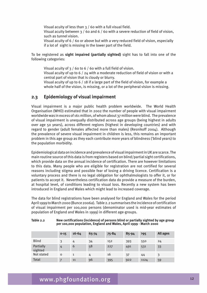
The data for blind registrations have been analysed for England and Wales for the period April 1999 to March 2000 (Bunce 2006a). Table 2.2 summarises the incidence of certification of visual impairment per 100,000 persons (denominator used is mid-year estimates of population of England and Wales in 1999) in different age-groups.
Table 2.2 New certifications (incidence) of persons blind or partially sighted by age group per 100,000 population, England and Wales, April 1999 - March 2000
0-15 16-64 65-74 75-84 85-94 >95 All ages
Blind � � �� 152 �9� 550 2�
Partially sighted
� 6 58 227 �91 5�1 ��
Not stated 0 1 � 16 �7 �� �
Total 7 11 96 �95 922 112� 59
1�www.phgfoundation.org
Incidence and prevalence of visual impairment and blindness in children
A study published by the British Childhood Visual Impairment Study Group (BCVISG) which, undertook active surveillance in the year 2000 through the British Paediatrics Surveillance Unit and the British Ophthalmological Surveillance Units, found the yearly incidence for severe visual impairment and blindness was highest in the first year of life, at �.0 per 10,000 (95% CI �.6-�.5), with a cumulative incidence by age 16 years of 5.9 per 10,000 (5.�-6.5) (Rahi 200�). During the year of the study (2000) there were ��9 children under the age of 16 who were diagnosed with severe visual impairment or blindness in the UK2.1
Rahi notes an average yearly incidence of 1.2 per 10,000 children aged 0-15 years of severe visual impairment and blindness among the South Asian population in the UK compared to an average yearly incidence of 0.2 per 10,000 children among the white population. Within the South Asian category, it is particularly the Pakistani and Bangladeshi community who experience this high incidence, with an average yearly incidence of 1.6 per 10,000 children (an eight fold increase compared to the white population).
It was estimated by Rahi that known hereditary disorders account for severe visual impairment/blindness in a third of all children. Thus in the UK around 150 children will be newly diagnosed each year with severe visual impairment/blindness due to a hereditary disorder. The prevalence of visual impairment, severe visual impairment and blindness in industrialised countries is estimated to be 10-22 per 10,000 children under age 16 years (Gilbert 1999).
2.4 Main causes of certifiable blindness and visual impairment
The main causes of certifiable visual impairment in England and Wales are macular degeneration (57%), glaucoma (11%) diabetic retinopathy (6%), optic atrophy (�%) and hereditary retinal disorders (�%) (Bunce 2006a). The causes differ in different age groups.
Children
Data from blind registrations in England and Wales show that visual pathway disorders and hereditary retinal disease are the major causes of childhood visual impairment with hereditary retinal disease accounting for 1�.1% of certifications in children (Bunce 2006a), optic atrophy 18.9 % and disorders of visual cortex 15.9 % of cases. See figure 2.1. Similarly, data from partial sight certification show that hereditary retinal disease accounts for 11.�% of certification and optic atrophy for 11.2%.
2 This is substantially higher than the incidence for new certifications in this age group (Table 2.2) which would suggest a figure of only 0.7 per 10,000, indicating that there may be substantially lower ascertainment in certification.
1�www.phgfoundation.org
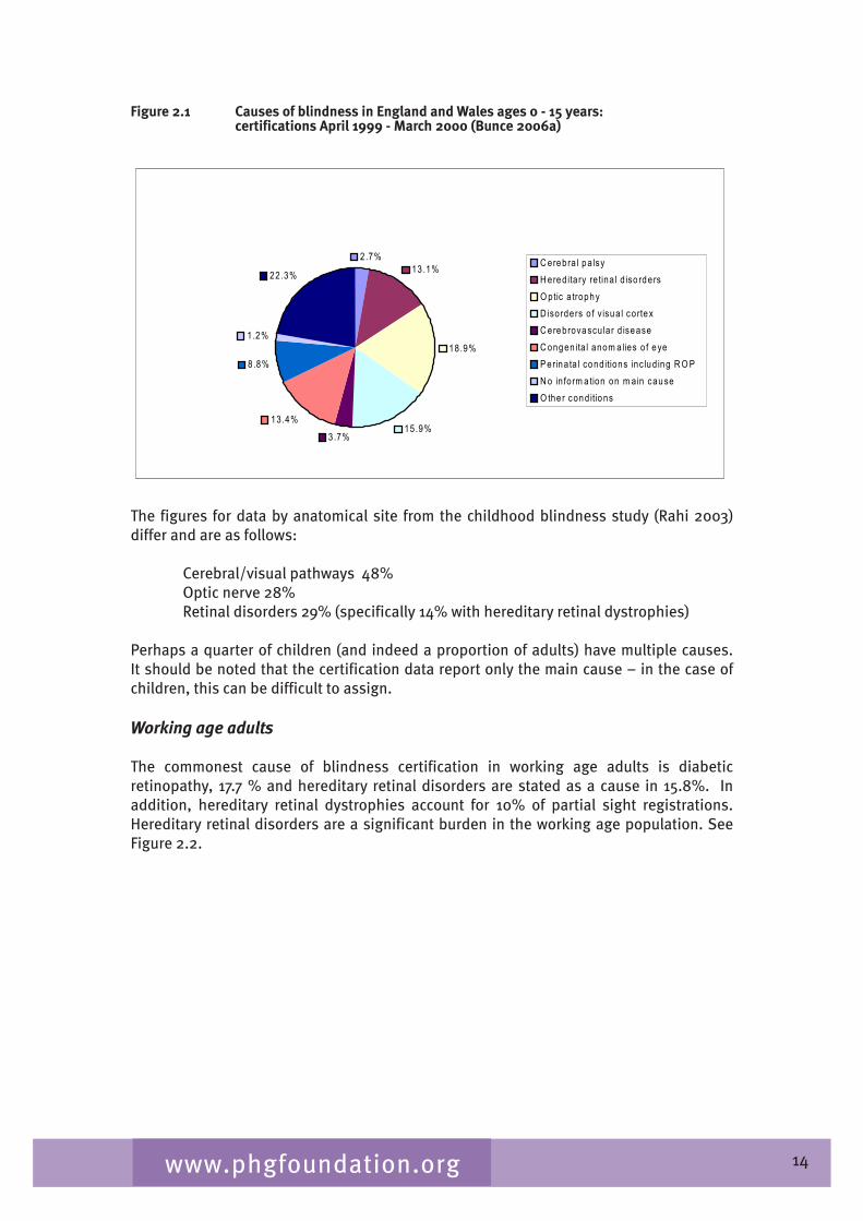
Figure 2.1 Causes of blindness in England and Wales ages 0 - 15 years: certifications April 1999 - March 2000 (Bunce 2006a)
2.7% 13.1%
18.9%
15.9% 3.7%
13.4%
8.8%
1.2%
22.3% C erebra l pa lsy H ered ita ry re tina l d iso rders O ptic a trophy D isorders o f v isua l cortex C erebrovascu lar d isease C ongenita l anom alies o f eye P erinata l cond itions inc lud ing R O P N o in form a tion on m ain cause O the r cond itions
The figures for data by anatomical site from the childhood blindness study (Rahi 200�) differ and are as follows:
Cerebral/visual pathways �8% Optic nerve 28% Retinal disorders 29% (specifically 1�% with hereditary retinal dystrophies)
Perhaps a quarter of children (and indeed a proportion of adults) have multiple causes. It should be noted that the certification data report only the main cause – in the case of children, this can be difficult to assign.
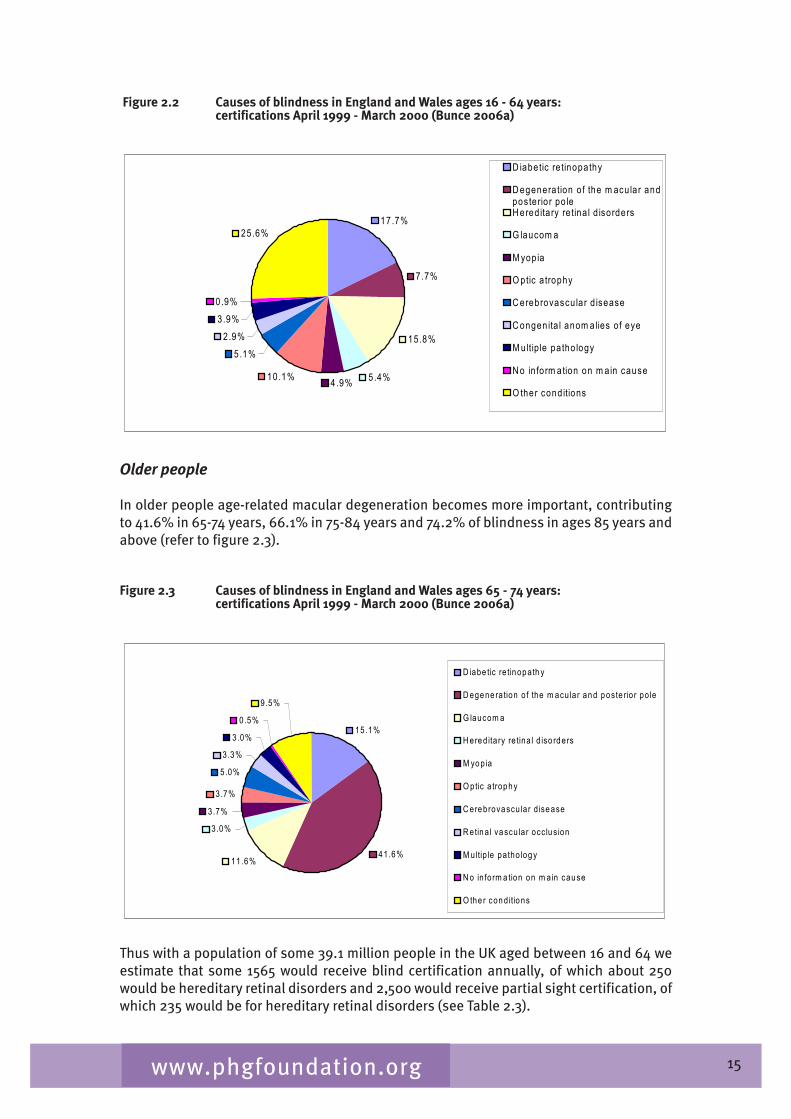
Working age adults
The commonest cause of blindness certification in working age adults is diabetic retinopathy, 17.7 % and hereditary retinal disorders are stated as a cause in 15.8%. In addition, hereditary retinal dystrophies account for 10% of partial sight registrations. Hereditary retinal disorders are a significant burden in the working age population. See Figure 2.2.
15www.phgfoundation.org
Figure 2.2 Causes of blindness in England and Wales ages 16 - 64 years: certifications April 1999 - March 2000 (Bunce 2006a)
17.7%
7.7%
15.8%
5.4% 4.9% 10.1% 5.1%
2.9% 3.9% 0.9%
25.6%
D iabetic re tinopathy D egenera tion o f the m acu lar and posterio r po le H ered ita ry re tina l d isorders G laucom a M yopia O ptic a trophy C erebrovascu lar d isease C ongenita l anom alies of eye M ultip le patho logy N o in form ation on m ain cause O ther cond itions
Older people
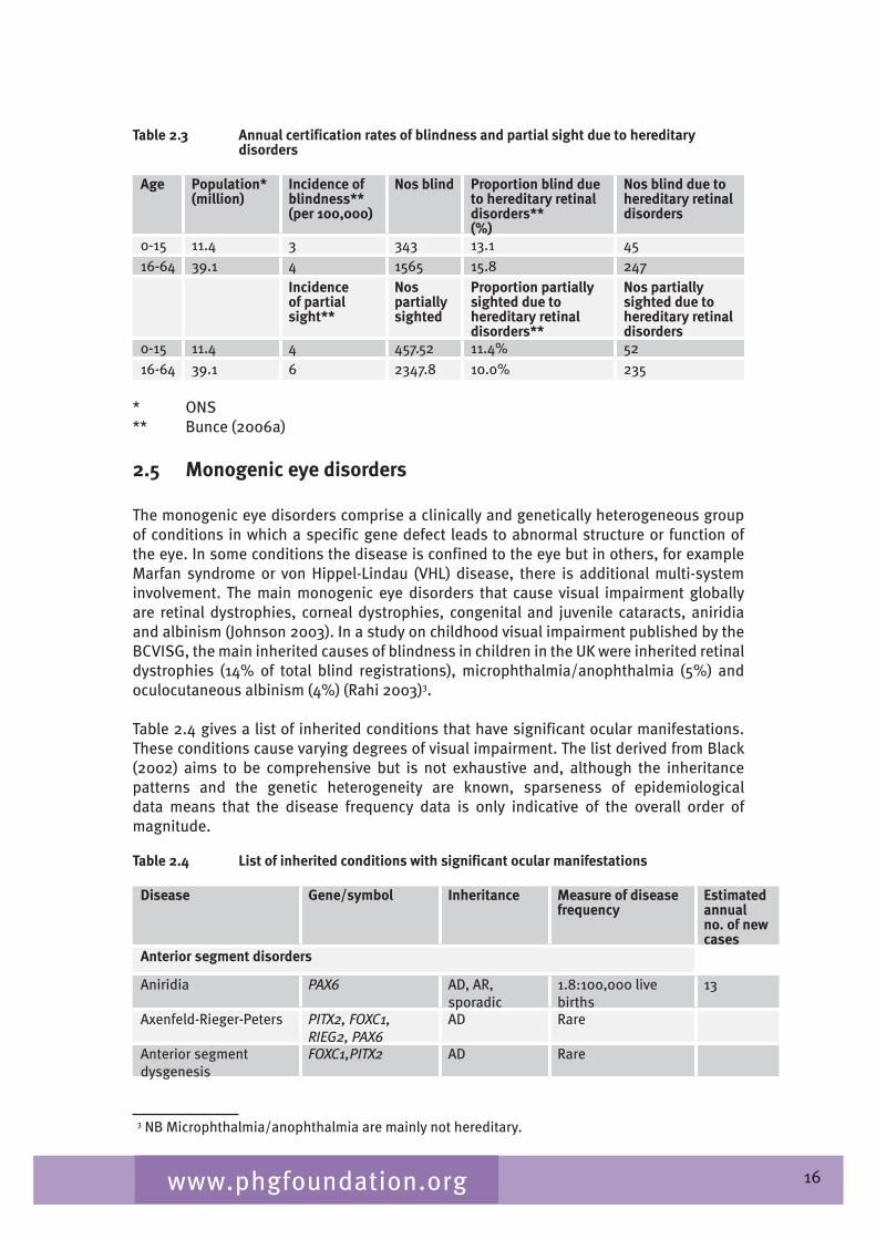
In older people age-related macular degeneration becomes more important, contributing to �1.6% in 65-7� years, 66.1% in 75-8� years and 7�.2% of blindness in ages 85 years and above (refer to figure 2.�).
Figure 2.3 Causes of blindness in England and Wales ages 65 - 74 years: certifications April 1999 - March 2000 (Bunce 2006a)
15.1%
41.6% 11.6%
3.0% 3.7%
3.7% 5.0% 3.3%
3.0% 0.5%
9.5%
D iabe tic re tinopath y D egene ra tion o f the m acu lar and poste rio r po le G laucom a H ered ita ry re tina l d iso rders M yopia O ptic a trophy C erebrovascu lar d isease R etina l vascu lar occ lus ion M ultip le patho logy N o in form a tion on m ain cause O the r cond itions
Thus with a population of some �9.1 million people in the UK aged between 16 and 6� we estimate that some 1565 would receive blind certification annually, of which about 250 would be hereditary retinal disorders and 2,500 would receive partial sight certification, of which 2�5 would be for hereditary retinal disorders (see Table 2.�).
16www.phgfoundation.org
Table 2.3 Annual certification rates of blindness and partial sight due to hereditary disorders
Age Population*(million)
Incidence of blindness**(per 100,000)
Nos blind Proportion blind due to hereditary retinal disorders**(%)
Nos blind due to hereditary retinal disorders
0-15 11.� � ��� 1�.1 �516-6� �9.1 � 1565 15.8 2�7
Incidence of partial sight**
Nos partially sighted
Proportion partially sighted due to hereditary retinal disorders**
Nos partially sighted due to hereditary retinal disorders
0-15 11.� � �57.52 11.�% 5216-6� �9.1 6 2��7.8 10.0% 2�5
* ONS** Bunce (2006a)
2.5 Monogenic eye disorders
The monogenic eye disorders comprise a clinically and genetically heterogeneous group of conditions in which a specific gene defect leads to abnormal structure or function of the eye. In some conditions the disease is confined to the eye but in others, for example Marfan syndrome or von Hippel-Lindau (VHL) disease, there is additional multi-system involvement. The main monogenic eye disorders that cause visual impairment globally are retinal dystrophies, corneal dystrophies, congenital and juvenile cataracts, aniridia and albinism (Johnson 200�). In a study on childhood visual impairment published by the BCVISG, the main inherited causes of blindness in children in the UK were inherited retinal dystrophies (1�% of total blind registrations), microphthalmia/anophthalmia (5%) and oculocutaneous albinism (�%) (Rahi 200�)�.2
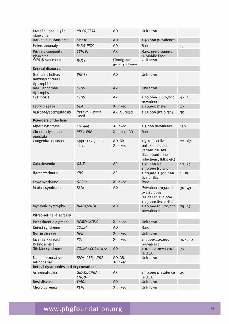
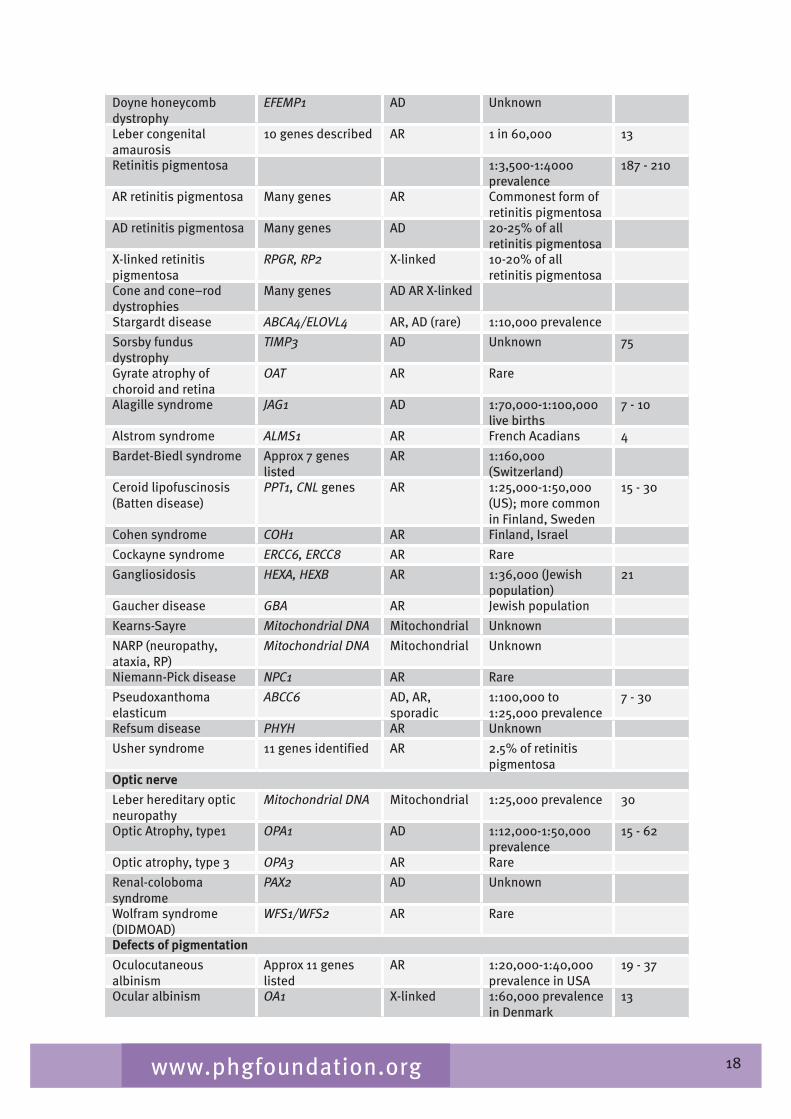
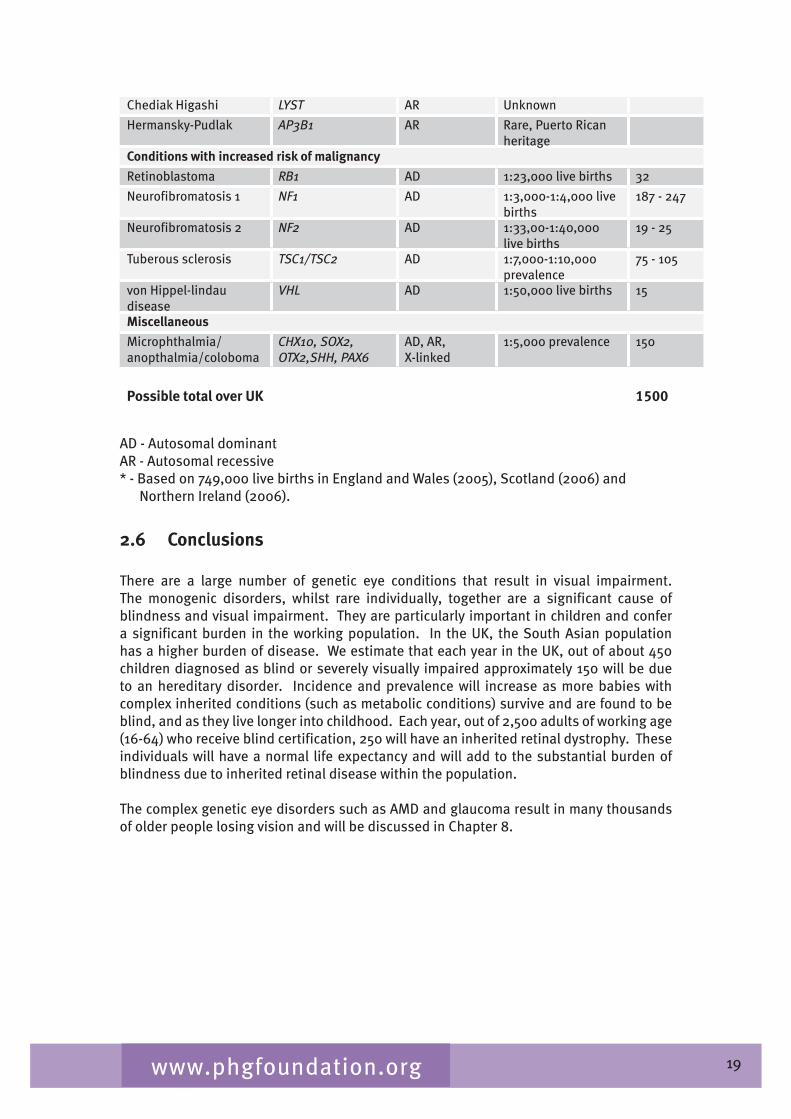
Table 2.� gives a list of inherited conditions that have significant ocular manifestations. These conditions cause varying degrees of visual impairment. The list derived from Black (2002) aims to be comprehensive but is not exhaustive and, although the inheritance patterns and the genetic heterogeneity are known, sparseness of epidemiological data means that the disease frequency data is only indicative of the overall order of magnitude.
Table 2.4 List of inherited conditions with significant ocular manifestations
Disease Gene/symbol Inheritance Measure of disease frequency
Estimated annual no. of new cases
Anterior segment disorders
Aniridia PAX6 AD, AR, sporadic
1.8:100,000 live births
1�
Axenfeld-Rieger-Peters PITX2, FOXC1, RIEG2, PAX6
AD Rare
Anterior segment dysgenesis
FOXC1,PITX2 AD Rare
2� NB Microphthalmia/anophthalmia are mainly not hereditary.
17www.phgfoundation.org
Juvenile open angle glaucoma
MYCO/TIGR AD Unknown
Nail-patella syndrome LMX1B AD 1:50,000 prevalence
Peters anomaly PAX6, PITX2 AD Rare 15
Primary congenital glaucoma
CYP1B1 AR Rare, more common in Middle East
WAGR syndrome PAX 6 Contiguous gene syndrome
Unknown
Corneal diseases
Granular, lattice, Bowman corneal dystrophies
BIGH3 AD Unknown
Macular corneal dystrophy
CTNS AR Unknown
Cystinosis CTNS AR 1:50,000- 1:180,000 prevalence
� - 15
Fabry disease GLA X-linked 1:�0,000 males 19
Mucopolysaccharidosis Approx 5 genes listed
AR, X-linked 1:25,000 live births �0
Disorders of the lens
Alport syndrome COL4A5 X-linked 1:5,000 prevalence 150
Chondrodysplasia punctata
PEX7, EBP X-linked, AD Rare
Congenital cataract Approx 12 genes listed
AD, AR,X-linked
1-�:10,000 live births (includes various causes like intrauterine infections, IMDs etc)
22 - 67
Galactosemia GALT AR 1:70,000 UK, 1:�0,000 Ireland
10 - 25
Homocystinuria CBS AR 1:�0,000-1:500,000 live births
1 - 19
Lowe syndrome OCRL1 X-linked Rare
Marfan syndrome FBN1 AD Prevalence 1:�,000 to 1:10,000; incidence 1:15,000-1:25,000 live births
�0 - �9
Myotonic dystrophy DMPK/ZNF9 AD 1:�0,000 to 1:20,000 prevalence
25 - �7
Vitreo-retinal disorders
Incontinentia pigmenti NEMO/IKBKG X-linked Unknown
Kniest syndrome COL2A AD Rare
Norrie disease NPD X-linked Unknown
Juvenile X-linkedRetinoschisis
RS1 X-linked 1:5,000-1:25,000 prevalence
�0 - 150
Stickler syndrome COL2A1,COL11A1/2 AD 1:10,000 prevalence in USA
75
Familial exudative retinopathy
FZD4, LRP5, NDP AD, AR, X-linked
Unknown
Retinal dystrophies and degenerations
Achromatopsia GNAT2,CNGA3, CNGB3
AR 1:�0,000 prevalence in USA
25
Best disease VMD2 AD Unknown
Choroideremia REP1 X-linked Unknown
18www.phgfoundation.org
Doyne honeycomb dystrophy
EFEMP1 AD Unknown
Leber congenital amaurosis
10 genes described AR 1 in 60,000 1�
Retinitis pigmentosa 1:�,500-1:�000 prevalence
187 - 210
AR retinitis pigmentosa Many genes AR Commonest form of retinitis pigmentosa
AD retinitis pigmentosa Many genes AD 20-25% of all retinitis pigmentosa
X-linked retinitis pigmentosa
RPGR, RP2 X-linked 10-20% of all retinitis pigmentosa
Cone and cone–rod dystrophies
Many genes AD AR X-linked
Stargardt disease ABCA4/ELOVL4 AR, AD (rare) 1:10,000 prevalence
Sorsby fundus dystrophy
TIMP3 AD Unknown 75
Gyrate atrophy of choroid and retina
OAT AR Rare
Alagille syndrome JAG1 AD 1:70,000-1:100,000 live births
7 - 10
Alstrom syndrome ALMS1 AR French Acadians �
Bardet-Biedl syndrome Approx 7 genes listed
AR 1:160,000 (Switzerland)
Ceroid lipofuscinosis (Batten disease)
PPT1, CNL genes AR 1:25,000-1:50,000 (US); more common in Finland, Sweden
15 - �0
Cohen syndrome COH1 AR Finland, Israel
Cockayne syndrome ERCC6, ERCC8 AR Rare
Gangliosidosis HEXA, HEXB AR 1:�6,000 (Jewish population)
21
Gaucher disease GBA AR Jewish population
Kearns-Sayre Mitochondrial DNA Mitochondrial Unknown
NARP (neuropathy, ataxia, RP)
Mitochondrial DNA Mitochondrial Unknown
Niemann-Pick disease NPC1 AR Rare
Pseudoxanthoma elasticum
ABCC6 AD, AR, sporadic
1:100,000 to 1:25,000 prevalence
7 - �0
Refsum disease PHYH AR Unknown
Usher syndrome 11 genes identified AR 2.5% of retinitis pigmentosa
Optic nerve
Leber hereditary optic neuropathy
Mitochondrial DNA Mitochondrial 1:25,000 prevalence �0
Optic Atrophy, type1 OPA1 AD 1:12,000-1:50,000 prevalence
15 - 62
Optic atrophy, type � OPA3 AR Rare
Renal-coloboma syndrome
PAX2 AD Unknown
Wolfram syndrome (DIDMOAD)
WFS1/WFS2 AR Rare
Defects of pigmentation
Oculocutaneous albinism
Approx 11 genes listed
AR 1:20,000-1:�0,000 prevalence in USA
19 - �7
Ocular albinism OA1 X-linked 1:60,000 prevalence in Denmark
1�
19www.phgfoundation.org
Chediak Higashi LYST AR Unknown
Hermansky-Pudlak AP3B1 AR Rare, Puerto Rican heritage
Conditions with increased risk of malignancy
Retinoblastoma RB1 AD 1:2�,000 live births �2
Neurofibromatosis 1 NF1 AD 1:�,000-1:�,000 live births
187 - 2�7
Neurofibromatosis 2 NF2 AD 1:��,00-1:�0,000 live births
19 - 25
Tuberous sclerosis TSC1/TSC2 AD 1:7,000-1:10,000 prevalence
75 - 105
von Hippel-lindau disease
VHL AD 1:50,000 live births 15
Miscellaneous
Microphthalmia/anopthalmia/coloboma
CHX10, SOX2, OTX2,SHH, PAX6
AD, AR, X-linked
1:5,000 prevalence 150
Possible total over UK 1500
AD - Autosomal dominantAR - Autosomal recessive* - Based on 7�9,000 live births in England and Wales (2005), Scotland (2006) and Northern Ireland (2006).
2.6 Conclusions
There are a large number of genetic eye conditions that result in visual impairment. The monogenic disorders, whilst rare individually, together are a significant cause of blindness and visual impairment. They are particularly important in children and confer a significant burden in the working population. In the UK, the South Asian population has a higher burden of disease. We estimate that each year in the UK, out of about �50 children diagnosed as blind or severely visually impaired approximately 150 will be due to an hereditary disorder. Incidence and prevalence will increase as more babies with complex inherited conditions (such as metabolic conditions) survive and are found to be blind, and as they live longer into childhood. Each year, out of 2,500 adults of working age (16-6�) who receive blind certification, 250 will have an inherited retinal dystrophy. These individuals will have a normal life expectancy and will add to the substantial burden of blindness due to inherited retinal disease within the population.
The complex genetic eye disorders such as AMD and glaucoma result in many thousands of older people losing vision and will be discussed in Chapter 8.
20www.phgfoundation.org
Chapter 3 The patient viewpoint
Patients’ views on the characteristics of a good ophthalmology genetics service and on genetic testing were obtained through two focus group meetings. The first meeting was held on 6th November 2006 at Moorfields Hospital, London and participants were from a number of patient representative organisations from around the UK (see Table �.1). The second group was held on 2�th January 2007 at the Churchill Hospital, Oxford, and the participants were patients with genetic eye disorders who were mostly receiving care from the Oxford Ophthalmic Genetics Service. The following chapter is an amalgamation of the views of participants from both consultation exercises. However, it should be noted that, since patients and representative organisations were informed by their experiences arising from a number of services, the comments do not necessarily reflect experiences gained at the Moorfields or Oxford Genetics Services.
Table 3.1 Organisations involved in the focus group held at Moorfields
RP Society (Retinitis Pigmentosa) RNIB (Royal National Institute for the Blind) Contact-A-FamilyMacular Disease SocietyInternational Glaucoma AssociationAction for Blind PeopleChildhood Eye Cancer TrustUsher ResearchSense the National Deafblind and Rubella Association
3.1 Features of a good service
Communication
Clear communication is a defining issue of a good service as identified by both groups. Patients want information on their diagnosis delivered in a manner that they can understand. Once patients have been given the diagnosis, they need information on available treatment (if any), the likely progression of the condition and how it may affect key aspects of their life such as employment and mobility, including the ability to retain a driving licence. Patients also require information on inheritance patterns so that their children and siblings receive appropriate advice and care, and can make informed decisions about their futures when deciding on, for example, their careers and their reproductive options. Most patients will also wish to discuss current research and possible future advances in clinical management, including novel therapies.
The importance of the communication process was highlighted by both groups. Most patient care is long term since genetic ophthalmic conditions often result in a gradual deterioration of vision. Patients want to remain in the system with agreed follow up care and have a named person to contact. They also want to be informed of new developments. A good service is a robust service where individuals, regardless of where in the UK they live, receive a quality service and, once they have been identified as requiring care, their contact details are available for follow-up and not lost in the system. In ophthalmic departments where there is no specific ophthalmic genetics service, patients may suffer from a lack of expertise and pertinent information, with no cohesive linking of delivery of
21www.phgfoundation.org
diagnosis. Furthemore, links with relevant services such as social services, visual aids and follow up review may not be offered. The following comments from patients have expressed these concerns:
“They never link us together- you kind of drop off - I now just go to the low vision clinic but I have got no more information on what’s happening with updates on what is happening with dominant optic atrophy…I am sure there have been tremendous updates in genetics from the last time I was seen and it would be nice to have some recap over what’s happened in the last years…..I was told to buy vitamin B because that was what they were working on to do with nerve damage but I was told that 15 years ago – I don’t know if the results of those tests were any good or not. Are there such things that you can eat to improve your eyesight or is that just a myth?”
“There is no consistency….. It comes down to luck – who you see.”
Particular aspects of communication were also highlighted as important. Service providers need to have an understanding of the cultural and religious backgrounds of patients. Patients understanding of and reaction to their condition may be affected by their cultural background. Services need to be sensitive to the appropriateness of certain options such as termination of pregnancy. All patients should have equal access to services, and information may need to be given via interpreters or presented in different formats for those patients with sensory impairment.
Genetic counselling
Both groups of patients and patients’ representatives highlighted the value of genetic counselling but reported that it is often not offered in eye clinics. Many patients need emotional support to enable them to come to terms with the diagnosis. Patients note that only once the person has psychologically adjusted to the condition can he or she move forward and start to deal with the implications. Many patients commented on the shock they had experienced on being given a diagnosis and that no support was offered to help them adjust to the information and its implications.
“Sometimes people leave the consultation and they are in a state of shock and sometimes you are left for weeks without anything. They need to be able to be given a contact number so that they can ring… People need information immediately. Anybody in shock needs help immediately and support and they are not given that.”
“For me having genetic counselling would be top of my list because I have never had it and I would really like it – I have had information from the eye hospital but it has been very patchy and the way it's been delivered is either in a way you can’t understand as it is way over your head or they have just given it to you without thinking of the impact that it might actually have.”
In marked contrast, patients who had received genetic counselling found it very valuable, in terms of understanding the diagnosis, discussing the implications and being signposted to services that can help the individual make the adjustments necessary to lead as full and independent a life as possible.
“When you know an awful lot about it you can go into a lot of jargon which can totally go over the heads of people – when I first encountered (name of counsellor) she couched it in simple terms, easy for the lay person to understand and I understood very, very well and my daughter was given the option that any time if she had any concern just to phone up.”
22www.phgfoundation.org
“The fear of the unknown is greater than the fear of the known… at least if you know you can start making plans and organise the rest of your life appropriately - How long can I work? How long can I drive? That clarity in my own mind has been very helpful.”
Integrated service
The importance of an integrated service was a key theme. Many patients will have complex needs and their sight problems may be one of a range of symptoms they experience as a result of their genetic condition, as occurs in, for example, Marfan syndrome. Other senses may be affected such as in Usher disease, where hearing as well as sight is affected.
Another aspect of an integrated service is communication between primary, secondary and tertiary services so that there is a clear pathway of care. GPs’ knowledge of specialist genetic ophthalmic services is very variable and so patients may not be referred appropriately. Similarly the district general hospital may not have clear referral pathways to the specialist service. As a result there is uneven access to specialist services.
A third aspect of an integrated service is communication between health and other services such as social services, disability support services, employment support services, housing services, careers advice services and the voluntary sector etc, so that the needs of patients are addressed. It would be helpful to have a key individual who takes responsibility to make those links. This could be the genetic counsellor, specialist nurse or family support worker.
Patients also highlighted the importance of the integration of services between paediatric and adult services. Patients with long-term conditions may be lost to the service as they make the transition between adult and child services. Some services have transitional clinics to oversee the transfer of care from paediatric to adult services.
Service that caters for the extended family
By their nature, genetic disorders may affect other family members, and services should be able to encompass the whole family rather than solely the index individual referred. This may require some flexibility as family members may live across regional boundaries or there may be differing referral arrangements between the different secondary services that refer to a particular regional specialist service.
The quality of the service is more important to patients than the distance they have to travel to access the service – patients are often willing to travel further if that results in being seen by a more expert service.
Open access
There was some discussion on whether patients should have open access to services. Patients feel that the benefit of having the option to be seen again needs to be balanced by services not being overwhelmed by the ‘worried well’. Overall patients feel they should be able to make contact with services if they are concerned about a deterioration in their condition.
2�www.phgfoundation.org
3.2 Genetic testing
Patients’ representatives were asked to comment on the benefits and disadvantages of having a genetic test and the timescale that is acceptable for the process. Their responses are outlined below.
Benefits of having a genetic test
(a) To decrease morbidity and mortality
The main advantage in having a genetic test for most patients is if the confirmation of a diagnosis results in preventive options which can decrease morbidity and mortality. This is further discussed on the Chapter 5 on evaluation of genetic testing.
(b) Information for family members, particularly children
Some of the patients did not perceive a personal benefit in having a genetic test but felt that it might yield useful information for their children or other family members. The identification of the mutation causing the disorder would provide information so that family members could be tested for specific mutations in the future when more treatment options might be available.
“You will be leaving a legacy for your children and your children’s children.”
(c) Gene therapy trial
For some patients, the main benefit of genetic testing is the identification of the mutation causing their specific disorder so that they can participate in trials of novel therapy such as gene therapy. This is particularly true for disorders such as RP that can show both genetic and allelic heterogeneity. As more clinical trials are undertaken, the demand for genetic testing is likely to increase. Other patients felt their main motivation for undergoing genetic testing would be if it benefited research.
(d) To inform lifestyle decision making
A genetic test may result in a diagnosis that indicates that the individual’s vision is likely to deteriorate in the future. This information may influence decisions such as career choices or a change in direction in one’s career. An individual whose job is dependent on the ability to drive may need to think of an alternative career. The choice of housing may be influenced – for example a bungalow might be more suitable than a home with stairs, or a home located close to public transport links and shops. The result may also influence planning a family.
“If you can find out what is going to happen to you over the next 10 years or 20 years it gives you the opportunity of thinking… Have I got another 10 years of doing the job I’ve currently got or do I have to find another job within the organization I’m currently working in or do I have to look at perhaps trying to get early retirement or find alternative employment where the rest of my skills or abilities are going to used.”
“I now know my condition - I plan to be on good bus routes.”
2�www.phgfoundation.org
(d) To either rule out or confirm the presence of a condition
Patients also identified the value of genetic tests in either ruling out or confirming the presence of a condition that is present in a family. For example if the mutation causing a particular type of RP has been identified in a child, other siblings could be tested to discover whether they too have the mutation. There should be guidelines/protocols in place for predictive genetic or ophthalmic testing of children.
Disadvantages of having a genetic test
The main disadvantage that patients identified is the psychological impact of having a positive result, particularly for an asymptomatic individual. This, however, needs to be balanced against the state of anxiety that exists where an individual already knows from family history that he or she has a high risk of visual loss (usually 50% as most tests are done for autosomal dominant disease). The identification of a mutation might make the person live in a state of anxiety waiting for the symptoms to become manifest. Future ‘blindness’ may dominate one’s life and stop the individual living in the present. Patients felt that genetic counselling should minimise the anxiety caused and help the individual recognise the benefits of having advance information. Patients should be made aware that having a genetic test may affect their position with insurance companies.
“When you give this information, it is to try and bring the family back into the present rather than what is going to happen on the future. So many negative things come as a result, the child is protected, is rushed off to Disney Land, is given things and the rest of the family is neglected, the brothers and sisters, it is always, always, always him or her. So this goes back a lot to the quality of the information that is given about a condition. We do acres of work on the telephone with distressed families who have just been given this diagnosis, and try and bring them back into the present, and give them a sense of future for themselves and their child. So an awful lot more work needs to be done on how we impart information, the quality of the information, how we follow up, and the skill of the people who do this.”
Timescale for delivering the results of genetic tests
Patients felt that as short a timescale as possible is desirable but that the accuracy and quality of the information is more important than the speed of delivering the results. Since the process can be lengthy it is important to keep patients informed of the progress towards getting a result.
3.3 Conclusions and recommendations
Patients and their representative organisations were concerned at the lack of a consistent, high quality, robust service across the UK. Both the patients’ group and the patients’ representatives group highlighted the variability across the UK as to whether there was a good regional service to be referred to and whether the GP/optometrist/district general hospital service knew of the specialist service.
The patients’ groups were consistent in their support for molecular genetic testing because of its value to the patient and their family. Molecular genetic testing can provide accuracy of diagnosis and inheritance patterns, giving routes to potential therapy and, with counselling, properly informed decisions on life choices.
25www.phgfoundation.org
Good communication and counselling are key attributes of a good genetic service. The patients want to be kept fully informed of advances in testing and treatment. They want counselling to understand diagnosis, for emotional support and to make decisions on life choices such as reproductive choices, education, employment and mobility.
The genetic service must be properly integrated with a clear, simple and robust management process that the patient can follow easily. The present service is viewed by the patients as inconsistent. The quality of service is more important than its speed or ease of location.
It was the general view that patients want to be kept informed of advances in research, and want to be part of the research effort. They understand that helping research will lead to more accurate diagnosis and therapies for themselves and their families. Patients who have participated in research want to be informed of the outcome and, if appropriate, be referred to the appropriate clinical services when the research trial has finished.
Voluntary organisations should play a key role in keeping patients informed of developments in genetic research, clinical trials and therapies.
Patients should be encouraged to seek assistance from other organisations such as social services, disability support services, employment support services, housing services and careers advice services, as appropriate, and to develop and maintain links with the RNIB and other appropriate voluntary organisations.
26www.phgfoundation.org
Chapter 4 Genetic ophthalmology services
4.1 Introduction
Patients with inherited eye disease causing visual impairment usually first present to their general practitioner or optometrist and are then referred to their local eye clinic. From there, patients are often referred on to consultant ophthalmologists with a subspecialty interest. A specific diagnosis may require further investigation including ocular imaging and electrophysiology. Genetic counselling may involve a further referral to the clinical genetics service although some specialist ophthalmologists offer counselling as part of the clinical service. The majority of regional clinical services now run joint ophthalmology/genetics clinics that include access to a consultant from both specialities. From the patient perspective this is a lengthy and often uncertain pathway.
4.2 The need for specialist services
Specialist services in ophthalmic genetics are needed to provide high quality care for patients with genetic eye disease for a number of reasons:
Individually the disorders are rare and general ophthalmologists are likely to have little experience of diagnosis and counselling;
Diagnosis may require specialist investigations such as ocular electrophysiology or specific ocular imaging, which are not widely available;
Counselling patients and their families requires more time than is available in routine ophthalmology clinics;
Clinical geneticists do not have expertise in eye examination and investigation for diagnosis and carrier detection, or in advising on treatment options and prognosis in ophthalmology;
There have been rapid advances in understanding the molecular basis of genetic
eye disease;
There is strong patient demand to consult clinicians with experience of these rare disorders;
Such clinics are suitable settings for clinical research including clinical trials of novel therapies;
Time and expertise are required for counselling, support and referral to appropriate support services such as low vision and social services;
The need is particularly great for children as genetic eye diseases are a significant cause of blindness in this age-group. The need for specialist services is rising and will continue to do so because of a combination of patient demand and new technologies;
27www.phgfoundation.org
Demand for genetic counselling amongst individuals with visual impairment is rising, and services are seeing a significant increase in referrals. With dissemination of information about genetic advances through voluntary organisations and the internet, the public are becoming better informed and are asking to be referred for specialist management and genetic advice.
Although many of the genes responsible for inherited ophthalmic conditions have been identified, routine genetic testing is only available for a small number of these. However, it is likely that this will expand with more genetic diagnostic tests moving from research into standard clinical diagnostics in accredited laboratories. As new treatments become available, it will increasingly be important that patients who might benefit are identified and referred to appropriate services.
4.3 Roles within the specialist service
The role of the specialist ophthalmologist is in making an accurate diagnosis, offering advice on prognosis, and developing a treatment plan that may involve other specialists in clinical medicine, social work and education. For those conditions (perhaps the majority) that are not amenable to specific treatment, management is focussed on making best use of residual vision by, for example, using low vision optical aids and computer software. Advice about retraining, acquiring new and different skills, education about driving and information about access to work schemes is important for adults. In the case of visually impaired children, there is a strong focus on providing educational support in school. The ophthalmologist may also be able to put the family in touch with the patient support groups for patients with specific genetic eye conditions.
Most specialist ophthalmologists work very closely with their clinical genetic colleagues, with the latter providing expertise in making a diagnosis, for example in a child with multiple congenital abnormalities that include eye anomalies. Such expertise encompasses dysmorphology and the diagnosis of syndromic and genetic conditions with complex genetic aetiologies. In addition it may be that a clinical geneticist will be required in discussing areas including prenatal diagnosis, ethical issues relating to genetic testing and recurrence risks to the wider family. Genetic counselling and interpretation of results of molecular genetic testing may be carried out by the ophthalmologist with a special interest in ophthalmic genetics.
Genetic counsellors have a number of important roles to play in the ophthalmology genetics service. Where they are employed in specialist services they provide particular support to families before, during and after the appointment. In pre-clinic phases, they ensure that the patient and family understand what will happen and have realistic expectations, and collect information in advance to draw an accurate family tree. The special expertise of the genetic counsellor is also used to help patients understand the significance of genetic testing and deciding on testing. Finally, genetic counsellors who work in this area have a key role in bringing together not only their understanding of rare genetic disease but also the particular context of loss of vision; they are thus well-placed to help patients to access specialist support from both statutory and voluntary sectors.
Electrophysiology tests are objective, non-invasive methods for measuring the function of the retina or optic nerve. They are thus an important aid to the ophthalmologist in establishing an accurate diagnosis in patients with suspected inherited retinal disease. The tests may also be used to investigate asymptomatic family members who are at risk of inheriting the disease; this may help identify sub-clinical abnormality and also assist
28www.phgfoundation.org
in determining the mode of inheritance and so aid genetic counselling of the family. Electrophysiology departments are generally established in large regional teaching hospitals and are headed by a consultant clinical scientist experienced in carrying out and interpreting the results of the specialised procedures required for testing this rare group of patients. Referral of patients may be made by consultant ophthalmologists or geneticists on an ad hoc basis or the diagnostic service may be integrated into a joint eye–genetic clinic session.
It is the role of the laboratory scientist to use professional judgment in advising clinicians on the appropriate investigations for individual patients as a member of the clinical team caring for that patient and to give detailed clinical interpretation of results for individual patients, which may include guidance on additional investigations. It is essential that, wherever possible, molecular genetic services are performed in a routine laboratory environment working under appropriate governance. These services should be managed and executed by scientific staff with the appropriate qualifications. Many countries, including the UK, have in place a system of mandatory State Registration ensuring a valuable level of professional oversight.
4.4 Models of services
Most inherited eye disorders are individually uncommon and it is therefore unrealistic to expect most consultant ophthalmologists to have the experience and expertise to make a specific diagnosis or to be able to give advice about prognosis. Within ophthalmology there should be well established referral networks within each region so that patients with, or suspected of having, rare genetic eye disorders can be referred to an ophthalmologist with a special interest in such disorders.
Most regions in the UK have regional genetic eye disease clinics jointly run by ophthalmologists and clinical geneticists. In some large units the genetic counselling is provided by ophthalmologists with a specialist interest in genetics but even then there will be strong links to the local clinical genetic services. Whatever model is used the specialist units allow the concentration of core facilities such as ocular electrophysiology, low vision services and educational support at a single site and allows a holistic approach to the care of patients with visual impairment.
One or two such specialists within each region is usually sufficient to cope with the work load. For very rare disorders such as retinoblastoma referrals may be made supra-regionally.
4.5 Conclusion
A need for accurate diagnosis, prognosis and counselling and the advent of probable new therapies for ophthalmic genetic conditions makes it imperative that ophthalmic genetics is firmly established as a specialist service with enough specialists to serve regionalas a specialist service with enough specialists to serve regionalwith enough specialists to serve regional populations, and with well-recognised referral pathways.
29www.phgfoundation.org
Chapter 5 Evaluation of genetic tests in ophthalmology: exploring clinical utility
5.1 Introduction
In this chapter we focus on genetic tests and particularly those used in ophthalmology. We aim to develop an understanding of the ways in which genetic tests are evaluated, and, in particular, the ACCE framework, which is a model process for evaluation. We focus on the various parameters of clinical utility and look for examples that illustrate clinical utility in ophthalmology genetic tests including some published evidence in the management of retinoblastoma.
5.2 Background
Definition of a genetic test
There are various definitions of 'genetic test' and the question of definition has provoked discussion in the literature (Burke 2002). The main point of debate centres on whether a genetic test gives information about an inherited disorder (in which case examination of the eye to diagnose RP might be construed as a genetic test) or whether it is based on either DNA or DNA-related technologies. The US Task Force on Genetic Testing uses a very broad definition of a genetic test as ‘the analysis of human DNA, RNA, chromosomes, proteins, and certain metabolites in order to detect heritable disease-related genotypes, mutations, phenotypes or karyotypes for clinical purposes.’ The Genetics and Insurance Committee (GIAC), by contrast, defines a genetic test as ‘a test to detect the presence or absence of, or change in a particular gene or chromosome’.
A more recent definition distinguishes first the assay, which is 'the method for determining the presence of quantity of a component' and goes on to define the genetic test as a laboratory assay that is used to identify a particular genotype, for a particular disease in a particular population for a particular purpose’ (Zimmern 2007). For the purpose of this report we use this last definition, which is based on analysis of an individual’s DNA.
Evaluation
Evaluation is defined as ‘a process that attempts to determine as systematically and objectively as possible the relevance, effectiveness and impact of activities in the light of their objectives’. In order to carry out an evaluation we need to define the activity (in this case the genetic test), its purpose and how effectively and efficiently the purpose is achieved. The ACCE framework provides a model process for evaluating genetic tests.
5.3 The ACCE framework for evaluation
The ACCE framework was developed in the United States by the Foundation for Blood Research through a cooperative agreement with the Centers for Disease Control and Prevention (CDC) (Haddow 200�).
�0www.phgfoundation.org
The acronym ACCE stands for the four key elements needed to evaluate any genetic test: Analytic validity; Clinical validity; Clinical utility; and Ethical, legal, and social implications.
Analytic validity defines the ability of a test to measure the genotype of interest both accurately and reliably.
Clinical validity defines the ability of a test to detect or predict the associated disorder (ie phenotype).
Clinical utility defines the risks and benefits associated with the introduction of a test into practice. Specifically, clinical utility focuses on the health outcomes, both positive and negative, associated with testing.
Ethical, legal, and social implications of the testing process include those inherent in any medical technology as well as those specific to genetic tests.
A framework has been developed for reviewing gene tests using this process. This involves first defining the disorder, purpose of test, clinical setting and the precise test to be used. The process then involves collecting information to answer a series of �� questions, setting these out under the standard framework and, at the same time, identifying information gaps (questions set out in Burke 2007). The evidence required is extensive. In view of the high number and rarity of conditions for which genetic tests are undertaken, it is not surprising that there are very few completed evaluations of genetic tests and none in the area of ophthalmology. This situation is being addressed through the 'EGAPP'�1project, which aims to support the development of a coordinated process for evaluating genetic tests and other genomic applications.
The ACCE framework has been expanded recently (Burke 2007), in particular the dimensions of clinical utility, noting that this must be related to the different purposes of the test as well as to the way in which the genotype contributes to the causes of the disease, and the dimensions of health care quality, such as whether the health service is able to deliver the preventive services that may be required for those who test positive.
5.4 Clinical utility of genetic tests in ophthalmology
Purpose of genetic tests
Genetic tests have a range of different purposes in health care. Although often described generally as being for purposes of diagnosis, predictive testing, susceptibility testing or screening, further exploration in a paper by Burke and Zimmern (Burke 2007) concluded that these serve only as ‘intermediate purposes’ as they do not get to the heart of why we do a test. They argue that genetic tests should have one of the following ultimate goals:
�4 Evaluation of Genomic Applications in Practice and Prevention (EGAPP): Implementation and Evaluation of a Model Approach. EGAPP is a pilot project initiated by the CDC National Office of Public Health Genomics in the autumn of 200�.
�1www.phgfoundation.org
Reduce morbidity or mortalityProvide salient information on the care of patients or family membersAssist the patient or family members with reproductive decision making
Such outcomes may be achieved for the proband or for family members in whom cascade testing becomes possible when the underlying molecular pathology is identified. They may also be achieved by providing information to assist the process of healthcare.
The key question is what value a genetic test adds to the information derived from full clinical work-up including family history. In the following paragraphs we describe some of the purposes and outcomes of genetic tests in genetic eye disease. However, the individual tests do not fall into neat 'purpose' categories. The same test may fulfil a number of purposes for patients, family and health services, and its potential benefit(s) will differ according to the multiple and varying needs of all these 'stakeholders'.
The literature contains very few examples of formal evaluations of the clinical utility of genetic tests and a literature search found examples in ophthalmology only in the area of retinoblastoma with studies of genetic testing on the process of care.
Further consideration of the evaluation of genetic tests as part of an exploration of prioritisation has led to a provisional finding that it is necessary to judge the effectiveness of a given test in achieving a range of possible purposes, for a patient and family with a particular condition and in a particular clinical, social and psychological situation.
5.5 Benefits of genetic testing in ophthalmology in achieving test purposes
There are many ophthalmic conditions where a strategy of genetic testing can provide benefits to patients, their families and the process of care (often also benefiting the healthcare system by increased efficiency). The benefits will vary according to:
The condition in question, its severity, and its underlying genetic pathologyThe possibilities for prevention, treatment or amelioration of the conditionWhether management decisions are related to specific genotypesCrucial decisions that the individual may need to make (eg about career)The needs of the family – in particular their need to consider risk to future offspring
These benefits and some of the factors that affect them are illustrated for particular conditions in the paragraphs below. Two case histories provided as an appendix to this chapter amply show the complexity of benefits that genetic tests may afford to patients, their extended families and the health services.
Benefits of information gained from better diagnosis and prognosis
In many inherited eye disorders the diagnosis can be made clinically by detailed clinical history, family history, clinical examination and investigations such as ocular electrophysiology. This might include examination of relatives. If this is the case it might be questioned why a genetic test would be undertaken. For example, a diagnosis in a young male of X-linked retinoschisis (XLRS) can be made on the basis of reduced visual acuity, characteristic appearance of fundus (foveal or peripheral schisis), a characteristic electroretinogram with selective reduction of amplitude of dark-adapted b-wave amplitude and a family history consistent with X-linked inheritance. However, in the absence of a
•••
•••••
�2www.phgfoundation.org
family history, and where the fundus appearance is atypical the diagnosis can be more difficult and molecular genetic testing can provide a definitive diagnosis. Only one gene, RS1, which is well characterised, has been associated with XLRS, and mutations are found in nearly 90% of males with a clinical diagnosis.
Confirmation of the diagnosis allows better information to be given to the patient about the prognosis. XLRS has a generally good long-term visual prognosis. However, the prognosis is still limited to a general one pertaining to XLRS overall. In XLRS the course of disease can be very variable even within the family and there is no good correlation between genotype and phenotype. Knowledge of the specific mutation is, therefore, of limited value to the proband. However, it does allow more accurate genetic counselling of other family members – specifically female relatives who are at risk of being a carrier of this disorder as the carrier state cannot be recognised clinically. (This benefit is discussed under a further heading of information to assist reproductive choice.)
One of the more obvious conditions in which a genetic test might lead to a diagnosis and more precise information about prognosis is Usher syndrome. This condition presents as a congenital, bilateral, profound hearing loss and is often thought to be non-syndromal deafness until the early signs of RP - tunnel vision and night blindness - become noticeable either to parents, teachers or the individual. Early diagnosis is important as a child with Usher syndrome will have priority for cochlear implantation and may, therefore, receive bilateral surgery at a young age when there will be maximum benefit for development of communication. Without genetic testing it is difficult to make this timely diagnosis as the retinal changes associated with the syndrome are not apparent at an early age. Thus genetic testing can confirm the diagnosis of Usher syndrome and indicate the most appropriate treatment option. An early diagnosis will also have an impact on education, allowing this to be tailored to the child’s future needs.
The relative complexity of the genetics underlying RP makes the additional value of genetic testing ‘simply to obtain information’ more problematic. RP is a group of inherited disorders in which abnormalities of the photoreceptors (rods and cones) or the retinal pigment epithelium (RPE) of the retina lead to progressive visual loss. The inheritance of the disorder is very varied and can be autosomal dominant (15-25%), autosomal recessive (5-20%), X-linked (5-15%), and some digenic and mitochondrial forms have also been described. Many of the cases seen in the clinic are simplex cases (ie a single occurrence in a family). Such cases are often due to autosomal recessive or X-linked recessive inheritance but can also be the result of an autosomal dominant mutation arising de novo or associated with incomplete penetrance in a parent. The molecular genetic causes of RP are unusually complicated as there is considerable genetic heterogeneity and, for most genes, there can be many different disease causing mutations. Given this extensive genetic heterogeneity, molecular genetic testing using the currently available methods is not feasible, but this may change in the future as resequencing technology is incorporated into practice (see Chapter 8).
Alström disease is a good example of a condition in which an early diagnosis can inform the subsequent assessment and surveillance programme by providing information about the likely associated abnormalities. Alström syndrome is a rare autosomal recessive disorder that presents in infancy with a severe rod cone dystrophy. Affected individuals may later develop hearing impairment, cardiomyopathy, diabetes and obesity. The confirmation of this diagnosis in infancy by molecular genetic testing leads to regular surveillance for the systemic abnormalities, which can have a benefit both in terms of survival and reducing morbidity.
��www.phgfoundation.org
When a clinical diagnosis is already made, an important reason for genetic testing of a proband would be to allow the possibility of molecular diagnosis to provide information for family members.
In some adult onset dominant disorders, the offspring of affected individuals may wish to have pre-symptomatic molecular genetic diagnosis to inform career or other lifestyle choices. This is relevant, for example, in milder forms of autosomal dominant RP and macular disorders such as Sorsby fundus dystrophy and dominant drusen. Patients need to be carefully counselled before making a decision to have pre-symptomatic testing. A negative test result would be expected to have psychological benefits in terms of freedom from worry for the individual’s life and concern about their offspring. However, the benefits of testing must be weighed against the deleterious psychological impact of a positive test when the individual will know that vision is expected to deteriorate, with no possibility of preventing this occurring, and resulting limitation of prospects for insurance, employment and other aspects of life.
Decreasing morbidity and mortality by preventive care
Genetic testing can be an important part of clinical management in determining the best means of preventing loss of vision and other morbidity or even mortality. Retinoblastoma (RB) is a malignant tumour originating in embryonic retinal cells. With a birth prevalence of 1 in 18 000 live births, it is the most common intra-ocular cancer of infants. RB has both genetic and non-hereditary forms. The genetic form of RB is inherited as an autosomal dominant trait; it may be familial but many cases represent new mutations. Patients with bilateral disease, multifocal tumours and associated, non-ocular tumours have, by definition, the genetic form of RB with a germline mutation of the RB1 gene. The non-genetic form of RB is caused by somatic mutations in the RB1 gene occurring in a single developing retinal cell. All non-genetic forms of RB have single, unilateral tumours. However, about 15% of patients with unilateral tumours do have germline mutations. RB is uniformly fatal without treatment. Treatment options include local treatments such as cryotherapy and laser for early tumours and chemotherapy, radiotherapy and enucleation of the eye for advanced disease. Modern treatments have resulted in greatly improved survival rates but there is significant morbidity associated with treatment. The best results are achieved with early diagnosis, which allows focal treatment to be given with preservation of the eye and good vision.
In the management of RB, molecular genetic testing is of value in two situations. In patients with unilateral RB, genetic testing is extremely useful in determining whether they have a germline mutation and will need further careful surveillance. This requires chromosomal and molecular testing of the proband, including testing of tumour tissue and DNA from white blood cells. Identification of individuals with germline mutations is important because they have an increased risk of developing tumours in the unaffected eye, and outside the eye, including pinealomas and neuroectodermal tumours, osteosarcomas, soft tissue sarcomas and melanomas.
Conversely, if RB1 mutant alleles identified in the tumour are not detected in leukocyte DNA, although there is a small (1%) chance that the individual has low-level mosaicism (involving less than 20% of blood cells) for the mutant allele this risk is small enough that examination under anaesthesia may not be justified, and may be replaced with regular clinical examination of the eyes. Thus, genetic testing of tumour and peripheral blood can help the patient by ensuring that only those who are at risk have the inconvenience and potential morbidity associated with repeated examination under anaesthetic.
��www.phgfoundation.org
In known genetic cases, determination of the mutation also allows testing of relatives, ensuring that only those who are mutation-positive require surveillance and prenatal diagnosis. These are discussed further in the sections on process of care and reproductive choice (below).
Improving the process of care
Continuing with our example of RB, genetic testing translates into more effective and efficient clinical management for the patient and the family as a whole. It is usual practice to screen all patients with germline tumours and other at-risk children in the family with regular examinations under anaesthetic. Molecular genetic diagnosis means that only those with the germline mutation need to be screened, which saves morbidity, anxiety and money. In 2002 a study was published by Raizis et al (2002) of the relative benefits of RB1 gene testing, more particularly, the use of testing of tumour tissue over conventional ophthalmological testing. It showed that genetic testing of peripheral blood DNA is useful in identifying mutations that allow further family testing. However, mutations are only found in about 62% of clinically heritable cases (the remaining being due to unidentified deletions or mutations within introns). The absence of apparent mutations in peripheral blood DNA means that germline status cannot be resolved – had tumour tissue been available for initial identification of mutations, their presence or absence in the blood DNA can then easily be determined. Raizis et al found that, in three cases, gene testing using tumour analysis as well as blood analysis allowed four siblings to be discharged from follow-up with an estimated lifetime cost saving of at least NZ$100,000. Similarly, Joseph et al (200�) showed that adopting a genetic testing strategy provided a �.5 fold cost-saving for a proband and a 6-fold saving for a family with two siblings compared to the cost of clinical examination.
In RB, clarification of the presence of a germline mutation also gives families the opportunity to consider testing future children by early analysis for the RB1 mutation through either CVS at 11 weeks or by amniocentesis. This can allow termination of affected fetuses or the parents to be aware of status to allow early delivery and commencement of surveillance before term.
Decreasing morbidity and mortality by informing treatment decisions
Outside the inherited cancer syndromes such as RB and Von Hippel-Lindau disease there is as yet no evidence that molecular diagnosis has any impact on management decisions. However with the development of new biological based therapies this situation is likely to change. Some novel therapies such as gene therapy will be gene specific and knowledge of the disease–causing mutation will be essential. For other therapies the early diagnosis provided by molecular testing may be equally important. For example the development of new biological treatments for choroidal neovascularisation (CNV) may impact on inherited forms of macular degeneration where visual loss is due to CNV.
Sorsby fundus dystrophy, for example, is a rare autosomal dominant disorder in which new blood vessels grow under the fovea, resulting in fluid accumulation and haemorrhage in the macular region. Usually symptoms do not appear until after the age of �0 when there may be a rapid decrease in vision. The gene for this disorder has been identified as TIMP3, which is located on the long arm of chromosome 22. Molecular diagnosis of at risk family members is now possible and it is speculated that a surveillance program in those carrying a TIMP3 mutation and early recognition and treatment of CNV could lead
�5www.phgfoundation.org
to improved prognosis. However, to date, there is no evidence that such a programme would be effective and, more importantly, there is no treatment that can be administered prophylactically, that is, before blood vessels or atrophic changes supervene.
Information to assist reproductive choice
There are many examples in ophthalmology where disease and disability are sufficiently serious for parents and other family members to wish to understand and reduce the risk of recurrence. Here, genetic counselling and consideration of genetic testing are required. Genetic testing provides supplementary information to that obtained from the individual’s history, including family history and clinical examination in terms of diagnosis and mode of inheritance. For example, Norrie disease (ND) is the most severe phenotype of NDP gene-related retinopathies that are characterised by a spectrum of fibrous and vascular changes of the retina at birth. In this phenotype, there is total bilateral inoperable retinal detachment present from birth, which results in total blindness. Some males with ND have developmental delay/mental retardation and behavioural abnormalities and some later develop sensorineural hearing loss. Carrier females have abnormal eye examination. NDP is the only gene involved in ND (although cases resembling ND can be seen with mutations in LRPS and FZD4), and 85% of individuals with NDP gene-related retinopathies have sequence alterations with the remainder having sub-microscopic deletions (http://www.geneclinics.org/). Thus there is a high likelihood that genetic testing will allow identification of a mutation and enable family testing to take place. ND is inherited as an X-linked trait but in many cases there is no known family history of other affected males. Where there is a family history, the mother will be a carrier but molecular diagnosis will allow the status of other at risk females in the family to be determined. Where there is no family history, the mother is usually a carrier but de novo mutations can occur. The identification of the ND mutation in the child can allow the status of the mother and her female relatives to be determined. Carrier females can then go on to consider pre-implantation genetic diagnosis or prenatal testing to avoid recurrence in their children.
Other examples where prenatal testing and consideration of termination of pregnancy may be considered include RB, as discussed above, Leber congenital amaurosis, choroideremia and X-linked RP. Alternatively, in RB, because the condition is treatable, parents might use prenatal testing to determine whether the child is affected in order to allow early delivery of the child and commencement of eye screening.
Genetic testing for research
Genetic testing to find a molecular diagnosis can also be important in research of novel treatments that might be genotype specific. A recurring theme amongst patients and voluntary organisations was the importance of testing to aid research. However, although this will indeed be important and may benefit patients as a group in the long run, such testing should be provided within the research setting and it should not be a reason for undertaking testing within the health service.
5.6 Conclusion
There is very limited published research on clinical utility for genetic tests, both in general, and in the context of ophthalmology. This chapter has begun to explore some of the dimensions of clinical utility that would need to be considered in a formal evaluation. Undoubtedly some of the parameters, such as the value of information and, in particular,
�6www.phgfoundation.org
the added value of a molecular diagnosis over a clinical diagnosis will be hard to measure. Evaluation of these, and the more concrete outcomes, such as the use and outcomes of testing to provide reproductive choice, or the contribution of genetic testing to more efficient process of care will require prospective study and a multi-disciplinary design that includes psychology, sociology and health economics as well as clinical and laboratory disciplines.
The relative rarity of the conditions means that such evaluations could not be undertaken for all. However, one way forward might be to take a small number of conditions as exemplars and study in detail the added value of a molecular diagnosis in the various areas of utility outlined in this chapter. The development of data on the value gained across various dimensions for a cohort of patients might then contribute to the development of criteria for test provision according to different clinical needs.
�7www.phgfoundation.org
Case history 1 Sorsby fundus dystrophy (SFD)
Sorsby fundus dystrophy (SFD) is an autosomal dominant dystrophy affecting central vision. It is highly penetrant and those who have the genetic mutation tend to present in their late �0s to early 50s with choroidal neovascularisation, which affects their central vision. Although the choroidal neovascularisation associated with this condition has been reported to respond to steroid or photodynamic therapy treatment, and may well respond to the new antivascular endothelial growth factor agents, currently there is no cure for SFD. If a mutation in the TIMP-3 gene is identified, then family members can be offered testing. Patients with the mutation are educated that they must assess their vision on a regular basis for distortion, and self-refer for assessment and treatment as appropriate.
Mr B, aged �8, was first diagnosed with SFD in his early forties. Mr B is now registered as severely sight impaired and has recently begun to use a guide-dog. Mr B has two adult sons (both in their early twenties) and identical twin daughters (in their early teens). Each of Mr B’s children has a 50% risk of inheriting the condition.
On first contacting the ophthalmic genetic counsellor, Mr B had questions about how to get his children tested for the TIMP-3 gene mutation. He felt particularly keen that his children “should know, as soon as possible, so they can be prepared and in case steroid or laser treatment could help them in the future”. Such testing in family members who are at 50% risk with no signs or symptoms is known as predictive genetic testing. For late-onset genetic conditions, such as SFD, that do not have an impact on health until much later in life, predictive genetic testing is generally offered to adults and not children. This testing is offered after pre-test counselling. Predictive genetic testing also requires that the familial, disease-causing mutation has been identified previously in a family member with the condition.
Our ophthalmic genetic counsellor discussed the process with Mr B. She was also able to highlight the potential implications of genetic testing for Mr B’s identical twin daughters in the future. The genetic counsellor arranged for a blood sample to be taken and a mutation in TIMP-3 was identified in Mr B, thus confirming the diagnosis and the causative mutation in SFD. Since the gene mutation was identified, predictive genetic testing was offered to Mr B’s adult children. Mr B’s eldest son has since been referred and seen in clinic for pre-test counselling. Following his appointment, this son has decided not to take predictive genetic testing any further, at this stage.
The possibility of genetic testing in this family offers diagnostic certainty, identifies individuals who are risk of developing choroidal neovascularisation, and provides information about long-term visual prognosis.
�8www.phgfoundation.org
Case history 2 Pattern dystrophy and peripherin/RDS mutations
The term pattern dystrophy encompasses a range of autosomal dominantly inherited dystrophies characterised by abnormal pigment deposition at the level of the RPE. This condition can be associated with various degrees of central visual loss. Commonly visual acuity is preserved until late adult life, but driving may be affected, so that monitoring this is important, as is checking vision for distortion as this condition can be complicated by the development of choroidal neovascularisation, which requires intervention. Mutations in the peripherin/RDS gene have been also described in association with autosomal dominant RP, cone and cone-rod dystrophy and macular dystrophies. It has also been described in families with extreme phenotypic diversity (from retinitis pigmentosa to pattern dystrophy) in the same family.
A 68 year old lady who first presented with distortion in the vision and small central scotoma in the left eye re-presented nine years later when she developed similar symptoms in the right eye. She reported that she had a son who had been noted to have abnormalities at both maculae, but was asymptomatic at the age of �5 despite the abnormal retinal findings. She wished to know if they had the same condition and wanted more information for her son as well as any information regarding future therapies. Discussion regarding testing in pattern dystrophy was undertaken, and she elected to go ahead with testing for the above reasons, although she understood that no current intervention/treatment was available. A mutation was identified in the peripherin/RDS gene and this provided the possibility of predictive genetic testing for adult members of the family and confirmatory testing in her son.
Knowledge of the mutation and subsequent testing in the family prevented further need for clinical testing and information was disseminated in the family who were geographically separated and seeing different doctors, which saved exhaustive investigations. An annual review to check that vision was adequate for driving was arranged for the proband and advice regarding urgent review if central distortion developed was emphasised to both the proband and her son.
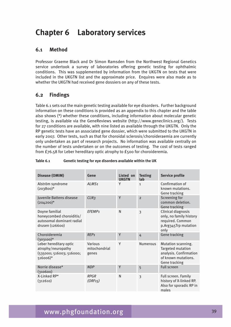
�9www.phgfoundation.org
Chapter 6 Laboratory services
6.1 Method
Professor Graeme Black and Dr Simon Ramsden from the Northwest Regional Genetics service undertook a survey of laboratories offering genetic testing for ophthalmic conditions. This was supplemented by information from the UKGTN on tests that were included in the UKGTN list and the approximate price. Enquires were also made as to whether the UKGTN had received gene dossiers on any of these tests.
6.2 Findings
Table 6.1 sets out the main genetic testing available for eye disorders. Further background information on these conditions is provided as an appendix to this chapter and the table also shows (*) whether these conditions, including information about molecular genetic testing, is available via the GeneReviews website (http://www.geneclinics.org/). Tests for 27 conditions are available, with nine listed as available through the UKGTN. Only the RP genetic tests have an associated gene dossier, which were submitted to the UKGTN in early 2007. Other tests, such as that for choroidal sclerosis/choroideraemia are currently only undertaken as part of research projects. No information was available centrally on the number of tests undertaken or on the outcomes of testing. The cost of tests ranged from £76.58 for Leber hereditary optic atrophy to £500 for choroideremia.
Table 6.1 Genetic testing for eye disorders available within the UK
Disease (OMIM) Gene Listed on UKGTN
Testing lab
Service profile
Alström syndrome(20�800)*
ALMS1 Y 1 Confirmation of known mutations.Gene tracking
Juvenile Battens disease(20�200)*
CLN3 Y 2 Screening for common deletion.Gene tracking
Doyne familial honeycombed choroiditis/autosomal dominant radial drusen (126600)
EFEMP1 N � Clinical diagnosis only, no family history required. Common p.Arg��5Trp mutation only
Choroideremia(�0�100)*
REP1 Y � Gene tracking
Leber hereditary optic atrophy/neuropathy (5�5000; 51600�; 516000; 516006)*
Various mitochondrial genes
Y Numerous Mutation scanning. Targeted mutation analysis. Confirmation of known mutations. Gene tracking
Norrie disease*(�10600)
NDP Y 5 Full screen
X-Linked RP*(�12610)
RPGR (ORF15)
N � Full screen. Family history of X-linked RP. Also for sporadic RP in males
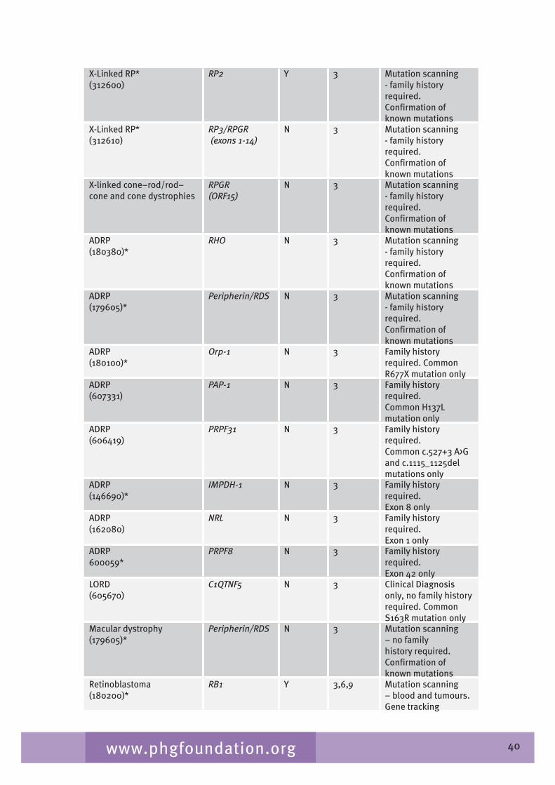
�0www.phgfoundation.org
X-Linked RP*(�12600)
RP2 Y � Mutation scanning - family history required.Confirmation of known mutations
X-Linked RP*(�12610)
RP3/RPGR (exons 1-14)
N � Mutation scanning - family history required.Confirmation of known mutations
X-linked cone–rod/rod–cone and cone dystrophies
RPGR (ORF15)
N � Mutation scanning - family history required.Confirmation of known mutations
ADRP(180�80)*
RHO N � Mutation scanning - family history required.Confirmation of known mutations
ADRP(179605)*
Peripherin/RDS N � Mutation scanning - family history required.Confirmation of known mutations
ADRP(180100)*
Orp-1 N � Family history required. Common R677X mutation only
ADRP(607��1)
PAP-1 N � Family history required.Common H1�7L mutation only
ADRP(606�19)
PRPF31 N � Family history required.Common c.527+� A>G and c.1115_1125del mutations only
ADRP(1�6690)*
IMPDH-1 N � Family history required.Exon 8 only
ADRP(162080)
NRL N � Family history required.Exon 1 only
ADRP600059*
PRPF8 N � Family history required.Exon �2 only
LORD(605670)
C1QTNF5 N � Clinical Diagnosis only, no family history required. Common S16�R mutation only
Macular dystrophy(179605)*
Peripherin/RDS N � Mutation scanning – no family history required. Confirmation of known mutations
Retinoblastoma(180200)*
RB1 Y �,6,9 Mutation scanning – blood and tumours.Gene tracking
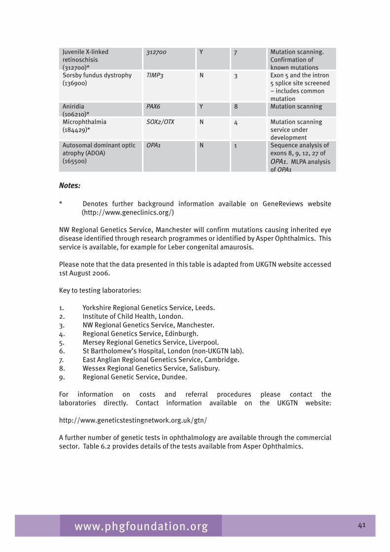
�1www.phgfoundation.org
Juvenile X-linked retinoschisis(�12700)*
312700 Y 7 Mutation scanning.Confirmation of known mutations
Sorsby fundus dystrophy (1�6900)
TIMP3 N � Exon 5 and the intron 5 splice site screened – includes common mutation
Aniridia(106210)*
PAX6 Y 8 Mutation scanning
Microphthalmia(18��29)*
SOX2/OTX N � Mutation scanning service under development
Autosomal dominant optic atrophy (ADOA)(165500)
OPA1 N 1 Sequence analysis of exons 8, 9, 12, 27 of OPA1. MLPA analysis of OPA1
Notes:
* Denotes further background information available on GeneReviews website (http://www.geneclinics.org/)
NW Regional Genetics Service, Manchester will confirm mutations causing inherited eye disease identified through research programmes or identified by Asper Ophthalmics. This service is available, for example for Leber congenital amaurosis.
Please note that the data presented in this table is adapted from UKGTN website accessed 1st August 2006.
Key to testing laboratories:
1. Yorkshire Regional Genetics Service, Leeds.2. Institute of Child Health, London.�. NW Regional Genetics Service, Manchester.�. Regional Genetics Service, Edinburgh.5. Mersey Regional Genetics Service, Liverpool.6. St Bartholomew’s Hospital, London (non-UKGTN lab).7. East Anglian Regional Genetics Service, Cambridge.8. Wessex Regional Genetics Service, Salisbury.9. Regional Genetic Service, Dundee.
For information on costs and referral procedures please contact the laboratories directly. Contact information available on the UKGTN website: http://www.geneticstestingnetwork.org.uk/gtn/
A further number of genetic tests in ophthalmology are available through the commercial sector. Table 6.2 provides details of the tests available from Asper Ophthalmics.
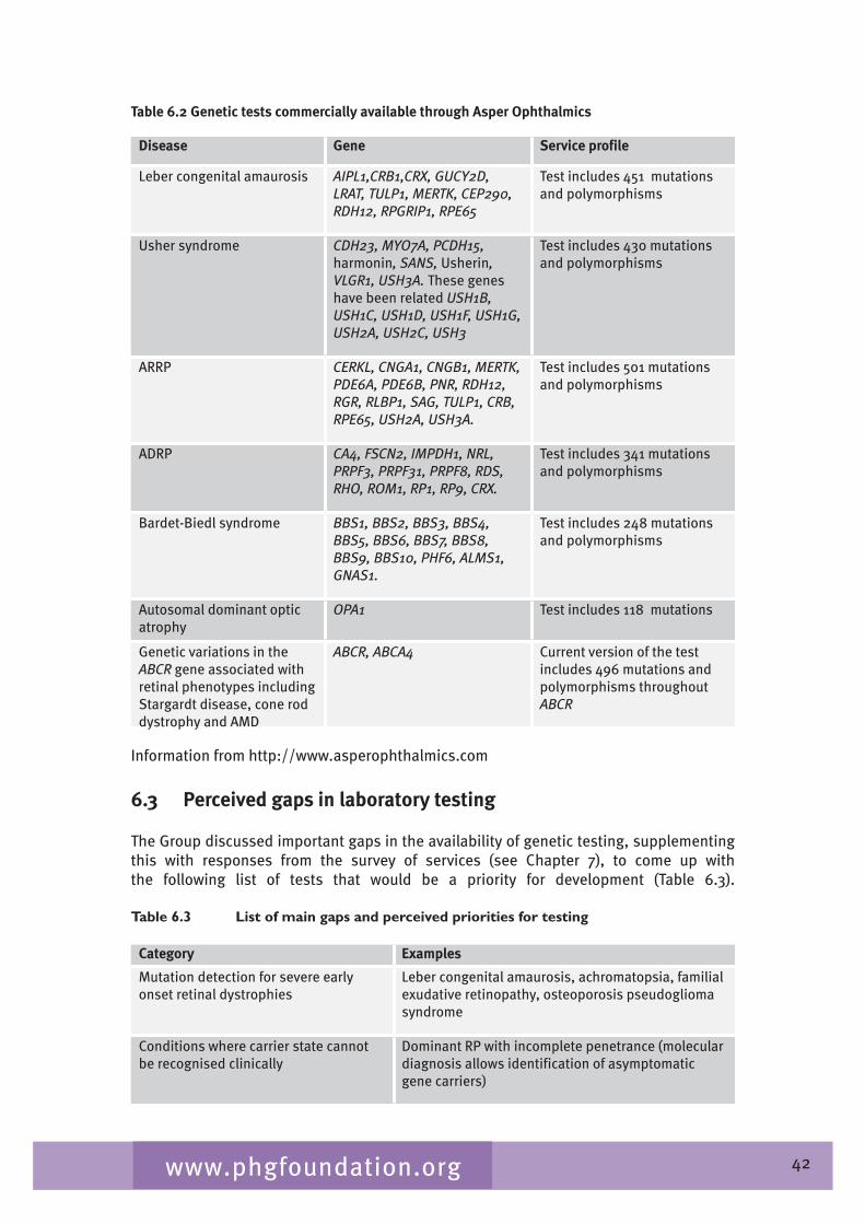
�2www.phgfoundation.org
Table 6.2 Genetic tests commercially available through Asper Ophthalmics
Disease Gene Service profile
Leber congenital amaurosis AIPL1,CRB1,CRX, GUCY2D, LRAT, TULP1, MERTK, CEP290, RDH12, RPGRIP1, RPE65
Test includes �51 mutations and polymorphisms
Usher syndrome CDH23, MYO7A, PCDH15, harmonin, SANS, Usherin, VLGR1, USH3A. These genes have been related USH1B, USH1C, USH1D, USH1F, USH1G, USH2A, USH2C, USH3
Test includes ��0 mutations and polymorphisms
ARRP CERKL, CNGA1, CNGB1, MERTK, PDE6A, PDE6B, PNR, RDH12, RGR, RLBP1, SAG, TULP1, CRB, RPE65, USH2A, USH3A.
Test includes 501 mutations and polymorphisms
ADRP CA4, FSCN2, IMPDH1, NRL, PRPF3, PRPF31, PRPF8, RDS, RHO, ROM1, RP1, RP9, CRX.
Test includes ��1 mutations and polymorphisms
Bardet-Biedl syndrome BBS1, BBS2, BBS3, BBS4, BBS5, BBS6, BBS7, BBS8, BBS9, BBS10, PHF6, ALMS1, GNAS1.
Test includes 2�8 mutations and polymorphisms
Autosomal dominant optic atrophy
OPA1 Test includes 118 mutations
Genetic variations in the ABCR gene associated with retinal phenotypes including Stargardt disease, cone rod dystrophy and AMD
ABCR, ABCA4 Current version of the test includes �96 mutations and polymorphisms throughout ABCR
Information from http://www.asperophthalmics.com
6.3 Perceived gaps in laboratory testing
The Group discussed important gaps in the availability of genetic testing, supplementing this with responses from the survey of services (see Chapter 7), to come up with the following list of tests that would be a priority for development (Table 6.�). Table 6.3 List of main gaps and perceived priorities for testing
Category Examples
Mutation detection for severe early onset retinal dystrophies
Leber congenital amaurosis, achromatopsia, familial exudative retinopathy, osteoporosis pseudoglioma syndrome
Conditions where carrier state cannot be recognised clinically
Dominant RP with incomplete penetrance (molecular diagnosis allows identification of asymptomatic gene carriers)
��www.phgfoundation.org
Mutation detection in severe developmental eye disorders
Congenital glaucoma, anophthalmia
Conditions where genetic testing can confirm the inheritance pattern
Simplex RP
Disorders where the eye disease is associated with other systemic abnormalities
Ushers syndrome, albinism, Bardet-Biedl syndrome
6.4 Barriers to testing
In the past, the number of known gene mutations causing disease was limited, thus, molecular genetic analysis was not widely available and different testing mechanisms were used. In some cases, testing relied upon indirect methods such as linkage analyses. This is seldom the case now and the majority of analyses test for mutations in known genes. Today’s methods rely upon the identification of a sequence variation in affected individuals in a family and the demonstration that this is disease causing. Once the causative mutation has been identified in affected members of a family, molecular diagnosis can be offered, after careful counselling, to family members at risk of inheriting the causative gene.
For the majority of genes that are known to underlie Mendelian disorders, the identification of pathogenic mutation is labour-intensive and time-consuming and requires a detailed analysis of the whole gene. In some cases there is a strong relationship between phenotype and genotype. This exists amongst certain of the monogenic macular dystrophies. The majority of cases of Sorsby dystrophy and Doyne Honeycomb dystrophy (Malattia Leventinese) result from single point mutations in the TIMP3 and EFEMP1 genes, respectively (Weber 199�; Gregory 1996; Stone 1999). There is a similar situation for the stromal corneal dystrophies that are linked to chromosome 5q�1 and caused by mutations in the BIGH3 gene, where the range of mutations causing granular, lattice type I and Bowman’s layer (Thiel-Behnke and Reis-Buckler) dystrophies is very limited (Munier 2002). In these circumstances, the molecular identification of this point mutation is straightforward (although identifying a laboratory willing to do it might be less so). In other disorders there is considerable allelic heterogeneity and the identification of the causative mutations is much more difficult.
General barriers to molecular testing and the development role of laboratories
Technological
In many cases where mutation identification is attempted, the task is not simple. Usually, where a mutation is suspected, the whole gene must be screened. In the case of ABCA4, which is mutated in Stargardt disease and encompasses 51 exons and 6,000–7,000 base pairs of DNA, this is an enormous task that is beyond the scope of most diagnostic laboratories (Webster 2001). Furthermore the pick-up rate amongst those known to harbour mutations in ABCA4 is considerably less than 100% (Briggs 2001). This means that a negative result is of limited value as it neither excludes a mutation in ABCA4 nor in any other gene. Finally, for many molecules, there is a significant degree of normal variation in both the gene and, importantly, its encoded protein. The task of defining whether a variation that alters a single amino acid is pathogenic is onerous and, in the absence of a functional protein assay, may even be impossible.
��www.phgfoundation.org
In conditions where mutation of a number of genes can cause an identical phenotype (RP is the most obvious example) there is often no way to choose one from a number of genes: this may make testing impractical in a clinical setting using current techniques. Thus for conditions like ARRP or Leiber congenital amaurosis currently it is impossible to get a comprehensive and equitable service for mutation detection. This may change with the development of new technologies for molecular genetic testing.
Cost and gate-keeper functions
There are limited budgets for genetic testing and the usual pattern is for these to be managed within an overall budget held by the genetic service. The genetics service, thus, acts as gatekeeper to this budget and will determine priority based on clinical utility. With limited budgets, this means that tests might be refused, particularly where patients are from a different geographical area (eg family members) or from a different service such as ophthalmology. The latter might be exacerbated where genetics departments have more limited understanding of this specialist area. In general, such problems might be alleviated by the specialist services themselves demonstrating clinical utility and cost effectiveness within their own service, and thereby arguing for inclusion of genetic testing in their own budgets.
It is common for research programmes involved in the identification of novel disease-causing genes to continue screening patients within the target population for a short period but then to stop due to change in research priorities or lack of funding. It is not always possible for NHS diagnostic laboratories to take over the testing of such genes although the Manchester service will confirm mutations identified on a research basis. Currently the provision of molecular services for ophthalmology is patchy and service laboratories are unwilling and/or unable to take on testing of genes for which the demands for testing are low or where the chances of picking up a mutation are low. Therefore for conditions such as anterior segment dysgenesis (low mutation pick-up rate), ARRP or Usher and Bardet-Biedl syndrome syndromes (high genetic heterogeneity) there has, to date, been a general low rate of transfer from research advances to diagnostics and this has resulted in problems in providing molecular services for patients.
Clinical utility
The assessment of genuine clinical utility of molecular testing is difficult. In terms of making a clinical/financial case for developing testing this is becoming increasingly recognised as important. Overall the ability to find a mutation in a patient does not necessarily amount to information which will change either patient decision-making or clinical management. For a large number of conditions (Rieger syndrome, ADRP, glaucoma, macular dystrophy) it may be difficult to justify the development of molecular testing on the basis of clinical utility. Tests that have limited utility are unlikely to make their way into service.
The UKGTN has adopted the ACCE methodology for genetic test evaluation (Analytical validity, Clinical validity, Clinical utility and Ethical, legal and social issues) first developed in the United States. The UKGTN panel that considers Gene Dossiers weight the impact of the test on therapy and management decisions highly but also recognises the value of a precise diagnosis and information to patients and their family as a valid parameter in judging the cost/benefit of a test. Tests that are approved by the UKGTN Steering Group are considered for approval by the Genetic Commissioning Advisory Group (GENCAG). The test of the UKGTN/GENCAG process may be its responsiveness to the rapid changes in the utility of genetic tests as research and development progresses. In particular we can begin to see a shift in the reason for requesting a genetic test from help in accurate diagnosis and
�5www.phgfoundation.org
personal/life decision making to using the tests to inform the entry of patients into clinical trials and thereafter for access to improved management, treatment and prescribing.
Geography/race
Barriers to accessing health care will, by definition, act as barriers to molecular testing although there is no obvious additional barrier for the process. For patients where there is limited understanding of English, counselling may be problematic especially when complex issues may need to be discussed via an interpreter. Obtaining truly informed consent may be a challenge.
Methods of funding for developments
The introduction of an internal market in the NHS in 1990-91 created the current system of healthcare purchasers (commissioners) and providers and also encouraged the development of provider to provider services within the NHS with money for services directly changing hands. This underpinned the growth of the professionally led specialist network for genetic tests which became the Department of Health supported UKGTN in 200�.
This encouraged molecular genetic laboratories to introduce new services in the confidence that they would be supported directly through income external to their local core contract. Service growth was weaker where trusts did not pass on income to the laboratories providing the services.
However, there have been and remain a number of barriers or inhibitors. The first is the development time to put a new test in place. This activity is under-resourced and often falls between the gap between research and service development. Trusts are generally risk-averse and may regard the initial support for the test development and launch as too speculative. This is particularly true where genetic tests are relevant to rare conditions, technically complex (due to locus heterogeneity or in large genes with no founder mutations), expensive, and where the clinical utility of the test is largely informational. Ophthalmic genetic tests often share some or all of these features, which probably explains their slow development and submission to the UKGTN for national provision. Challenging genetic testing services like this required significant research-led support to tackle difficult technological barriers and to demonstrate their viability before services could be established. The DH funding for National Genetics Reference Laboratory (Manchester) supplemented by the British Retinitis Pigmentosa Society helped to establish the technological platform and was a model to meet this type of development need. Genetic testing in cardiology shares features of complexity and cost and was similarly supported by the Genetic Knowledge Parks in Oxford and London.
An optimistic picture for funding of testing for genetic eye conditions can be envisaged if a firm link is established between genetic testing and entry into therapeutic trials and thereafter improved treatment for eye conditions. Under these conditions funding for genetic testing and further development money will be found if it is clear that over time the societal burden of visual handicap is lessened.
Current tensions in the NHS funding system include some threats to the network approach to specialised genetic testing in the UK. The tendency of centres to repatriate or ‘claw back’ referrals for specialised tests is caused by a combination of the increased analytical capacity in the system due to the Genetics White Paper investment, pressure on trust budgets due to the overall NHS budget deficit in 2005-6 and 2006-7, and the more individualised and
�6www.phgfoundation.org
profit-centred approach of foundation trusts. This threatens to damage specialisation in the network and is not helpful in developing expertise and overall efficiency for the NHS.The development of all genetic testing continues in a period of very rapid change with new and potentially transforming technologies becoming available. In particular the availability of (cheap) multiplex SNP typing and (expensive) whole genome DNA sequencing opens the possibility for the parallel screening of many genes from a single patient. These are the technologies that could transform genetic testing for ophthalmic disorders. The adoption of these technologies by the NHS will require careful assessment of the technologies and evidence-led, medium-scale investment decisions, and may be a driver in service re-configuration.
6.5 Issues for discussion
The following issues must be addressed if the laboratory services are to be able to meet the current and likely anticipated need for testing:
The need for more tests
Limitations of volume and capacity. Estimates of current activity in genetic testing are a large underestimate of need because they are currently limited by expressed lack of funding for genetic tests. Beneath this is a further level of need that is not expressed but results from the lack of established clinical services that would provide a pathway from routine ophthalmology services to the specialist genetic service.
Budgets and gatekeeper functions
Supporting laboratories to develop new tests
A suitable model for molecular genetics services. The need to take a strategic view on the best way model for molecular genetics services, and in particular where the testing services will need to be provided and whether a single, two, or more sites is the most appropriate.
The role of commercial providers and how this will be integrated with the health service provision.
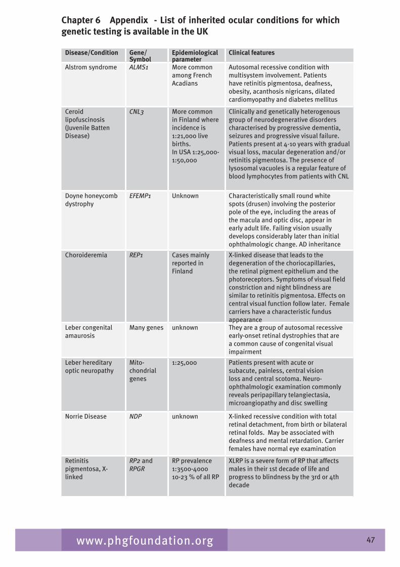
�7www.phgfoundation.org
Chapter 6 Appendix - List of inherited ocular conditions for which genetic testing is available in the UK
Disease/Condition Gene/Symbol
Epidemiological parameter
Clinical features
Alstrom syndrome ALMS1 More common among French Acadians
Autosomal recessive condition with multisystem involvement. Patients have retinitis pigmentosa, deafness, obesity, acanthosis nigricans, dilated cardiomyopathy and diabetes mellitus
Ceroid lipofuscinosis (Juvenile Batten Disease)
CNL3 More common in Finland where incidence is 1:21,000 live births.In USA 1:25,000-1:50,000
Clinically and genetically heterogenous group of neurodegenerative disorders characterised by progressive dementia, seizures and progressive visual failure. Patients present at �-10 years with gradual visual loss, macular degeneration and/or retinitis pigmentosa. The presence of lysosomal vacuoles is a regular feature of blood lymphocytes from patients with CNL
Doyne honeycomb dystrophy
EFEMP1 Unknown Characteristically small round white spots (drusen) involving the posterior pole of the eye, including the areas of the macula and optic disc, appear in early adult life. Failing vision usually develops considerably later than initial ophthalmologic change. AD inheritance
Choroideremia REP1 Cases mainly reported in Finland
X-linked disease that leads to the degeneration of the choriocapillaries, the retinal pigment epithelium and the photoreceptors. Symptoms of visual field constriction and night blindness are similar to retinitis pigmentosa. Effects on central visual function follow later. Female carriers have a characteristic fundus appearance
Leber congenital amaurosis
Many genes unknown They are a group of autosomal recessive early-onset retinal dystrophies that are a common cause of congenital visual impairment
Leber hereditary optic neuropathy
Mito-chondrialgenes
1:25,000 Patients present with acute or subacute, painless, central vision loss and central scotoma. Neuro-ophthalmologic examination commonly reveals peripapillary telangiectasia, microangiopathy and disc swelling
Norrie Disease NDP unknown X-linked recessive condition with total retinal detachment, from birth or bilateral retinal folds. May be associated with deafness and mental retardation. Carrier females have normal eye examination
Retinitis pigmentosa, X-linked
RP2 and RPGR
RP prevalence 1:�500-�00010-2� % of all RP
XLRP is a severe form of RP that affects males in their 1st decade of life and progress to blindness by the �rd or �th decade
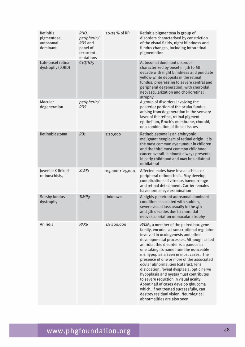
�8www.phgfoundation.org
Retinitis pigmentosa, autosomal dominant
RHO,peripherin/RDS and panel of recurrent mutations
20-25 % of RP Retinitis pigmentosa is group of disorders characterised by constriction of the visual fields, night blindness and fundus changes, including intraretinal pigmentation
Late-onset retinal dystrophy (LORD)
C1QTNF5 Autosomal dominant disorder characterized by onset in 5th to 6th decade with night blindness and punctate yellow-white deposits in the retinal fundus, progressing to severe central and peripheral degeneration, with choroidal neovascularization and chorioretinal atrophy
Macular degeneration
peripherin/RDS
A group of disorders involving the posterior portion of the ocular fundus, arising from degeneration in the sensory layer of the retina, retinal pigment epithelium, Bruch's membrane, choroid, or a combination of these tissues
Retinoblastoma RB1 1:20,000 Retinoblastoma is an embryonic malignant neoplasm of retinal origin. It is the most common eye tumour in children and the third most common childhood cancer overall. It almost always presents in early childhood and may be unilateral or bilateral
Juvenile X-linked-retinoschisis,
XLRS1 1:5,000-1:25,000 Affected males have foveal schisis or peripheral retinoschisis. May develop complications of vitreous haemorrhage and retinal detachment. Carrier females have normal eye examination
Sorsby fundus dystrophy
TIMP3 Unknown A highly penetrant autosomal dominant condition associated with sudden, severe visual loss usually in the �th and 5th decades due to choroidal neovasculariation or macular atrophy
Aniridia PAX6 1.8:100,000 PAX6, a member of the paired box gene family, encodes a transcriptional regulator involved in oculogenesis and other developmental processes. Although called aniridia, this disorder is a panocular one taking its name from the noticeable iris hypoplasia seen in most cases. The presence of one or more of the associated ocular abnormalities (cataract, lens dislocation, foveal dysplasia, optic nerve hypoplasia and nystagmus) contributes to severe reduction in visual acuity. About half of cases develop glaucoma which, if not treated successfully, can destroy residual vision. Neurological abnormalities are also seen
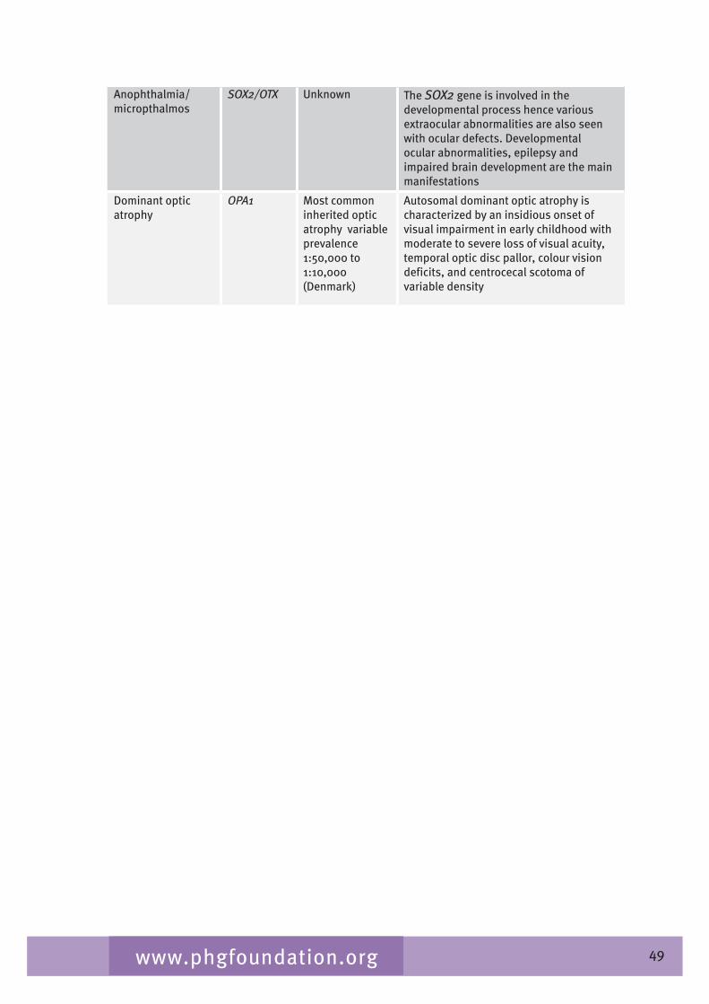
�9www.phgfoundation.org
Anophthalmia/micropthalmos
SOX2/OTX Unknown The SOX2 gene is involved in the developmental process hence various extraocular abnormalities are also seen with ocular defects. Developmental ocular abnormalities, epilepsy and impaired brain development are the main manifestations
Dominant optic atrophy
OPA1 Most common inherited optic atrophy variable prevalence 1:50,000 to 1:10,000 (Denmark)
Autosomal dominant optic atrophy is characterized by an insidious onset of visual impairment in early childhood with moderate to severe loss of visual acuity, temporal optic disc pallor, colour vision deficits, and centrocecal scotoma of variable density
50www.phgfoundation.org
Chapter 7 Survey of clinical genetic ophthalmology services
7.1 Introduction
The purpose of the survey was to identify the main providers of specialist genetic ophthalmology services throughout the UK and to obtain information on the services offered, structure, organisation and activity and any information on service shortfall.
7.2 Method
The questionnaire was sent out in November 2006 to 2� centres in the UK identified as possibly providing specialist genetic ophthalmology services through an initial query to all regional genetics services the previous July. A reminder was sent to centres who had not replied in January 2007.
Information was collected on the general organisation of services for children and adults, levels of specialist staffing, provision of specialist clinics, how the services were accessed, conditions managed, availability of special diagnostic facilities and other services, and perceived gaps in provision.
7.3 Results
Replies were received from all services. Full questionnaire responses were received from 20 specialist ophthalmology genetics services including one service based at Musgrove Park Hospital, Taunton that had not been identified in the initial survey. This was part of the Bristol regional service. Four services (West Scotland, Trent, NW Thames and NE Thames) reported that they did not operate a specialist ophthalmic genetics service. Full contact details provided by each service are given in Appendix 1. The findings are detailed in the following sections. For consistency services are displayed in all tables in the standard order of Strategic Health Authority area, followed by Wales, Scotland and Northern Ireland.
Services and populations served
Nineteen specialist services were identified and provided detailed information. These are set out by Strategic Health Authority area in Table 7.1. It can thus be seen that services are available in conjunction with most regional genetic services and therefore in the majority of the Strategic Health Authority (SHA) areas of England and in Wales, Scotland and Northern Ireland. There is no specialist service available from Glasgow and Nottingham, NE Thames and NW Thames. NW Thames noted that ophthalmology patients are seen in normal genetics clinics. However, the specialist Eye Hospital at Moorfields in London has an established Genetic Eye service with an extensive service serving London and Southeast and, to a lesser extent, the whole of the UK.
Catchment populations range from approximately 500,000 (Tayside and North Fife) to 5.2 million (West Midlands). Three services, all in Scotland serve populations of under 1 million, nine serve populations of between 1-2.9 million while seven serve populations of between � and 5.2 million. The four regional centres that do not provide specialist services serve populations of 2.8-�.5 million.
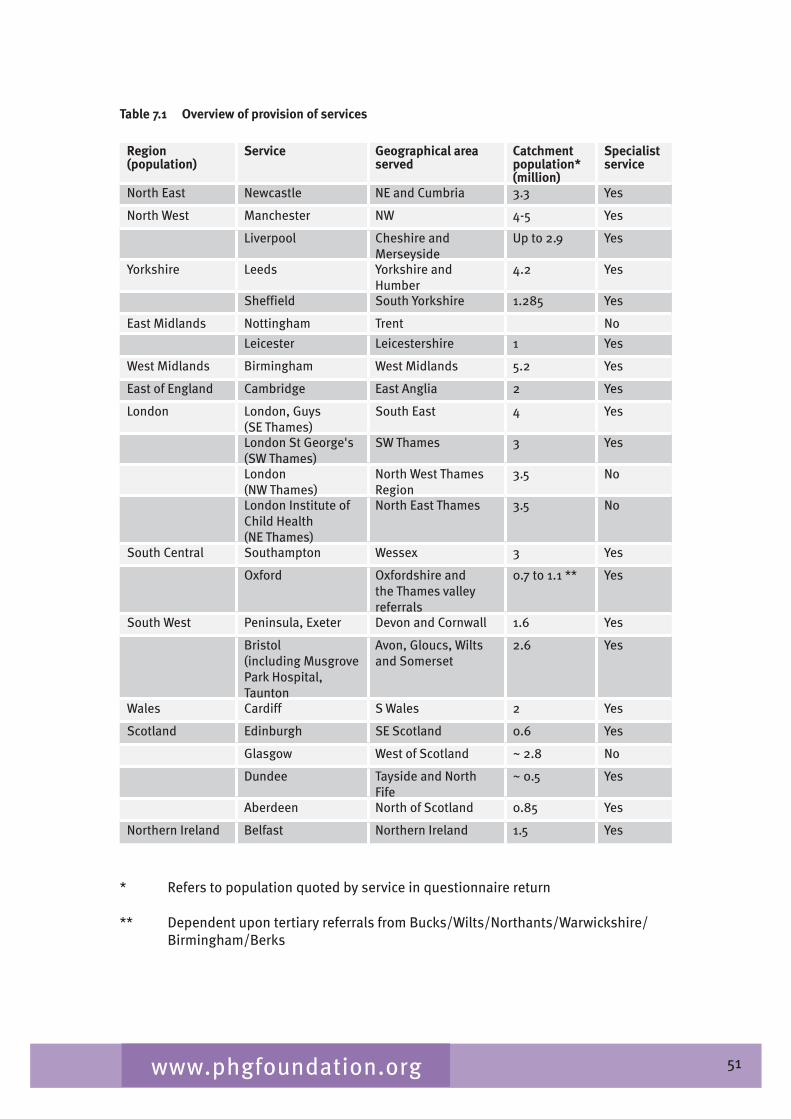
51www.phgfoundation.org
Table 7.1 Overview of provision of services
Region(population)
Service Geographical area served
Catchmentpopulation* (million)
Specialist service
North East Newcastle NE and Cumbria �.� Yes
North West Manchester NW �-5 Yes
Liverpool Cheshire and Merseyside
Up to 2.9 Yes
Yorkshire Leeds Yorkshire and Humber
�.2 Yes
Sheffield South Yorkshire 1.285 Yes
East Midlands Nottingham Trent No
Leicester Leicestershire 1 Yes
West Midlands Birmingham West Midlands 5.2 Yes
East of England Cambridge East Anglia 2 Yes
London London, Guys (SE Thames)
South East � Yes
London St George's (SW Thames)
SW Thames � Yes
London (NW Thames)
North West Thames Region
�.5 No
London Institute of Child Health (NE Thames)
North East Thames �.5 No
South Central Southampton Wessex � Yes
Oxford Oxfordshire and the Thames valley referrals
0.7 to 1.1 ** Yes
South West Peninsula, Exeter Devon and Cornwall 1.6 Yes
Bristol(including Musgrove Park Hospital, Taunton
Avon, Gloucs, Wilts and Somerset
2.6 Yes
Wales Cardiff S Wales 2 Yes
Scotland Edinburgh SE Scotland 0.6 Yes
Glasgow West of Scotland ~ 2.8 No
Dundee Tayside and North Fife
~ 0.5 Yes
Aberdeen North of Scotland 0.85 Yes
Northern Ireland Belfast Northern Ireland 1.5 Yes
* Refers to population quoted by service in questionnaire return
** Dependent upon tertiary referrals from Bucks/Wilts/Northants/Warwickshire/ Birmingham/Berks
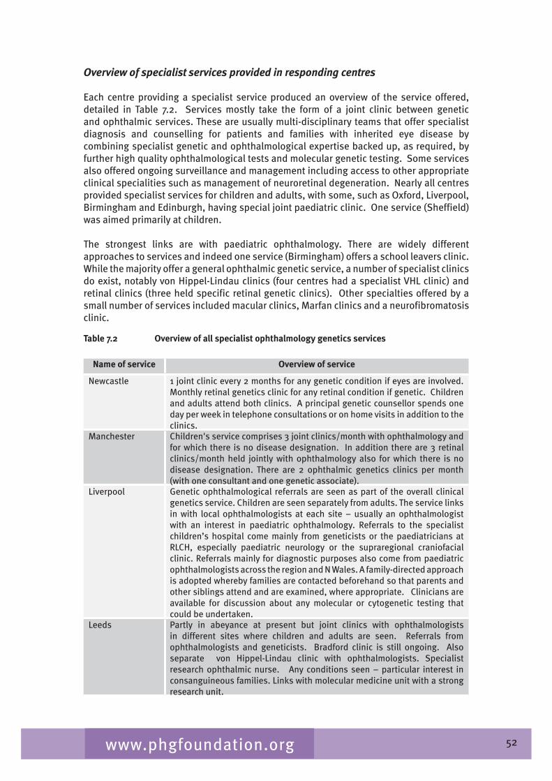
52www.phgfoundation.org
Overview of specialist services provided in responding centres
Each centre providing a specialist service produced an overview of the service offered, detailed in Table 7.2. Services mostly take the form of a joint clinic between genetic and ophthalmic services. These are usually multi-disciplinary teams that offer specialist diagnosis and counselling for patients and families with inherited eye disease by combining specialist genetic and ophthalmological expertise backed up, as required, by further high quality ophthalmological tests and molecular genetic testing. Some services also offered ongoing surveillance and management including access to other appropriate clinical specialities such as management of neuroretinal degeneration. Nearly all centres provided specialist services for children and adults, with some, such as Oxford, Liverpool, Birmingham and Edinburgh, having special joint paediatric clinic. One service (Sheffield) was aimed primarily at children.
The strongest links are with paediatric ophthalmology. There are widely different approaches to services and indeed one service (Birmingham) offers a school leavers clinic. While the majority offer a general ophthalmic genetic service, a number of specialist clinics do exist, notably von Hippel-Lindau clinics (four centres had a specialist VHL clinic) and retinal clinics (three held specific retinal genetic clinics). Other specialties offered by a small number of services included macular clinics, Marfan clinics and a neurofibromatosis clinic.
Table 7.2 Overview of all specialist ophthalmology genetics services
Name of service Overview of service
Newcastle 1 joint clinic every 2 months for any genetic condition if eyes are involved. Monthly retinal genetics clinic for any retinal condition if genetic. Children and adults attend both clinics. A principal genetic counsellor spends one day per week in telephone consultations or on home visits in addition to the clinics.
Manchester Children's service comprises � joint clinics/month with ophthalmology and for which there is no disease designation. In addition there are � retinal clinics/month held jointly with ophthalmology also for which there is no disease designation. There are 2 ophthalmic genetics clinics per month (with one consultant and one genetic associate).
Liverpool Genetic ophthalmological referrals are seen as part of the overall clinical genetics service. Children are seen separately from adults. The service links in with local ophthalmologists at each site – usually an ophthalmologist with an interest in paediatric ophthalmology. Referrals to the specialist children’s hospital come mainly from geneticists or the paediatricians at RLCH, especially paediatric neurology or the supraregional craniofacial clinic. Referrals mainly for diagnostic purposes also come from paediatric ophthalmologists across the region and N Wales. A family-directed approach is adopted whereby families are contacted beforehand so that parents and other siblings attend and are examined, where appropriate. Clinicians are available for discussion about any molecular or cytogenetic testing that could be undertaken.
Leeds Partly in abeyance at present but joint clinics with ophthalmologists in different sites where children and adults are seen. Referrals from ophthalmologists and geneticists. Bradford clinic is still ongoing. Also separate von Hippel-Lindau clinic with ophthalmologists. Specialist research ophthalmic nurse. Any conditions seen – particular interest in consanguineous families. Links with molecular medicine unit with a strong research unit.
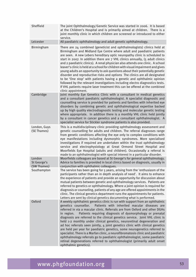
5�www.phgfoundation.org
Sheffield The joint Ophthalmology/Genetic Service was started in 2006. It is based at the Children’s Hospital and is primarily aimed at children. There is a joint monthly clinic in which children are screened or introduced to either service.
Leicester All paediatric ophthalmology and adult genetic ophthalmology.
Birmingham There are 2� combined (geneticist and ophthalmologists) clinics held at Birmingham and Midland Eye Centre where adult and paediatric patients are seen. A new Lebers hereditary optic neuropathy clinic is scheduled to start in 2007. In addition there are 7 VHL clinics annually, (5 adult clinics and 2 paediatric clinics). A renal physician also attends one clinic. A school leaver’s clinic is held at a school for children with visual impairment and gives young adults an opportunity to ask questions about their potentially genetic disorder and reproductive risks and options The clinics are all designated to be ‘One stop’ with patients having a genetic and ophthalmic opinion followed by the relevant investigations including electro diagnostics tests. If VHL patients require laser treatment this can be offered at the combined clinic appointment.
Cambridge Joint monthly Eye Genetics Clinic with a consultant in medical genetics and a consultant paediatric ophthalmologist. A specialist diagnostic and counselling service is provided for patients and families with inherited eye disorders by combining genetic and ophthalmological expertise backed up by high quality electrodiagnostic testing and molecular genetic testing where appropriate. In addition there is a monthly VHL clinic held jointly by a consultant in cancer genetics and a consultant ophthalmologist. A specialist service for Stickler syndrome patients is also provided.
London, Guys (SE Thames)
This is a multidisciplinary clinic providing ophthalmology assessment and genetic counselling for adults and children. The referral diagnoses range from genetic conditions affecting the eye only to complex conditions with eye manifestations including dysmorphic syndromes. More specialised investigations if required are undertaken within the trust ophthalmology service and electrophysiology at Great Ormond Street Hospital and Moorfields Eye Hospital (adults and children). Occasionally a referral is made to an ophthalmologist with special interest in a particular disorder.
London St George’s (SW Thames)
Moorfields colleagues are based at St George’s for general ophthalmology. Advice to families is provided in local clinics based on diagnosis, usually in conjunction with ophthalmic colleagues.
Southampton The service has been going for 5 years, arising from the 'enthusiasm of the participants rather than an in depth analysis of need'. It aims to enhance the experience of patients and provide an opportunity for discussion about mutual patients between genetic and ophthalmology services. Patients are referred to genetics or ophthalmology. Where a joint opinion is required for diagnosis or counseling, patients of any age are offered appointments in the clinic. The clinical genetics department runs the appointments for patients. Letters are sent by clinical genetics documenting what is performed.
Oxford A weekly ophthalmic genetics clinic is run with support from an ophthalmic genetics counsellor. Patients with inherited macular diseases are referred in via a macular clinic. Referrals are from Oxford and other DGHs in region. Patients requiring diagnosis of dysmorphology or prenatal diagnosis are referred to the clinical genetics service. Joint VHL clinic is held 1-2 monthly under clinical genetics, neuroretinal degeneration and ad hoc referrals seen jointly, � joint genetics clinic with clinical genetics are held per year for paediatric genetics, some neurogenetics referred to specialist. There is a Marfan clinic, a neurofibromatosis clinic and paediatric ophthalmology referrals go to paediatric ophthalmologist, some paediatric retinal degenerations referred to ophthalmologist (primarily adult onset ophthalmic genetics).
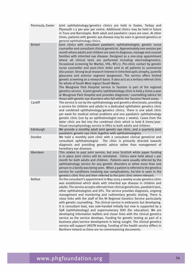
5�www.phgfoundation.org
Peninsula, Exeter Joint ophthalmology/genetics clinics are held in Exeter, Torbay and Plymouth 1-2 per year per centre. Additional clinics may be held in future in Truro and Barnstaple. Both adult and paediatric cases are seen. At other times, patients with genetic eye disease may be seen in general genetics or general ophthalmology clinics.
Bristol Joint clinics with consultant paediatric ophthalmologist, genetic nurse counsellor and consultant clinical geneticist. Approximately one session per month where adults and children are seen to diagnose, manage and counsel families with inherited eye disease. Designed as a one-stop appointment where all clinical tests are performed including electrodiagnostics. Occasional screening for Marfan, VHL, NF1/2. Pre-clinic contact by genetic nurse counsellor and post-clinic letter sent to all patients to summarise discussion. Strong local research interest in inherited optic atrophy, juvenile glaucoma and anterior segment dysgenesis. The service offers limited genetic screening on a research basis. It also acts as a tertiary referral clinic for whole of South West region/South Wales.The Musgrove Park Hospital service in Taunton is part of the regional genetics service. A joint genetic/ophthalmology clinic is held � times a year at Musgrove Park Hospital and provides diagnosis/ counselling/advice for those with genetic eye disorders who fall within the Taunton/Yeovil areas.
Cardiff The service is run by the ophthalmology and genetics directorate, providing a service for children and adults in a dedicated ophthalmic genetics clinic and combined ophthalmology/genetics clinics. There is one adult clinic per week for medical retinal problems and one paediatric ophthalmology genetic clinic (run by an ophthalmologist every 2 weeks). Cases from the latter clinic are fed into the combined clinic which is held 8 times/year. Visual neurophysiology service ie ERGs to both adults and children.
Edinburgh We provide a monthly adult joint genetic eye clinic, and a quarterly joint paediatric genetic eye clinic together with ophthalmologists.
Dundee We hold a monthly joint clinic with a consultant clinical geneticist and consultant ophthalmologist. The clinic is primarily directed towards diagnosis and providing genetic advice rather than management of hereditary eye diseases.
Aberdeen This relates to past joint service, but once Scottish white paper funding is in place joint clinics will be reinstated. Clinics were held about 1 per month for both adults and children. Patients were usually referred by the ophthalmology service for any genetic disorders or when more than one person in a family was being seen. When a patient is referred to the genetics service for conditions involving eye complications, he/she is seen in the genetics clinic first and then referred to the joint clinic where relevant.
Belfast On the consultant’s appointment in May 2005 a weekly ocular genetics clinic was established which deals with inherited eye disease in children and adults. The service accepts referrals from clinical geneticists, paediatricians, other ophthalmologists and GPs. The service provides diagnosis, ongoing management and monitoring and rudimentary genetic testing. There is close links with the staff of the NI Regional Genetics Service particularly with genetic counselling. The clinical service is embryonic but developing. It is consultant lead, was sole-handed initially but now is supported by a SpR (ophthalmology) and supernumerary SHO (for education). We are developing information leaflets and closer links with the clinical genetics service as the service develops. Funding for genetic testing as part of a business plan/service development is being sought. The clinical genetics service will support UKGTN testing. Funding of the health service differs in Northern Ireland so there are no commissioning documents.
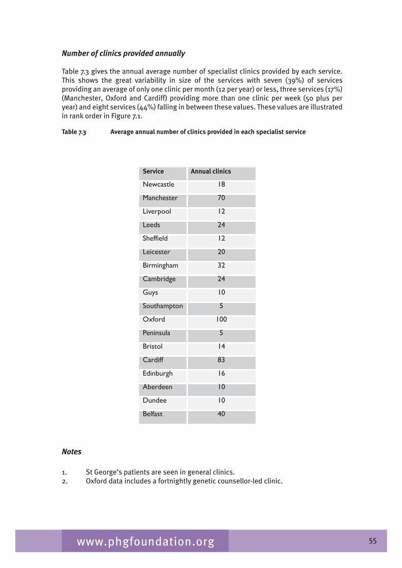
55www.phgfoundation.org
Number of clinics provided annually
Table 7.� gives the annual average number of specialist clinics provided by each service. This shows the great variability in size of the services with seven (�9%) of services providing an average of only one clinic per month (12 per year) or less, three services (17%) (Manchester, Oxford and Cardiff) providing more than one clinic per week (50 plus per year) and eight services (��%) falling in between these values. These values are illustrated in rank order in Figure 7.1.
Table 7.3 Average annual number of clinics provided in each specialist service
Notes
1. St George’s patients are seen in general clinics.2. Oxford data includes a fortnightly genetic counsellor-led clinic.
Service Annual clinics
Newcastle 18
Manchester 70
Liverpool 12
Leeds 24
Sheffield 12
Leicester 20
Birmingham 32
Cambridge 24
Guys 10
Southampton 5
Oxford 100
Peninsula 5
Bristol 14
Cardiff 83
Edinburgh 16
Aberdeen 10
Dundee 10
Belfast 40
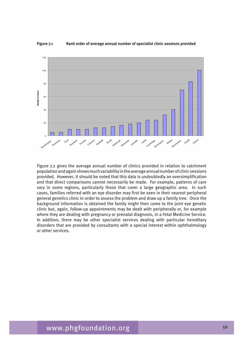
56www.phgfoundation.org
Figure 7.1 Rank order of average annual number of specialist clinic sessions provided
0
20
40
60
80
100
120
Southampton
Pen insu la G uys
AberdeenDundee
L iverpoo l
Sheffield
B ris to l
Ed inbu rgh
Newcastle
Le icesterLeeds
Cambridge
B irmingham
Be lfas t
M anchesterCard iff
O xford
Numb
er of
clini
cs
Figure 7.2 gives the average annual number of clinics provided in relation to catchment population and again shows much variability in the average annual number of clinic sessions provided. However, it should be noted that this data is undoubtedly an oversimplification and that direct comparisons cannot necessarily be made. For example, patterns of care vary in some regions, particularly those that cover a large geographic area. In such cases, families referred with an eye disorder may first be seen in their nearest peripheral general genetics clinic in order to assess the problem and draw up a family tree. Once the background information is obtained the family might then come to the joint eye genetic clinic but, again, follow-up appointments may be dealt with peripherally or, for example where they are dealing with pregnancy or prenatal diagnosis, in a Fetal Medicine Service. In addition, there may be other specialist services dealing with particular hereditary disorders that are provided by consultants with a special interest within ophthalmology or other services.
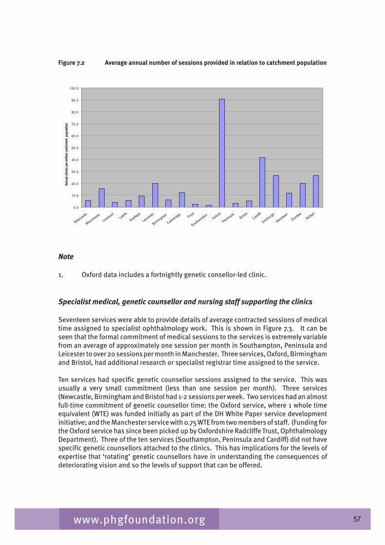
57www.phgfoundation.org
Figure 7.2 Average annual number of sessions provided in relation to catchment population
0.0
10.0
20.0
30.0
40.0
50.0
60.0
70.0
80.0
90.0
100.0
Newcastle
M anchester
L iverpoo lLeeds
Sheffield
Le icester
B irmingham
Cambridge Guys
SouthamptonO xford
Pen insu laB ris to l
Card iff
Ed inbu rgh
AberdeenDundee
Be lfas t
Annu
al cli
nics p
er mi
llion c
atchm
ent p
opula
tion
Note
1. Oxford data includes a fortnightly genetic consellor-led clinic.
Specialist medical, genetic counsellor and nursing staff supporting the clinics
Seventeen services were able to provide details of average contracted sessions of medical time assigned to specialist ophthalmology work. This is shown in Figure 7.�. It can be seen that the formal commitment of medical sessions to the services is extremely variable from an average of approximately one session per month in Southampton, Peninsula and Leicester to over 20 sessions per month in Manchester. Three services, Oxford, Birmingham and Bristol, had additional research or specialist registrar time assigned to the service.
Ten services had specific genetic counsellor sessions assigned to the service. This was usually a very small commitment (less than one session per month). Three services (Newcastle, Birmingham and Bristol had 1-2 sessions per week. Two services had an almost full-time commitment of genetic counsellor time: the Oxford service, where 1 whole time equivalent (WTE) was funded initially as part of the DH White Paper service development initiative; and the Manchester service with 0.75 WTE from two members of staff. (Funding for the Oxford service has since been picked up by Oxfordshire Radcliffe Trust, Ophthalmology Department). Three of the ten services (Southampton, Peninsula and Cardiff) did not have specific genetic counsellors attached to the clinics. This has implications for the levels of expertise that ‘rotating’ genetic counsellors have in understanding the consequences of deteriorating vision and so the levels of support that can be offered.
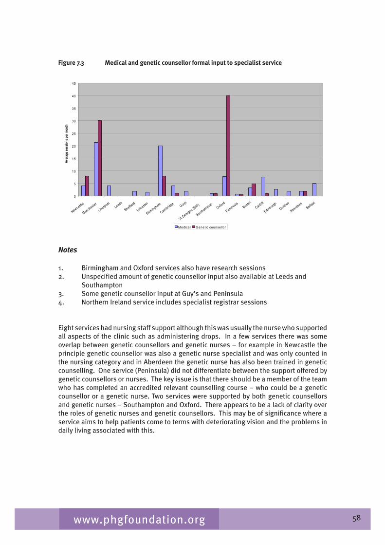
58www.phgfoundation.org
Figure 7.3 Medical and genetic counsellor formal input to specialist service
0
5
10
15
20
25
30
35
40
45
Newcastle
M anchester
L iverpoo lLeeds
Sheffield
Le icester
B irmingham
Cambridge Guys
S t Georges (S
W )
SouthamptonO xford
Pen insu laB ris to l
Card iff
Ed inbu rghDundee
AberdeenBe lfas t
Aver
age s
essio
ns pe
r mon
th
M edica l G enetic counse llo r
Notes
1. Birmingham and Oxford services also have research sessions2. Unspecified amount of genetic counsellor input also available at Leeds and Southampton�. Some genetic counsellor input at Guy’s and Peninsula�. Northern Ireland service includes specialist registrar sessions
Eight services had nursing staff support although this was usually the nurse who supported all aspects of the clinic such as administering drops. In a few services there was some overlap between genetic counsellors and genetic nurses – for example in Newcastle the principle genetic counsellor was also a genetic nurse specialist and was only counted in the nursing category and in Aberdeen the genetic nurse has also been trained in genetic counselling. One service (Peninsula) did not differentiate between the support offered by genetic counsellors or nurses. The key issue is that there should be a member of the team who has completed an accredited relevant counselling course – who could be a genetic counsellor or a genetic nurse. Two services were supported by both genetic counsellors and genetic nurses – Southampton and Oxford. There appears to be a lack of clarity over the roles of genetic nurses and genetic counsellors. This may be of significance where a service aims to help patients come to terms with deteriorating vision and the problems in daily living associated with this.
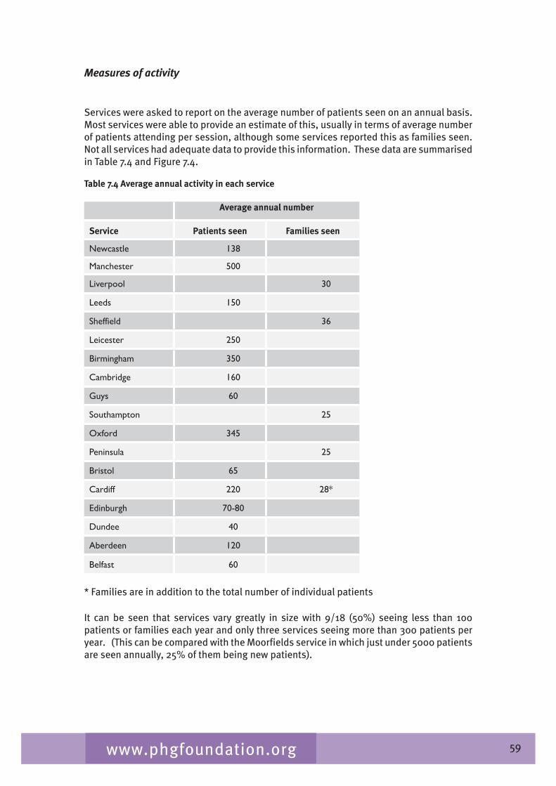
59www.phgfoundation.org
Measures of activity
Services were asked to report on the average number of patients seen on an annual basis. Most services were able to provide an estimate of this, usually in terms of average number of patients attending per session, although some services reported this as families seen. Not all services had adequate data to provide this information. These data are summarised in Table 7.� and Figure 7.�.
Table 7.4 Average annual activity in each service
Average annual number
Service Patients seen Families seen
Newcastle 138
Manchester 500
Liverpool 30
Leeds 150
Sheffield 36
Leicester 250
Birmingham 350
Cambridge 160
Guys 60
Southampton 25
Oxford 345
Peninsula 25
Bristol 65
Cardiff 220 28*
Edinburgh 70-80
Dundee 40
Aberdeen 120
Belfast 60
* Families are in addition to the total number of individual patients
It can be seen that services vary greatly in size with 9/18 (50%) seeing less than 100 patients or families each year and only three services seeing more than �00 patients per year. (This can be compared with the Moorfields service in which just under 5000 patients are seen annually, 25% of them being new patients).
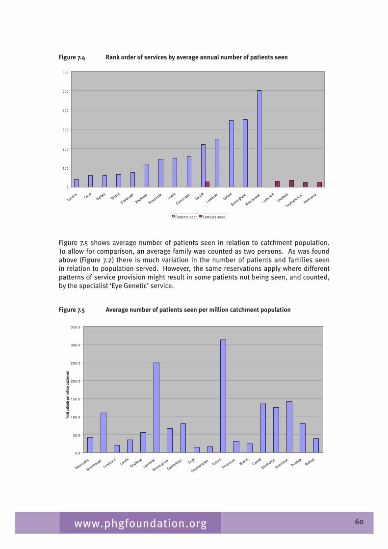
60www.phgfoundation.org
Figure 7.4 Rank order of services by average annual number of patients seen
0
100
200
300
400
500
600
DundeeGuys
Be lfas tB ris to l
Ed inbu rgh
Aberdeen
NewcastleLeeds
CambridgeCard iff
Le icesterO xford
B irmingham
Manchester
L iverpoo l
Sheffield
Southampton
Pen insu la
P atien ts seen Fam ilies seen
Figure 7.5 shows average number of patients seen in relation to catchment population. To allow for comparison, an average family was counted as two persons. As was found above (Figure 7.2) there is much variation in the number of patients and families seen in relation to population served. However, the same reservations apply where different patterns of service provision might result in some patients not being seen, and counted, by the specialist ‘Eye Genetic’ service.
Figure 7.5 Average number of patients seen per million catchment population
0.0
50.0
100.0
150.0
200.0
250.0
300.0
350.0
Newcastle
M anchester
L iverpoo lLeeds
Sheffield
Le icester
B irmingham
Cambridge Guys
SouthamptonO xford
Pen insu laB ris to l
Card iff
Ed inbu rgh
AberdeenDundee
Be lfas t
Total
patie
nts pe
r milli
on ca
tchme
nt
61www.phgfoundation.org
Referral information
Referrals to ophthalmic genetic clinics are mainly from ophthalmic and clinical genetics services although some referrals from other specialties (eg paediatrics) do occur. There is variability from area to area in the levels of GP referrals and while most do receive referrals from GPs (80% of those who responded to this question) some noted that these were occasional. Two services also stated that they received referrals from optometrists or opticians. This variability in referral reflects the differences in formal and informal patient pathways that exist across the country.
Referral protocols and advertising of specialist service
Only one service (Cardiff) reported having a referral protocol. A further two services (London Guys and Oxford) stated that there was a recognised care pathway. In Oxford, this was directly to the ophthalmic genetics service. Where designated, referrals were made to the service via the clinical genetics department which acted as gatekeepers to the combined clinic. Most services had not undertaken any specific ‘marketing’ of the service and relied on word of mouth and knowledge of the service that they hoped was gained through teaching and informal discussions. The Oxford service used patient summary letters, sent to referring consultants to raise awareness of what the specialist service could provide. Information about three services (Cambridge, London Guys and Bristol) was provided on their trust website. Others (Cambridge, Oxford and Northern Ireland) had written letters to clinicians. The Oxford service used their programme of educational sessions to raise awareness about the service amongst local clinicians. This educational programme was also supported by the genetic counsellor. They had recently also provided information to patients about the services through the Adult Low Vision Services leaflet. One service (Aberdeen) stated that it did not advertise as it was already running at full capacity.
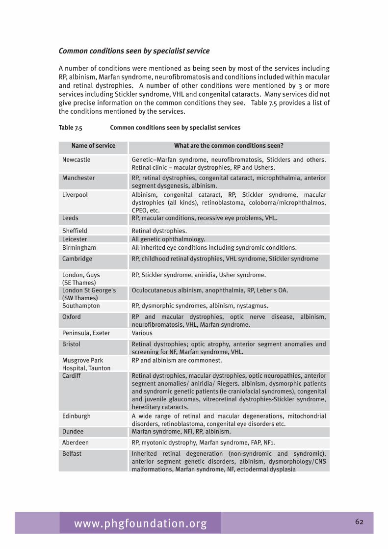
62www.phgfoundation.org
Common conditions seen by specialist service
A number of conditions were mentioned as being seen by most of the services including RP, albinism, Marfan syndrome, neurofibromatosis and conditions included within macular and retinal dystrophies. A number of other conditions were mentioned by � or more services including Stickler syndrome, VHL and congenital cataracts. Many services did not give precise information on the common conditions they see. Table 7.5 provides a list of the conditions mentioned by the services.
Table 7.5 Common conditions seen by specialist services
Name of service What are the common conditions seen?
Newcastle Genetic–Marfan syndrome, neurofibromatosis, Sticklers and others. Retinal clinic – macular dystrophies, RP and Ushers.
Manchester RP, retinal dystrophies, congenital cataract, microphthalmia, anterior segment dysgenesis, albinism.
Liverpool Albinism, congenital cataract, RP, Stickler syndrome, macular dystrophies (all kinds), retinoblastoma, coloboma/microphthalmos, CPEO, etc.
Leeds RP, macular conditions, recessive eye problems, VHL.
Sheffield Retinal dystrophies.Leicester All genetic ophthalmology.Birmingham All inherited eye conditions including syndromic conditions.
Cambridge RP, childhood retinal dystrophies, VHL syndrome, Stickler syndrome
London, Guys (SE Thames)
RP, Stickler syndrome, aniridia, Usher syndrome.
London St George's (SW Thames)
Oculocutaneous albinism, anophthalmia, RP, Leber's OA.
Southampton RP, dysmorphic syndromes, albinism, nystagmus.
Oxford RP and macular dystrophies, optic nerve disease, albinism, neurofibromatosis, VHL, Marfan syndrome.
Peninsula, Exeter Various
Bristol Retinal dystrophies; optic atrophy, anterior segment anomalies and screening for NF, Marfan syndrome, VHL.
Musgrove Park Hospital, Taunton
RP and albinism are commonest.
Cardiff Retinal dystrophies, macular dystrophies, optic neuropathies, anterior segment anomalies/ aniridia/ Riegers. albinism, dysmorphic patients and syndromic genetic patients (ie craniofacial syndromes), congenital and juvenile glaucomas, vitreoretinal dystrophies-Stickler syndrome, hereditary cataracts.
Edinburgh A wide range of retinal and macular degenerations, mitochondrial disorders, retinoblastoma, congenital eye disorders etc.
Dundee Marfan syndrome, NFI, RP, albinism.
Aberdeen RP, myotonic dystrophy, Marfan syndrome, FAP, NF1.
Belfast Inherited retinal degeneration (non-syndromic and syndromic), anterior segment genetic disorders, albinism, dysmorphology/CNS malformations, Marfan syndrome, NF, ectodermal dysplasia
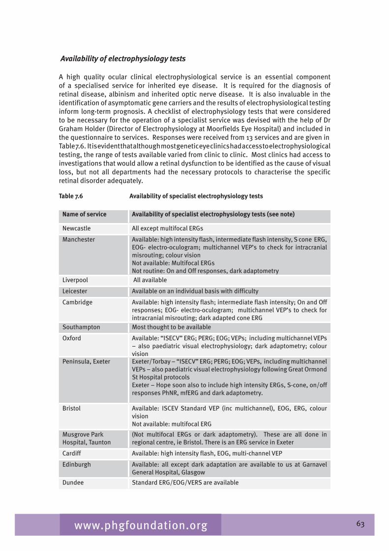
6�www.phgfoundation.org
Availability of electrophysiology tests
A high quality ocular clinical electrophysiological service is an essential component of a specialised service for inherited eye disease. It is required for the diagnosis of retinal disease, albinism and inherited optic nerve disease. It is also invaluable in the identification of asymptomatic gene carriers and the results of electrophysiological testing inform long-term prognosis. A checklist of electrophysiology tests that were considered to be necessary for the operation of a specialist service was devised with the help of Dr Graham Holder (Director of Electrophysiology at Moorfields Eye Hospital) and included in the questionnaire to services. Responses were received from 1� services and are given in Table 7.6. It is evident that although most genetic eye clinics had access to electrophysiological testing, the range of tests available varied from clinic to clinic. Most clinics had access to investigations that would allow a retinal dysfunction to be identified as the cause of visual loss, but not all departments had the necessary protocols to characterise the specific retinal disorder adequately.
Table 7.6 Availability of specialist electrophysiology tests
Name of service Availability of specialist electrophysiology tests (see note)
Newcastle All except multifocal ERGs
Manchester Available: high intensity flash, intermediate flash intensity, S cone ERG, EOG- electro-oculogram; multichannel VEP’s to check for intracranial misrouting; colour visionNot available: Multifocal ERGsNot routine: On and Off responses, dark adaptometry
Liverpool All available
Leicester Available on an individual basis with difficulty
Cambridge Available: high intensity flash; intermediate flash intensity; On and Off responses; EOG- electro-oculogram; multichannel VEP’s to check for intracranial misrouting; dark adapted cone ERG
Southampton Most thought to be available
Oxford Available: “ISECV” ERG; PERG; EOG; VEPs; including multichannel VEPs – also paediatric visual electrophysiology; dark adaptometry; colour vision
Peninsula, Exeter Exeter/Torbay – “ISECV” ERG; PERG; EOG; VEPs, including multichannel VEPs – also paediatric visual electrophysiology following Great Ormond St Hospital protocolsExeter – Hope soon also to include high intensity ERGs, S-cone, on/off responses PhNR, mfERG and dark adaptometry.
Bristol Available: ISCEV Standard VEP (inc multichannel), EOG, ERG, colour vision Not available: multifocal ERG
Musgrove Park Hospital, Taunton
(Not multifocal ERGs or dark adaptometry). These are all done in regional centre, ie Bristol. There is an ERG service in Exeter
Cardiff Available: high intensity flash, EOG, multi-channel VEP
Edinburgh Available: all except dark adaptation are available to us at Garnavel General Hospital, Glasgow
Dundee Standard ERG/EOG/VERS are available
6�www.phgfoundation.org
NoteNote
Services were asked about the availability of the following specialist tests: high intensity flash; intermediate flash intensity; S cone ERG; On and Off responses; Multifocal ERGs; EOG (electro-oculogram); multichannel VEP’s to check for intracranial misrouting; dark adaptometry; colour vision testing.
Gaps in services
Fourteen services responded on their perception of the main gaps in service provision for inherited eye disease. Three services mentioned the lack of support services to administer and run the service whilst a further service mentioned complete lack of interest from commissioners and organisational gaps in engaging 'two departments separately funded'. Sustainability of the service, particularly where they were dependent on individuals with a special interest was also mentioned here and in other sections of the questionnaire. Two services identified a lack of electrophysiology testing, one service noted lack of genetic counselling and a further service noted lack of nursing support. Gaps in provision and funding for genetic testing were also noted. Those related to particular tests are discussed in Chapter 6. Services described the lack of funding for adequate genetic testing, and the lack of centres to send samples while others focused on the technological barriers to effective genetic testing where many genes are involved in highly heterogeneous conditions. A further theme was the variability in referral rates from different districts within the region suggesting that some patients were not being referred to the service.
Patient information
Many services used patient information produced by charities such as the Retinitis Pigmentosa Society and Macular Disease Society and referred patients to relevant websites or support groups through the charity Contact-A-Family. Seven services preferred to use their letter to patients to provide the primary information that patients required supplemented as appropriate by information produced by the charities. No services had produced their own patient information leaflets.
Payment by results (PbR)
At the time of completing the survey, payment by results had not had an impact on the specialist ophthalmic genetics service. In parts of the UK such as Scotland, Wales and Northern Ireland it had not been introduced. In other areas, the genetics service is not included in the payment by results system. One service did raise concerns that in general it may make it harder to set up joint clinics.
65www.phgfoundation.org
7.4 Provision of specialist services for genetic eye disease in London and Moorfields Eye Hospital
The provision of services for genetic eye disease in London is complicated by the pattern of NHS service provision in the capital, particularly the presence of many different teaching hospitals and specialist hospitals such as Moorfields Eye Hospital (MEH), the Hospital for Children, Great Ormond Street and the National Hospital for Nervous Diseases. This diversity of providers has made it more difficult to develop multi-speciality clinics such as Eye Genetic Clinics. Moorfields Eye Hospital has had established Genetic Eye clinics and a genetic register for more than �0 years. The genetic register has more than �0,000 entries. MEH has two consultant ophthalmologists with a special interest in inherited eye disease who run � clinics a week that are concerned with genetic eye disease. These clinics are supported by genetic counsellors, family support workers and a social worker. In 2005-6 some �777 patients were seen by the two consultants in the genetic eye clinics of whom approximately 25% were new patients and the rest follow-up. There is a large department of ocular electrophysiology with two senior scientists and several technicians. The electrophysiology department tests over 2000 patients a year, the majority of which have inherited eye disease. There is also a very strong research programme in inherited eye disease. MEH is one of the new NHS Biomedical Research Centres and one of the research themes relates to inherited eye disease.
The MEH model differs from much of the rest of the UK. Here most diagnosis and genetic counselling is carried out in specialised clinics by ophthalmologists with a special interest. Some patients, for example those with undiagnosed multi-system disease, complex developmental abnormalities or those wishing to consider prenatal diagnosis are referred to their local clinical genetic services. The MEH service receives referrals from across the UK with the majority coming from London and the South East. Many families with genetic eye disease have a long association with MEH and prefer to be seen at the hospital rather than access local services. The presence of the MEH service in London has meant that there has been little incentive for Regional Genetic Services in London to develop joint eye genetic clinics.
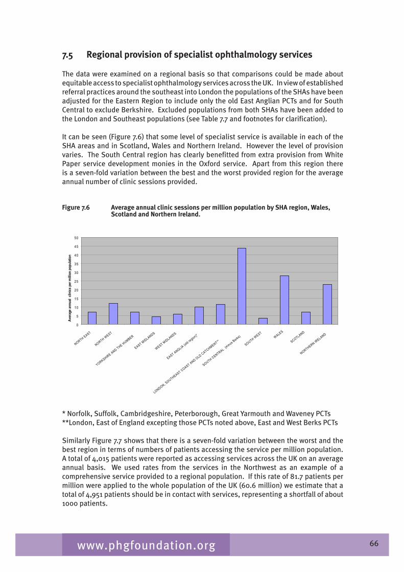
66www.phgfoundation.org
7.5 Regional provision of specialist ophthalmology services
The data were examined on a regional basis so that comparisons could be made about equitable access to specialist ophthalmology services across the UK. In view of established referral practices around the southeast into London the populations of the SHAs have been adjusted for the Eastern Region to include only the old East Anglian PCTs and for South Central to exclude Berkshire. Excluded populations from both SHAs have been added to the London and Southeast populations (see Table 7.7 and footnotes for clarification).
It can be seen (Figure 7.6) that some level of specialist service is available in each of the SHA areas and in Scotland, Wales and Northern Ireland. However the level of provision varies. The South Central region has clearly benefitted from extra provision from White Paper service development monies in the Oxford service. Apart from this region there is a seven-fold variation between the best and the worst provided region for the average annual number of clinic sessions provided.
Figure 7.6 Average annual clinic sessions per million population by SHA region, Wales, Scotland and Northern Ireland.
0
5
10
15
20
25
30
35
40
45
50
NORTH EAST
NORTH WEST
YORKSH IRE AND THE HUMBER
EAST M IDLANDS
W EST M IDLANDS
EAST ANGLIA (o ld reg ion )*
LONDON , SOUTHEAST COAST AND OLD CATCHMENT**
SOUTH CENTRAL (minus Berks)
SOUTH WEST
W ALES
SCOTLAND
NORTHERN IRELAND
Aver
age a
nnua
l clin
ics p
er m
illion
pop
ulat
ion
* Norfolk, Suffolk, Cambridgeshire, Peterborough, Great Yarmouth and Waveney PCTs**London, East of England excepting those PCTs noted above, East and West Berks PCTs
Similarly Figure 7.7 shows that there is a seven-fold variation between the worst and the best region in terms of numbers of patients accessing the service per million population. A total of �,015 patients were reported as accessing services across the UK on an average annual basis. We used rates from the services in the Northwest as an example of a comprehensive service provided to a regional population. If this rate of 81.7 patients per million were applied to the whole population of the UK (60.6 million) we estimate that a total of �,951 patients should be in contact with services, representing a shortfall of about 1000 patients.
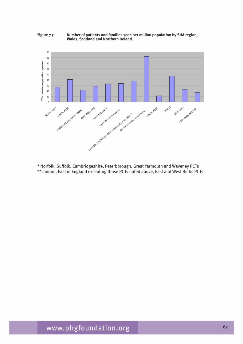
67www.phgfoundation.org
Figure 7.7 Number of patients and families seen per million population by SHA region, Wales, Scotland and Northern Ireland.
0
20
40
60
80
100
120
140
160
180
NORTH EAST
NORTH WEST
YORKSH IRE AND THE HUMBER
EAST M IDLANDS
W EST M IDLANDS
EAST ANGLIA (o ld reg ion)*
LONDON , SOUTHEAST COAST AND OLD CATCHMENT**
SOUTH CENTRAL (minus Berks)
SOUTH WEST
W ALES
SCOTLAND
NORTHERN IRELAND
TOTA
L pa
tient
s see
n pe
r milli
on p
opul
atio
n
* Norfolk, Suffolk, Cambridgeshire, Peterborough, Great Yarmouth and Waveney PCTs**London, East of England excepting those PCTs noted above, East and West Berks PCTs

68www.phgfoundation.org
Table 7.7 Data for clinics provided and patients and families seen
Population (thousands) Service Annual clinics Patients seen
Total clinics
Total patients and families
Clinics/ million
Total patients/million
NORTH EAST 2,555.70 Newcastle 18 138 18 138 7.0 54.0
NORTH WEST 6,853.20 Manchester 70 500 82 560 12.0 81.7Liverpool 12 30 families (est)
YORKSHIRE AND THE HUMBER 5,142.40 Leeds 24 150 36 222 7.0 43.2
Sheffield 12
EAST MIDLANDS 4,364.20 Nottingham No service 20 250 4.6 57.3Leicester 20 250
WEST MIDLANDS 5,366.70 Birmingham 32 350 32 350 6.0 65.2
OLD EAST ANGLIA*2,399.00 Cambridge 24 160 24 160 10.0 66.7
LONDON, SOUTHEAST COAST, BERKS** 16,557.80 Guys 10 50-70 190 1254 11.5 75.7
Moorfields 180 1194***
SOUTH CENTRAL (excl Berks) 2,400.00 Southampton 5 25 families 105 395 43.8 164.6
Oxford 100 290-400
SOUTH WEST 5,124.10 Peninsula 5 25 families 19 115 3.7 22.4Bristol 14 60-70
WALES 2892.7Cardiff 83
220 plus 28 families 83 276 28.0 93.1
SCOTLAND 5094.8 Edinburgh 16 70-80 36 235 7.1 46.1Dundee 10 40Aberdeen 10 120
NORTHERN IRELAND
1724.4Belfast 40 60 40 60 23.0 34.5
Sources for population data TOTAL 4015Table 15 - Provisional Mid-2006 Population Estimates: Quinary age groups for PCOS in England; estimated resident population ONSTable A: Resident population estimates mid-2006: quinary age groups by sex Northern Ireland Statistics and Research AgencyMid-2005 population estimates Scotland (revised 2007). General Register Office for ScotlandStatistics for Wales mid 2006 estimates ONS*Norfolk, Suffolk, Cambridgeshire and Peterborough, Great Yarmouth and Waveney** London, Southeast Coast, East of England except * above, E and W Berks***new patients only (25% of total)
SHA/REGION
69www.phgfoundation.org
7.6 Conclusions and recommendations
The data on service provision have some weaknesses but are the best we have available. In particular, we may have missed activity related to eye genetics that takes place in ‘general’ genetics clinics, some paediatric ophthalmology services and a few ophthalmology clinics that are devoted to specific inherited conditions.
However, we can conclude that there is some level of specialist service provision in each SHA area in the UK, although the comprehensive nature of provision and the actual level of provision in relation to population size are variable.
There is inequity in population access to specialist services with a seven-fold variation in numbers of clinic sessions provided within each SHA per million population and a seven-fold variation in numbers of patients seen in specialist services from worst- to best-served region. This indicates that there may be a substantial level of unmet need in many regions. We estimate this to be around 1000 patients across the UK. The specialist service is highly focussed on the specialist hospital in London and there is a small number of other well established regional centres.
There are a large number of small services throughout the UK and only three services that see more then �00 patients per year. Nine services see under 100 patients and/or families per year. This may reflect different patterns of provision for this patient group with some activity not being captured. However it raises questions about the critical mass of the service and whether enough investment would be made in developing the necessary organisational structure, staff expertise and supporting services to provide a truly specialist multi-disciplinary service.
All services do not have full access to a multi-disciplinary team including geneticist, ophthalmologist, genetic counsellor, laboratory scientist and electrophysiologist. Again this suggests that there is an overall lack of suitably trained and experienced professionals across the UK.
Access to molecular genetic tests is variable.
Access to electrophysiological testing is variable.
Relationships with local ophthalmology community, paediatric services and other relevant health communities have not usually been formalised in terms of referral guidelines and protocols. Most services have not attempted to outreach to their communities in a systematic way and it is thus possible/likely that many ophthalmology and other services are unaware of the existence of specialist genetic ophthalmology services.
Recommendations
Specialist services should set out agreed standards of care that include appropriate structure, standards and function of multi-disciplinary teams including access to specialist services and equipment and to genetic testing. Services will need to consider how they link to other specialties as they care for the needs of patients with complex disorders whose sight problems are part of a range of symptoms.
Consideration should be given to the best configuration of specialist ophthalmology
70www.phgfoundation.org
genetics services and their funding. In particular, a model of services should be devised that builds on the experience of the more established services and provides equity of access to highly specialist clinical and laboratory expertise and facilities.
Consideration should be given to the possibility of forming a national network of specialist services - possibly based on the current EGG group, but with a focus on service development rather than research.
Consideration should be given to the possibility of accrediting services to provide specialist genetic ophthalmology - or at least accreditation of individuals for ordering genetic tests.
In order to increase capacity, the development of appropriate education and training for those who will provide the specialist services in each discipline needs to be formally considered by the responsible professional organisations.
Specialist genetic ophthalmology services should work with district ophthalmology services to develop and implement protocols for referral and systems for shared care.
Services and commissioners should review how protocols and guidelines are brought to the attention of relevant professionals and in particular how reminders and information can be accessible to the professional at the time of need. This might ideally be through electronic based prompts such as through the Electronic Care Record and Connecting for Health systems or via an up-to-date website.
Specialist genetic ophthalmology services should work in conjunction with voluntary organisations to raise awareness of the service with patients and their families.
In order to properly assess service provision against population need, services should audit referrals by geographic area of residence. Formal work on awareness raising and education should be pursued by engagement with the National Centre for Genetics Education and Training in Birmingham, which works on competences and the development of appropriate resources for different professional groups and specialities.
Less formally, opportunities should be sought to raise awareness through conference appearances, publications, contribution to local and national educational programmes, etc, and at different professional levels.
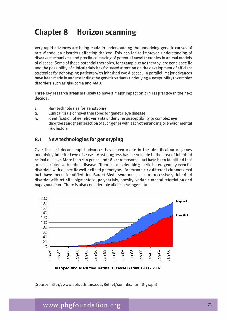
71www.phgfoundation.org
Chapter 8 Horizon scanning
Very rapid advances are being made in understanding the underlying genetic causes of rare Mendelian disorders affecting the eye. This has led to improved understanding of disease mechanisms and preclinical testing of potential novel therapies in animal models of disease. Some of these potential therapies, for example gene therapy, are gene specific and the possibility of clinical trials has focussed attention on the development of efficient strategies for genotyping patients with inherited eye disease. In parallel, major advances have been made in understanding the genetic variants underlying susceptibility to complex disorders such as glaucoma and AMD.
Three key research areas are likely to have a major impact on clinical practice in the next decade:
1. New technologies for genotyping 2. Clinical trials of novel therapies for genetic eye disease �. Identification of genetic variants underlying susceptibility to complex eye
disorders and the interaction of such genes with each other and major environmental risk factors
8.1 New technologies for genotyping
Over the last decade rapid advances have been made in the identification of genes underlying inherited eye disease. Most progress has been made in the area of inherited retinal disease. More than 1�0 genes and 180 chromosomal loci have been identified that are associated with retinal disease. There is considerable genetic heterogeneity even for disorders with a specific well-defined phenotype. For example 12 different chromosomal loci have been identified for Bardet-Biedl syndrome, a rare recessively inherited disorder with retinitis pigmentosa, polydactyly, obesity, variable mental retardation and hypogonadism. There is also considerable allelic heterogeneity.
(Source: http://www.sph.uth.tmc.edu/Retnet/sum-dis.htm#D-graph)
72www.phgfoundation.org
Such diversity presents major problems for developing a service for the identification of mutations underlying genetic eye disease. To date, NHS genetic services provide only a limited number of tests for inherited eye disease and it is unlikely, given the large numbers of genes involved, that comprehensive molecular genetic testing can be provided by laboratories using conventional mutation analysis. There are a number of possible solutions to this problem (Koenekoop 2007).
1. Utilise information about the phenotype to direct molecular genetic testing
In some disorders, for example vitelliform dystrophy, dominant drusen, Sorsby fundus dystrophy and juvenile retinoschisis there is good genotype phenotype correlation and little genetic heterogeneity, so molecular genetic testing can be targeted to single genes or even specific mutations. However this is not possible for most disorders at present.
2. Strategy based on the relative frequency of mutations causing disease in the community
In this approach to genetically heterogenous disorders, molecular genetic testing starts with screening of the most common genes or most common mutations found in a particular ethnic group. This is the approach used by the Manchester regional laboratory for screening for mutations causing retinitis pigmentosa (RP). For example, in X-linked RP exon ORF15 of RPGR is screened first followed, if necessary, by screening of other exons of RPGR and then the RP2 gene. Similarly, in autosomal dominant RP the gene encoding rhodopsin is screened first as it accounts for about 25% of all cases,
3. Microarray technology
i. APEX technology
Microarray (‘gene chips’) methods for the detection of mutations in a number of eye disorders are now commercially available (www.asperbio.com). These microarrays are designed to detect mutations and polymorphisms in disease genes that have been previously identified and published. Microarrays are designed that use a technique called arrayed primer extension (APEX). Oligonucleotides are designed for each mutation/polymorphism and fixed on a glass slide. The patient’s DNA is then amplified for each DNA segment using the polymerase chain reaction (PCR) and then annealed to the glass slide. DNA polymerase is added with dye labelled dideoxynucleotides so that an extension of one nucleotide takes place at the �’ end of the oligonucleotide using the patient's DNA as a template. A laser and computer analysis is used to detect the dye signal. Many different oligonucleotides can be arrayed on the chip and multiple genes screened simultaneously. One commercial company, Asper Ophthalmics (www.asperophthalmics.com), has chips available for a number of inherited eye diseases including Stargardt disease, Leber congenital amaurosis, Usher syndrome, autosomal dominant optic atrophy, autosomal dominant RP, Bardet-Biedl syndrome and autosomal recessive RP. The service is reliable and relatively cheap and the chip is updated as more mutations and gene are identified. This microarray technology is a useful first pass screening method that are genetically heterogeneous. For example, in Leber’s congenital amaurosis, the chip will identify the causative gene in more than �0% of cases using a single test that costs 1�0 euros. The disadvantage of this technology is that it can only identify mutations that are on the chip (ie those previously reported) and in recessive disease much of the time will only identify one
7�www.phgfoundation.org
of the two mutant alleles meaning that further sequencing of that gene is needed. Finally, in an NHS setting, the findings will need to be confirmed in an accredited NHS molecular genetics laboratory and segregation of the disease allele within the family investigated before the results can be used in clinical practice.
ii. Other methods of parallel genotyping
In order to be able to identify with confidence the precise genetic mutation(s) causing a disorder, the coding sequence of all genes known to cause a particular phenotype needs to be sequenced. As discussed above, this is a very costly and time intensive process using conventional sequencing. A number of resequencing technologies are now available that utilise microarrays to probe hundreds of thousands of specific DNA sequences and could be used to screen for mutations in multiple genes simultaneously. Several different technologies are commercially available including the Affymetrix (www.affymetrix.com) and Illumina (www.illumina.com) platforms. Although no commercial resequencing chips specifically designed for eye disease are available currently, it is likely that they will become available in the near future.
8.2 Clinical trials for inherited retinal dystrophies
Most forms of progressive retinal dystrophies are not amenable to treatment. A number of different treatment strategies have been explored in animal models including retinal and retinal pigment epithelial transplantation, the use of growth factors, retinal implants (artificial retina), stem cell therapy and gene therapy. These therapies can be grouped into those which are disease- and gene-specific, for example gene therapy or biochemical treatments, and general treatments that aim to slow photoreceptor degeneration or replace photoreceptors. Specific therapies require molecular diagnosis but even in trials relating to generic treatments, such as the use of growth factors, it is important to know the underlying genetic mutation as particular therapies may work better in some retinal disorders than others. Patients who are recruited into these trials will need to have a confirmed molecular diagnosis. This fact is driving in large part the interest of patient groups in genotyping.
Two clinical trials are currently underway. The first is a trial of gene therapy for a severe recessively inherited infantile rod-cone dystrophy caused by mutations in the gene RPE65. Gene-replacement therapy has proved effective in mice and dogs that lack RPE65. The human trial involves sub-retinal injection of an AAV vector containing the human RPE65 gene driven by an RPE65 promoter. In preparation for the trial, DNA from a large panel of patients with severe infantile onset rod-cone dystrophies has been sequenced to identify suitable patients. If, as hoped, this trial is successful, trials of gene therapy in other recessively inherited retinal dystrophies will follow. Such trials of gene therapy will be confined to patients with mutations in a specific gene. The second clinical trial is investigating the use of a growth factor, ciliary neurotrophic growth factor in adults with RP. A phase two trial is underway. This generic treatment is applicable to most geneticgeneric treatment is applicable to most geneticis applicable to most geneticapplicable to most geneticto most genetic forms of RP. It is likely that further clinical trials of novel therapies will start over the next 5 years, increasing the need for efficient genotyping of our patient population.
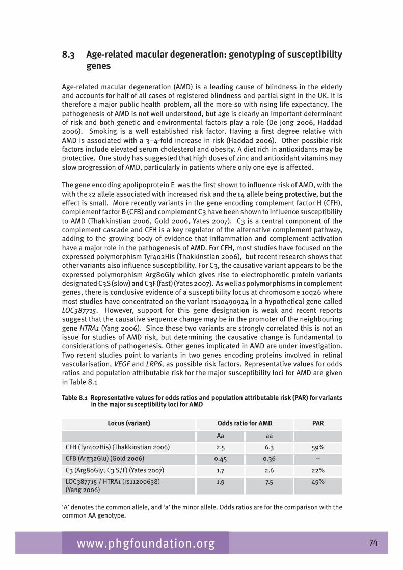
7�www.phgfoundation.org
8.3 Age-related macular degeneration: genotyping of susceptibility genes
Age-related macular degeneration (AMD) is a leading cause of blindness in the elderly and accounts for half of all cases of registered blindness and partial sight in the UK. It is therefore a major public health problem, all the more so with rising life expectancy. The pathogenesis of AMD is not well understood, but age is clearly an important determinant of risk and both genetic and environmental factors play a role (De Jong 2006, Haddad 2006). Smoking is a well established risk factor. Having a first degree relative with AMD is associated with a �–�-fold increase in risk (Haddad 2006). Other possible risk factors include elevated serum cholesterol and obesity. A diet rich in antioxidants may be protective. One study has suggested that high doses of zinc and antioxidant vitamins may slow progression of AMD, particularly in patients where only one eye is affected.
The gene encoding apolipoprotein E was the first shown to influence risk of AMD, with the with the ε2 allele associated with increased risk and the ε� allele being protective, but the being protective, but the effect is small. More recently variants in the gene encoding complement factor H (CFH), complement factor B (CFB) and complement C� have been shown to influence susceptibility to AMD (Thakkinstian 2006, Gold 2006, Yates 2007). C� is a central component of the complement cascade and CFH is a key regulator of the alternative complement pathway, adding to the growing body of evidence that inflammation and complement activation have a major role in the pathogenesis of AMD. For CFH, most studies have focused on the expressed polymorphism Tyr�02His (Thakkinstian 2006), but recent research shows that other variants also influence susceptibility. For C�, the causative variant appears to be the expressed polymorphism Arg80Gly which gives rise to electrophoretic protein variants designated C�S (slow) and C�F (fast) (Yates 2007). As well as polymorphisms in complement genes, there is conclusive evidence of a susceptibility locus at chromosome 10q26 where most studies have concentrated on the variant rs10�9092� in a hypothetical gene called LOC387715. However, support for this gene designation is weak and recent reports suggest that the causative sequence change may be in the promoter of the neighbouring gene HTRA1 (Yang 2006). Since these two variants are strongly correlated this is not an issue for studies of AMD risk, but determining the causative change is fundamental to considerations of pathogenesis. Other genes implicated in AMD are under investigation. Two recent studies point to variants in two genes encoding proteins involved in retinal vascularisation, VEGF and LRP6, as possible risk factors. Representative values for odds ratios and population attributable risk for the major susceptibility loci for AMD are given in Table 8.1
Table 8.1 Representative values for odds ratios and population attributable risk (PAR) for variants in the major susceptibility loci for AMD
Locus (variant) Odds ratio for AMD PAR
Aa aa
CFH (Tyr�02His) (Thakkinstian 2006) 2.5 6.� 59%
CFB (Arg�2Glu) (Gold 2006) 0.�5 0.�6 --
C� (Arg80Gly; C� S/F) (Yates 2007) 1.7 2.6 22%
LOC�87715 / HTRA1 (rs112006�8)(Yang 2006)
1.9 7.5 �9%
‘A’ denotes the common allele, and ‘a’ the minor allele. Odds ratios are for the comparison with the common AA genotype.
75www.phgfoundation.org
A substantial part of the genetic variation influencing susceptibility to AMD has now been identified and this raises the question of whether genotyping has a role in diagnosis, management or identification of those at increased risk. Diagnosis of AMD currently relies on clinical examination, if necessary supplemented by fluorescein angiography, and is usually straightforward. The modest conditional information provided by genotyping would not usefully contribute to this. With regard to management, there is no evidence as yet that genotype influences the response to treatment, but this will undoubtedly be an important focus of future research. Identification of subjects at increased risk of AMD on the basis of their genotype is certainly now possible, either in close relatives of index cases or in the general population. However, this is only of value if interventions can be offered to reduce their risk of developing the disease. There is no conclusive evidence to support the use of nutritional supplements in this situation. Currently, the only advice one could offer would be to eat a healthy diet rich in green vegetables and not smoke, but this is good advice regardless of genotype.
8.4 Glaucoma
Glaucoma is an important cause of registered blindness and partial sight in the UK second only to AMD (Bunce 2006a). Defined as a group of heterogeneous disorders, glaucoma is generally characterised by a progressive excavation of the optic disc and corresponding loss of the visual field. Glaucoma can be classified into primary, secondary and developmental glaucoma with primary open-angle glaucoma (POAG) accounting for more than half of all forms of the disease. POAG is often, but not always, associated with elevated intraocular pressure. The lack of a consensus case definition until recently has resulted in considerable uncertainty in the diagnosis of this condition and hindered the accurate estimation of the burden of illness in the general population.
The pathogenesis of glaucoma remains unclear and understanding of the disease mechanism at a molecular level is relatively poor. Risk factors such as advancing age, positive family history, African descent and smoking are currently known (Hewitt 2006). Evidence for a genetic component to POAG has also been observed in twin studies and family studies (recently reviewed by Hewitt 2006). The prevalence of POAG is 10 times higher in first-degree relatives of patients than that in the general population.
Links to at least 20 genetic loci have been described in the literature with 11 chromosomal loci designated GLC1A to GLC1K (listed in Table 1 and in Table 2 – available in supplementary information via PHG Foundation Website) (Fan 2006A, Fan 2006B). The myocilin gene (MYOC) [formerly referred to as the trabecular meshwork induced glucocorticoid response (TIGR) gene] on chromosome 1q2�-q25 (GLC1A locus) was identified as the first candidate gene for POAG. More than �0 disease-associated mutations have been identified in MYOC and one study has suggested that approximately one in �0 POAG patients has a mutation in this gene with the Gln�68STOP mutation being most common.
The second candidate gene to be identified for POAG was the optineurin (referring to optic neuropathy-inducing) gene (OPTN) located on chromosome 10p1� (GLC1E locus) with the Glu50Lys mutation being most commonly observed. Mutations were found within OPTN in 16.7% (9 out of 5�) of the families initially studied.
More recently, a third candidate gene at the GLC1G locus on chromosome 5q��-q�5, WD repeat-containing protein �6 (WDR�6), has been implicated, although this finding requires further investigation as subsequent work has failed to replicate the initial findings.In addition to these three candidate genes, at least 16 other POAG-associated genes
76www.phgfoundation.org
have been reported in the literature, although most have either only been reported once or conflicting findings make the aetiological role of these genes in POAG unclear or controversial (Fan 2006A, Fan 2006B). With mutations in MYOC, OPTN and WDR36 accounting for no more than 10% of all POAG patients, other POAG genes accounting for most cases still remain to be identified (Hewitt 2006, Fan 2006A, Fan 2006B). Much work remains to assess the contribution of these genes. From this, the strength of association with disease for each of these genes and how much of the population attributable risk is conferred by these variants can be estimated.
Population-based clinical screening is believed to currently miss approximately half of all cases with POAG, including those with advanced disease (reviewed by Hewitt 2006). If POAG can be detected earlier and appropriate therapeutic treatment obtained, blindness from glaucoma can be prevented. Early diagnosis is the key to an effective screening program to identify individuals with no obvious signs or symptoms of the disease prior to damage occurring. Once a POAG patient is identified as having a disease-causing mutation, all first degree relatives can then be tested and closely monitored for early clinical symptoms, so allowing treatment to prevent any further loss of functional vision.

77www.phgfoundation.org
Chapter 9 Discussion and recommendations
9.1 Epidemiology
Blindness is a very significant disability. In children it affects development, education and the care needed from families and professionals. In adults it has many implications for a person's independence, ability to work, and social and psychological well-being. It may have many causes in children. These include treatable or preventable causes such as retinopathy of prematurity, various tumours, congenital cataract, infection or untreatable conditions such as cerebral visual impairment (eg cortex, sub cortical structures and visual pathways, optic nerve atrophy and hypoplasia as well as hereditary retinal dystrophies). In adults the principle underlying causes are age related macular degeneration, glaucoma and diabetic retinopathy.
Eye disease and visual impairment are very common within the population. Our review (Chapter 2) shows that genetic factors contribute to this both as single gene disorders and as factors in complex chronic conditions. In children and younger age groups, a higher proportion of those with visual impairment and blindness have disorders with a genetic basis. Although individual conditions are rare, across the population of the UK each year some 150 children under the age of 16 will be newly diagnosed with severe visual impairment or blindness caused by inherited eye disease (see Section 2.�) and around 250 adults of working age with a genetic disorder will receive blind certification (see Table 2.�). Incidence and prevalence may increase in the future with increasing survival of children with complex genetic conditions that affect the eye, such as some of the inherited metabolic conditions. In the adult population near normal adult survival for those with inherited eye conditions means that the prevalence of certified blindness due to genetic eye diseases within the population is probably several thousand cases with many more individuals either being uncertified or having partial sight. Our research demonstrates that the epidemiology of genetic eye disease remains poor.
Figures for new cases provided in Chapter 2 give an absolute minimum number of those requiring input from specialist services each year. Such statistics do not include patients who have genetic eye disease but do not become blind or severely visually impaired (for example, those with treatable disease or disease in only one eye) and do not include adults who do not seek certification. As well as dealing with new cases, services will also be involved with patients in whom genetic eye disease is suspected but eventually not confirmed; this will include at risk family members. All of these individuals represent a need for specialist services.
9.2 Patients' views
When faced with undiagnosed visual deterioration or blindness, particularly where a number of family members might be affected, patients need and demand access to and care from a specialist service with experience in diagnosing and managing their condition. They need to be able to trust that the service is robust, up-to-date and expert. It must be connected with the necessary specialist investigative facilities and associated specialist clinical services and be integrated with their local health services and, where relevant, social services, educational or employment services and voluntary organisations.
78www.phgfoundation.org
Patients and their representative organizations have highlighted a number of concerns (Chapter �). They noted a lack of a consistent, high quality, services across the UK. Both the patients’ group and the patients’ representatives group highlighted the variability across the UK in access for patients to a good regional specialist service including whether their local GP, optometrist or DGH service knew about it.
In terms of a genetic counselling service the patient groups felt that good communication and counselling are key attributes of a good genetic testing service. The patients wanted to be kept fully informed of advances in testing and treatment. They want counselling to help them to understand the diagnosis, for emotional support, and to make decisions on life choices such as reproductive choices, education, employment and mobility.
The patients’ groups were consistent in their support for provision of genetic testing because of its value to the patient and family. Genetic testing can provide accuracy of diagnosis and inheritance patterns giving routes to therapy and, with counselling, properly informed decisions on life choices. In terms of current processes the family groups felt that a genetic testing service must be properly integrated with a clear, simple and robust management process that the patient could follow easily. They felt that the present service was inconsistent. Importantly they felt that the quality of service is more important than its speed or ease of location.
9.3 Clinical and laboratory services
The clinical and laboratory diagnosis and subsequent management of patients with genetic eye disease and their families is a specialist area within ophthalmology requiring a multidisciplinary team of ophthalmologist, geneticist, genetic counsellor, molecular genetic scientist and electro-physiologist with experience of the particular conditions and with the necessary specialist facilities and relevant genetic testing. These services and the current provision in the UK are described in Chapters �, 6 and 7. As more possibilities for diagnosis and management become available the requirements of clinical governance and the demands of better informed and mobile patients will necessitate the development of more specialist clinical services.
Increasingly it is possible to make a precise diagnosis of genetic eye disease through molecular genetic testing. Both patients and clinicians value this capability for a number of reasons - which differ for different tests and in different situations. We have shown in Chapter 5 that in ophthalmology a genetic test might allow clinicians to circumvent alternative complex, expensive or time-consuming diagnostic procedures or programmes and lead directly to a definitive diagnosis; it might give further information about prognosis or inform possible treatment or surveillance programmes and, thus, reduce morbidity or mortality; it might provide other family members with testing options to assess their own risk of disease; and it might provide parents with options for prenatal testing for subsequent children. Increasingly a molecular diagnosis is needed for entry into treatment trials; some novel treatments will be genotype specific but even in trials for treatments with general applicability knowledge of genotype may be necessary when assessing efficacy in different groups of patients.
Our horizon scanning in Chapter 8 shows that the need for genetic testing is set to expand in the near future for a variety of reasons. Research is already yielding new treatments, such as gene therapy and biochemical treatments that are gene specific and require prior genotyping. At present this is driving patient demand for testing to enable possible entry to clinical trials but it is likely that, as these treatments become part of the therapeutic
79www.phgfoundation.org
repertoire, there will be a greater need for confirmed molecular diagnoses within NHS services. New technologies are becoming available for genotyping making it possible to reach a molecular diagnosis for a wider range of disorders. Finally, there is the real expectation that, in the future, research findings on gene disease association might be able to confirm the importance of genetic factors in the prevention, early diagnosis and management of some of the important multi-factorial conditions that are the main cause of visual loss in the general population. When this happens molecular testing will be important within the general ophthalmology service as well as across services dealing specifically with genetic eye disease.
Despite the expected increase in need for services for genetic conditions within ophthalmology, in the context of the overall service these disorders are likely still to remain a comparatively small and specialist part of the services. It is necessary, therefore, for commissioners and providers to consider how ophthalmic genetics can develop to meet current and future needs, both by expanding capacity and by developing overall service configuration that balances specialist expertise with local accessibility.
9.4 Recommendations
Commissioning specialist genetic ophthalmology services
The services in the UK for genetic eye disease have not, for the most part, been formally planned and commissioned to meet the needs of a given population, but have arisen through the interest and persistence of enthusiasts. We have evidence (Chapter 7) of patchy service provision, inequality of access and small services that lack access to necessary specialist elements. Our review has shown that there is some provision of a specialist ophthalmic genetics service in each of the Strategic Health Authority areas for England and in Wales, Scotland and Northern Ireland but the level of this is quite variable. There is a seven-fold variation in the number of clinic sessions provided per million population and a similar variation in the number of patients seen per million population between the regions with most and least service provision. Some of these differences may be accounted for by patient flows across SHA boundaries arising from longstanding patterns of patient referral. This happens particularly around London, the Southeast and Eastern regions. Otherwise, there is no epidemiological evidence that the difference in provision reflects differing population need. By contrast there is anecdotal evidence from patient groups that many families are not in contact with specialist services.
80www.phgfoundation.org
Provision of specialist genetic ophthalmology services
A need for accurate diagnosis, prognosis and counselling, and the advent of new therapies for ophthalmic genetic conditions makes it imperative that ophthalmic genetics is firmly established with the appropriate multi-disciplinary specialists and sufficient national laboratory support to serve regional populations with well-recognised referral pathways.
Specialist services were extremely variable in the amount and range of services offered. Some regions such as Manchester were able to provide a very comprehensive service, whereas other services comprised simply a joint clinic held between a geneticist and interested ophthalmologist on an occasional basis, which might only be once or twice per year. Only half the services had dedicated genetic counsellor time as a formal commitment. There is thus likely to be a significant unmet need and hence a requirement to increase the capacity of specialist services.
There are 19 providers of services in the UK but the volume of patients and families seen each year in individual services ranged from 20 to 500 with five services seeing 50 or less patients/families per year and only five services seeing 200 patients/families or more. It is unlikely that small services have the critical mass that is necessary to gain sufficient experience of these rare conditions and to enable them to formalise the necessary organisational structures (such as patient pathways, referral mechanisms. outreach programmes, audit and research) that will ensure high quality care for patients.
Two models for specialist services exist:
The provision of joint genetic/ophthalmic clinics. This is the model of service currently provided in most centres in the UK and is recognised as being very successful. To address the current underprovision of service and anticipated future demands using this model would require a significant increase in resources, including more sessions for both clinical geneticists, genetic counsellors and ophthalmologists.
Recommendations
1. Mechanisms should be developed to support the commissioning by PCTs of specialist genetic ophthalmic services on a population basis with coverage for the entire population. This will require:
a) Commissioners of ophthalmology services to work together to ensure provision of specialised elements of the service
b) involvement of clinical, laboratory and other providersc) involvement of patient groups d) the development of information systems to audit activity with respect to geographic
area of residence.
2. A service specification should be developed that sets out the services that should be provided and the necessary standards.
�. Commissioners with responsibility for ophthalmology in areas where there is little or no specialist service for genetic eye disease should urgently review how their patients with these conditions are managed.
81www.phgfoundation.org
The provision of an ophthalmologist with a special interest and training in genetics. In this model the ophthalmologist manages most of the patients with the support of a genetic counsellor and passes on to the geneticist only those who have particular complex ethical, counselling or interpretive issues. This model is currently only in operation at Moorfields hospital and, if expanded to other centres, would require increased education and training of ophthalmologists, with possible issues of accreditation and further provision of genetic counsellors. In such a model it would be important to ensure close working relationships with a clinical genetics department so that patients and their families have access to the full range of genetic services. If such a service were to be sited primarily in an ophthalmology department this would require establishment of robust methods for managing the familial elements of care including family-based record keeping and family follow up.
At present there is no evidence for which model is more effective or cost-effective.
Recommendations
�. Services need to develop an integrated service model to ensure multi-disciplinary comprehensive provision now and the future ability to respond effectively to new needs. In the short term this is likely to be based on a limited number of regional or supra-regional centres where ophthalmologists, possibly with a sub-specialty interest in genetics, work alongside clinical geneticists in joint clinics. There needs to be a careful evaluation of alternative service models for example the development of specialised genetic eye clinics run by ophthalmologists who will liaise closely with clinical genetic colleagues.
5. Within the service model, care pathways should be set out for access to the specialist service and appropriate new technologies ensured and regulated by the development and implementation of care pathways, referral criteria, systems of shared care, appropriate information systems audit and monitoring.
6. Services need to devise and set out referral pathways between themselves and other relevant specialist as they manage the needs of patients with complex disorders whose sight problems are part of a range of symptoms.
7. Specialist genetic ophthalmology services should develop and publish agreed standards of care that include appropriate structure, standards and function of multi-disciplinary teams including access to specialist services and equipment, and to genetic testing.
8. The services should be configured and coordinated so as to harness the disparate expertise and energy in all of the services, whilst bringing them together informally to support smaller services in service organisational aspects.
9. Overall the service needs to increase capacity including medical, surgical, nursing, genetic counselling, electrophysiology and other specialist support services. Capacity and organisation of laboratory services must be kept under review in the light of likely increased demand as well as changes in technologies.
82www.phgfoundation.org
Recommendations
10. Services should embrace the support offered by voluntary organisations particularly in assistance with coping with genetic eye disease and in keeping patients and families informed of research advances.
11. A formal network of services should be set up to support practice. This could be based on the current Eye Genetics Group (EGG), but with a service focus.
Laboratory molecular genetics services
Tests for 18 conditions are available across the United Kingdom in nine different laboratories. Most laboratories provide only one or two tests, with the exception of Manchester, which provides testing for nine conditions. In addition, all but one test (retinoblastoma) are provided by only one laboratory. Not all these tests are listed by the UKGTN and only those related to retinitis pigmentosa (X-linked and AD) have associated Gene Dossiers. From our survey of services there also seemed to be lack of clarity about what tests could be requested, for what purpose, and by whom. Frequently tests had to be requested by the local genetics service, which provided a gate-keeper function. Many difficulties of funding were experienced especially where funding for genetic testing would come from a different local budget, or a different area.
Tests for at least seven conditions are provided by one commercial provider (Asper Ophthalmics based in Estonia). Some UK laboratories are developing experience using these services, particularly as an initial screen. However, their wider utility, particularly when possibly used by laboratories or clinicians with more limited skills in interpretation is questioned as is the assumption that gains of efficiency achieved by commercial companies using new technologies could not be achieved within the NHS laboratory setting.
Our work on prioritisation51shows that a method could be developed for prioritisation of tests - and that the criteria outlined in the evaluation chapter of this Report (Chapter 5) would be suitable. However, UKGTN needs to make a decision on whether it would be useful to develop this further.
15 See separate report available from PHG Foundation
8�www.phgfoundation.org
Recommendations
12. The UKGTN should continue to hold the list of available genetic tests for genetic eye disease. This should be kept up to date and include details of how the test can be accessed, cost, criteria for testing, etc. Where possible, tests should be developed for UKGTN listing with submission of Gene Dossiers for new tests. Gene Dossiers should be developed for available tests as far as possible to provide underpinning evaluation information for specialists and, potentially, for commissioners (and for the public).
1�. Laboratories should work with the UKGTN to decide the best model for genetic test provision (eg one, two or more laboratories). This should balance the need for efficiency against the need for peer support and laboratory back-up.
1�. Services should be prepared to increase the volume of testing as new genes are identified and tests become available.
15. Further work needs to be done to determine how all services access testing and what gate-keeper roles are needed now and in the future. Such work must be integrated closely with consideration of the best model for services. The role of the clinical or laboratory service in this, including a ‘gate-keeper function’, the possibility of development of criteria, and the use of accreditation to determine who can order tests are all areas to consider. In the long run, as more tests become available, and as they become cheaper, it may not be practical for clinical genetics to provide gate-keeper functions. This will particularly be the case if genetics develops further within the speciality of ophthalmology (eg if it becomes relevant for surveillance and early treatment of glaucoma) - it would be worth preparing educationally and organisationally for this now.
16. There needs to be an urgent review of the ways in which genetic tests are funded, including the problems that arise when the funding for genetic tests in ophthalmology is incorporated into the budget of a separate and much smaller service (genetics) rather than within the clinical ophthalmology budget as a whole.
17. The UKGTN should work with laboratories to develop clinical criteria for genetic testing in ophthalmology with priority for those listed by the UKGTN or subject to new approval through the Gene Dossier process.
18. There needs to be urgent consideration of the use that the NHS should make of commercial providers and, as appropriate, the ways that such services should be integrated with NHS systems.
8�www.phgfoundation.org
Ensuring access to specialist services
Patients and their family will not accept that the often 'serendipitous' way in which they find their way to a specialist represents good practice within the system. They are concerned at the injustice that a person who searches the internet, contacts the voluntary organisation or puts pressure on their local GP or consultant, or finds out by chance and demands a referral, might get to the specialist service whereas an individual with less personal resources might not. This situation comes about as local clinicians lack knowledge of both the rare conditions and of the appropriate services.
The services cannot rely on the usual mechanisms for patient referrals based solely on the knowledge and initiative of the referring physician as it is likely that a) these patients will not be recognised and b) the health professionals will not know that specialist services exist.
Most services were reactive and operated in the time-honoured way of receiving referrals from their 'community', mainly ophthalmology, geneticists, paediatricians and primary care. Some operated a system of picking out or triaging patients for a joint clinic through the use of clinical geneticist, paediatric ophthalmologist or ophthalmologist with a special interest in genetic disease. In general, services had not made formal links with District General Hospitals to ensure that patients were identified, although two services had extensive outreach educational sessions to raise awareness of the service and how to use it. Occasional websites and letters were described, but, in general, it seemed that services relied on word of mouth and patient summary letters to encourage appropriate referral. Only one service commented that 'there are probably some districts that do not refer as many patients as we might expect', but there had been no systematic attempt to address this at any point. Another actively did not advertise the service as it would not be able to meet the demand generated.
Recommendations
19. Specialist genetic ophthalmology services should work with district ophthalmology services to develop and implement protocols for referral and systems for shared care.
20. Services and commissioners should review how protocols and guidelines should be brought to the attention of relevant professionals and, in particular, how reminders and information can be accessible to the professional at the time of need. This might ideally be through electronic based prompts (such as through the Electronic care record and Connecting for Health systems or via an up-to-date website).
21. Specialist genetic ophthalmology services should work in conjunction with voluntary organisations to raise awareness of the service with patients and their families.
22. Services should audit referrals by geographic area of residence.
85www.phgfoundation.org
Education and training for specialists and for health professionals in mainstream medicine
As a main element of the drive to increase capacity, the issue of development of specialist education and training must be addressed for each of the professional groups involved. There is a need for individuals in the following professional groups to gain special knowledge and experience in the management of genetic eye disease: geneticists, ophthalmologists, genetic counsellors, ophthalmology nurses and electrophysiologists.
Recommendations
2�. It is recommended that the need for specialty training for geneticists, ophthalmologists, genetic counsellors, ophthalmology nurses and electro- physiologists be raised with the relevant professional organisations and those responsible for training. Mechanisms might include trainee-selected components, and the availability of special training modules or placements within a specialist training programme.
Within ophthalmology all should have a basic understanding of genetics relevant to that specialty. The curriculum currently states:
“All trainees must understand and apply knowledge of clinical genetics relevant to ophthalmic practice. They must be able to use this knowledge when advising patients about patterns of inheritance. They must recognise when it is appropriate to refer a patient for genetic counselling. They must recognise when it is important to offer a consultation with family members”.
Developing and delivering suitable curricula requires suitable educational and assessment resources as well as teachers that are confident in teaching genetics at an appropriate level.
Individuals who undertake higher training should be encouraged to specialise in genetic conditions in order to develop genetic knowledge and skills that are specific to their speciality. Although the number involved in such specialist training will be low initially, this should increase as the genetic aspects of services become better established. Ideally, individuals with specialist training should work together in centres where there is critical mass as well as links to specialist genetics services and relevant training and research programmes. It is proposed that the Royal College of Ophthalmologists should be encouraged to consider establishing genetics as a sub-speciality and providing genetics as an option for trainee-selected components. In this case, it will be important to establish how many sub-specialists should be trained and to provide formal special training posts in appropriate centres.
In genetic ophthalmology the problem of recognising and managing rare diseases is compounded by the genetic aspect, which adds the extra level of complexity of dealing with a qualitatively different set of diagnostic tests and of managing familial aspects of the condition. From the literature we know that most health professionals in primary care, the community and hospital specialties have very little understanding of genetics and would
86www.phgfoundation.org
not automatically recognise the possibilities and implications of genetic testing or be competent to discuss, order and interpret genetic tests. It is, thus, important that leaders in ophthalmology genetics work at various levels to improve the level of understanding of genetics amongst all those who deal with eye disease. This will include GPs, opticians/optometrists, ophthalmologists, ophthalmic surgeons and ophthalmic nurses. It will be important to ensure that education in genetics gets into the appropriate undergraduate and postgraduate training and continuing professional development.
Recommendations
2�. Formal work on awareness raising and education should be pursued by engagement with the National Centre for Genetics Education and Training in Birmingham, which works on competences and the development of appropriate resources for different professional groups and specialities.
25. Less formally, opportunities should be sought to raise awareness at different professional levels through, for example, conference appearances, publications, contribution to local and national educational programmes.
9.5 Conclusion and final recommendation
Finally, those with a stake in specialist genetic ophthalmology services as identified in this Report must find a way to maintain momentum and continue to shape their services to meet the needs of patients and their families and prepare for the future. This chapter has listed a set of recommendations, each of which are potentially time-consuming and need both professional and expert leadership and coordination.
Recommendations
26. It is recommended that an Implementation Board be set up with appropriate and representative membership in order to oversee the next steps. This Board should have dedicated expert professional and coordinator time.
87www.phgfoundation.org
Chapter 10 Lessons for mainstream medicine The devolution of genetic services from specialist to generalist was first signalled in April 2001 by the Secretary of State for Health who noted that the NHS needed to 'change and adapt its services' to meet this challenge. Some elements of genetic services would need to spread from specialist centres and into GP surgeries, health centres and local hospitals. In other words, genetic services would become more 'mainstream'. In 200� developing genetics in mainstream services was one of the main themes of the Genetics White Paper with substantial initiatives in modernising laboratories to provide essential infrastructure, engaging other specialties through service development pilots, developing GPs with a Special Interest in Genetics (GPSIs in Genetics) and the development of genetic screening programmes. It was thought that specialist genetics centres would play a leading role in the diffusion of genetics advances across the rest of the NHS.
10.1 Key issues in mainstreaming genetics
Four years on and using our experience in genetic ophthalmology (which included representation from the Oxford ophthalmology service development pilot project) two key issues arise that are likely to be relevant to all specialties.
The lack of strategic planning in specialist areas
1 There is rarely any strategic planning in these areas. Service developments in genetics related to mainstream specialties have tended to be led by enthusiastic providers, resulting in patchy and vulnerable services.
2 It is usually difficult to engage commissioners in such planning for a variety of reasons that are inherent in the nature of the work:
• conditions are rare• benefits of services and tests are usually not evidence-based • the technologies are seen as complex• potential benefits are not immediate (prevention rather than treatment)• likely benefits will be for other people in other geographical areas (family members) or in other services (eg prevention of consequences of severe visual impairment)• expected benefits might be linked to improved reproductive choice, which is a difficult area that raises questions about termination of pregnancy, disability rights, etc.
� Genetics is a small specialty with dispersed specialist providers who have not had the resources to raise the profile of the work and provide the necessary pressure nationwide to develop the service.
� Diagnostic services (molecular and cytogenetics laboratories) are funded separately from mainstream pathology, and commissioners of specialised services working with the genetics services have been the gate-keepers for genetic testing
88www.phgfoundation.org
Expected expansion in volume of work in genetics elements of clinical care
The requirements for increased volume of genetics elements of mainstream services will increase as:
the number of available tests for diagnosis increasetreatment options based on particular genotypes become available including gene therapy (thus increasing clinical utility of molecular tests)genetic tests become cheaper and start to increasingly replace other diagnostic procedures knowledge increases about genetics in common conditions such as glaucoma and screening options become availableclinical governance requires agreed standards of care, and increasingly knowledgeable and mobile patients demand referral to the specialist centresgenetic tests to determine treatment options (eg testing of cancer cells to guide chemotherapy) and to personalise treatment (pharmacogenetics) become part of routine practice.
10.2 Some policy options
The key question is how strategic planning and an expansion of services involving genetics can be achieved in the NHS in the most effective and cost effective way.
Specialist genetic services and mainstream specialities should consider developing policies in the following areas.
Increasing capacity and capability to engage with commissioners
It is probably unrealistic to expect that commissioners will, of their own accord, wish to examine and develop services in the area of genetic disease. Opportunities should be sought to raise awareness with commissioners in the context of work identified as national priority, such as the current work on sudden cardiac death as part of the National Service Framework. The ability to capitalise on such opportunities would be greatly enhanced by having networks of individuals with a special interest in the genetics of each specialty, underlying programmes of developing commissioner understanding and capacity and the enhancement of mechanisms for improving communication between providers and commissioners.
Increasing involvement of mainstream specialists in the provision of care that integrates genetics elements
Diagnosis and management of genetic disease is largely undertaken by specialists in system specific areas of medicine such as cardiology or ophthalmology, and, in the case of developmental disorders, in paediatrics. Molecular genetic testing is increasingly being used as a means of confirming a diagnosis. The skills of the geneticist are invoked particularly in assisting with the diagnosis (eg investigation of possible modes of inheritance), interpreting genetic tests, genetic counselling, considering family cascade testing, or where there is a need for counselling around pre-symptomatic testing or prenatal testing. Thus it is important that patients with these conditions are managed in services that have access to a multi-disciplinary team involving geneticists.
••
•
•
•
•
89www.phgfoundation.org
In the future, it is envisaged that use of genetics in medicine will inform understanding of disease mechanisms as well as understanding patterns of inheritance - and therefore the agenda will be progressed by physicians across a range of specialities with variable involvement of specialists in clinical genetics
Developing relationships between specialist genetics services and mainstream service that support but do not take over the genetics elements of care
As prevention or treatment options based on molecular diagnosis become more numerous, and, thus, the demand for ‘genetic’ management of disease arises across many specialties, the capacity for further development of the current, most prevalent model of ‘joint genetics/specialist clinics’ may be challenged. Whilst the joint genetic/specialist clinic has been widely recognised as a model of excellence there will be a tension between the desire to maintain this excellence and the need to provide an increased capacity for more patients and families over a wider range of conditions.
Specialist genetics services should therefore work with relevant specialities to determine how they can best support the management of inherited disease within mainstream medicine in a way that is sustainable as the volume of work multiplies and is most cost effective. There needs to be much greater collaboration and communication between geneticists and communities in the various specialities. In service terms, the key will be to use geneticists for what only they can do and to develop other members of the service to act more autonomously in genetic aspects.
Further research is needed into what service models are the most cost effective.
Establishing formal or informal sub-specialty interest in genetic aspects of disease
Undoubtedly the rarity of genetic conditions and their relative complexity mean that all specialists in a particular field would not have the necessary expertise or access to the relevant special diagnostic tests and other support services. However, all should have a basic understanding of genetics relevant to that specialty. In ophthalmology, for example, the curriculum states:
“All trainees must understand and apply knowledge of clinical genetics relevant to ophthalmic practice. They must be able to use this knowledge when advising patients about patterns of inheritance. They must recognise when it is appropriate to refer a patient for genetic counselling. They must recognise when it is important to offer a consultation with family members”
If this is to be achieved, there will be a need for those responsible for curriculum delivery to ensure that there are suitable educational and assessment resources and that teachers are confident in dealing with genetics.
A small, but increasing number of those in higher training should be encouraged to specialise in genetic conditions within their specialist area and develop genetic knowledge and skills tailored to that specialty. Thereafter they should work in centres where there is critical mass as well as links to specialist genetics services and relevant training and research programmes. The colleges will need to develop genetics as an option for trainee selected components and to consider whether sub-specialty status for genetics within a specialty would be appropriate. If so, an important consideration would be to establish
90www.phgfoundation.org
the number of sub-specialists who should be trained and formal special training posts in one or two centres.
The establishment of provider networks
In order to ensure high quality and comprehensive services with equity of access, services may benefit from the development of informal supra-regional or even national support networks. These networks would not be for direct provision of care but would enable the following activities to be supported and developed by enabling specialist to work together and coordinating their efforts: advocacy for the service; advice to commissioners; a directory of designated services; strategic planning; standards and governance; coordination; expert advice; professional education and training; liaison and communication; information for patients and families; research ; IT; and monitoring and audit and reporting functions. An outline for a specification for a network is available in a prototype document prepared for the implementation of recommendations in the area of Inherited Metabolic Disease (http://www.bimdg.org.uk/IMD.asp).
Establishing system for accessing genetic tests
It will not be possible or desirable for specialist genetic services in the longer term to provide gate-keeper functions for most genetic tests. This will increasingly become the case as:
Genetic tests get relatively cheaperDiagnoses at a molecular level becomes more useful for determining treatment optionsGenetic disease becomes more treatable overallTesting for variants associated with common chronic disease becomes more usefulClinicians in other specialties become better trained and more knowledgeableCommercial organisations develop high-throughput testing.
Genetic laboratories and clinical services should therefore work with the specialist mainstream services to set out:
Agreed criteria for each test in terms of person, purpose, etcAgreed personnel 'accredited' to order tests; the necessary education and training for this would need to be agreedProcess for monitoring/audit genetic test requestsAvailability of expert backup for difficult questions, interpretation, etc from clinical geneticist and laboratory scientists.
The UKGTN should have a role in coordinating this.
For education
If mainstream specialist services are to increase their input to the management of genetic disorders, there must be a significant increase in expertise within the specialty. Consideration should therefore be given to the development of the following:
••••••
••
••

91www.phgfoundation.org
Inclusion of genetics aspects in all areas of the speciality training (eg medical, nursing and other associated specialities such as dietetics)Establishment of sub-specialty training (eg fellowships)Access to specialist genetics departments for mentoring and continuing education of any genetic counsellors or nurse counsellors working within the specialityAccess to specialist genetics departments, ethical groups (eg Ethox) for discussion and education around ethical dilemmas and other aspects of genetic counselling.
Specialist geneticists should have a key role in training specialists alongside other key disciplines such as epidemiology, public health and the social sciences.
•
••
•
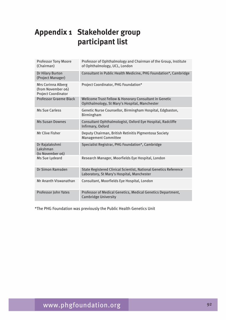
92www.phgfoundation.org
Appendix 1 Stakeholder group participant list
Professor Tony Moore (Chairman)
Professor of Ophthalmology and Chairman of the Group, Institute of Ophthalmology, UCL, London
Dr Hilary Burton(Project Manager)
Consultant in Public Health Medicine, PHG Foundation*, Cambridge
Mrs Corinna Alberg(from November 06) Project Coordinator
Project Coordinator, PHG Foundation*
Professor Graeme Black Wellcome Trust Fellow & Honorary Consultant in Genetic Ophthalmology, St Mary's Hospital, Manchester
Ms Sue Carless Genetic Nurse Counsellor, Birmingham Hospital, Edgbaston, Birmingham
Ms Susan Downes Consultant Ophthalmologist, Oxford Eye Hospital, Radcliffe Infirmary, Oxford
Mr Clive Fisher Deputy Chairman, British Retinitis Pigmentosa Society Management Committee
Dr Rajalakshmi Lakshman(to November 06)
Specialist Registrar, PHG Foundation*, Cambridge
Ms Sue Lydeard Research Manager, Moorfields Eye Hospital, London
Dr Simon Ramsden State Registered Clinical Scientist, National Genetics Reference Laboratory, St Mary's Hospital, Manchester
Mr Ananth Viswanathan Consultant, Moorfields Eye Hospital, London
Professor John Yates Professor of Medical Genetics, Medical Genetics Department, Cambridge University
*The PHG Foundation was previously the Public Health Genetics Unit
9�www.phgfoundation.org
Appendix 2 References
Chapter 2
Black 2002. Black G. Genetics for ophthalmologists. Remedica publishing; London 2002.Bunce 2006a. Bunce C. Causes of blindness and partial sight certifications in England and Wales 1999- 2000. Report submitted to Department of Health 2006.Bunce 2006b. Bunce C and Wormald R. Leading causes of certification for blindness and partial sight in England and Wales. BMC Public Health 2006, 6:58Gilbert 1999. Gilbert C, Anderton L, Dandona L, Foster A. Prevalence of visual impairment in children: a review of available data. Ophthalmic Epidemiology 1999; 6, 1: 7�-82. Johnson 2003. Johnson, GL, Weale R, Minassian DC. The epidemiology of eye disease. 2nd Edition; Arnold Publishers; London 200�.Rahi 2003. Rahi J. Severe visual impairment and blindness in children in the UK. The Lancet 200�; �62: 1�59-1�65. Resnikoff 2004. Resnikoff S. Global data on visual impairment in the year 2002: Policy and Practice. Bulletin of the WHO 200�.
Chapter 5
Burke 2002. Burke, W. "Genetic testing." New Engl J Med, Vol. ��7, 2002, pp. 1867-1875Burke 2007. Burke W and Zimmern R. Moving beyond ACCE: An Expanded Framework for Genetic Test Evaluation. http://www.phgfoundation.org/pages/work7.htm#acceHaddow 2004. Haddow J, Palomaki G. ACCE: a model process for evaluating data on emerging genetic tests, in Khoury M, Little J, Burke W, eds. Human Genome Epidemiology, Oxford University Press, Oxford 200�, pp. 217-2��Joseph 2004. Joseph B, Shanmugam MP, Srinivasan MK, Kumaramanickavel G. Retinoblastoma: genetic testing versus conventional clinical screening in India. Mol Diagn. 200�;8(�):2�7-��. Raizis 2002. Raizis A, Clemett R, Corbett R, McGaughran J, Evans J, George P. Improved clinical management of retinoblastoma through gene testing. N Z Med J. 2002 May 2�;115(115�): 2�1-�.Zimmern 2007. Zimmern RL, Kroese M. The evaluation of genetic tests. J Public Health (Oxf). 2007 29:2�6-50
9�www.phgfoundation.org
Chapter 6 Laboratory services
Briggs 2001. Briggs CE, Rucinski D, Rosenfeld PJ, Hirose T, Berson EL, Dryja TP. Mutations in ABCR (ABCA�) in patients with Stargardt macular degeneration or cone-rod degeneration. Invest Ophthalmol Vis Sci. 2001 Sep;�2(10):2229-�6. Gregory 1996. Gregory CY, Evans K, Wijesuriya SD, Kermani S, Jay MR, Plant C, Cox N, Bird AC, Bhattacharya SS. The gene responsible for autosomal dominant Doyne's honeycomb retinal dystrophy (DHRD) maps to chromosome 2p16Hum Mol Genet. 1996; Jul;5(7):1055-9.Munier, 2002. Munier FL, Frueh BE, Othenin-Girard P, Uffer S, Cousin P, Wang MX, Héon E, Black GC, Blasi MA, Balestrazzi E, Lorenz B, Escoto R, Barraquer R, Hoeltzenbein M, Gloor B, Fossarello M, Singh AD, Arsenijevic Y, Zografos L, Schorderet DF. BIGH� mutation spectrum in corneal dystrophies. Invest Ophthalmol Vis Sci. 2002 Apr;��(�):9�9-5�. Stone 1999. Stone EM, Lotery AJ, Munier FL, Héon E, Piguet B, Guymer RH, Vandenburgh K, Cousin P, Nishimura D, Swiderski RE, Silvestri G, Mackey DA, Hageman GS, Bird AC, Sheffield VC, Schorderet DF. A single EFEMP1 mutation associated with both Malattia Leventinese and Doyne honeycomb retinal dystrophy. Nat Genet. 1999 Jun;22(2):199-202. Weber 1994. Weber BH, Vogt G, Pruett RC, Stöhr H, Felbor U. Mutations in the tissue inhibitor of metalloproteinases-� (TIMP�) in patients with Sorsby's fundus dystrophy. Nat Genet. 199� Dec;8(�):�52-6. Webster 2001. Webster AR, Héon E, Lotery AJ, Vandenburgh K, Casavant TL, Oh KT, Beck G, Fishman GA, Lam BL, Levin A, Heckenlively JR, Jacobson SG, Weleber RG, Sheffield VC, Stone EM An analysis of allelic variation in the ABCA� gene. Invest Ophthalmol Vis Sci. 2001 May;�2(6):1179-89.
Chapter 8 Horizon scanning
Bunce 2006. Bunce C. Causes of blindness and partial sight certifications in England and Wales 1999- 2000. Report submitted to Department of Health 2006.De Jong 2006. De Jong PTMV. Age-related macular degeneration. N Engl J Med 2006;�55:1�7�-85.Fan BJ 2006A. Fan BJ, Tam PO, Choy KW, Wang DY, Lam DS, Pang CP. Molecular diagnostics of genetic eye diseases. Clin Biochem 2006;�9:2�1-9.Fan BJ 2006B. Fan BJ, Wang DY, Lam DS, Pang CP. Gene mapping for primary open angle glaucoma. Clin Biochem 2006;�9:2�9-58.Gold 2006. Gold B, Merriam JE, Zernant J, Hancox LS, Taiber AJ, Gehrs K et al. Variation in factor B (BF) and complement component 2 (C2) genes is associated with age-related macular degeneration. Nat Genet 2006;�8:�58-62.Haddad 2006. Haddad S, Chen CA, Santangelo SL, Seddon JM. The genetics of age-related macular degeneration: a review of progress to date. Surv Ophthalmol 2006;51:�16-6�.Hewitt 2006. Hewitt AW, Craig JE, Mackey DA. Complex genetics of complex traits: the case of primary open-angle glaucoma. Clin Experiment.Ophthalmol. 2006;��:�72-8�.Koenekoop RK 2007. Koenekoop RK et al. Genetic testing for retinal dystrophies and dysfunctions:benefits dilemmas and solutions. Clinical and Experimental Ophthalmology 2007; �5:�7�-�85)Thakkinstian 2006. Thakkinstian A, Han P, McEvoy M, Smith W, Hoh J, Magnusson K, ZhangThakkinstian A, Han P, McEvoy M, Smith W, Hoh J, Magnusson K, Zhang K, Attia J. Systematic review and meta-analysis of the association between complement factor H Y�02H polymorphisms and age-related macular degeneration. Hum Mol Genet 2006;15:278�-90.

95www.phgfoundation.org
Yang 2006. Yang Z, Camp NJ, Sun H, Tong Z, Gibbs D, Cameron DJ, Chen H, Zhao Y, Pearson E, Li X, Chien J, Dewan A, Harmon J, Bernstein PS, Shridhar V, Zabriskie NA, Hoh J, Howes K, Zhang K. A variant of the HTRA1 gene increases susceptibility to age-related macular degeneration. Science. 2006;�1�:992-�.Yates 2007. Yates JRW, Sepp T, Matharu BK, Khan JC, Thurlby DA, Shahid H, Clayton DG, Hayward C, Morgan J, Wright AF, Armbrecht AM, Dhillon B, Deary IJ, Redmond E, Bird AC, Moore AT for the Genetic Factors in AMD Study Group. Complement C� variant and the risk of age-related macular degeneration. N Engl J Med 2007;�57:55�-61.
96www.phgfoundation.org
Appendix 3 List of abbreviations used
ACCE Analytic validity, Clinical validity, Clinical utility, Ethical, Legal and Social Issues (ELSI)
AD Autosomal dominant
ADRP Autosomal dominant retinitis pigmentosa
AMD Age-related macular degeneration
AR Autosomal recessive
ARRP Autosomal recessive retinitis pigmentosa
BBS Bardet-Biedl syndrome
BCVISG British Childhood Visual Impairment Study Group
BL Blind
CDC Centers for Disease Control and Prevention
CNV Choroidal neovascularisation
CPD Continuous professional development
CPEO Chronic progressive external ophthalmoplegia
ECG Electrocardiograph
EGAPP Evaluation of Genomic Applications in Practice and Prevention
EOG Electro-oculography
ERG Electroretinogram
FAP Familial adenomatous polyposis
FEVR Familial Exudative Vitreoretinopathy
GenCAG Genetic Commissioning Advisory Group
GIAC Genetics and Insurance Committee
ICD International Classification of Diseases, Injury and Causes of Death
ISCEV International Society for Clinical Electrophysiology of Vision
LCA Leber congenital amaurosis
97www.phgfoundation.org
mFERG Multifocal electroretinograms
ND Norrie disease
NF Neurofibromatosis
OCT Optical coherence tomography
PbR Payment by results
PERG Pattern electroretinogram
PHGU Public Health Genetics Unit
POAG Primary open angle glaucoma
PVEP Pattern visual evoked potentials
PhNR The photopic negative response (PhNR) is a negative component of the photopic electroretinogram (ERG)
RB Retinoblastoma
RNIB Royal National Institute of Blind People
RP Retinitis pigmentosa
RPE Retinal pigment epithelium
SFD Sorsby fundus dystrophy
SVI Severe visual impairment
UKGTN UK Genetic Testing Network
VEP Visual evoked potential
VHL von Hippel-Lindau disease
VI Visual impairment
WHO World Health Authority
WTE Whole time equivalent
XLRS X-linked retinoschisis

98www.phgfoundation.org

PHG FoundationStrangeways Laboratory
2 Worts CausewayCambridge
CambsCB1 8RN
UK
Registered charity no. 1118664
Company no. 5823194