Embed Size (px)
Citation preview

MICROBIOLOGICAL REVIEWS, Sept. 1988, p. 327-3360146-0749/88/030327-10$02.00/0Copyright ©D 1988, American Society for Microbiology
Vol. 52, No. 3
Genetic Mechanisms of Bacterial Antigenic VariationH. STEVEN SEIFERT'* AND MAGDALENE SO2
Department of Microbiology and Immunology, Northwestern Medical and Dental Schools, Chicago, Illinois 60611,and Department of Molecuilar- Biology, Scripps Clinic and Reseairch Foundation, La Jolla, California 920372
INTRODUCTION ........................................................ 327
MECHANISMS OF ANTIGENIC VARIATION ........................................................ 328
N. gonorrhoeae Pilin ......................................................... 328
Pilus function ........................................................ 328
Evidence for pilin antigenic variation ......................................................... 328
Two models of pilin antigenic variation ........................................................ 328
N. gonorrhoeae P.11 ......................................................... 331
P.11 function ........................................................ 331
P.11 genetics ........................................................ 331
Models for P.11 regulation........................................................ 332
Borrelia VMP Variation........................................................ 332
VMP antigenic variation ........................................................ 332
Genetics of VMP antigenic variation ........................................................ 332
Models of VMP antigenic variation ........................................................ 333
OTHER SYSTEMS THAT MAY UNDERGO ANTIGENIC VARIATION ........................................333
SUMMARY AND OVERVIEW........................................................ 334
ACKNOWLEDGMENTS ........................................................ 334
LITERATURE CITED ........................................................ 334
INTRODUCTION
The ability of an organism to respond and adapt to theenvironment is the keystone to its survival as a species.Pathogenic microorganisms have evolved a variety of mech-anisms to deal with the rigors of the host immune response.These mechanisms, depending on the life cycle of thepathogen, include mimicry of host antigens, survival withinprofessional phagocytes, and antigenic variation of majorsurface antigens (see reference 7). Not surprisingly, manypathogenic microbes which are constantly exposed to anti-bodies within the host have evolved the latter mechanism todeal with the immune response. The purpose of this article isto discuss in detail the genetic mechanisms of bacterialantigenic variation. For a review on antigenic variation ofthe African trypanosomes, we refer the reader to Borst andCross (8).
Broadly defined, antigenic variation refers to the ability ofa microbe to alter the antigenic character of its surfacecomponents. The ability of a microbe to undergo antigenicvariation is defined here as the ability of a single strain toexpress several antigenic variants of a cellular component,with the rate of change of the component being significantlyhigher than the mutation rate. The simplest form of antigenicvariation involves a biphasic transition resulting from a rapidon-and-off switching of gene expression modulated by re-combination at the deoxyribonucleic acid (DNA) level. Suchswitches occur for the type 1 pili of Escherichia coli (1), pilinof Neisseria gonorrhoeae (66, 69), and protein II (P.11) of N.gonorrhoeae (75). Some bacteria alternately express onlytwo antigenic types of one protein. Salmonella typhimurium,the best-studied example, alternately expresses two flagellingenes, Hi and H2. Differential expression of Hi and H2(also known as the flagellar phase transition) is controlled atthe DNA level, involving an invertible segment of DNA, a
* Corresponding author.
process which ultimately allows the expression of one fla-gellin gene while preventing the expression of the other (68,94). DNA inversion events also control the alternate expres-sion of genes encoding pilins of Moraxella bovis (48b), aswell as genes encoding the tail fiber components of bacterio-phages Mu and P1 (see reference 58).
All bacterial species show interstrain variation of surfacedeterminants. This is largely due to antigenic drift or theslow accumulation of mutations in the structural genesencoding the surface components. Examples of systemswhich show antigenic drift are the streptococcal M protein(20, 45) and the lipopolysaccharide of Salmonella spp. (55).Antigenic drift is postulated also to contribute to the emer-gence of hemagglutinin and neuraminidase variants of the fluvirus (see reference 93). Viral pathogens also show antigenicshift, the exchange of major antigenic determinants betweenstrains in mixed infections. Antigenic shift is analogous tobacterial antigenic variation as defined here, as the former isbelieved to result from recombination between partiallyhomologous DNA sequences in mixed infections. The twoprocesses differ in that antigenic variation occurs during anyinfection with a single strain, while antigenic shift requires amixed infection with two variant strains.
In contrast, some bacterial pathogens can express insuccession numerous variants of a protein or other surfacecomponent which are distinguishable from each other sero-logically. The most intensely studied systems are the pilinand P.11 proteins of N. gonorrhoeae and the variable majorprotein (VMP) of Borrelia hermsii. In these cases, a singlecell has the genetic information necessary for production ofa large number of each of these variant proteins. In N.gonorrhoeae, variation of both pilin and P.11 proteins havebeen observed in vitro and in vivo.Thus far, we have discussed antigenic variation in the
context of immune surveillance. It is often assumed thatpathogenic microbes have evolved antigenic variation for the
327
on Septem
ber 17, 2018 by guesthttp://m
mbr.asm
.org/D
ownloaded from

328 SEIFERT AND SO
sole purpose of evading the host immune response. How-ever, we must keep in mind that antigenic variation ofsurface components could also serve other purposes, such as
adaptation of a microbe to various environments. This ismost clearly demonstrated by the i-antigen system of Para-mecium aurelia (see below). Pathogens may also use anti-genic variation to modulate their host range. N. gonorrhoeaestrains producing gamma or delta pilins apparently differ intheir binding affinities for different epithelial cell types (44).Variant papG proteins on the pili of E. coli urinary tractisolates attach to different cell types (48a; D. Low, personalcommunication; see also below).
MECHANISMS OF ANTIGENIC VARIATION
N. gonorrhoeae Pilin
Pilus function. The pilus is an outer membrane organellecomposed of repeating subunits of the 18 to 24-kilodaltonpilin protein and possibly several other proteins in minoramounts. It facilitates adherence of the bacterium to a
variety of eucaryotic cell types (10, 33, 79, 92), and isthought to play a role in bacterial interaction with neutro-phils (34, 54, 61, 87, 88). Piliated gonococci are naturallycompetent for DNA transformation (73), although transfor-mation with Neisseria DNA occurs preferentially (17). Pilia-ted cells will transform chromosomal markers at a frequency3 to 4 logs higher than their nonpiliated counterparts (73),although the pilus component involved in these interactionshas not been identified.
Evidence for pilin antigenic variation. Results of earlystudies suggested that pilin undergoes extensive antigenicvariation. Pilins from different clinical isolates vary in size,isoelectric point, and amino acid composition (41). They alsodiffer in their binding affinities for epithelial cells (44) and are
immunologically distinct (11). Antisera raised against pilifrom one isolate will cross-react best with homologous pili,but only minimally with pili from other strains. Not surpris-ingly, pili of one strain protect human volunteers againstchallenge only by the homologous strain (9). Partial aminoacid sequences obtained for pilins from two clinical isolatesindicate that the pilins share extensive homology within therange of residues determined (approximately 23 residues[27]).
In vitro studies have shown that a single cell of one
gonococcal strain is capable of giving rise to many progeny
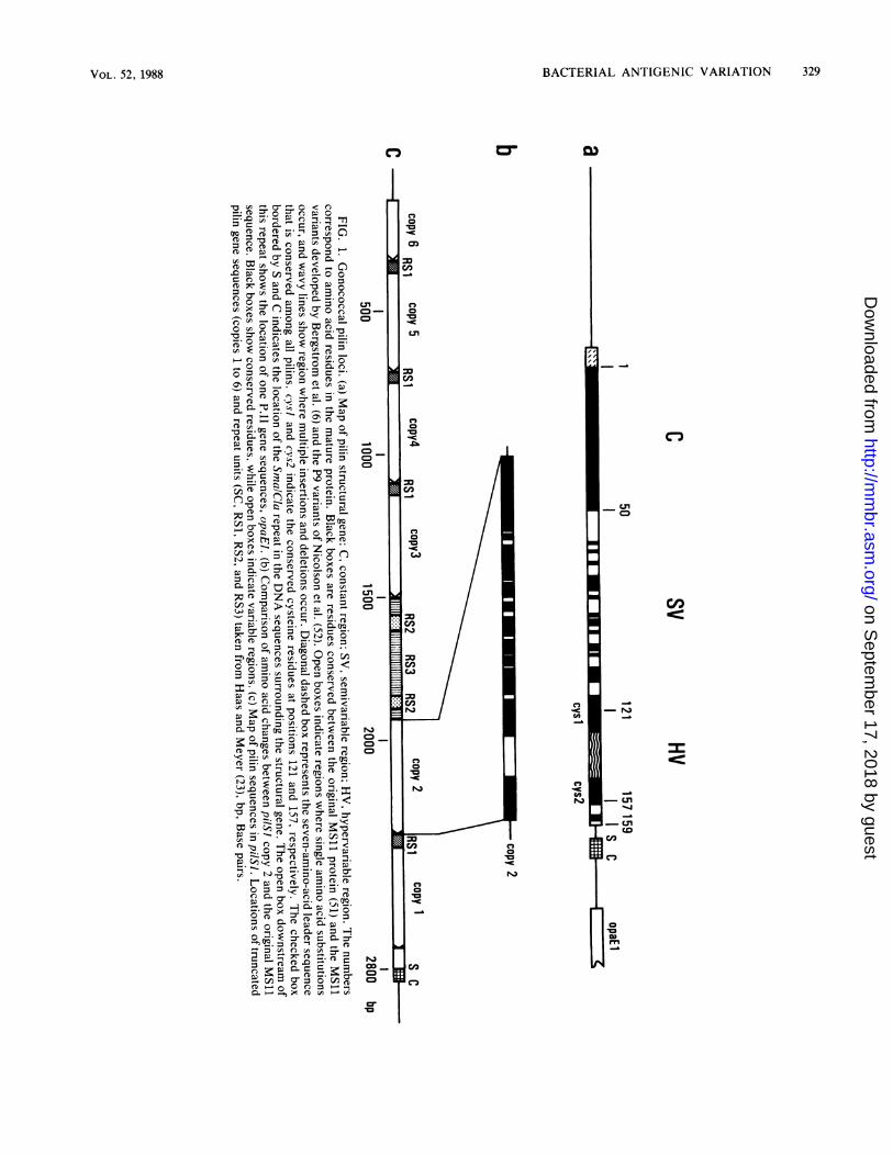
which now produce pilins with new epitopes (24, 52). Acomparison of expressed variant pilin sequences from thesestrains indicated that the pilin gene can be divided intoconstant (C), semivariable (SV), and hypervariable (HV)regions (Fig. la). The C region encodes approximately thefirst 50 residues of each mature pilin and is invariant insequence in all cases examined. The variant pilin sequences
differ from each other to a small degree in the central, or SV,portion of the gene. This region is characterized mainly bysingle base changes which result in single amino acid
changes of the protein. In the last third of the pilin gene (the
HV region) are found insertions and deletions of one or more
codons in multiple sites, as well as single codon changes.Epitope mapping studies with pilin-specific monoclonal an-
tibodies indicate that this region encodes the most antigenic
portion of pilin (52). Although the latter two-thirds of the
pilin gene is considered variable, it does contain small
invariant regions. The two most striking of these have been
termed cys-J and cys-2, after the cysteine residue each
encodes, which flank the HV region. Presumably, the invari-
ant amino acids in pilin are critical to the maintenance ofpilus structure and perhaps to other functions as well.Two approaches have been used to examine the arrange-
ment of pilin sequences in the gonococcal chromosome.DNA sequencing of pilSI, a major pilin-hybridizing locusnear the two expression sites, showed that this region ofabout 3 kilobase pairs contains six variant pilin sequencesoriented in the same direction as the expressed gene(s) (23)(Fig. lc). Each gene copy contains the entire variable regionup to, but not always including, the termination codon ofpilin. Each also contains some constant 5' sequences,though the 5'-endpoints differ from one gene to the other. Nopilin promoter sequences are found in the locus. A compar-ison of the pil variant sequences in pilSI shows that thesepartial genes can also be divided into C, SV, and HVregions. The comparison of silent sequences allowed Haasand Meyer (23) to propose that minicassettes of variable pilinsequences found in silent pilin loci are introduced as discreteunits into an intact variant pilin gene in the expression site.One such minicassette sequence (from copy 1 or 3) from amajor silent pil locus, pilSI, has been observed to havedonated its sequence to the expression site during pilinantigenic variation. Others have also shown recombinationof other partial pilSi gene copies (from copy 5) into anexpression locus (85). The conserved sequences within thepilin structural gene are probably used as crossover pointsfor the recombination reactions and are most likely requiredto maintain the structural integrity of the pilus.The arrangement of pilin sequences in the gonococcal
chromosome also has been examined by Southern hybrid-ization, using synthetic oligonucleotide probes specific forseveral regions of two variant pilin genes. These studies (67)essentially support the DNA sequence findings describedabove and further identify other silent pil loci whose variantsequences are used by the cell to produce antigenicallyvariant pilins. The above studies have identified seven silentpil loci (termed pilSI-7), although there may be additionalundiscovered silent loci. It is now accepted that pilin anti-genic variation results from the replacement of the expressedpilin sequence in an expression locus by part or all of avariant sequence from a silent copy. The requirement for thegonococcal recA homolog in promoting the recombinationreactions between the silent and expressed genes (38-40)suggests that the exchange involves the formation of hetero-duplex DNA.
In addition to copies of variant pil genes, the silent locishare other sequence homologies with the expression sitespilEl and pilE2. Of note is the Sma/Cla repeat, which ispresent at the 3'-untranscribed region of the complete pilinsequence in each expression site, as well as in each silentlocus (23, 56b, 67; T. F. Meyer, personal communication)(Fig. 1). All loci that contain pilin sequences also contain theSma/Cla repeat. Other DNA repeats have also been identi-fied in silent and expressed pilin loci. These include RS1,RS2, and RS3 (23, 56b) (Fig. lc). The function of theserepeats has not been determined. However, it is probablethat some or all of them will be involved in the recombina-tional process which leads to pilin antigenic variation.Two models of pilin antigenic variation. Southern hybrid-
ization studies based on oligonucleotide probes have givenrise to one model of pilin antigenic variation. Each HV-specific oligonucleotide hybridizes to one silent locus in allderivatives of strain MS11. In those progeny which nowactively express the variant pilin gene from which this HVsequence is derived, the probe also hybridizes to the pilinexpression locus. Such data (23, 66, 84) indicate that pilin
MICROBIOL. REV.
on Septem
ber 17, 2018 by guesthttp://m
mbr.asm
.org/D
ownloaded from
BACTERIAL ANTIGENIC VARIATION
C,
CA,
C,2
g,
-.,
-U,CoI.cceC:
m_0
VOL. 52, 1988 329
m
on Septem
ber 17, 2018 by guesthttp://m
mbr.asm
.org/D
ownloaded from

330 SEIFERT AND SO
antigenic variation results when a copy of a variant pilin genein a silent pil locus is recombined into an expression site.However, the pilin sequence displaced from the expressionsite during recombination is not found in the silent locuswhich has actively donated its variant pil gene, nor is itfound anywhere else outside its own silent locus. Theseresults suggest that recombination between the silent andexpression loci is a nonreciprocal event. This unidirectionaltransfer of genetic material within a cell would be classifiedas gene conversion.Gene conversion occurs in a number of eucaryotes such as
yeasts (29, 30, 36, 37), African trypanosomes (46), andmammalian cells (18, 64, 69). However, this reaction occursat a low frequency in procaryotes. In S. typhimurium, geneconversion occurs at a frequency of approximately 10-6 (J.Roth, personal communication). If pilin-related gene conver-sion events in the gonococcus were to occur by generalrecombination, they may be expected to occur at a fre-quency similar to that of S. typhimurium. Such a frequencyis lower than that observed for pilin variation. Therefore, ifgene conversion is responsible for pilin recombination, itwould have to be a specialized system which gives a highrate of recombination, but which only allows for nonrecip-rocal recombination. Such a system has been described formating type interconversion in Saccharomyces cerevisiae(see reference 28).Two aspects of the gonococcal life cycle lend support to
an alternate model of pilin antigenic variation. As mentionedearlier, the gonococcus is naturally competent for DNAtransformation (73) and has a strong preference for its ownDNA (17). For piliated cells the frequency of transformationof chromosomal markers such as antibiotic resistance andauxotrophic markers is approximately 10-2 to 10-3. Thetransformation frequency for nonpiliated cells is 3 to 4 logslower (73). The gonococcus also undergoes autolysis readily(25). In an actively growing culture, a certain percentage ofcells have already lysed. Thus, living gonococci are con-stantly exposed to free DNA, and transformation of chro-mosomal markers between strains in mixed culture has beenreported (63). These observations suggest that the gono-coccus has evolved the process of DNA transformation as aspecific rapid adaptive response, rather than as a long-termmechanism to acquire new traits for the species in general.Thus, DNA transformation very well could be used bygonococci to undergo pilin antigenic variation. According tothe DNA transformation model, antigenic variation beginswhen DNA from a lysed cell is taken up by a living cell. Thegonococcal chromosome contains many silent variant pilgenes. If the incoming DNA contains a silent pil locus, itwould preferentially recombine at a site with which it shareshomology, such as the pil gene in the expression site. Theproduct of such a recombination event would be a variantcell that now expresses a new pilin gene. Figure 2 is adiagrammatic representation of both models of pilin anti-genic variation.To examine pilin-related recombination events, an assay
has been developed to allow quantitation of recombinationfrequencies between pilin loci in the gonococcus (H. S.Seifert, R. A. Ajioka, C. Marchal, P. F. Sparling, and M. So,submitted for publication). An operon fusion was created, inthe gonococcus, in which the chloramphenicol acetyltrans-ferase structural gene (the CAT cartridge; 13) was inserteddownstream of the pilin promoter sequences and upstreamof the intact pilin structural gene in pilEl (Fig. 3, strainMS11CAT8). Translation of each gene is regulated by itsown ribosomebinding site, and transcription of both genes is
Gene Conversion AutolysislTransformation
/
FIG. 2. Diagrammatic representation of extracellular versus in-tercellular models of pilin antigenic variation. Open box representsthe pilin gene being expressed by the gonococcal cell. Hatched andstippled boxes represent variant pilin sequences in the gonococcalchromosome. The cell on the left is switching its pilin variant by anintracellular gene conversion mechanism. The cell on the right isswitching its pilin gene sequence by uptake and recombination withfree DNA released from a neighboring cell that has autolyzed. Forsimplicity of representing the models, the gonococcal cells are notshown as diplococci.
controlled by the pilin promoter. The other expression locusexpresses a wild-type pilin gene. Nonreciprocal recombina-tion events involving pilin loci can be scored by plating cellson chloramphenicol levels that select against those with onecopy of CAT, but allow the growth of those with two copiesof CAT. The rate at which the wild-type expression sitereceives the CAT gene from the other site would reflect thefrequency of gene conversion, and this approach can be usedto quantitate such events.To test the hypothesis that DNA transformation is the
basis of pilin antigenic variation, the frequency of the above"gene conversion" event was measured in a strain deficientin the DNA uptake step of transformation. The DNA uptakemutation from strain FA660 (dud-i; G. Biswas and P. F.
MS11 AP+ CmS
pilE2
MS1 1CAT8P+ CmR
MS1l CAT8-1P+ CmR
{IACATPIL11
1 ~ ~~~~~ ATIPRFIG. 3. Model for pilin gene conversion. A diagram showing the
structure of pilin expression loci, pilEIand piIE2, in three strains,MS11-A, MS11CAT8, and MS11CAT8-1. PIL indicates pilin struc-tural gene, while CAT represents the chloramphenicol acetyltrans-ferase structural gene. The uppercase P indicates the location of thepilin promoter relative to the structural genes. P+denotes piliatedphenotype and CmS denotes sensitivity to chloramphenicol, whileCmRdenotes resistance to chloramphenicol.
MICROBIOL. REV.
on Septem
ber 17, 2018 by guesthttp://m
mbr.asm
.org/D
ownloaded from
BACTERIAL ANTIGENIC VARIATION 331
(CTCTT)n
HV1 HV2
Leader Mature Protein
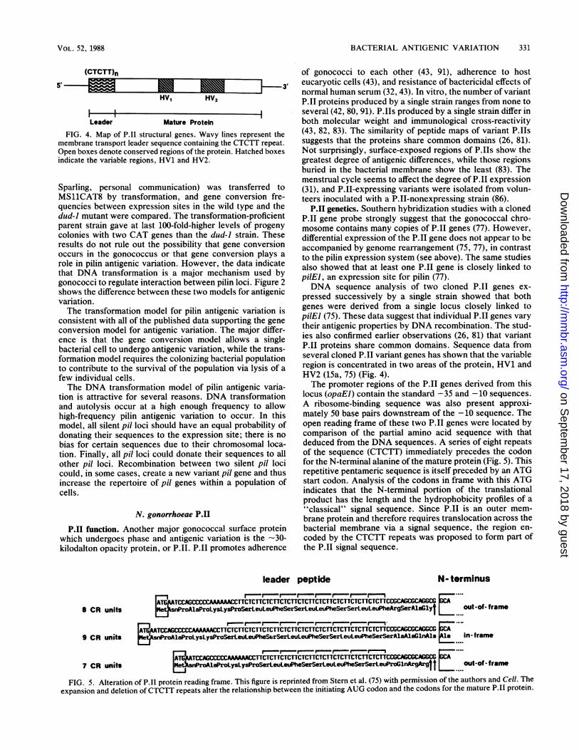
FIG. 4. Map of P.11 structural genes. Wavy lines represent themembrane transport leader sequence containing the CTCTT repeat.Open boxes denote conserved regions of the protein. Hatched boxesindicate the variable regions, HV1 and HV2.
Sparling, personal communication) was transferred toMS11CAT8 by transformation, and gene conversion fre-quencies between expression sites in the wild type and thedud-i mutant were compared. The transformation-proficientparent strain gave at last 100-fold-higher levels of progenycolonies with two CAT genes than the dud-i strain. Theseresults do not rule out the possibility that gene conversionoccurs in the gonococcus or that gene conversion plays arole in pilin antigenic variation. However, the data indicatethat DNA transformation is a major mechanism used bygonococci to regulate interaction between pilin loci. Figure 2shows the difference between these two models for antigenicvariation.The transformation model for pilin antigenic variation is
consistent with all of the published data supporting the geneconversion model for antigenic variation. The major differ-ence is that the gene conversion model allows a singlebacterial cell to undergo antigenic variation, while the trans-formation model requires the colonizing bacterial populationto contribute to the survival of the population via lysis of afew individual cells.The DNA transformation model of pilin antigenic varia-
tion is attractive for several reasons. DNA transformationand autolysis occur at a high enough frequency to allowhigh-frequency pilin antigenic variation to occur. In thismodel, all silent pil loci should have an equal probability ofdonating their sequences to the expression site; there is nobias for certain sequences due to their chromosomal loca-tion. Finally, all pil loci could donate their sequences to allother pil loci. Recombination between two silent pil locicould, in some cases, create a new variant pil gene and thusincrease the repertoire of pil genes within a population ofcells.
N. gonorrhoeae P.I1
P.I1 function. Another major gonococcal surface proteinwhich undergoes phase and antigenic variation is the -30-kilodalton opacity protein, or P.II. P.II promotes adherence
of gonococci to each other (43, 91), adherence to hosteucaryotic cells (43), and resistance of bactericidal effects ofnormal human serum (32, 43). In vitro, the number of variantP.II proteins produced by a single strain ranges from none toseveral (42, 80, 91). P.IIs produced by a single strain differ inboth molecular weight and immunological cross-reactivity(43, 82, 83). The similarity of peptide maps of variant P.IIssuggests that the proteins share common domains (26, 81).Not surprisingly, surface-exposed regions of P.IIs show thegreatest degree of antigenic differences, while those regionsburied in the bacterial membrane show the least (83). Themenstrual cycle seems to affect the degree of P.11 expression(31), and P.II-expressing variants were isolated from volun-teers inoculated with a P.II-nonexpressing strain (86).
P.11 genetics. Southern hybridization studies with a clonedP.1I gene probe strongly suggest that the gonococcal chro-mosome contains many copies of P.11 genes (77). However,differential expression of the P.11 gene does not appear to beaccompanied by genome rearrangement (75, 77), in contrastto the pilin expression system (see above). The same studiesalso showed that at least one P.11 gene is closely linked topilEl, an expression site for pilin (77).DNA sequence analysis of two cloned P.11 genes ex-
pressed successively by a single strain showed that bothgenes were derived from a single locus closely linked topilEl (75). These data suggest that individual P.II genes varytheir antigenic properties by DNA recombination. The stud-ies also confirmed earlier observations (26, 81) that variantP.11 proteins share common domains. Sequence data fromseveral cloned P.II variant genes has shown that the variableregion is concentrated in two areas of the protein, HV1 andHV2 (1Sa, 75) (Fig. 4).The promoter regions of the P.II genes derived from this
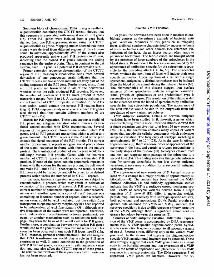
locus (opaEI) contain the standard -35 and -10 sequences.A ribosome-binding sequence was also present approxi-mately 50 base pairs downstream of the -10 sequence. Theopen reading frame of these two P.11 genes were located bycomparison of the partial amino acid sequence with thatdeduced from the DNA sequences. A series of eight repeatsof the sequence (CTCTT) immediately precedes the codonfor the N-terminal alanine of the mature protein (Fig. 5). Thisrepetitive pentameric sequence is itself preceded by an ATGstart codon. Analysis of the codons in frame with this ATGindicates that the N-terminal portion of the translationalproduct has the length and the hydrophobicity profiles of a"classical" signal sequence. Since P.II is an outer mem-brane protein and therefore requires translocation across thebacterial membrane via a signal sequence, the region en-coded by the CTCTT repeats was proposed to form part ofthe P.11 signal sequence.
leader peptide N- terminus
8 CR unitsARAl5WfD I.t,I42IALAIVVIVVLI.* WII.I IL.*.* I t I.... ..LW.. I I ._.II.IwIesnProAlaProLysLysProSerLeuLeuPheSerSerLeuLeuPheSerSerLeuLeuPheArgSerAlaGlyt .....out -of- frame
r - r - . - r r - , r - , - r_ , X- ....l-....A---r- - - ---rrrTrrTrTTrTrTTrrTrTTrT'Tr TrTrTTrTrTTrTrrrPrArYYfP'-EA klr.rA
9 CR units PsrPrOoLysLysProSerLeuLeuPheSerSerLeuLeuPheSerSerLeuLeuPheSerSerAlSAlaGInAla A in-frame
_ ~~~~~r-r- .- X- e_ .~ r,. ....
ATb,uTCCAAAAAACCTTCTCTTCTCTTCTCTTCTCTTCTCTTCTCTTCTCTTCCCACGCAGGCG7 CR units nPoA1aProLysLysProSerLeuLeuPheSerSerLeuLeuPheSerSerLeuProG1lngArgttL.... out-Of-frame
FIG. 5. Alteration of P.11 protein reading frame. This figure is reprinted from Stem et al. (75) with permission of the authors and Cell. Theexpansion and deletion ofCTCTT repeats alter the relationship between the initiating AUG codon and the codons for the mature P.11 protein.
VOL. 52, 1988
r-In
on Septem
ber 17, 2018 by guesthttp://m
mbr.asm
.org/D
ownloaded from
332 SEIFERT AND SO
Southern blots of chromosomal DNA, using a syntheticoligonucleotide containing the CTCTT repeat, showed thatthis sequence is associated with many if not all P.II genes(75). Other P.11 genes were isolated from a gene bankderived from one P.JI-expressing strain, using the CTCTToligonucleotide as probe. Mapping studies showed that theseclones were derived from different regions of the chromo-some. In addition, approximately 25% of the clones alsoproduced apparently native sized P.11 proteins in E. coli,indicating that the cloned P.II genes contain the codingsequence for the entire protein. Thus, in contrast to the pilsystem, each P.II gene is a potential P.11 expression site.Primer extension sequence determination of the CTCTT
region of P.II messenger ribonucleic acids from severalderivatives of one gonococcal strain indicates that theCTCTT repeats are transcribed and thus are truly part of thecoding sequence of the P.II gene. Furthermore, most, if notall, P.11 genes are transcribed in all of the derivativeswhether or not the cells produced P.II proteins. However,the number of pentameric CTCTT units present in thesetranscripts ranged from 8 to 13. A transcript containing thecorrect number of CTCTT repeats, in relation to the ATGstart codon, would contain the correct P.11 reading frame(Fig. 5). DNA sequence analysis of several cloned P.II genesalso showed that they contain different numbers of theCTCTT unit (1Sa).Models for P.11 regulation. These data support a model of
P.11 phase and antigenic variation which involves transla-tional control coupled with DNA recombination. Severalregions of the gonococcal chromosome contain intact P.AIgenes, and all P.II genes are transcribed within a cell at anygiven moment. The CTCTT repeats at the 5' end of the geneencode the signal sequence of the P.11 protein. The correctnumber of pentameric repeats in a gene would place codonsof the signal sequence in frame with those of the matureprotein. The translational product of such a gene would be afull-length variant P.II protein. Genes with the incorrectnumber of CTCTT repeats would encode a truncated P.11product. If none of the genes contain pentameric repeats inframe with the codons for the mature protein, the cell wouldnot make P.11, and a P.II- phenotype is the result. A singleP.11 gene could be turned on and off by a yet to be definedprocess which varies the number of its CTCTT repeats.
In bacteria, tandemly repeated sequences are subject torecombination, a process which may result in deletion orexpansion of the number of repeats. A P.11 gene with thecorrect number of pentameric repeats could, after recombi-nation with another gene or a copy of itself, become onecontaining an incorrect number of repeats. Such a recombi-nation event could be recA mediated, but the switch fromtransparent to opaque colony morphology has been reportedto be independent of recA functionality (38), although rela-tive frequencies of the switch were not reported. Therefore,recA independent recombination between pentameric re-peats, or another mechanism such as replication fork slip-page, may form the basis of the on/off switch for P.11 genes.Furthermore, recombination between two variant P.1I geneswould lead to the generation of new variant sequences. Thiseven has been observed in one such P.11 locus, opaEl (15a,75; C. Marchal, personal communication). One can imaginethat DNA transformation would play a role in P.11 geneexpression as well. It could contribute to the generation ofnew P.II variant genes, as occurs with pilin antigenic varia-tion, and may also affect the CTCTT region of these genes.The relative contribution of these processes to P.II variationhas not been reported.
Borrelia VMP Variation
For years, the borrelias have been cited in medical micro-biology courses as the primary example of bacterial anti-genic variation. Members of this genus cause relapsingfever, a clinical syndrome characterized by successive boutsof fever in humans and other animals (see reference 19).Infection of the host, via an insect vector, often leads topersistent bacteremia. The febrile crises are brought aboutby the presence of large numbers of the spirochetes in theblood stream. Resolution of the fevers is accompanied by theappearance of antibodies specific for the spirochete respon-sible for the preceding attack (14, 16, 50). The spirocheteswhich produce the next bout of fever will induce their ownspecific antibodies. Upon injection of a rat with a singlespirochete, antigenically distinct spirochetes can be isolatedfrom the blood of the animal during the relapse phases (65).The characteristics of this disease suggest that surfaceantigens of the spirochetes undergo antigenic variation.Thus, growth of spirochetes of one predominant type isresponsible for a relapse. The resolution of the relapse is dueto the clearance from the blood of spirochetes by antibodiesspecific for that spirochete population. The appearance ofthe next relapse would be due to the outgrowth from thispopulation of new variant spirochetes.VMP antigenic variation. Details of borrelia antigenic
variation have been studied in B. hermsii, a genus whichcauses relapsing fever in mice. Infection studies showed thata single organism can give rise to at least 24 new serotypes(78). Thus, the bacterium contains many copies of variantgenes that encode the cellular component which undergoesantigenic variation. The frequency of serotype switching isapproximately 1o-4 to 10-3 (78). As is the case in Africantrypanosomes (8), there is a loose order of appearance of theserotypes in the host, and certain serotypes predominate inthe early stages of the disease (78). Serotypes cleared fromone host can reappear upon passage of the spirochete to asecond host (15). This finding indicates that genetic informa-tion for serotype specificity is not lost during antigenicvariation, a necessary condition for a successful antigenicvariation system.The appearance of new serotypes of B. hermsii is corre-
lated with a change in a major protein of approximately 40kilodaltons (4). The antigen has been named the VMP.Surface iodination (4) and antibody agglutination studiesindicate that the VMP is a surface-exposed membrane pro-tein. VMPs of serotypic variants derived from a singleorganism of B. hermsii HS1 differ in molecular weight,peptide map, and reactivity with serotype-specific antisera,both polyclonal and monoclonal (3, 4). Partial protein se-quence data obtained for VMP7 and VMP21 indicate thatserotype specificity is due to differences in primary structureof the VMPs, although there is significant amino acid se-quence homology between the proteins (5).
Genetics of VMP antigenic variation. Differential expres-sion of the VMP genes is accompanied by DNA rearrange-ments (49). A VMP-specific oligonucleotide probe hybrid-izes to a restriction fragment common to all isogenic variantsof one B. hermsii strain, differing only in the variant VMPproduced. In the strains that express the oligonucleotide-specific VMP, an additional fragment can be detected. Thesedata strongly suggest that each VMP gene exists as a silentcopy in the borrelial genome and that expression of a VMPgene is the result of duplication and placement of this VMPsequence into an expression site. The DNA sequences 5' ofexpressed VMP genes are identical. However, the 5'-
MICROBIOL. REV.
on Septem
ber 17, 2018 by guesthttp://m
mbr.asm
.org/D
ownloaded from
BACTERIAL ANTIGENIC VARIATION 333
A B
SP SP _
EP t = 1 EP
SP {}SP{
EP3 EP 3
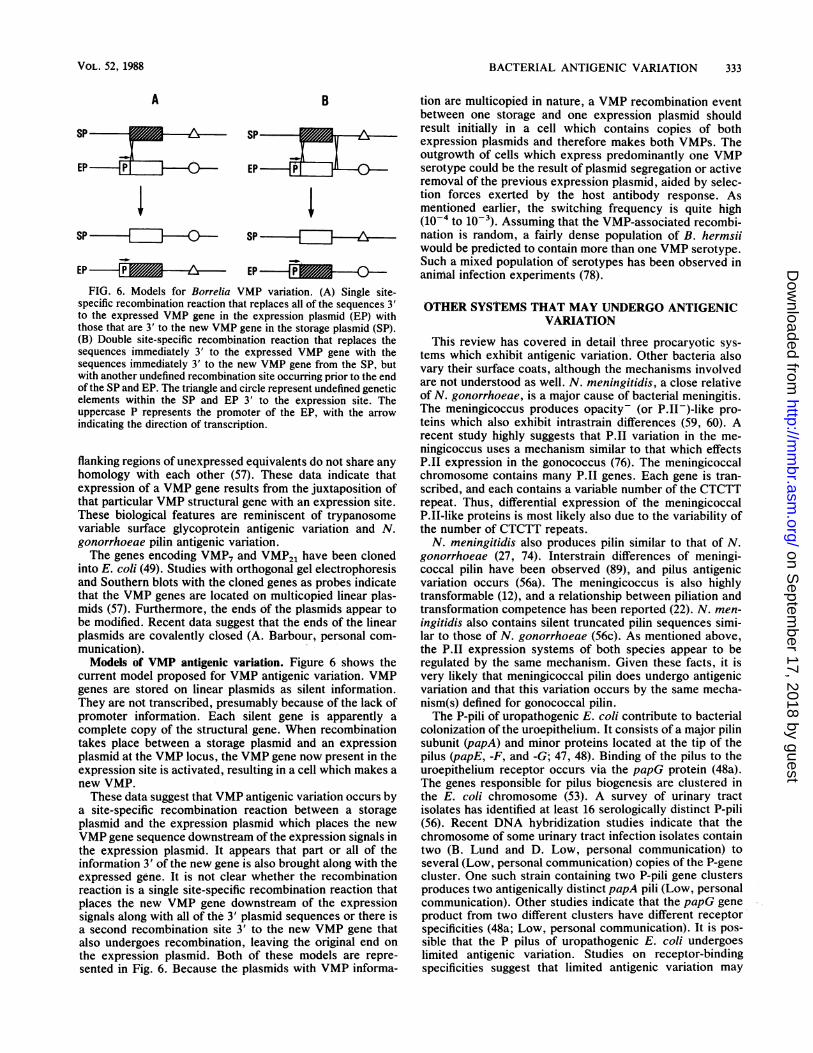
FIG. 6. Models for Borrelia VMP variation. (A) Single site-specific recombination reaction that replaces all of the sequences 3'to the expressed VMP gene in the expression plasmid (EP) withthose that are 3' to the new VMP gene in the storage plasmid (SP).(B) Double site-specific recombination reaction that replaces thesequences immediately 3' to the expressed VMP gene with thesequences immediately 3' to the new VMP gene from the SP, butwith another undefined recombination site occurring prior to the endof the SP and EP. The triangle and circle represent undefined geneticelements within the SP and EP 3' to the expression site. Theuppercase P represents the promoter of the EP, with the arrow
indicating the direction of transcription.
flanking regions of unexpressed equivalents do not share any
homology with each other (57). These data indicate thatexpression of a VMP gene results from the juxtaposition ofthat particular VMP structural gene with an expression site.These biological features are reminiscent of trypanosomevariable surface glycoprotein antigenic variation and N.gonorrhoeae pilin antigenic variation.The genes encoding VMP7 and VMP21 have been cloned
into E. coli (49). Studies with orthogonal gel electrophoresisand Southern blots with the cloned genes as probes indicatethat the VMP genes are located on multicopied linear plas-mids (57). Furthermore, the ends of the plasmids appear tobe modified. Recent data suggest that the ends of the linearplasmids are covalently closed (A. Barbour, personal com-
munication).Models of VMP antigenic variation. Figure 6 shows the
current model proposed for VMP antigenic variation. VMPgenes are stored on linear plasmids as silent information.They are not transcribed, presumably because of the lack ofpromoter information. Each silent gene is apparently acomplete copy of the structural gene. When recombinationtakes place between a storage plasmid and an expressionplasmid at the VMP locus, the VMP gene now present in theexpression site is activated, resulting in a cell which makes a
new VMP.These data suggest that VMP antigenic variation occurs by
a site-specific recombination reaction between a storageplasmid and the expression plasmid which places the newVMP gene sequence downstream of the expression signals inthe expression plasmid. It appears that part or all of theinformation 3' of the new gene is also brought along with theexpressed gene. It is not clear whether the recombinationreaction is a single site-specific recombination reaction thatplaces the new VMP gene downstream of the expressionsignals along with all of the 3' plasmid sequences or there isa second recombination site 3' to the new VMP gene thatalso undergoes recombination, leaving the original end onthe expression plasmid. Both of these models are repre-sented in Fig. 6. Because the plasmids with VMP informa-
tion are multicopied in nature, a VMP recombination eventbetween one storage and one expression plasmid shouldresult initially in a cell which contains copies of bothexpression plasmids and therefore makes both VMPs. Theoutgrowth of cells which express predominantly one VMPserotype could be the result of plasmid segregation or activeremoval of the previous expression plasmid, aided by selec-tion forces exerted by the host antibody response. Asmentioned earlier, the switching frequency is quite high(10-4 to 10-3). Assuming that the VMP-associated recombi-nation is random, a fairly dense population of B. hermsiiwould be predicted to contain more than one VMP serotype.Such a mixed population of serotypes has been observed inanimal infection experiments (78).
OTHER SYSTEMS THAT MAY UNDERGO ANTIGENICVARIATION
This review has covered in detail three procaryotic sys-tems which exhibit antigenic variation. Other bacteria alsovary their surface coats, although the mechanisms involvedare not understood as well. N. meningitidis, a close relativeof N. gonorrhoeae, is a major cause of bacterial meningitis.The meningicoccus produces opacity- (or P.II-)-like pro-teins which also exhibit intrastrain differences (59, 60). Arecent study highly suggests that P.11 variation in the me-ningicoccus uses a mechanism similar to that which effectsP.JI expression in the gonococcus (76). The meningicoccalchromosome contains many P.11 genes. Each gene is tran-scribed, and each contains a variable number of the CTCTTrepeat. Thus, differential expression of the meningicoccalP.II-like proteins is most likely also due to the variability ofthe number of CTCTT repeats.N. meningitidis also produces pilin similar to that of N.
gonorrhoeae (27, 74). Interstrain differences of meningi-coccal pilin have been observed (89), and pilus antigenicvariation occurs (56a). The meningicoccus is also highlytransformable (12), and a relationship between piliation andtransformation competence has been reported (22). N. men-ingitidis also contains silent truncated pilin sequences simi-lar to those of N. gonorrhoeae (56c). As mentioned above,the P.II expression systems of both species appear to beregulated by the same mechanism. Given these facts, it isvery likely that meningicoccal pilin does undergo antigenicvariation and that this variation occurs by the same mecha-nism(s) defined for gonococcal pilin.The P-pili of uropathogenic E. coli contribute to bacterial
colonization of the uroepithelium. It consists of a major pilinsubunit (papA) and minor proteins located at the tip of thepilus (papE, -F, and -G; 47, 48). Binding of the pilus to theuroepithelium receptor occurs via the papG protein (48a).The genes responsible for pilus biogenesis are clustered inthe E. coli chromosome (53). A survey of urinary tractisolates has identified at least 16 serologically distinct P-pili(56). Recent DNA hybridization studies indicate that thechromosome of some urinary tract infection isolates containtwo (B. Lund and D. Low, personal communication) toseveral (Low, personal communication) copies of the P-genecluster. One such strain containing two P-pili gene clustersproduces two antigenically distinct papA pili (Low, personalcommunication). Other studies indicate that the papG geneproduct from two different clusters have different receptorspecificities (48a; Low, personal communication). It is pos-sible that the P pilus of uropathogenic E. coli undergoeslimited antigenic variation. Studies on receptor-bindingspecificities suggest that limited antigenic variation may
VOL. 52, 1988
on Septem
ber 17, 2018 by guesthttp://m
mbr.asm
.org/D
ownloaded from

334 SEIFERT AND SO
occur to allow the bacterial pathogen to modulate its hostrange.
Perhaps the most striking example of the evolution ofantigenic variation for purposes other than escape from hostimmune response is P. aurelia. The outer surface of thisfree,-living protozoan consists of a major protein termed thei-?-htigen (72). Serotyping of P. aurelia is based on theantigenic characteristics of this high-molecular-weight pro-tein. A stock of P. aurelia is capable of expressing severalantigenically distinct i-antigens in succession (71), althoughi-antigen variation, like the P.1 system, does not appear toinvolve genome rearrangement (21). i-Antigen variation oc-
curs at a rapid rate in response to a variety of environmentalsignals such as pH, temperature, and food supply (70). Thefunction of the i-antigen is unclear but is thought to beimportant for the life cycle of the paramecium, as variantslacking the protein have not been obtained.
SUMMARY AND OVERVIEW
The studies described above indicate that procaryoteshave evolved a variety of mechanisms to vary their surfacecoats. N. gonorrhoeae primarily uses DNA transformationto effect pilus antigenic variation at the recombinationallevel. It also uses recombination (and perhaps also DNAtransformation) to bring about P.11 antigenic variation at thetranslational level. Finally, Borrelia organisms have evolveda plasmid recombination system to undergo VMP antigenicvariation.To place procaryotic antigenic variation into proper per-
spective, we end this review with a brief consideration of thehost immune system. Mammals have also evolved whatcould be considered an antigenic variation system, i.e., thegeneration of antibodies with different antigen-binding spec-
ificities. The arrangement of multiple copies of V, D, and Jgene segments in the mammalian genome is reminiscent ofthe arrangement of silent pilin gene segments in the gono-
coccal chromosome. However, unlike pilin, P.11, and VMPexpression, the generation of a functional expressing immu-noglobulin gene does not involve expression sites. Instead, a
complete immunoglobulin gene is created by recombina-tional joining of various gene segments, with concomitantdeletion of intervening sequences. A system that appears to
resemble the gonococcal pilin mechanism has been de-scribed for chicken immunoglobulin light chains (62). Thelight chain variants all are derived from a unique V-Jrearrangement, with diversification occurring by gene con-
version from other V gene copies to this single expressedgene within the Bursa of Fabricius.
Four main processes appear to be responsible for thegeneration of antibody diversity in mammalian cells (seereference 90). The first, known as "combinational diver-sity," is the joining of V and J gene segments in various
combinations. Diversity could also be generated by impre-cise joining at V-J, V-D, and D-J junctions. In addition,joining of the VH-D and D-JH segments could lead to
insertion of one to several nucleotides at these junctions.Finally, sequence changes could occur in immunoglobulingene segments by somatic mutation. Whether these fourprocesses also contribute to antigenic variation in procary-
otic systems is not known at present. Since both the procary-
otic and eucaryotic systems operate at the recombinationallevel, it is possible that the first three processes whichcontribute to immunoglobulin diversity also play a role in
procaryotic antigenic variation. As for somatic mutations, it
is clear that antigenic drift contributes significantly to the
generation of hemagglutinin and neuraminidase variants ofthe flu virus. It is therefore likely that this process alsocontributes to sequence variability of the pilin, P.11, andVMP genes. In addition, gene conversion is thought tocontribute to the generation of somatic mutation in immu-noglobin genes (2).
In summary, it is interesting to note that the systems ofantigenic variation and immunoglobulin diversification haveevolved in a similar and complementary fashion, with DNArecombination playing a central mechanistic role. It is highlylikely that the two systems developed together, with eachproviding the evolutionary pressure needed by the other.Finally, the examples of antigenic variation covered in thisreview illustrate the fascinating and diverse ways microbeshave found to regulate and alter gene expression.
ACKNOWLEDGMENTS
We thank all of our colleagues who communicated results prior topublication, as noted in the text.
LITERATURE CITED1. Abraham, J. M., C. S. Freitag, J. R. Clements, and B. I.
Eisenstein. 1985. An invertible element of DNA controls phasevariation of type 1 fimbriae of Escherichia coli. Proc. NatI.Acad. Sci. USA 82:5724-5727.
2. Baltimore, D. 1981. Gene conversion: some implications forimmunoglobin genes. Cell 4:592-594.
3. Barbour, A. G., 0. Barrera, and R. Judd. 1983. Structuralanalysis of the variable major proteins of Borrellia hermsii. J.Exp. Med. 158:2127-2140.
4. Barbour, A. G., S. L. Tessier, and H. G. Stoenner. 1982.Variable major proteins of Borrelia hermsii. J. Exp. Med. 156:1312-1324.
5. Barstad, P. A., J. E. Coligan, M. G. Raum, and A. G. Barbour.1985. Variable major proteins of Borrelia hermsii. J. Exp. Med.161:1302-1314.
6. Bergstrom, S., K. Robbins, J. M. Koomey, and J. Swanson.1986. Piliation control mechanisms in Neisseria gonorrhoeae.Proc. Natl. Acad. Sci. USA 83:3890-3894.
7. Bloom, B. R. 1979. Games parasites play: how parasites evadeimmune surveillance. Nature (London) 279:21-26.
8. Borst, P., and G. A. M. Cross. 1982. Molecular basis fortrypanosome antigenic variation. Cell 29:291-303.
9. Brinton, C. C., J. Bryan, J. Dillon, N. Guerina, J. L. Jacobson,A. Labik, S. Lee, A. Levine, S. Lim, J. McMichael, S. Polen, K.Rogers, A. C.-C. To, and S. C.-C. To. 1978. Uses of pili ingonorrhea control: role of bacterial pili in disease, purificationand properties of gonococcal pili, and progress in the develop-ment of a gonococcal pilus vaccine for gonorrhea, p. 155-178. InG. F. Brooks, E. C. Gotschlich, K. K. Holmes, W. D. Sawyer,and F. E. Young (ed.), Immunobiology of Neisseria gonor-rhoeae. American Society for Microbiology, Washington, D.C.
10. Buchanan, T., and W. A. Pearce. 1976. Pili as a mediator of theattachment of gonococci to human erythrocytes. Infect. Immun.13:1483-1489.
11. Buchanan, T. M. 1975. Antigenic heterogeneity of gonococcalpili. J. Exp. Med. 141:1470-1475.
12. Catlin, B. W. 1960. Transformation of Neisseria meningitidis bydeoxyrobinucleates from cells and from culture slime. J. Bac-teriol. 79:579-590.
13. Close, T. J., and R. L. Rodriguez. 1982. Construction andcharacterization of the chloramphenicol-resistance gene car-tridge: a new approach to the transcriptional mapping of extra-chromosomal elements. Gene 20:305-316.
14. Coffey, E. M., and W. C. Eveland. 1967. Experimental relapsingfever initiated by Borrelia hermsii. I. Identification of majorserotypes by immunofluorescence. J. Infect. Dis. 117:23-28.
15. Coffey, E. M., and W. C. Eveland. 1967. Experimental relapsingfever initiated by Borrelia hermsii. II. Sequential appearance ofmajor serotypes in the rat. J. Infect. Dis. 117:29-34.
MICROBIOL. REV.
on Septem
ber 17, 2018 by guesthttp://m
mbr.asm
.org/D
ownloaded from

BACTERIAL ANTIGENIC VARIATION 335
15a.Connell, T. D., W. J. Black, T. H. Kawula, D. S. Barritt, J. A.Dempsey, K. Keverneland, Jr., A. Stephenson, B. S. Schepurt,G. L. Murphy, and J. G. Cannon. 1988. Recombination amongprotein II genes of Neisseria gonorrhoeae generates new codingsequences and increases structural variability in the protein IIfamily. Mol. Microbiol. 2:227-230.
16. Cunningham, J. 1925. Serological observations on relapsingfever in Madras. Trans. R. Soc. Trop. Med. Hyg. 19:11-17.
17. Dougherty, T. J., A. Asmus, and A. Tomasz. 1979. Specificity ofDNA uptake in genetic transformation of gonococci. Biochem.Biophys. Res. Commun. 86:97-104.
18. Evans, G. A., D. H. Margulies, R. D. Camerini-Otero, K. Ozato,and J. G. Seidman. 1982. Structure and expression of a mousemajor histocompatibility antigen gene, H-2Ld. Proc. Natl. Acad.Sci. USA 79:1994-1998.
19. Felsenfeld, 0. 1965. Borrelia, human relapsing fever, and para-site-vector-host relationships. Bacteriol. Rev. 29:46-74.
20. Fischetti, V. A., M. Jarymowycz, K. F. Jones, and J. R. Scott.1986. Streptococcal M protein size mutants occur at highfrequency within a single strain. J. Exp. Med. 164:971-980.
21. Forney, J. D., L. M. Epstein, L. B. Preere, B. M. Rudman, D. J.Widmayer, W. H. Klein, and J. R. Preer. 1983. Structure andexpression of genes for surface proteins in Paramecium. Mol.Cell. Biol. 3:466-474.
22. Froholm, 0. F., K. Jyssum, and K. Bovre. 1973. Electronmicroscopical and cultural features of Neisseria meningitidiscompetence variants. Acta Pathol. Microbiol. Scand. Sect. B81:525-537.
23. Haas, R., and T. F. Meyer. 1986. The repertoire of silent pilusgenes in Neisseria gonorrhoeae: evidence for gene conversion.Cell 44:107-115.
24. Hagblom, P., E. Segal, E. Billyard, and M. So. 1985. Intragenicrecombination leads to N. gonorrhoeae pilus antigenic varia-tion. Nature (London) 315:156-158.
25. Hebeler, B. H., and F. E. Young. 1975. Autolysis of Neisseriagonorrhoeae. J. Bacteriol. 122:385-392.
26. Heckels, J. E. 1981. Structural comparison of Neisseria gonor-rhoeae outer membrane proteins. J. Bacteriol. 145:736-742.
27. Hermodsen, M. A., K. C. S. Chen, and T. M. Buchanan. 1978.Neisseria pili proteins: amino-terminal amino acid sequencesand identification of an unusual amino acid. Biochemistry 17:442-445.
28. Herskowitz, I. 1983. Cellular differentiation, cell lineages, andtransposable genetic cassettes in yeast. Curr. Top. Dev. Biol.18:1-14.
29. Hicks, J. B., J. N. Strathern, and A. J. S. Klar. 1979. Transpos-able mating type genes in Saccharomyces cerevisiae. Nature(London) 282:478-483.
30. Jackson, J. A., and G. R. Fink. 1981. Gene conversion betweenduplicated genes in yeast. Nature (London) 292:306-311.
31. James, J. F., and J. Swanson. 1978. Studies of gonococcusinfection. XIII. Occurrence of color/opacity colonial variants inclinical cultures. Infect. Immun. 19:332-340.
32. James, J. F., E. Zurlingen, C. J. Lammel, and G. F. Brooks.1982. Relationship of protein I and colonial opacity to serumkilling of Neisseria gonorrhoeae. J. Infect. Dis. 145:37-44.
33. James-Holmequest, A. N., J. Swanson, T. M. Buchanan, R. D.Wende, and R. P. Williams. 1975. Differential attachment bypiliated and nonpiliated Neisseria gonorrhoeae to humansperm. Infect. Immun. 9:897-902.
34. King, G., J. F. James, and J. Swanson. 1978. Studies ofgonococcus infection. XI. Comparison of in vivo and in vitroassociation of Neisseria gonorrhoeae with human neutrophils.J. Infect. Dis. 137:38-43.
35. King, G. J., and J. Swanson. 1978. Studies on gonococcusinfection. XV. Identification and surface proteins of Neisseriagonorrhoeae correlated with leukocyte association. Infect. Im-mun. 21:575-584.
36. Klar, A. J. S., and J. N. Strathern. 1984. Resolution of recom-bination intermediates generated during yeast mating typeswitching. Nature (London) 310:744-748.
37. Klein, H. L. 1984. Lack of association between intrachromo-somal gene conversion and reciprocal exchange. Nature (Lon-
don) 310:748-753.38. Koomey, J. M., and S. Falkow. 1985. Pilus/pilin expression and
homologous recombination in Neisseria gonorrhoeae, p. 180-187. In G. K. Schoolnik (ed.), The pathogenic neisseriae.American Society for Microbiology, Washington, D.C.
39. Koomey, J. M., and S. Falkow. 1987. Cloning of the recA geneof Neisseria gonorrhoeae and construction of gonococcal recAmutants. J. Bacteriol. 169:790-795.
40. Koomey, M., E. C. Gotschlich, K. Robbins, S. Bergstrom, and J.Swanson. 1987. Effects of recA mutations on pilus antigenicvariation and phase transitions in Neisseria gonorrhoeae. Ge-netics 117:391-398.
41. Lambden, P. R. 1982. Biochemical comparison of pili fromvariants of Neisseria gonorrhoeae P9. J. Gen. Microbiol. 128:2105-2111.
42. Lambden, P. R., and J. E. Heckels. 1979. Outer membraneprotein composition and colonial morphology of Neisseria gon-orrhoeae strain P9. FEMS Microbiol. Lett. 5:263-265.
43. Lambden, P. R., J. E. Heckels, L. T. James, and P. J. Watt.1979. Variations in surface protein composition associated withvirulence properties in opacity types of Neisseria gonorrhoeae.J. Gen. Microbiol. 114:305-312.
44. Lambden, P. R., J. N. Robertson, and P. J. Watt. 1980.Biological properties of two distinct pilus types produced byisogenic variants of Neisseria gonorrhoeae. J. Bacteriol. 141:393-396.
45. Lancefield, R. C. 1928. The antigenic complex of Streptococcushaemolyticus. J. Exp. Med. 47:469-480.
46. Laurent, M., E. Pays, E. Magnos, N. V. Meirveene, M. Gaston,R. 0. Williams, and M. Steinert. 1983. DNA rearrangementslinked to expression of a predominant surface antigen gene oftrypanosomes. Nature (London) 302:263-266.
47. Lindberg, F. P., B. Lund, and S. Normark. 1984. Genes ofpyelonephritis E. coli required for digalactoside-specific agglu-tination of human cells. EMBO J. 3:1167-1173.
48. Lindberg, F. P., B. Lund, and S. Normark. 1986. Localization ofthe receptor-binding protein adhesin at the top of the bacterialpilus. Nature (London) 328:84-87.
48a.Lund, B., F. Lindberg, B.-I. Marklund, and S. Normark. 1987.The PapG protein is the a-D-galactopyranosyl-(1--*4)-p-D-ga-lactopyranose binding adhesin of uropathogenic Escherichiacoli. Proc. Natl. Acad. Sci. USA 84:5898-5902.
48b.Marrs, C. F., W. W. Ruehl, G. K. Schoolnik, and S. Falkow.1988. Pilin gene phase variation of Moraxella bovis is caused byan inversion of the pilin genes. J. Bacteriol. 170:3032-3039.
49. Meier, J. T., M. I. Simon, and A. G. Barbour. 1985. Antigenicvariation is associated with DNA rearrangements in a relapsingfever Borrelia. Cell 41:403-409.
50. Meleney, H. E. 1928. Relapse phenomena of Spironema recur-rentis. J. Exp. Med. 48:65-75.
51. Meyer, T. F., E. Billyard, R. Haas, S. Storzbach, and M. So.Pilus genes of Neisseria gonorrhoeae: chromosomal organiza-tion and DNA sequence. Proc. Natl. Acad. Sci. USA 81:6110-6114.
52. Nicolson, I. J., A. C. F. Perry, M. Virji, J. E. Heckels, and J. R.Saunders. 1987. Localization of antibody binding sites by se-quence analysis of cloned pilin genes from Neisseria gonor-rhoeae. J. Gen. Microbiol. 133:825-833.
53. Norgren, M., S. Normark, D. Lark, P. O'Hanley, G. K. School-nik, S. Falkow, C. Svanborg-Eden, M. Baga, and B. E. Uhlin.1984. Mutation in E. coli cistrons affecting adhesion to humancells do not abolish Pap pili fiber formation. EMBO J. 3:1159-12165.
54. Ofek, I., E. H. Beachey, and A. L. Bisno. 1974. Resistance ofNeisseria gonorrhoeae to phagocytosis: relationship to colonialmorphology and surface pili. J. Infect. Dis. 129:310-316.
55. Orskov, F., and I. Orskov. 1986. Enterobacteriaceae, p. 292-294. In A. 1. Braude, C. E. Davis, and J. Fierer (ed.), Infectiousdiseases and medical microbiology. The W. B. Saunders Co.,Philadelphia.
56. Orskov, I., and F. Orskov. 1983. Serology of Escherichia colifimbriae. Prog. Allergy 33:88-105.
56a.Perry, A. C. F., C. A. Hart, I. J. Nicolson, J. R. Heckels, and
VOL. 52, 1988
on Septem
ber 17, 2018 by guesthttp://m
mbr.asm
.org/D
ownloaded from
336 SEIFERT AND SO
J. R. Saunders. 1987. Inter-strain homology of pilin sequencesin Neisseria meningitidis isolates that express markedly dif-ferent antigenic pilus types. J. Gen. Microbiol. 133:1409-1418.
56b.Perry, A. C. F., I. J. Nicolson, and J. R. Saunders. 1987.Structural analysis of the pilE region of Neisseria gonorrhoeaeP9. Gene 60:85-92.
56c.Perry, A. C. F., I. J. Nicolson, and J. R. Saunders. 1988.Neisseria meningitidis C114 contains silent truncated pilingenes that are homologous to Neisseria gonorrhoeae pil se-quences. J. Bacteriol. 170:1691-1697.
57. Plasterk, R. H. A., M. I. Simon, and A. G. Barbour. 1985.Transposition of structural genes to an expression sequence ona linear plasmid causes antigenic variation in the bacteriumBorrelia hermsii. Nature (London) 318:257-263.
58. Plasterk, R. H. A., and P. van de Putte. 1984. Genetic switchesby DNA inversions in procaryotes. Biochim. Biophys. Acta782:111-119.
59. Poolman, J. T., S. de Marie, and H. C. Zanen. 1980. Variabilityof low-molecular-weight, heat-modifiable outer membrane pro-teins of Neisseria meningitidis. Infect. Immun. 30:642-648.
60. Poolman, J. T., C. T. P. Hopman, and H. C. Zanen. 1985.Colony variants of Neisseria meningitidis strain 2996(B:2b:P1.2): influence of class-5 outer membrane proteins and lipo-polysaccharides. J. Med. Microbiol. 19:203-209.
61. Punsalang, A. P., Jr., and W. D. Sawyer. 1973. Role of pili in thevirulence of Neisseria gonorrhoeae. Infect. Immun. 8:255-263.
62. Reynaud, C.-A., V. Anquez, H. Grimal, and J.-C. Weill. 1987. Ahyperconversion mechanism generates the chicken light chainpreimmune repertoire. Cell 48:379-388.
63. Sarubbi, F. A., Jr., and P. F. Sparling 1974. Transfer ofantibiotic resistance in mixed cultures of Neisseria gonor-rhoeae. J. Infect. Dis. 130:660-663.
64. Schreier, P. H., A. L. M. Bothwell, B. Mueller-Hill, and D.Baltimore. 1981. Multiple differences between the nucleic acidsequences of the IgG2aa and IgG2ab alleles of the mouse. Proc.Natl. Acad. Sci. USA 78:4495-4499.
65. Schuhardt, V. T., and M. Wilkerson. 1951. Relapse phenomenain rats infected with single spirochetes (Borrelia recurrentis var.turicate). J. Bacteriol. 62:215-221.
66. Segal, E., E. Billyard, M. So, S. Storzbach, and T. F. Meyer.1985. Role of chromosomal rearrangement in N. gonorrhoeaepilus phase variation. Cell 40:293-300.
67. Segal, E., P. Hagblom, H. S. Seifert, and M. So. 1986. Antigenicvariation of gonococcal pilus involves assembly of separatedsilent gene segments. Proc. Nati. Acad. Sci. USA 83:2177-2181.
68. Silverman, M., and M. Simon. 1980. Phase variation: geneticanalyses of switching mutants. Cell 19:845-851.
69. Slightom, J. L., A. E. Blechl, and 0. Smithies. 1980. Human fetalGgamma-and Agamma-globin genes: complete nucleotide se-quences suggest that DNA can be exchanged between theseduplicated genes. Cell 21:627-638.
70. Sommerville, J. 1970. Serotype expression in Paramecium.Adv. Microbiol. Physiol. 4:131-178.
71. Sonnenborn, T. M. 1970. The cytoplasm in heredity. Heredity 4:11-36.
72. Sonnenborn, T. M. 1975. Paramecium aurelia, p. 469-594. InR. C. King (ed.), Handbook of genetics, vol. 2. Plenum Pub-lishing Corp., New York.
73. Sparling, P. F. 1966. Genetic transformation of Neisseria gon-orrhoeae to streptomycin resistance. J. Bacteriol. 92:1364-1371.
74. Stephens, D. S., A. M. Whitney, J. Rothbard, and G. K.Schoolnik. 1985. Pili of Neisseria gonorrhoeae. Analysis ofstructure and investigation of structural and antigenic relation-ships to gonococcal pili. J. Exp. Med. 161:1539-1553.
75. Stern, A., M. Brown, P. Nickel, and T. F. Meyer. 1986. Opacitygenes in Neisseria gonorrhoeae: control of phase and antigenicvariation. Cell 47:61-71.
76. Stern, A., and T. F. Meyer. 1987. Common mechanism control-ling phase and antigenic variation in pathogenic Neisseria. Mol.Microbiol. 1:5-12.
77. Stern, A., P. Nickel, T. F. Meyer, and M. So. 1984. Opacitydeterminants of Neisseria gonorrhoeae: gene expression andchromosomal linkage to the gonococcal pilus gene. Cell 37:447-456.
78. Stoenner, H. G., T. Dodd, and C. Larsen. 1982. Antigenicvariation of Borrelia hermsii. J. Exp. Med. 156:1297-1311.
79. Swanson, J. 1973. Studies on gonococcus infection. IV. Pili:their role in attachment of gonococci to tissue culture cells. J.Exp. Med. 127:571-589.
80. Swanson, J. 1978. Studies of gonococcus infection. XIV. Cellwall protein differences among color/opacity colony variants ofNeisseria gonorrhoeae. Infect. Immun. 21:292-302.
81. Swanson, J. 1980. 125I-labeled peptide mapping of some heat-modifiable proteins of the gonococcal outer membrane. Infect.Immun. 28:54 64.
82. Swanson, J. 1982. Colony opacity and protein II compositions ofgonococci. Infect. Immun. 37:359-368.
83. Swanson, J., and 0. Barrera. 1983. Immunological characteris-tics of gonococcal outer membrane protein II assessed byimmunoprecipitation, immunoblotting, and coagglutination. J.Exp. Med. 157:1405-1420.
84. Swanson, J., S. Bergstrom, 0. Barrera, K. Robbins, and D.Corwin. 1985. Pilus- gonococcal variants. Evidence for multipleforms of piliation control. J. Exp. Med. 162:729-744.
85. Swanson, J., S. Bergstrom, K. Robbins, A. 0. Barrera, D.Corwin, and J. M. Koomey. 1986. Gene conversion involvingthe pilin structural gene correlates with pilus+ to pilus- changesin Neisseria gonorrhoeae. Cell 47:267-276.
86. Swanson, J., K. Robbins, 0. Barrera, D. Corwin, J. Boslego, J.Ciak, M. Blake, and J. M. Koomey. 1987. Gonococcal pilinvariants in experimental gonorrhea. J. Exp. Med. 165:1344-1357.
87. Swanson, J., E. Sparks, B. Keligs, M. A. Siam, and C. Parrott.1974. Studies on gonococcus infection. V. Observations on invitro interactions of gonococci and human neutrophils. Infect.Immun. 10:633 644.
88. Thongthai, C., and W. D. Sawyer. 1973. Studies on the virulenceof Neisseria gonorrhoeae. I. Relation of colonial morphologyand resistance to phagocytosis by polymorphonuclear leuko-cytes. Infect. Imun. 7:373-379.
89. Tinsley, C. R., and J. E. Heckels. 1986. Variation in theexpression of pili and outer membrane protein by Neisseriameningitidis during the course of meningicoccal infection. J.Gen. Microbiol. 132:106-113.
90. Tonegawa, S. 1983. Somatic generation of antibody diversity.Nature (London) 302:575-581.
91. Virji, M., and J. S. Everson. 1981. Comparative virulence ofopacity variants of Neisseria gonorrhoeae strain P9. Infect.Immun. 31:965-970.
92. Ward, M. E., P. J. Watt, and J. N. Robertson. 1974. The humanfallopian tube: a laboratory model for gonococcal infection. J.Infect. Dis. 129:650-659.
93. Webster, R. G., W. G. Laver, G. M. Air, and G. C. Schild. 1982.Molecular mechanisms of variation in influenza virus. Nature(London) 296:115-121.
94. Zeig, J., M. Silverman, H. Hilmen, and M. Simon. 1977.Recombinational switching for gene expression. Science 196:170-175.
MICROBIOL. REV.
on Septem
ber 17, 2018 by guesthttp://m
mbr.asm
.org/D
ownloaded from