Embed Size (px)
Citation preview
General rights Copyright and moral rights for the publications made accessible in the public portal are retained by the authors and/or other copyright owners and it is a condition of accessing publications that users recognise and abide by the legal requirements associated with these rights.
• Users may download and print one copy of any publication from the public portal for the purpose of private study or research. • You may not further distribute the material or use it for any profit-making activity or commercial gain • You may freely distribute the URL identifying the publication in the public portal
If you believe that this document breaches copyright please contact us providing details, and we will remove access to the work immediately and investigate your claim.
Downloaded from orbit.dtu.dk on: Jun 13, 2018
Genetic dissection of complete genomes of Type 2 PRRS viruses isolated in Denmarkover a period of 15 years
Kvisgaard, Lise Kirstine; Hjulsager, Charlotte Kristiane; Brar, Manreetpal Singh; Leung, Frederick C. C.;Larsen, Lars ErikPublished in:Veterinary Microbiology
Link to article, DOI:10.1016/j.vetmic.2013.09.023
Publication date:2013
Document VersionPublisher's PDF, also known as Version of record
Link back to DTU Orbit
Citation (APA):Kvisgaard, L. K., Hjulsager, C. K., Brar, M. S., Leung, F. C. C., & Larsen, L. E. (2013). Genetic dissection ofcomplete genomes of Type 2 PRRS viruses isolated in Denmark over a period of 15 years. VeterinaryMicrobiology, 167(3-4), 334-344. DOI: 10.1016/j.vetmic.2013.09.023
Genetic dissection of complete genomes of Type 2 PRRSviruses isolated in Denmark over a period of 15 years§
Lise K. Kvisgaard a,*, Charlotte K. Hjulsager a, Manreetpal Singh Brar b,Frederick C.C. Leung b,c, Lars E. Larsen a
a National Veterinary Institute, Technical University of Denmark, DK-1870 Frederiksberg C, Denmarkb School of Biological Sciences, University of Hong Kong, Hong Kong, Chinac Bioinformatics Center, Nanjing Agriculture University, Nanjing, China
1. Introduction
A mystery swine disease causing reproductive failure insows and severe pneumonia in piglets was first describedin North America in the late 1980s and was a few yearslater also observed in Europe (Keffaber, 1989; Wensvoort
et al., 1991). The etiological agent of the disease was foundto be viral and finally named Porcine Reproductive andRespiratory Syndrome Virus (PRRSV) based on the clinicalsigns (Terpstra et al., 1991; Wensvoort et al., 1991; Collinset al., 1992). Isolation and characterization of virusesobtained from the two continents indicated a pronounceddegree of genetic heterogeneity with only 50–60%nucleotide identity (Wensvoort et al., 1991; Collinset al., 1992; Allende et al., 1999). Subsequently, PRRSviruses were divided into two major genotypes namedType 1 for the genotype initially described in Europe andType 2 for the North American genotype (Allende et al.,1999). Nowadays, both genotypes circulate worldwide (Shiet al., 2010a).
PRRSV is an enveloped single-stranded positive-senseRNA virus, belonging to the Arteriviridae family withinthe order Nidovirales (Cavanagh, 1997). The genome is
Veterinary Microbiology 167 (2013) 334–344
A R T I C L E I N F O
Article history:
Received 25 July 2013
Received in revised form 13 September 2013
Accepted 17 September 2013
Keywords:
Porcine Reproductive and Respiratory Syn-
drome Virus (PRRSV)
Genotype 2/Type 2
Full genome/complete genome
Non-structural protein 2 (nsp2)
Amino acid variations ORF5
A B S T R A C T
Type 2 Porcine Reproductive and Respiratory Syndrome Virus (PRRSV) was first detected
in Europe in 1996 co-incident with the introduction of a live attenuated vaccine. Since
then, only limited ORF5 and ORF7 sequences of Type 2 PRRS viruses have been reported
throughout Europe. In the present study, the genetic and antigenic diversity of 11
complete genomes and 49 ORF5 and 55 ORF7 nucleotide sequences obtained from 57
viruses in Denmark from 2003 to 2012 were examined. The genetic identity of the 11
complete genomes to the vaccine strain (Ingelvac PRRS MLV) ranged between 93.6 and
99.6% while the 49 ORF5 sequences examined were 94.0–99.8% identical to the vaccine
strain. Among the Danish sequences, the pairwise nucleotide identity was 90.9–100% and
93.0–100.0% for ORF5 and ORF7, respectively. Analysis of the genetic region encoding
NSP2 revealed high diversity among the Danish viruses with an 86.6–98.9% range in
similarity. Furthermore, several of the sequenced viruses harbored deletions in the NSP2
coding region. Phylogenetic analysis in a global Type 2 PRRSV framework classified all
Danish isolates to a single cluster (sub-lineage 5.1) which comprised strains closely-
related to the Type 2 prototype isolate VR2332.
� 2013 The Authors. Published by Elsevier B.V. All rights reserved.
§ This is an open-access article distributed under the terms of the
Creative Commons Attribution-NonCommercial-No Derivative Works
License, which permits non-commercial use, distribution, and reproduc-
tion in any medium, provided the original author and source are credited.* Corresponding author at: National Veterinary Institute, Technical
University of Denmark, Bulowsvej 27, DK-1870 Frederiksberg C, Den-
mark. Tel.: +45 35886606; fax: +45 35886340.
E-mail addresses: [email protected] (L.K. Kvisgaard), [email protected]
(C.K. Hjulsager), [email protected] (M.S. Brar), [email protected]
(Frederick C.C. Leung), [email protected] (L.E. Larsen).
Contents lists available at ScienceDirect
Veterinary Microbiology
jou r nal h o mep ag e: w ww .e ls evier . co m/lo c ate /vetm i c
0378-1135/$ – see front matter � 2013 The Authors. Published by Elsevier B.V. All rights reserved.
http://dx.doi.org/10.1016/j.vetmic.2013.09.023
15–15.5 kb long and encodes 10 ORFs (Meulenberg et al.,1993; Wu et al., 2001; Firth et al., 2011; Johnson et al.,2011). ORF5, encoding the glycoprotein GP5, is one of themost variable genes and is therefore often used inphylogenetic analysis (Conzelmann et al., 1993; Keyet al., 2001; Shi et al., 2010b). A serological surveyperformed in 1996 documented that Denmark was freeof antibodies against Type 2 PRRSV prior to the introduc-tion of a modified live vaccine (Botner et al., 1997; Madsenet al., 1998). Since then, Type 2 PRRSV infections have beensporadically reported throughout Europe, however, ORF5and ORF7 sequences of European Type 2 PRRSVs are scarce.Apart from a Hungarian Type 2 PRRSV strain (Balka et al.,2008), all published European Type 2 viruses shared a highdegree of identity to the PRRSV MLV vaccine strain (>98%)which is based on the VR2332 isolate. In 2006, Chinaexperienced the emergence of a highly pathogenic strain ofPRRSV which had a unique genomic structure (Tian et al.,2007; An et al., 2010). The PRRSV epidemic in Chinaaffected more than 2,000,000 pigs with about 400,000 fatalcases. This abrupt occurrence of highly pathogenic PRRSVstrains emphasizes the significance of monitoring thediversity of circulating strains around the world both inrespect to the sensitivity and specificity of diagnostic testsas well as efficacy of available vaccines.
Today – more than 15 years after the initial introduc-tion – Type 2 PRRSV still causes significant clinicalproblems in Danish herds. Previously sequenced DanishType 2 PRRSV isolates were reported to have a high level ofidentity to the vaccine strain (Madsen et al., 1998;Storgaard et al., 1999). However, since only a few DanishPRRS viruses have been sequenced during the last 15 years,the diversity of circulating Type 2 viruses in Denmark andin the rest of Europe is unknown. This represents animportant void especially since the export of Danish pigs toEastern and central Europe is significant. For example, in2012 Denmark exported more than 9 million live pigswhereas the import of breeding animals was much less(n < 100). Despite this limited import of live pigs, it is stillpossible that foreign PRRSV isolates may be introducedinto Denmark by contaminated transport carriers orpersons as recently seen in Sweden (Carlsson et al.,2009). Similarly, insight into PRRSV diversity in Denmark isof mutual interest for a range of countries. Accordingly, themain objective of the present study was to close theknowledge gap on the temporal diversity of circulatingType 2 PRRS viruses in Danish pigs. A comprehensivesequence analysis of ORF5 and its gene product, GP5, ofDanish Type 2 PRRSV viruses isolated in the years 2003–2012 was performed and compared to the early introducedDanish Type 2 viruses and globally isolated Type 2 viruses.Additionally, for the first time, a thorough genetic andantigenic analysis of 11 complete genome sequences ofType 2 PRRSV isolated in Europe was conducted.
2. Material and methods
2.1. Sample material
Lung tissue, serum, oral fluid, and nasal swabs wereobtained from 35 Danish swine herds in the years
2003–2012. A number of strains were propagated forone passage in Marc-145 cells using the general cell cultureprocedure (Kim et al., 1993).
2.2. RNA extraction
Total RNA was extracted from serum, nasal swabs, cellculture supernatant, and lung tissue. Lung tissue wasprepared as a 5% homogenate in RLT buffer (QIAGEN)containing 1% b-mercaptoethanol (Sigma–Aldrich). RNAwas extracted from lung homogenate and nasal swabsusing RNeasy Minikit (QIAGEN) according to the manu-facturer’s instructions. Total RNA from serum and cellculture supernatant was purified using QIAamp Viral RNAMini Kit (QIAGEN). Elution volume for both extractionsmethods was 60 ml. The RNA was stored at �80 8C untiluse.
2.3. Real time RT-PCR
Purified RNA was screened for PRRSV using a modifica-tion of the Primer Probe Energy Transfer RT-PCR (PriProET-RT-PCR) assay described by Balka et al. (2009).
2.4. cDNA synthesis and PCR amplification
Full-genome cDNA synthesis was performed by Super-Script1 III First-Strand Synthesis System (Invitrogen)following the recommendations by the manufacturerexcept that the cDNA synthesis step was extended to90 min. A poly(dT) RT-primer was used as cDNA primer (50-CAG GAA ACA GCT ATG ACA CCT GAT CTC TAG AAA CGTT(T)38-30) (Nielsen et al., 2003). PCR amplification of ORF5and ORF7 was carried out using the AccuPrimeTM Taq DNAPolymerase High Fidelity Kit (Invitrogen). The PCR mixturewas prepared as recommended by the supplier except thatthe amount of AccuPrimeTM Taq High Fidelity wasincreased to 0.5 ml. The PCR amplification was conductedon a T3 thermo cycler (Biometra) with the followingconditions: [94 8C for 15 s], 45 cycles: [94 8C 15 s, 55 8C for30 s, 68 8C for 60 s] then finalize with 68 8C for 5 min andcool down to 4 8C. PCR primers for amplifying ORF5 andORF7 were from Oleksiewicz et al. (1998). PCR productswere analyzed on 2% agarose gels (E-gels, Invitrogen).
Long range PCR amplification was performed using thefull-genome cDNA as template with AccuPrimeTM Taq HighFidelity kit as described by Kvisgaard et al. (2013).
2.5. Cycle sequencing and next generation sequencing
PCR products of the ORF5 and ORF7 amplifications weresequenced by cycling sequencing using the Sanger method(Sanger et al., 1977) with the ORF5 and ORF7 primers usedas sequencing primers (Sanger et al., 1977). In total, 39ORF5 sequences (accession no. KC506625; KC506628-30;KC506632-35; KC506637-41; KC506643-59; KC506661-662; KC506665; KC506667-69; KC506671-72; KC577601)and 45 ORF7 sequences (accession no. KC506674;KC506677-79; KC506681-84; KC506686-90; KC506692-712; KC506714-15; KC506718; KC506720-22; KC506724-26; KC577602-3) were generated.
L.K. Kvisgaard et al. / Veterinary Microbiology 167 (2013) 334–344 335
Full genome sequences were generated by nextgeneration sequencing technologies (NGS) using longrange PCR amplicons covering the full genome of PRRSVin two or four fragments as templates. For a detaileddescription of the procedure and next generation sequen-cing see Kvisgaard et al. (2013). In short, equimolarconcentration of the PCR amplicons covering the fullgenome of PRRSV were prepared for sequencing on Roche/454 Genome Sequencer FLX + Titanium (LGC GenomicsGmbH, Berlin, Germany) and Ion Torrent GPM sequencer(DTU Multi-Assay Core (DMAC), Kgs. Lyngby, Denmark).Additionally, full length sequences of two Danish viruses(DK-2010-10-2-1 and DK-2010-10-7-1) were obtainedusing the 454 GS Jr. platform. DNA template for these twoisolates was generated using a random PCR methodpreviously described (Van Doorsselaere et al., 2011).
2.6. Sequence analysis
Data analysis of ORF5 and ORF7 sequences obtainedfrom the cycle sequencing was carried out using thecommercial software CLC Main Workbench v. 6.6.2 (CLCBIO, Arhus, Denmark). Contigs of ORF5 and ORF7 wereproduced from assembling the raw data obtained fromcycle sequencing against the reference sequence VR2332(PRU87392). Nucleotide and amino acid sequences werealigned using MUSCLE (MUltiple Sequence Comparison byLog-Expectation) (Edgar, 2004). The ORF5 sequences wereclassified based on a globally representative ORF5 phylo-genetic framework (Shi et al., 2010b). The ORF5 phyloge-netic tree of the newly sequenced Danish isolates andreference sequences was constructed using a BayesianMarkov chain Monte Carlo (BMCMC) method implemen-ted in MrBayes v3.2 (Ronquist et al., 2012) under settingspreviously described (Shi et al., 2010b).
Mapping of reads obtained from full genome sequen-cing was performed by the Burrows-Wheeler aligner(BWA) using the bwasw algorithm for Roche 454 FLXand Ion Torrent PGM sequencer data (Kvisgaard et al.,2013). Amino acid sequences were predicted from thenucleotide sequences using CLC Main Workbench v. 6.6.2.Nucleotide and amino acid sequences were aligned usingMUSCLE (MUltiple Sequence Comparison by Log-Expecta-tion). The phylogenetic tree constituting the completegenomes were constructed from Neighbor Joining Algo-rithm with Bootstrap: 1000 replicates (CLC Main Work-bench v. 6.6.2, CLC BIO, Arhus, Denmark).
Potential N-glycosylation sites were determined forGP2, GP3, GP4, and GP5 using NetNGlyc 1.0 Server (http://www.cbs.dtu.dk/services/NetNGlyc/). Using a neural net-work, N-glycosylation sites were selected when potentialabove the threshold 0.5 was reached (http://www.cbs.dtu.dk/services/NetNGlyc/output.php).
3. Results
3.1. Analysis of ORF5 and ORF7 sequences
A total of 49 ORF5 and 55 ORF7 nucleotide sequenceswere obtained from 57 viruses collected in the years 2003–2012. The lengths of the ORF5 and ORF7 sequences were
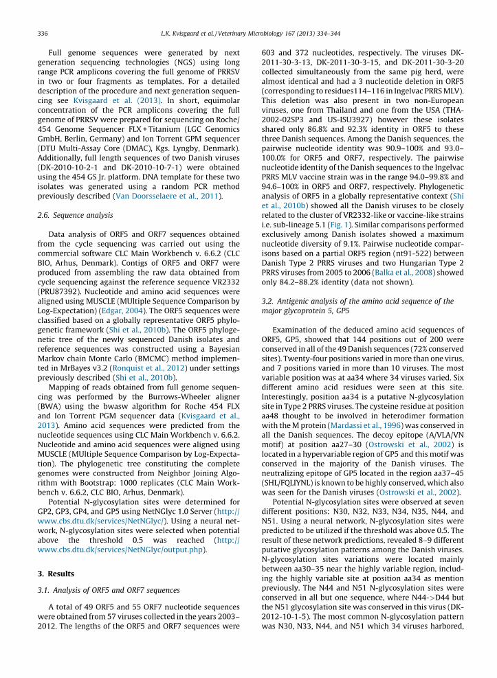
603 and 372 nucleotides, respectively. The viruses DK-2011-30-3-13, DK-2011-30-3-15, and DK-2011-30-3-20collected simultaneously from the same pig herd, werealmost identical and had a 3 nucleotide deletion in ORF5(corresponding to residues114–116 in Ingelvac PRRS MLV).This deletion was also present in two non-Europeanviruses, one from Thailand and one from the USA (THA-2002-02SP3 and US-ISU3927) however these isolatesshared only 86.8% and 92.3% identity in ORF5 to thesethree Danish sequences. Among the Danish sequences, thepairwise nucleotide identity was 90.9–100% and 93.0–100.0% for ORF5 and ORF7, respectively. The pairwisenucleotide identity of the Danish sequences to the IngelvacPRRS MLV vaccine strain was in the range 94.0–99.8% and94.6–100% in ORF5 and ORF7, respectively. Phylogeneticanalysis of ORF5 in a globally representative context (Shiet al., 2010b) showed all the Danish viruses to be closelyrelated to the cluster of VR2332-like or vaccine-like strainsi.e. sub-lineage 5.1 (Fig. 1). Similar comparisons performedexclusively among Danish isolates showed a maximumnucleotide diversity of 9.1%. Pairwise nucleotide compar-isons based on a partial ORF5 region (nt91-522) betweenDanish Type 2 PRRS viruses and two Hungarian Type 2PRRS viruses from 2005 to 2006 (Balka et al., 2008) showedonly 84.2–88.2% identity (data not shown).
3.2. Antigenic analysis of the amino acid sequence of the
major glycoprotein 5, GP5
Examination of the deduced amino acid sequences ofORF5, GP5, showed that 144 positions out of 200 wereconserved in all of the 49 Danish sequences (72% conservedsites). Twenty-four positions varied in more than one virus,and 7 positions varied in more than 10 viruses. The mostvariable position was at aa34 where 34 viruses varied. Sixdifferent amino acid residues were seen at this site.Interestingly, position aa34 is a putative N-glycosylationsite in Type 2 PRRS viruses. The cysteine residue at positionaa48 thought to be involved in heterodimer formationwith the M protein (Mardassi et al., 1996) was conserved inall the Danish sequences. The decoy epitope (A/VLA/VNmotif) at position aa27–30 (Ostrowski et al., 2002) islocated in a hypervariable region of GP5 and this motif wasconserved in the majority of the Danish viruses. Theneutralizing epitope of GP5 located in the region aa37–45(SHL/FQLIYNL) is known to be highly conserved, which alsowas seen for the Danish viruses (Ostrowski et al., 2002).
Potential N-glycosylation sites were observed at sevendifferent positions: N30, N32, N33, N34, N35, N44, andN51. Using a neural network, N-glycosylation sites werepredicted to be utilized if the threshold was above 0.5. Theresult of these network predictions, revealed 8–9 differentputative glycosylation patterns among the Danish viruses.N-glycosylation sites variations were located mainlybetween aa30–35 near the highly variable region, includ-ing the highly variable site at position aa34 as mentionpreviously. The N44 and N51 N-glycosylation sites wereconserved in all but one sequence, where N44->D44 butthe N51 glycosylation site was conserved in this virus (DK-2012-10-1-5). The most common N-glycosylation patternwas N30, N33, N44, and N51 which 34 viruses harbored,
L.K. Kvisgaard et al. / Veterinary Microbiology 167 (2013) 334–344336
however, in 14 of these sequences N30 did not reach thepotential threshold of 0.5 and therefore was not predictedto be N-glycosylated by the neural network.
3.3. Genetic analysis of 11 complete Danish PRRSV Type 2
genomes
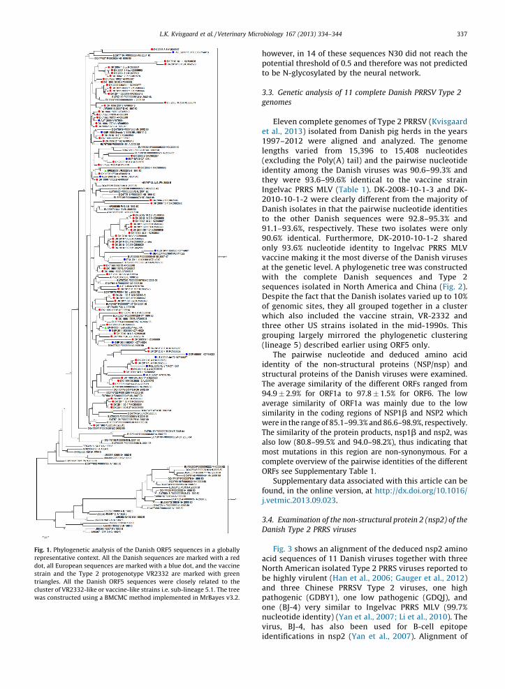
Eleven complete genomes of Type 2 PRRSV (Kvisgaardet al., 2013) isolated from Danish pig herds in the years1997–2012 were aligned and analyzed. The genomelengths varied from 15,396 to 15,408 nucleotides(excluding the Poly(A) tail) and the pairwise nucleotideidentity among the Danish viruses was 90.6–99.3% andthey were 93.6–99.6% identical to the vaccine strainIngelvac PRRS MLV (Table 1). DK-2008-10-1-3 and DK-2010-10-1-2 were clearly different from the majority ofDanish isolates in that the pairwise nucleotide identitiesto the other Danish sequences were 92.8–95.3% and91.1–93.6%, respectively. These two isolates were only90.6% identical. Furthermore, DK-2010-10-1-2 sharedonly 93.6% nucleotide identity to Ingelvac PRRS MLVvaccine making it the most diverse of the Danish virusesat the genetic level. A phylogenetic tree was constructedwith the complete Danish sequences and Type 2sequences isolated in North America and China (Fig. 2).Despite the fact that the Danish isolates varied up to 10%of genomic sites, they all grouped together in a clusterwhich also included the vaccine strain, VR-2332 andthree other US strains isolated in the mid-1990s. Thisgrouping largely mirrored the phylogenetic clustering(lineage 5) described earlier using ORF5 only.
The pairwise nucleotide and deduced amino acididentity of the non-structural proteins (NSP/nsp) andstructural proteins of the Danish viruses were examined.The average similarity of the different ORFs ranged from94.9 � 2.9% for ORF1a to 97.8 � 1.5% for ORF6. The lowaverage similarity of ORF1a was mainly due to the lowsimilarity in the coding regions of NSP1b and NSP2 whichwere in the range of 85.1–99.3% and 86.6–98.9%, respectively.The similarity of the protein products, nsp1b and nsp2, wasalso low (80.8–99.5% and 94.0–98.2%), thus indicating thatmost mutations in this region are non-synonymous. For acomplete overview of the pairwise identities of the differentORFs see Supplementary Table 1.
Supplementary data associated with this article can befound, in the online version, at http://dx.doi.org/10.1016/j.vetmic.2013.09.023.
3.4. Examination of the non-structural protein 2 (nsp2) of the
Danish Type 2 PRRS viruses
Fig. 3 shows an alignment of the deduced nsp2 aminoacid sequences of 11 Danish viruses together with threeNorth American isolated Type 2 PRRS viruses reported tobe highly virulent (Han et al., 2006; Gauger et al., 2012)and three Chinese PRRSV Type 2 viruses, one highpathogenic (GDBY1), one low pathogenic (GDQJ), andone (BJ-4) very similar to Ingelvac PRRS MLV (99.7%nucleotide identity) (Yan et al., 2007; Li et al., 2010). Thevirus, BJ-4, has also been used for B-cell epitopeidentifications in nsp2 (Yan et al., 2007). Alignment of
Fig. 1. Phylogenetic analysis of the Danish ORF5 sequences in a globally
representative context. All the Danish sequences are marked with a red
dot, all European sequences are marked with a blue dot, and the vaccine
strain and the Type 2 protogenotype VR2332 are marked with green
triangles. All the Danish ORF5 sequences were closely related to the
cluster of VR2332-like or vaccine-like strains i.e. sub-lineage 5.1. The tree
was constructed using a BMCMC method implemented in MrBayes v3.2.
L.K. Kvisgaard et al. / Veterinary Microbiology 167 (2013) 334–344 337
the predicted amino acid sequence of nsp2 of the Danishviruses with the vaccine strain revealed that several ofthe Danish viruses harbored deletions resulting invariation of the length of nsp2 from 1174 to 1096 aminoacids residues. One virus harbored a 4 amino aciddeletion following the amino acid at position 793 andfive other viruses harbored a 3 amino acid deletion at theposition corresponding to aa593–595 in nsp2 of IngelvacPRRS MLV. One of the viruses harboring the 3 amino aciddeletion was the virus DK-2010-10-1-2 and this virusalso had a 19 amino acid deletion corresponding to theposition aa498–516 in Ingelvac PRRS MLV nsp2. Thus,this virus encoded the shortest nsp2 of the Danishviruses with an amino acid length of 1174 residues. Allthe deletions observed in the Danish viruses werelocated at the hyper variable region between aa150–850 of nsp2. The nsp2 protein is known to be the mostdiverse protein of all the proteins encoded by PRRSV,nevertheless, the cysteine protease domain (PL2) locatedin the region aa46–146 of nsp2 is known to be conservedwithin the same genotype. Indeed, this region was alsohighly conserved in the Danish viruses with only 17substitution sites out of 101positions while the NorthAmerican and Chinese viruses had only 16 and 9substitution sites, respectively, in comparison to theIngelvac PRRS MLV nsp2 sequence. The amino acidresidues forming the putative catalytic triad C54, D88,and H123 (Han et al., 2009), were conserved in all theDanish viruses as well as in the American and Chineseviruses. All 6 putative B-cell epitopes identified by Yanet al. (2007) are highlighted in Fig. 3. Epitope sites SP1,SP2, and SP4 were highly conserved among the Danishviruses, whereas epitope sites SP5, SP6, and SP8 showeda high proportion of variable sites (60%, 64%, and 69%,respectively).
Comparison of the Danish nsp2 sequences to thevaccine strain showed high similarity for 7 of the viruses(DK-1997-19407B, DK-2004-1-7-Pl, DK-2010-10-2-1, DK-2010-10-4-1, DK-2010-10-7-1, DK-2011-030311-1, andDK-2012-01-11-3) with 96.2–99.3% amino acid identityand lower similarity for 4 viruses (DK-2003-2-3, DK-2004-2-1, DK-2008-2-1, and DK-2010-10-1-2) with amino acididentity in the range of 89.0–92.5%.
3.5. Antigenic analysis of the minor glycoproteins, GP2, GP3,
and GP4
From the pairwise amino acid examinations, GP3 wasshown to be the most diverse structural protein among theDanish Type 2 viruses with amino acid identity of 90.9–99.2%. Linear B-cell epitopes have been identified in all 3minor glycoproteins using Pepscan technologies using thesequence of the virus strain NVSL 97-7895 (de Lima et al.,2006). The GP2 comprised two putative linear B-cellepitopes at amino acid positions aa41–55 and 121–135,respectively. These regions were highly conserved amongthe Danish viruses; however, the epitope located at theregion aa41–55 harbored 4 substitutions sites compared tothe NVSL 97-7895 strain. For GP3, 4 overlapping con-secutive linear B-cell epitopes have been identified (aa61–75, aa71–85, aa81–95, and aa91–105) (de Lima et al.,2006). Even though GP3 was the most diverse structuralprotein among the Danish Type 2 viruses, the 4 epitopesites were all highly conserved with only 8 substitutionsites distributed throughout all 4 epitope regions (aa61–105) when compared to the NVSL 97-7895 strain GP3sequence (Supplementary Fig. 1). The putative epitope sitein GP4 at position aa51–65 was not conserved among theDanish viruses with 10 substitution sites out of a possibleof 15 (Supplementary Fig. 2).
Supplementary data associated with this article can befound, in the online version, at http://dx.doi.org/10.1016/j.vetmic.2013.09.023.
Examination of potential N-glycosylation sites revealed2 putative N-glycosylation sites at N178 and N184 in GP2,7 putative N-glycosylation sites (N29, N42, N50, N131,N152, N160, and N195) in GP3, and 4 putative N-glycosylation sites (N37, N84, N120, and N130) in GP4.The result of nine neural networks predicted all putative N-glycosylation sites in GP2 and GP4 to be N-glycosylatedhowever N37 in GP4 did not score a unanimous vote fromthe jury network to be N-glycosylated. For GP3 all 7putative N-glycosylation sites were predicted to be N-glycosylated in all the Danish viruses except for virus DK-2008-10-1-3 where the potential at this position did notreach the threshold of 0.5. A unanimous vote from the jurypredicted N-glycosylation at N195 however this position is
Table 1
Overview of the 11 Danish Type 2 PRRSV complete genomes.
Virus Accession no. Collection year Clinical signs (herd) Sequencing material Genome lengtha Nt identity%b
DK-1997-19407B KC862576 1997 Stillborn pigletc Marc-145 isolate 15,396 99.4
DK-2003-2-3 KC862584 2003 – Marc-145 isolate 15,408 96.1
DK-2004-2-1 KC862585 2004 – Marc-145 isolate 15,408 96.0
DK-2004-1-7-Pl KC862578 2004 – Marc-145 isolate 15,408 99.4
DK-2008-10-1-3 KC862582 2008 Reproductive failure Lung homogenate 15,408 95.4
DK-2010-10-1-2 KC862579 2010 Respiratory Lung homogenate 15,342 93.6
DK-2010-10-7-1 KC862580 2010 Respiratory Marc-145 isolate 15,399 98.8
DK-2010-10-2-1 KC862581 2010 Respiratory Marc-145 isolate 15,399 99.6
DK-2010-10-4-1 KC862583 2010 Respiratory Lung homogenate 15,399 98.1
DK-2011-030311-1 KC862577 2011 High fever Lung homogenate 15,408 99.3
DK-2012-01-11-3 KC862575 2012 Respiratory/
reproductive failure
Lung homogenate 15,399 98.3
a Excluding the Poly(A) tail.b Pairwise nucleotide identity to Ingelvac PRRS MLV (EF484033).c This virus was obtained from a non-vaccinated herd about 6 months after the Danish vaccination program had started in the fall 1996.
L.K. Kvisgaard et al. / Veterinary Microbiology 167 (2013) 334–344338
believed to be located in the transmembrane region of GP3,hence unlikely to be glycosylated (Dokland, 2010).
The cysteine residues important for protein folding andfunction was 100% conserved in all the minor structuralproteins of the examined Danish viruses.
4. Discussion
We report here for the first time the complete genomesequences obtained from Type 2 PRRS viruses isolated inEurope. With the 11 complete genomes sequenced fromviruses isolated in the years 1997–2012 (Kvisgaard et al.,2013), the evolution of Type 2 PRRSV since its introduction15 years ago in Denmark is now well described. The virusDK-1997-19407B was isolated from a non-vaccinated herdabout 6 months after the Danish PRRSV Type 2 vaccinationprogram first started in the fall of 1996 (Nielsen et al.,2001). The complete genome of this virus was 99.4%identical to the vaccine strain and to two other Type 2viruses isolated in the US in 1995 and 1997, respectively.These findings support epidemiological data indicatingthat Type 2 PRRSV was likely introduced into Denmark bythe MLV vaccine.
The sudden emergence of more pathogenic PRRSviruses in North America (Han et al., 2006; Gauger et al.,2012), Asia (Tian et al., 2007; An et al., 2010) and EasternEurope (Karniychuk et al., 2010; Weesendorp et al., 2013;Morgan et al., 2013) emphasize the importance ofmonitoring the genetic diversity of PRRSV. Despite thisobvious need, there has been only a limited focus on the
genetic diversity of Type 2 PRRSV in Europe (Stadejek et al.,2013). Thus the perception has been that all Type 2 virusesin Europe are closely related to the attenuated vaccinestrain and are only minor contributors to clinical PRRS inEurope. In Hungary, however, where the Ingelvac PRRSMLV vaccine has not been used (Balka et al., 2008), twoType 2 PRRS viruses were isolated in 2005 and 2006 thatshared higher level of identity to a Canadian strain than toany Type 2 MLV strains, indicating that they may havebeen introduced by other sources than by vaccination(Balka et al., 2008). Pairwise nucleotide comparison of thepartial ORF5 sequences of the Hungarian viruses to the 49Danish viruses examined in our study revealed that theyonly shared 84.2–88.2% nucleotide identity and thereforesuggests that they did not originate from Denmark, despitethe use of Danish boar semen in Hungarian herds.
The majority of the 11 complete genomes showed highlevel of similarity to the vaccine strain with pairwisenucleotide identity of 98.1–99.6%. This similarity corre-lated well with the diversity in ORF5 of the Danish viruseswhere all the ORF5 sequences clustered along with thevaccine strain in lineage 5 (Shi et al., 2010b). Interestingly,four viruses from 2003, 2004, 2008, and 2010 showed ahigher level of diversity to the vaccine strain with 93.6–96.1% complete genome similarity, however, they stillclustered within the lineage 5 in the phylogenetic analysis.Based on the ORF5 nucleotide sequences obtained from allDanish viruses isolated between 2003 and 2012, thediversity of the Danish viruses to the vaccine strain was inthe range of 94.0–99.8%, which is a greater range than thediversity of most European Type 2 viruses (>98% similarityto the vaccine strain) (Greiser-Wilke et al., 2010; Stadejeket al., 2013). The diversity among 1400 global Type 2 ORF5sequences retrieved from a database was 3.5% (Shi et al.,2010b) which also is less than the diversity of 9.1% seenamong the Danish ORF5 sequences. The reason for thisdifference is not clear, but it indicate that the selectionpressure on Danish PRRSV strains is different than on otherType 2 isolates maybe because it originated from anattenuated field strain. Most of the Danish samplesincluded in the present study originated from herds withsuspected acute outbreak of PRRSV. It is therefore veryunlikely that these herds vaccinated against PRRSV at thetime of submission, however, there are no reliable makersthat can be used to discriminate between the vaccinestrains and circulating Type 2 field strains.
Notwithstanding insights based on ORF5, the examina-tion of limited genomic regions may not be sufficient for amore complete understanding of PRRSV sequence hetero-geneity, as illustrated by the non-structural protein codingregions NSP1b and NSP2 of the Danish viruses that showeda very broad range of identity to the vaccine strain of 85.1–99.3% and 86.6–98.9%, respectively.
DK-2010-10-1-2 was the virus with the shortestgenome constituting 15,342 nucleotides resulting from adiscontinuous deletion in the coding region of NSP2 of 57plus 9 nucleotides (corresponding to 19 plus 3 amino acidsin the deduced amino acid sequence of nsp2, Fig. 3). Theconsecutive 9 nucleotide deletion was also seen in 4 otherviruses examined in this study. Although some of theseviruses were sequenced from RNA extracted from cell
Fig. 2. Phylogenetic analysis of 11 Danish complete PRRSV Type 2
genomes and 10 complete genomes of viruses isolated in China and North
America. The tree was constructed by the neighbor-joining method using
CLC Main workbench (v. 6.6.2). Bootstrap values were calculated from
1000 replicates and shown on branches. The scale bar represents 15%
nucleotide change. PRRSV Type 1 Lelystad is used as outgroup (set root
above node). CHN = China, DK = Denmark, US = United States of America.
L.K. Kvisgaard et al. / Veterinary Microbiology 167 (2013) 334–344 339
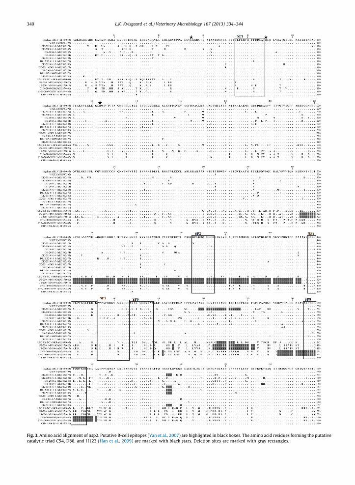
Fig. 3. Amino acid alignment of nsp2. Putative B-cell epitopes (Yan et al., 2007) are highlighted in black boxes. The amino acid residues forming the putative
catalytic triad C54, D88, and H123 (Han et al., 2009) are marked with black stars. Deletion sites are marked with gray rectangles.
L.K. Kvisgaard et al. / Veterinary Microbiology 167 (2013) 334–344340
culture supernatant, the deletion is naturally occurringsince it was also found in partial NSP2 sequences obtainedfrom the corresponding primary material (data notshown). Deletions are often seen in the NSP2 coding partof the PRRSV genome (Ropp et al., 2004; Han et al., 2006),and also in high virulent strains such as the MN184A andMN184B viruses and all the high pathogenic virusesisolated in China during the outbreak in 2006–2009 (An
et al., 2010). Based on these findings, one might presumethat deletions in NSP2 were linked to high virulence of thevirus. However, a low pathogenic virus, GDQJ (GQ374441),isolated in 2008 was shown to harbor the exact samedeletion as the high pathogenic Chinese viruses (Li et al.,2010). Additionally, using chimeric viruses, the NSP2deletion in the 2006 Chinese outbreak of PRRS was shownnot to be linked to virulence (Zhou et al., 2009). In this
Fig. 3. (Continued ).
L.K. Kvisgaard et al. / Veterinary Microbiology 167 (2013) 334–344 341
sense, the deletions found in NSP2 of the Danish virusescan be regarded as an ‘epidemiological marker’ and not as amarker of virulence.
The nsp2 is the viral protein containing the highestfrequency of B-cell epitopes (Oleksiewicz et al., 2001; deLima et al., 2006; Yan et al., 2007) and the humoralantibody response to nsp2 is more pronounced thantoward any other PRRSV protein. However, most of theantibodies are non-neutralizing (Han et al., 2007; Johnsonet al., 2007; Brown et al., 2009). Analysis of the Danishviruses showed that epitope sites SP1, SP2, and SP4identified by Yan et al. (2007) were highly conservedamong the Danish viruses, whereas epitope sites SP5, SP6,and SP8 showed a high proportion of variable sites of 60%,64%, and 69%, respectively (Fig. 3). de Lima et al. (2006),identified 18 B-cell epitopes in nsp2 of a North Americanvirus (NVSL 97-7895, acc. no. AY545985), but none of theseepitopes were conserved in the Danish nsp2 sequencesprobably as a consequence of the high level of amino acidvariation of nsp2 (amino acid comparison of the NorthAmerican nsp2 sequence to the Danish nsp2 sequenceswere 77.5–82.7% identity). The North American virus,NVSL 97-7895 was also used for screening of linear B-cellepitopes in the structural proteins and the epitopesidentified in GP2 and GP3 was highly conserved amongthe Danish viruses and to the North American strainalthough the overall amino acid variation was high in allthree minor glycoproteins compared to the Danish viruses.The B-cell epitope identified in GP4 at position aa51–65were highly variable with 10 substitutions out of 15possible. This fits well with previous findings that thisprotein only reacted with a small fraction of tested post-infection sera (de Lima et al., 2006). Taken together theseresults indicated that some – but not all – epitopes presentin the nonstructural and structural proteins of Type 2PRRSV are prone for changes – which again indicate thatantibodies generated against some epitopes may have noimpact on virus survival. On the other hand, the findingthat some of the epitopes were conserved despite decadesof drifts may actual indicate that conservation of thesesites favor viral survival maybe by directing immunolo-gical attention to sites that are not harmful for the virus.These considerations emphasize that comparisons ofcomplete genomes provides valuable information onconserved and variable regions and thereby can revealnew targets for immune intervention, the design of newvaccines and for the development of diagnostic tests.
The N-glycosylation of GP5 may be critical for properfunctioning of the protein. N-glycosylation, in general, isimportant for correct folding, targeting, and biologicalactivity of proteins (Ansari et al., 2006). In this study,potential N-glycosylation sites were observed at sevendifferent positions in GP5. The result of a neural networkspredictions lead to 8–9 different putative glycosylationpatterns among the Danish viruses, with the N-glycosyla-tion of N30, N33, N44, and N55 the most abundant pattern.However, even though the N-glycosylation motif at N30was present in 14 sequences it was not predicted to be N-glycosylated. The position N30 is located in the C-terminalpart of the putative signal peptide and glycosylation at thisposition may not have any influence on the mature protein,
but may contribute to other biological functions such asimmune evasion. The N-glycosylation at position N44 haspreviously been shown to be important for infectious virusproduction (Ansari et al., 2006) and only one of theglycosylation patterns found among the Danish GP5sequences failed to predict this site to be glycosylated.
Of the 11 complete genomes examined in this study, allof the putative N-glycosylation motifs in the minorglycoproteins GP2, GP3, and GP4 were present andpredicted to be N-glycosylated which strongly indicatesthat the glycosylation of these motifs are important for thefunctioning of the respective proteins.
In conclusion, this study presents for the first time in-depth analysis of complete genomes obtained from Type 2PRRS viruses isolated in Europe. From the completegenomes it was revealed that there was an overall highdiversity between the Danish viruses (90.6–99.3%) and onevirus was only 93.6% similar to the vaccine strain. Theseresults indicate that there has been pronounced geneticdrifts of the Danish Type 2 PRRSVs but the data do notsupport multiple introductions of different Type 2 strainsinto Denmark which fits well with a limited import ofliving animals and semen into Denmark. Lastly, Type 2viruses in Denmark seem to induce more severe clinicalsigns compared to other European countries but sharerelatively close evolutionary relationship in ORF5 to Type 2viruses found in other European countries. This couldindicate that the genetic determinants of viral virulence lieoutside ORF5 and emphasize that the generation ofcomplete genome sequences of European Type 2 isolatesshould be prioritized.
Acknowledgements
The research leading to these results has receivedfunding from the European Union Seventh FrameworkProgramme (FP7/2007–2013) under grant agreement no.245141 (New tools and approaches to control PorcineReproductive and Respiratory Syndrome in the EU and Asia(PoRRSCon) coordinated by Prof. H. Nauwynck) and theCOST Action FA902: Understanding and combating porcinereproductive and respiratory syndrome in Europe (Euro-PRRS.net).
References
Allende, R., Lewis, T.L., Lu, Z., Rock, D.L., Kutish, G.F., Ali, A., Doster, A.R.,Osorio, F.A., 1999. North American and European porcine reproduc-tive and respiratory syndrome viruses differ in non-structural proteincoding regions. J. Gen. Virol. 80 (Pt 2) 307–315.
An, T.Q., Tian, Z.J., Xiao, Y., Li, R., Peng, J.M., Wei, T.C., Zhang, Y., Zhou, Y.J.,Tong, G.Z., 2010. Origin of highly pathogenic porcine reproductive andrespiratory syndrome virus, China. Emerg. Infect. Dis. 16, 365–367.
Ansari, I.H., Kwon, B., Osorio, F.A., Pattnaik, A.K., 2006. Influence of N-linked glycosylation of porcine reproductive and respiratory syn-drome virus GP5 on virus infectivity, antigenicity, and ability toinduce neutralizing antibodies. J. Virol. 80, 3994–4004.
Balka, G., Hornyak, A., Balint, A., Benyeda, Z., Rusvai, M., 2009. Develop-ment of a one-step real-time quantitative PCR assay based on primer-probe energy transfer for the detection of porcine reproductive andrespiratory syndrome virus. J. Virol. Methods 158, 41–45.
Balka, G., Hornyak, A., Balint, A., Kiss, I., Kecskemeti, S., Bakonyi, T., Rusvai,M., 2008. Genetic diversity of porcine reproductive and respiratorysyndrome virus strains circulating in Hungarian swine herds. Vet.Microbiol. 127, 128–135.
L.K. Kvisgaard et al. / Veterinary Microbiology 167 (2013) 334–344342
Botner, A., Strandbygaard, B., Sorensen, K.J., Have, P., Madsen, K.G.,Madsen, E.S., Alexandersen, S., 1997. Appearance of acute PRRS-likesymptoms in sow herds after vaccination with a modified live PRRSvaccine. Vet. Rec. 141, 497–499.
Brown, E., Lawson, S., Welbon, C., Gnanandarajah, J., Li, J., Murtaugh, M.P.,Nelson, E.A., Molina, R.M., Zimmerman, J.J., Rowland, R.R., Fang, Y.,2009. Antibody response to porcine reproductive and respiratorysyndrome virus (PRRSV) nonstructural proteins and implicationsfor diagnostic detection and differentiation of PRRSV types I and II.Clin. Vaccine Immunol. 16, 628–635.
Carlsson, U., Wallgren, P., Renstrom, L.H., Lindberg, A., Eriksson, H.,Thoren, P., Eliasson-Selling, L., Lundeheim, N., Norregard, E., Thorn,C., Elvander, M., 2009. Emergence of porcine reproductive andrespiratory syndrome in Sweden: detection, response and eradica-tion. Transbound. Emerg. Dis. 56, 121–131.
Cavanagh, D., 1997. Nidovirales: a new order comprising Coronaviridaeand Arteriviridae. Arch. Virol. 142, 629–633.
Collins, J.E., Benfield, D.A., Christianson, W.T., Harris, L., Hennings, J.C.,Shaw, D.P., Goyal, S.M., McCullough, S., Morrison, R.B., Joo, H.S., 1992.Isolation of swine infertility and respiratory syndrome virus (isolateATCC VR-2332) in North America and experimental reproduction ofthe disease in gnotobiotic pigs. J. Vet. Diagn. Invest. 4, 117–126.
Conzelmann, K.K., Visser, N., Van Woensel, P., Thiel, H.J., 1993. Molecularcharacterization of porcine reproductive and respiratory syndromevirus, a member of the arterivirus group. Virology 193, 329–339.
de Lima, M., Pattnaik, A.K., Flores, E.F., Osorio, F.A., 2006. Serologic markercandidates identified among B-cell linear epitopes of Nsp2 andstructural proteins of a North American strain of porcine reproductiveand respiratory syndrome virus. Virology 353, 410–421.
Dokland, T., 2010. The structural biology of PRRSV. Virus Res. 154, 86–97.Edgar, R.C., 2004. MUSCLE: multiple sequence alignment with high
accuracy and high throughput. Nucleic Acids Res. 32, 1792–1797.Firth, A.E., Zevenhoven-Dobbe, J.C., Wills, N.M., Go, Y.Y., Balasuriya, U.B.,
Atkins, J.F., Snijder, E.J., Posthuma, C.C., 2011. Discovery of a smallarterivirus gene that overlaps the GP5 coding sequence and is impor-tant for virus production. J. Gen. Virol. 92, 1097–1106.
Gauger, P.C., Faaberg, K.S., Guo, B., Kappes, M.A., Opriessnig, T., 2012.Genetic and phenotypic characterization of a 2006 United Statesporcine reproductive and respiratory virus isolate associated withhigh morbidity and mortality in the field. Virus Res. 163, 98–107.
Greiser-Wilke, I., Fiebig, K., Drexler, C., grosse Beilage, E., 2010. Geneticdiversity of Porcine reproductive and respiratory syndrome virus(PRRSV) in selected herds in a pig-dense region of North-WesternGermany. Vet. Microbiol. 143, 213–223.
Han, J., Liu, G., Wang, Y., Faaberg, K.S., 2007. Identification of nonessentialregions of the nsp2 replicase protein of porcine reproductive andrespiratory syndrome virus strain VR-2332 for replication in cellculture. J. Virol. 81, 9878–9890.
Han, J., Rutherford, M.S., Faaberg, K.S., 2009. The porcine reproductive andrespiratory syndrome virus nsp2 cysteine protease domain possessesboth trans- and cis-cleavage activities. J. Virol. 83, 9449–9463.
Han, J., Wang, Y., Faaberg, K.S., 2006. Complete genome analysis of RFLP184 isolates of porcine reproductive and respiratory syndrome virus.Virus Res. 122, 175–182.
Johnson, C.R., Griggs, T.F., Gnanandarajah, J., Murtaugh, M.P., 2011. Novelstructural protein in porcine reproductive and respiratory syndromevirus encoded by an alternative ORF5 present in all arteriviruses. J.Gen. Virol. 92, 1107–1116.
Johnson, C.R., Yu, W., Murtaugh, M.P., 2007. Cross-reactive antibodyresponses to nsp1 and nsp2 of Porcine reproductive and respiratorysyndrome virus. J. Gen. Virol. 88, 1184–1195.
Karniychuk, U.U., Geldhof, M., Vanhee, M., Van Doorsselaere, J., Saveleva,T.A., Nauwynck, H.J., 2010. Pathogenesis and antigenic characteriza-tion of a new East European subtype 3 porcine reproductive andrespiratory syndrome virus isolate. BMC Vet. Res. 6, 30.
Keffaber, K.K., 1989. Reproductive failure of unknown etiology. Am. Assoc.Swine Pract. Newsl. 1, 1–10.
Key, K.F., Haqshenas, G., Guenette, D.K., Swenson, S.L., Toth, T.E., Meng,X.J., 2001. Genetic variation and phylogenetic analyses of the ORF5gene of acute porcine reproductive and respiratory syndrome virusisolates. Vet. Microbiol. 83, 249–263.
Kim, H.S., Kwang, J., Yoon, I.J., Joo, H.S., Frey, M.L., 1993. Enhancedreplication of porcine reproductive and respiratory syndrome (PRRS)virus in a homogeneous subpopulation of MA-104 cell line. Arch.Virol. 133, 477–483.
Kvisgaard, L.K., Hjulsager, C.K., Fahnoe, U., Breum, S.O., Ait-Ali, T., Larsen,L.E., 2013. A fast and robust method for full genome sequencing ofPorcine Reproductive and Respiratory Syndrome Virus (PRRSV) Type1 and Type 2. J. Virol. Methods 193, 697–705.
Li, Y., Xue, C., Wang, L., Chen, X., Chen, F., Cao, Y., 2010. Genomic analysis oftwo Chinese strains of porcine reproductive and respiratory syn-drome viruses with different virulence. Virus Genes 40, 374–381.
Madsen, K.G., Hansen, C.M., Madsen, E.S., Strandbygaard, B., Botner, A.,Sorensen, K.J., 1998. Sequence analysis of porcine reproductive andrespiratory syndrome virus of the American type collected fromDanish swine herds. Arch. Virol. 143, 1683–1700.
Mardassi, H., Massie, B., Dea, S., 1996. Intracellular synthesis, processing,and transport of proteins encoded by ORFs 5 to 7 of porcine repro-ductive and respiratory syndrome virus. Virology 221, 98–112.
Meulenberg, J.J., Hulst, M.M., de Meijer, E.J., Moonen, P.L., den Besten,A., de Kluyver, E.P., Wensvoort, G., Moormann, R.J., 1993. Lelystadvirus, the causative agent of porcine epidemic abortion andrespiratory syndrome (PEARS), is related to LDV and EAV. Virology192, 62–72.
Morgan, S.B., Graham, S.P., Salguero, F.J., Sanchez Cordon, P.J., Mokhtar, H.,Rebel, J.M., Weesendorp, E., Bodman-Smith, K.B., Steinbach, F., Fros-sard, J.P., 2013. Increased pathogenicity of European porcine repro-ductive and respiratory syndrome virus is associated with enhancedadaptive responses and viral clearance. Vet. Microbiol. 163, 13–22.
Nielsen, H.S., Liu, G., Nielsen, J., Oleksiewicz, M.B., Botner, A., Storgaard, T.,Faaberg, K.S., 2003. Generation of an infectious clone of VR-2332, ahighly virulent North American-type isolate of porcine reproductiveand respiratory syndrome virus. J. Virol. 77, 3702–3711.
Nielsen, H.S., Oleksiewicz, M.B., Forsberg, R., Stadejek, T., Botner, A.,Storgaard, T., 2001. Reversion of a live porcine reproductive andrespiratory syndrome virus vaccine investigated by parallel muta-tions. J. Gen. Virol. 82, 1263–1272.
Oleksiewicz, M.B., Botner, A., Madsen, K.G., Storgaard, T., 1998. Sensitivedetection and typing of porcine reproductive and respiratory syn-drome virus by RT-PCR amplification of whole viral genes. Vet.Microbiol. 64, 7–22.
Oleksiewicz, M.B., Botner, A., Toft, P., Normann, P., Storgaard, T., 2001.Epitope mapping porcine reproductive and respiratory syndromevirus by phage display: the nsp2 fragment of the replicase polyproteincontains a cluster of B-cell epitopes. J. Virol. 75, 3277–3290.
Ostrowski, M., Galeota, J.A., Jar, A.M., Platt, K.B., Osorio, F.A., Lopez, O.J.,2002. Identification of neutralizing and nonneutralizing epitopes inthe porcine reproductive and respiratory syndrome virus GP5 ecto-domain. J. Virol. 76, 4241–4250.
Ronquist, F., Teslenko, M., van der Mark, P., Ayres, D.L., Darling, A., Hohna,S., Larget, B., Liu, L., Suchard, M.A., Huelsenbeck, J.P., 2012. MrBayes3.2: efficient Bayesian phylogenetic inference and model choiceacross a large model space. Syst. Biol. 61, 539–542.
Ropp, S.L., Wees, C.E., Fang, Y., Nelson, E.A., Rossow, K.D., Bien, M., Arndt,B., Preszler, S., Steen, P., Christopher-Hennings, J., Collins, J.E., Ben-field, D.A., Faaberg, K.S., 2004. Characterization of emerging Eur-opean-like porcine reproductive and respiratory syndrome virusisolates in the United States. J. Virol. 78, 3684–3703.
Sanger, F., Nicklen, S., Coulson, A.R., 1977. DNA sequencing with chain-terminating inhibitors. Proc. Natl. Acad. Sci. U.S.A. 74, 5463–5467.
Shi, M., Lam, T.T., Hon, C.C., Hui, R.K., Faaberg, K.S., Wennblom, T.,Murtaugh, M.P., Stadejek, T., Leung, F.C., 2010a. Molecular epidemiol-ogy of PRRSV: a phylogenetic perspective. Virus Res. 154, 7–17.
Shi, M., Lam, T.T., Hon, C.C., Murtaugh, M.P., Davies, P.R., Hui, R.K., Li, J.,Wong, L.T., Yip, C.W., Jiang, J.W., Leung, F.C., 2010b. Phylogeny-basedevolutionary, demographical, and geographical dissection of NorthAmerican type 2 porcine reproductive and respiratory syndromeviruses. J. Virol. 84, 8700–8711.
Stadejek, T., Stankevicius, A., Murtaugh, M.P., Oleksiewicz, M.B., 2013.Molecular evolution of PRRSV in Europe: current state of play. Vet.Microbiol. 165 (July (1–2)) 21–28.
Storgaard, T., Oleksiewicz, M., Botner, A., 1999. Examination of theselective pressures on a live PRRS vaccine virus. Arch. Virol. 144,2389–2401.
Terpstra, C., Wensvoort, G., Pol, J.M., 1991. Experimental reproduction ofporcine epidemic abortion and respiratory syndrome (mystery swinedisease) by infection with Lelystad virus: Koch’s postulates fulfilled.Vet. Q. 13, 131–136.
Tian, K., Yu, X., Zhao, T., Feng, Y., Cao, Z., Wang, C., Hu, Y., Chen, X., Hu,D., Tian, X., Liu, D., Zhang, S., Deng, X., Ding, Y., Yang, L., Zhang, Y.,Xiao, H., Qiao, M., Wang, B., Hou, L., Wang, X., Yang, X., Kang, L.,Sun, M., Jin, P., Wang, S., Kitamura, Y., Yan, J., Gao, G.F., 2007.Emergence of fatal PRRSV variants: unparalleled outbreaks ofatypical PRRS in China and molecular dissection of the uniquehallmark. PLoS ONE 2, e526.
Van Doorsselaere, J., Geldhof, M., Nauwynck, H.J., Delputte, P.L., 2011.Characterization of a circulating PRRSV strain by means of randomPCR cloning and full genome sequencing. Virol. J. 8, 160.
L.K. Kvisgaard et al. / Veterinary Microbiology 167 (2013) 334–344 343
Weesendorp, E., Morgan, S., Stockhofe-Zurwieden, N., Popma-De Graaf,D.J., Graham, S.P., Rebel, J.M., 2013. Comparative analysis of immuneresponses following experimental infection of pigs with Europeanporcine reproductive and respiratory syndrome virus strains of dif-fering virulence. Vet. Microbiol. 163, 1–12.
Wensvoort, G., Terpstra, C., Pol, J.M., ter Laak, E.A., Bloemraad, M., deKluyver, E.P., Kragten, C., van Buiten, L., den Besten, A., Wagenaar, F.,1991. Mystery swine disease in The Netherlands: the isolation ofLelystad virus. Vet. Q. 13, 121–130.
Wu, W.H., Fang, Y., Farwell, R., Steffen-Bien, M., Rowland, R.R., Christo-pher-Hennings, J., Nelson, E.A., 2001. A 10-kDa structural protein of
porcine reproductive and respiratory syndrome virus encoded byORF2b. Virology 287, 183–191.
Yan, Y., Guo, X., Ge, X., Chen, Y., Cha, Z., Yang, H., 2007. Monoclonalantibody and porcine antisera recognized B-cell epitopes of Nsp2protein of a Chinese strain of porcine reproductive and respiratorysyndrome virus. Virus Res. 126, 207–215.
Zhou, L., Zhang, J., Zeng, J., Yin, S., Li, Y., Zheng, L., Guo, X., Ge, X.,Yang, H., 2009. The 30-amino-acid deletion in the Nsp2 ofhighly pathogenic porcine reproductive and respiratory syndromevirus emerging in China is not related to its virulence. J. Virol. 83,5156–5167.
L.K. Kvisgaard et al. / Veterinary Microbiology 167 (2013) 334–344344