Embed Size (px)
Citation preview

RESEARCH Open Access
Genetic deletion of muscle RANK orselective inhibition of RANKL is not aseffective as full-length OPG-fc in mitigatingmuscular dystrophySébastien S. Dufresne1, Antoine Boulanger-Piette1, Sabrina Bossé1, Anteneh Argaw1, Dounia Hamoudi1,Laetitia Marcadet1, Daniel Gamu2, Val A. Fajardo2, Hideo Yagita3, Josef M. Penninger4, A. Russell Tupling2
and Jérôme Frenette1,5*
Abstract
Although there is a strong association between osteoporosis and skeletal muscle atrophy/dysfunction, the functionalrelevance of a particular biological pathway that regulates synchronously bone and skeletal muscle physiopathology isstill elusive. Receptor-activator of nuclear factor κB (RANK), its ligand RANKL and the soluble decoy receptorosteoprotegerin (OPG) are the key regulators of osteoclast differentiation and bone remodelling. We thus hypothesizedthat RANK/RANKL/OPG, which is a key pathway for bone regulation, is involved in Duchenne muscular dystrophy (DMD)physiopathology. Our results show that muscle-specific RANK deletion (mdx-RANKmko) in dystrophin deficient mdx miceimproves significantly specific force [54% gain in force] of EDL muscles with no protective effect against eccentriccontraction-induced muscle dysfunction. In contrast, full-length OPG-Fc injections restore the force of dystrophicEDL muscles [162% gain in force], protect against eccentric contraction-induced muscle dysfunction ex vivo andsignificantly improve functional performance on downhill treadmill and post-exercise physical activity. Since OPGserves a soluble receptor for RANKL and as a decoy receptor for TRAIL, mdx mice were injected with anti-RANKLand anti-TRAIL antibodies to decipher the dual function of OPG. Injections of anti-RANKL and/or anti-TRAIL increasesignificantly the force of dystrophic EDL muscle [45% and 17% gains in force, respectively]. In agreement, truncatedOPG-Fc that contains only RANKL domains produces similar gains, in terms of force production, than anti-RANKLtreatments. To corroborate that full-length OPG-Fc also acts independently of RANK/RANKL pathway, dystrophin/RANKdouble-deficient mice were treated with full-length OPG-Fc for 10 days. Dystrophic EDL muscles exhibited a significantgain in force relative to untreated dystrophin/RANK double-deficient mice, indicating that the effect of full-lengthOPG-Fc is in part independent of the RANKL/RANK interaction. The sarco/endoplasmic reticulum Ca2+ ATPase (SERCA)activity is significantly depressed in dysfunctional and dystrophic muscles and full-length OPG-Fc treatment increasedSERCA activity and SERCA-2a expression. These findings demonstrate the superiority of full-length OPG-Fc treatmentrelative to truncated OPG-Fc, anti-RANKL, anti-TRAIL or muscle RANK deletion in improving dystrophic muscle function,(Continued on next page)
* Correspondence: [email protected] Hospitalier Universitaire de Québec–Centre de Recherche du CentreHospitalier de l’Université Laval (CHUQ-CRCHUL), Université Laval, 2705boulevard Laurier, RC-9500, Quebec City, QC G1V 4G2, Canada5Département de Réadaptation, Faculté de Médecine, Université Laval,Quebec City, QC G1V 4G2, CanadaFull list of author information is available at the end of the article
© The Author(s). 2018 Open Access This article is distributed under the terms of the Creative Commons Attribution 4.0International License (http://creativecommons.org/licenses/by/4.0/), which permits unrestricted use, distribution, andreproduction in any medium, provided you give appropriate credit to the original author(s) and the source, provide a link tothe Creative Commons license, and indicate if changes were made. The Creative Commons Public Domain Dedication waiver(http://creativecommons.org/publicdomain/zero/1.0/) applies to the data made available in this article, unless otherwise stated.
Dufresne et al. Acta Neuropathologica Communications (2018) 6:31 https://doi.org/10.1186/s40478-018-0533-1

(Continued from previous page)
integrity and protection against eccentric contractions. In conclusion, full-length OPG-Fc represents an efficientalternative in the development of new treatments for muscular dystrophy in which a single therapeutic approachmay be foreseeable to maintain both bone and skeletal muscle functions.
Keywords: Osteoprotegerin, Skeletal muscle, SERCA, Duchenne muscular dystrophy
IntroductionBone and muscle have the ability to adjust their struc-tures to meet their mechanical, hormonal, and metabolicenvironments. Osteoporosis and muscle atrophy/dys-function occur simultaneously in a number of condi-tions, including spaceflight, extended bed rest, andseveral muscular and neuromuscular diseases. Local andsystemic alterations in hormone and pro-inflammatorycytokine levels stimulate muscle and bone atrophy [25,32]. Changes in intracellular Ca2+ concentrationsregulate the physiological activities and expression ofspecific bone and muscle genes [15, 30]. Poor bonehealth and increased incidence of bone factures are wellrecognized clinically in Duchenne muscular dystrophy(DMD) patients suggesting cross-talks and mutual co-operative interactions between bones and dystrophicmuscles [11]. However, the potential cellular andmolecular mechanisms that may tie together bones andskeletal muscles during physiological and pathologicalconditions remain elusive.The receptor-activator of nuclear factor κB ligand
(RANKL), the membrane receptor RANK, and the sol-uble decoy receptor osteoprotegerin (OPG) are membersof the tumor necrosis factor (TNF) superfamily that reg-ulates bone remodelling [19, 27]. RANKL is expressedby osteoblasts, while RANK, its receptor, is expressed bypre-osteoclastic cells. The RANK/RANKL interaction in-duces the formation of multinucleated mature osteo-clasts, ultimately causing bone resorption [21]. OPG, thethird protagonist, is also produced by osteoblasts, bindsto RANKL and exerts an inhibitory effect on the pre-osteoclastic differentiation process [2]. Structurally, thenative OPG protein is highly conserved and containsfour TNFR-like domains (RANKL binding sites), twodeath domains (tumour necrosis factor-relatedapoptosis-inducing ligand [TRAIL binding sites]), and aheparin-binding domain [31]. Thus, OPG serves as adecoy receptor for the RANKL and TRAIL and is a veryefficient anti-resorptive and anti-apoptotic agent [3].The focus of research in our laboratory is to decipher
the potential cellular and molecular mechanisms thatmay tie together bones and skeletal muscles duringphysiological and pathological conditions. We first hy-pothesized that RANK/RANKL/OPG pathway, a keyregulator of bone homeostasis and Ca2+ storage, wouldcontribute in the regulation of skeletal muscle integrity
and function during the course of muscular dystrophy.We previously demonstrated that daily full-length OPG-Fc treatment markedly improved muscle function andintegrity in 5 week-old mdx mice [12]. The main object-ive of this study was to determine the specific contribu-tion of muscle RANK, RANKL and TRAIL in musculardystrophy. Using genetic and pharmacological ap-proaches in young and adult dystrophic mice, we areable to show the unequivocal superior effects of full-length OPG-Fc in rescuing dystrophic muscles relativeto selective muscle RANK deletion or anti-RANKL oranti-TRAIL treatments. Altogether, our results suggestthat full-length OPG-Fc is a multifunctional protein thathas the potential to impact on several different cellularprocesses with possibly profound implications for thetreatment of DMD.
Materials and methodsAnimalsMice carrying the RANKfloxed or RANKdel alleles andmuscle creatine kinase-cre (mck-cre) mice were back-crossed five times to a C57BL/6 background before gen-erating the mck-cre RANKdel/floxed (RANKmko) mice aspreviously described [13, 20]. Male wild-type (C57BL/6)and mdx dystrophic mice (C57BL/10ScSn-Dmdmdx/J)were purchased from the Jackson Laboratory (BarHarbor, ME, USA) and bred at our animal facility.RANKmko mice were also crossed with mdx-backgroundmice to generate double deficient mice (dystrophin andRANK). Mice were screened for the desired genotype byPCR analysis. PCR products were amplified using primerpairs as listed in Additional file 1: Table S2. Dystrophicmdx mice were injected daily with full-length OPG-Fc[12] [i.p., 1 mg/kg/d R&D systems, MN, USA], PBS,anti-RANKL [39] [1 mg/kg/ every 3 d, clone IK22–5],anti-TRAIL [22] [1 mg/kg/every 3 d, clone H2B2] ortruncated OPG-Fc [1 mg/kg/d, Syd Labs, MA, USA]from days 25 to 35 after birth. In another set of experi-ments, five six-month old mdx mice were injected daily,for 10 d, with full-length OPG-Fc [i.p. 1 mg/kg/d]followed by a downhill (eccentric) treadmill runningprotocol. C57BL/6 mice were used as a control andinjected daily with the same volume of phosphate-buffered saline (vehicle). At the end of the experimentalprocedures, mice were euthanized by cervical dislocationunder anesthesia and skeletal muscles [extensor
Dufresne et al. Acta Neuropathologica Communications (2018) 6:31 Page 2 of 10

digitorum longus (EDL), soleus (Sol) and diaphragm(Dia)] were removed and stored at − 80 °C for futureanalysis. All procedures were approved by the UniversitéLaval Research Center Animal Care and Use Committee,based on the Canadian Council on Animal Care guide-lines. All data generated or analysed during this study areincluded in this published article and its Additional file 1.
Immunofluorescence and stainingTransverse EDL muscle sections (10 μm) were cut (LeicaMicrosystems CM1850, Nussloch, Germany) in dupli-cate from the proximal and distal parts of the muscles.Tissue sections stained with hematoxylin and eosin(Sigma-Aldrich, St. Louis, MO, USA) were examinedwith an inverted microscope (Nikon, Ontario, Canada)and damage, regenerating and intact areas on approxi-mately 100 myofibers per muscle were quantified withImageJ software version 1.41 (National Institutes ofHealth, USA). The damaged area was defined as an areanot occupied by normal or regenerating muscle fibers.Image series of EDL were taken using a confocal micro-scope (Axio Observer.Z1; Carl Zeiss, Germany) and ac-quired using a Quorum WaveFX spinning disc confocalsystem (Quorum Technologies, Ontario, Canada). Solidstate laser lines 491 nm and 561 nm were used for exci-tation of green and red (Alexa-488 and Alexa-594), com-bined with appropriate BrightLine single-bandpassemission filters (536/40 nm and 624/40 nm, Semrock,NY, USA). z-series were acquired at the same time withDAPI fluorescence filter cube (Chroma Technology, VT,USA). The CCD camera used to capture the images wasa Hamamatsu Image EM C-9100. Images were acquiredand analyzed using Volocity software, version 4.2.1. It-erative restoration (deconvolution) was applied for theDAPI channel, using the same software.
Western blots and qPCRSkeletal muscles were homogenized in a lysis buffer con-taining 1 μg/ml, protease inhibitor cocktail (P8340; Sigma-Aldrich, Ontario, Canada), 20 mM Tris-base pH 7.5,140 mM NaCl, 1 mM MgCl2, 1 mM CaCl2, 10% glycerol,1% Igepal (Sigma-Aldrich, Ontario, Canada), 2 mMNa3VO4, 8.3 mM NaF and 0.2 mM PMSF. The proteincontent of the supernatant was measured using BCA pro-tein assay kit (EMD chemical, Nussloch Germany). Proteinhomogenates were electrophoretically separated on SDS-polyacrylamide gels, transferred to polyvinylidene difluoridemembranes (PVDF; Bio-Rad, CA, USA), blocked in 5%skim milk and incubated overnight at 4°C with the follow-ing primary antibodies (all from Santa Cruz Biotechnology):anti-SERCA-1a, anti-SERCA-2a anti-RANK and anti-GAPDH. The membranes were washed and incubated withappropriate HRP-conjugated secondary antibodies (SantaCruz Biotechnology, CA, USA). Bands were revealed using
the ECL-Plus chemiluminescent detection system (Perkin-Elmer, MA, USA). Images of the membranes were ac-quired, scanned, and analyzed using Quantity One software(v4.6.6, Bio-Rad). For RT-PCR analysis (Additional file 1:Table S2), skeletal muscles were rapidly put in RNAlaterRNA Stabilization Reagent (Qiagen, MD, USA). Total RNAwas isolated using the RNeasy Fibrous Tissue Mini Kit(Qiagen, MD, USA) according to manufacturer’s instruc-tions. During isolation process, RNA samples were treatedwith RNase-Free DNase Set (Qiagen, MD, USA). RNAquality was assessed using an Agilent 2100 Bioanalyzer(Agilent Technologies, CA, USA) and quantified using aNanoDrop 1000 Spectrophotometer (NanoDrop Technolo-gies, DE, USA). The expression of the RANK gene in eachsample was compared to the housekeeping gene GAPDH.Measurements were performed in duplicate for each stand-ard and muscle sample.
Contractile propertiesMice were injected with buprenorphine (i.p. 0.1 mg/kg)and were anaesthetized with pentobarbital sodium (i.p.50 mg/kg) 15 min later. The right Sol and EDL muscleswere resected and attached to a 305B-LR dual-mode leverarm system controlled by dynamic muscle control unitand data acquisition software (Aurora Scientific, Aurora,ON, Canada) as described by Dufresne et al. (2016) [13].For the eccentric contraction protocol, the muscles wereset at optimal L0 and stimulated at 150 Hz for 700 ms.Five hundred ms into the stimulation protocol, the mus-cles were lengthened to 10% of L0 at 0.5 L0/s for 200 ms.
Downhill running protocolSix month-old C57BL/6 and mdx mice were treated for10 days with PBS (n = 12) or full-length OPG-Fc [1 mg/kg/d] (n = 12). From days 7 to 9, mice were trained foracclimatization on an horizontal (0% grade) motorizedtreadmill at 6, 8 and then 10 m/min for 5 min. Followingthe training protocol, mice ran on a downhill sloped(14-degree decline) at 10 m/min for 45 min. Mice werecontinually observed during the running protocol.Exhausted mice showing physical signs of discomfortwere rested for 2 min. The running protocol was discon-tinued after 3 stops. Following the eccentric protocol,post-exercise mouse activity was measured by videotracking software in an open field for 24 h.
SERCA activityFrozen EDL, Sol and Dia muscles were homogenizedwith a ground-glass pestle in 5 volumes of 10 mM Tris/HCl (pH 8.3) supplemented with 0.3 M sucrose. SERCAactivity was measured in A. R. Tupling’s laboratory withan assay adapted for a spectrophotometric plate readeraccording to Duhamel and colleagues (2007) [14]
Dufresne et al. Acta Neuropathologica Communications (2018) 6:31 Page 3 of 10

following the oxidation of NADH at 340 nm in assaybuffer (pH 7.0) containing 1 mM EGTA, 10 mM phos-phoenolpyruvate, 18 U/mL pyruvate kinase and lactatedehydrogenase, 0.2 mM NADH, 20 mM HEPES,200 mM KCl, 15 mM MgCl2, 10 mM NaN3, and 5 mMATP. The homogenized muscles and assay buffer wereadded to tubes containing 15 different concentrations ofCa2+ (between 7.6 and 4.7 pCa units) in the presenceand absence of ionophore A23187 (4.2 μM). Theabsence of the ionophore causes back-inhibition ofSERCA pumps. Then 0.3 mM NADH was added to startthe reaction and the plate was read at a wavelength of340 nm for 30 min at 37 °C. The different concentra-tions of Ca2+ were used to determine the maximalenzyme activity (Vmax) and pCa50. Lastly, cyclopiazonic
acid (CPA; 40 μM), a highly specific SERCA inhibitor,was used to determine background activity.
Statistical analysesAll values are expressed as means +/− SEM. The datawere analyzed with Student’s t-test or one-way ANOVAwith Tukey post hoc test (InStat). The levels of signifi-cance was set at *p < 0.05, **p < 0.01, and ***p < 0.001 forPBS-treated mdx.
Results and discussionOPG-fc is superior to muscle-specific RANK deletion inmitigating muscular dystrophyWe previously showed that RANK is expressed in fullydifferentiated mouse myotubes, but not in proliferating
Fig. 1 RANK expression in dystrophic skeletal muscles. a A quantitative real-time PCR analysis revealed that muscle RANK expression is increasedby 5.5-fold in dystrophic EDL muscles relative to control. b Confocal images demonstrating the colocalization of dystrophin (green) and RANK(red) at the basal lamina in EDL muscles from C57BL/6mice. As expected, dystrophin is not expressed in EDL muscles from mdxmice while dystrophin andRANK proteins are absent in EDL muscles from mdx-RANKmko mice. H&E staining revealed that EDL muscles from mdx-RANKmko mice had a lesser numberof irregular fiber sizes and accumulation of perimysial connective tissue compared with mdx-RANKf/f muscles. Data are shown as mean +/−s.e.m. * Significantly different from control C57 EDL muscles, p < 0.05, n = 3, Student’s t-test. Scale bar = 100 μm
Dufresne et al. Acta Neuropathologica Communications (2018) 6:31 Page 4 of 10

C2C12 myoblasts [13]. Interestingly, RANK mRNA was5.5 fold higher in EDL muscles from dystrophic mdxmice relative to C57BL/6 mice (Fig. 1a). To exploreRANK’s contribution in dystrophic skeletal muscle andcompare it with OPG-Fc-mediated RANKL inhibition,we crossed dystrophic mdx mice with muscle-specificRANK deletion, thereafter named mdx-RANKmko. Mdx-RANKfloxed/floxed mice that do not carry the Crerecombinase, thereafter named mdx-RANKf/f whichappeared indistinguishable from mdx mice served aslittermate controls. Immunohistochemistry resultsconfirmed the presence of dystrophin in EDL musclesfrom C57BL/6 mice and RANK protein on themembrane of skeletal muscle fibers from C57BL/6 and
mdx mice. Dystrophin and RANK proteins were notdetected in skeletal muscles from mdx-RANKmko mice(Fig. 1b). We tested whether muscle-specific RANK de-letion improves structural integrity and muscle function.Hematoxylin/eosin staining showed that muscle RANKdeletion partially preserved muscle integrity in dys-trophin/RANK double-deficient mice (Fig. 1b andAdditional file 1: Figure S1). The specific and absoluteforce production of Sol, EDL and Dia muscles from mdxand mdx-RANKf/f mice were similar and significantlylower than C57BL/6 mice (Fig. 2a and Additional file 1:Figure S2). Muscle-specific RANK deletion significantlyincreased the specific and absolute forces of dystrophicEDL muscles by 87% and 54%, respectively, when
Fig. 2 Genetic deletion of muscle RANK or selective inhibitions of RANKL or TRAIL in dystrophic mice. a Maximum specific force analysis of theSol, EDL and Dia muscles were performed on 5-week-old C57BL/6, mdx-RANKmko, mdx-RANKf/f or mdx mice treated from days 25 to 35 withvehicle (PBS) or full-length OPG-Fc [1 mg/kg/d]. Uninjured muscles from C57BL/6 mice were used as controls. Full-length OPG-Fc [1 mg/kg/d]treatments and to a lesser extent, muscle-specific RANK deletion significantly preserved muscle force. b Schematic representations of full-lengthOPG-Fc, truncated OPG-Fc, RANKL and TRAIL antibodies. c Contractile properties of EDL muscles were performed on 5-week-old mdx mice treatedfrom days 25 to 35 with anti-RANKL or/and anti-TRAIL antibodies or truncated OPG-Fc. The inhibition of RANKL and TRAIL for 10 days increasedthe force production of dystrophic EDL muscles by 45% and 17% respectively. The truncated OPG-Fc had similar effects than anti-RANKL, increasingthe force production of EDL muscles by 43%, which were markedly lower than that of dystrophic EDL muscles from full-length OPG-Fc treated mdxmice (+ 162%). d To explore whether full-length OPG-Fc acts independently of RANKL/RANK, mdx-RANKmko mice were treated from days 25 to 35 withfull-length OPG-Fc [1 mg/kg/d], and showed additional gain in force relative to PBS-treated mdx-RANKmko mice. The dotted line is the visual representationof full-length OPG-Fc data. Data are shown as mean +/− s.e.m., one way ANOVA and Tukey’s post-hoc tests; significantly different from PBS-treated mdxmouse; * p< 0.05, ** p< 0.01, *** p< 0.001
Dufresne et al. Acta Neuropathologica Communications (2018) 6:31 Page 5 of 10

compared to the PBS-treated mdx mice (Fig. 2a andAdditional file 1: Figure S2). Although to a lower extentthan dystrophic EDL muscles, specific and absoluteforces of Sol and Dia muscles were also improved inmdx-RANKmko relative to mdx-RANKf/f mice (Fig. 2aand Additional file 1: Figure S2). Similar gains in forcewere observed in 15 month-old mdx-RANKmko relativeto mdx-RANKf/f mice (Additional file 1: Figure S3).Hematoxylin/eosin staining confirmed that muscleintegrity was strongly preserved in the full-length OPG-treated mdx mice (Additional file 1: Figure S4). Surpris-ingly, OPG-Fc almost completely restored EDL muscleforce to wild type levels and was significantly more ef-fective than muscle RANK deletion during the most se-vere phase of muscle degeneration in mdx mice (Fig. 2aand Additional file 1: Figure S2). Given the high respon-siveness of the EDL muscle to OPG-Fc treatment andthat fast-twitch muscles are vulnerable to DMD, wechose to use this muscle for the remaining of the study.Since full-length OPG-Fc was significantly more effect-
ive compared to muscle-specific RANK deletion in pre-serving muscle function during the most severe phase ofdegeneration in mdx mice, we next focused to determinethe contribution of the different OPG domains to the
preservation of muscle integrity (Fig. 2b). OPG is a sol-uble receptor for RANKL and a decoy receptor forTRAIL increasing cell survival by blocking the pro-apoptotic effects of the TRAIL/DR4–5 interaction [16,33]. To determine the dual function of OPG and the im-plication of the RANKL and TRAIL domains, mdx micewere injected with anti-RANKL and anti-TRAIL anti-bodies (Fig. 2b). The inhibition of RANKL and TRAILfor 10 days increased the specific force production ofdystrophic EDL muscles by 45% and 17%, respectively,which are markedly lower than full-length OPG-Fctreated mdx mice (162%) (Fig. 2c). Furthermore, thecombination of anti-RANKL and anti-TRAIL was notsuperior to anti-RANKL treatment alone (Fig. 2c). Toconfirm the exclusive efficiency of full-length OPG-Fc,mdx mice were also treated with the truncated form ofOPG-Fc that carries RANKL-binding domains (Fig. 2c).As expected, the truncated OPG-Fc had similar effectsto anti-RANKL treatment, increasing by 48% the forceproduction of dystrophic EDL muscles (Fig. 2c). To cor-roborate whether full-length OPG-Fc also acts independ-ently of RANK/RANKL pathway, dystrophin/RANKdouble-deficient mice were treated with full-lengthOPG-Fc for 10 days. Dystrophic EDL muscles exhibited
Fig. 3 Full-length OPG-Fc, but not muscle RANK deletion, prevents eccentric contraction-induced muscle dysfunction. a Ex vivo eccentric contractionprotocol of EDL muscles were performed on 5-week-old C57BL/6, mdx-RANKmko and mdx mice treated from days 25 to 35 with vehicle (PBS) orfull-length OPG-Fc [1 mg/kg/d]. Full-length OPG-Fc, but not muscle RANK deletion, significantly prevented the loss of force following repetitiveeccentric contractions. Functional performance was assessed on 5–6-month-old mdx mice treated with vehicle (PBS) or full-length OPG-Fc[1 mg/kg/d] for 10 days prior downhill running. b Only 10% of the PBS-treated versus 75% of the full-length OPG-Fc treated mdx mice wereable to complete the downhill running protocol at a speed of 10 m/s for 45 min. c The total distance travelled at 10 m/min for 45 min (450 m) for eachexperimental group. d The full-length OPG-Fc treatment rescued the voluntary cage activity post- exhausting eccentric downhill running. Data areshown as mean values +/− s.e.m.; one way ANOVA and Tukey’s post-hoc; significantly different at (a) each contraction or (b-d) each time point fromPBS-treated mdx mouse; * p < 0.05, ** p < 0.01, *** p < 0.001
Dufresne et al. Acta Neuropathologica Communications (2018) 6:31 Page 6 of 10
a significant gain in force (29%) relative to untreateddystrophin/RANK double-deficient mice, indicating thatfull-length OPG-Fc can act in part independently of theRANKL/RANK interaction (Fig. 2d). All treatments andmuscle RANK deletion did not change muscle mass(Additional file 1: Table S1). Our results show that anti-RANKL, anti-TRAIL, truncated OPG-Fc treatments orRANK deletion are much less effective than full-lengthOPG-Fc against muscular dystrophy (Additional file 1:Figure S5).
Full length OPG-fc, but not muscle RANK deletion, preventseccentric contraction-induced muscle dysfunction.Dystrophic muscles are vulnerable to repetitive eccen-tric contractions [6, 17] and as opposed to muscleRANK deletion, full-length OPG-Fc positively improvedEDL muscle resistance to repeated eccentric contrac-tions (Fig. 3a), conferring a role for full-length OPG-Fc
that is beyond the inhibition of RANKL/RANK interac-tions. To test whether the full-length OPG-Fc treat-ment provides a similar protection in vivo during aphysiological eccentric protocol, PBS- and OPG-Fctreated adult mdx mice were subjected to a 45-mindownhill eccentric running protocol. Ten percent ofPBS-treated versus 75% of the full-length OPG-Fctreated dystrophic mice were able to complete the en-tire downhill running protocol (Fig. 3b). The first stopfor the PBS-treated mdx mice occurred on average after17 min of downhill running, while full-length OPG-Fctreated mdx mice stopped for the first time after32 min (Additional file 1: Figure S6a). The PBS andfull-length OPG-Fc treated mice that failed to completethe eccentric protocol did, respectively, 63% and 91% ofthe expected distance (Additional file 1: Figure S6b).The total distance travelled for the PBS-treated mdxmice was 282 m, while full-length OPG-Fc treated mdx
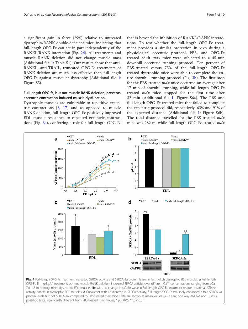
Fig. 4 Full-length OPG-Fc treatment increased SERCA activity and SERCA-2a protein levels in fast-twitch dystrophic EDL muscles. a Full-lengthOPG-Fc [1 mg/kg/d] treatment, but not muscle RANK deletion, increased SERCA activity over different Ca2+ concentrations ranging from pCa7,0–4,5 in homogenized dystrophic EDL muscles (b) with no change in pCa50 value. c Full-length OPG-Fc treatment rescued maximal ATPaseactivity (Vmax) in dystrophic EDL muscles. d Consistent with an increase in SERCA activity, full-length OPG-Fc markedly enhanced 6-fold SERCA-2aprotein levels but not SERCA-1a, compared to PBS-treated mdx mice. Data are shown as mean values +/− s.e.m.; one way ANOVA and Tukey’spost-hoc tests; significantly different from PBS-treated mdx mouse; * p < 0.05, ** p < 0.01
Dufresne et al. Acta Neuropathologica Communications (2018) 6:31 Page 7 of 10
mice completed 409 m (Fig. 3c). Following the strenu-ous eccentric protocol, mouse voluntary activity wasmeasured by video-tracking software for 24 h. Thefull-length OPG-Fc treatment enhanced cage activityby roughly 50% at any given time point during the24 h period (Fig. 3d and Additional file 1: Figure S7).Thus, our functional ex vivo and in vivo experimentsprovide physiological evidence that full-length OPG-Fc is very effective in protecting young and adult dys-trophic mice against eccentric contraction-inducedmuscle dysfunction.
Full-length OPG-fc, but not muscle-specific RANK deletion,increases SERCA activity and expression in dystrophic EDLmusclesWe previously showed that muscle RANK is importantin maintaining SERCA activity [13]. Since SERCA over-expression in skeletal muscles reduces susceptibility toeccentric contraction-induced muscle damage in dys-trophin and sarcoglycan-null mice and given that intrin-sic laryngeal muscles that overexpress SERCA areprotected against muscular dystrophy [18, 29], we testedwhether full-length OPG-Fc injections for 10 days mightalso enhance SERCA activity and SERCA-1a andSERCA-2a protein levels in fast-twitch dystrophic EDLmuscles. Maximal SERCA activity is significantly de-pressed in dystrophic EDL muscles and full-lengthOPG-Fc treatment restored almost completely its activ-ity (Fig. 4a-c). The protein levels of fast-twitch SERCA-1a were diminished in dystrophic EDL muscles and full-length OPG-Fc treatment selectively increased by 6-foldthe expression of the slow-twitch SERCA-2a. (Fig. 4d).However, muscle-specific RANK deletion did not in-crease maximal SERCA activity in dystrophic EDL, Soland Dia muscles (Fig. 4a-c and Additional file 1: FigureS8a-f ), providing additional evidence that full-lengthOPG-Fc could act through alternative pathways andmay potentially rescue Ca2+ cycling/homeostasisthrough SERCA-2a dependent mechanism.
Perspective and conclusionIn the early 2000s, truncated OPG-Fc (AMGN-0007)reached clinical trial for the treatment of osteoporosisand bone metastasis [5, 7]. Truncated and full-lengthOPG-Fc can interact with RANKL preventing the down-stream activation of NF-kB, a key controller of manygenes involved in inflammation. Our observations thatmuscle-specific RANK deletion, anti-RANKL or trun-cated OPG-Fc and full-length OPG-Fc treatment protectfast-twitch fibers are of paramount importance, sincethese powerful fibers are the first to disappear in manyforms of myopathies [28, 38]. However, we uncover aunique and superior role for full-length OPG-Fc in pro-tecting muscle function and integrity in the mdx model
of muscular dystrophy. Since full-length OPG-Fc rescuesSERCA activity in fast-twitch dystrophic skeletal mus-cles, we anticipate that full-length OPG-Fc treatmentwould contribute to normalize SR Ca2+ regulation inmuscular dystrophy by removing Ca2+ from themyoplasm and refilling the internal Ca2+ stores throughthe action of SERCA pumps. The stimulation of SERCApumps should lead to better Ca2+ mobilization breakingthe vicious cycle of muscle inflammation and Ca2+-dependent protease activation initiated by poor Ca2+
handling [9, 35].Current investigations are oriented toward the various
roles of full-length OPG-Fc. Bone, tumor cells, inflam-matory cells and vascular cells have given some insighton how full-length OPG-Fc may work to some extent in-dependently of RANKL inhibition in muscular dystrophy[4]. Native OPG possesses a heparin-binding domainthat may interact with various glycoaminoglycans andproteoglycans and ultimately integrins [23, 24, 26, 34,36, 40]. In addition, heparan-sulfate proteoglycans areabundant in skeletal muscles [8], increases in DMD [1]and may represent an additional target for full-lengthOPG-Fc [10]. On the other hand, it was recently foundthat integrin-linked kinase, the adaptor protein of integ-rins mediates force transduction in cardiomyocytesthrough SERCA-2a function [37]. It is thus tempting tospeculate that full-length OPG-Fc-induced SERCA2a
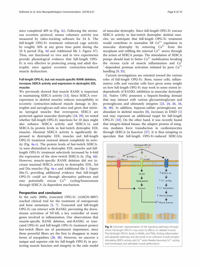
Fig. 5 Schematic representation of the signaling pathways throughwhich full-length OPG-Fc may exert its effects on skeletal muscle.The full-length OPG-Fc binds to RANKL and TRAIL limiting inflammationand apoptotic pathways and also binds to an unknown muscle receptorstimulating SERCA activity and Ca2+ entry thereby favouring Ca2+ cyclingand homeostasis and ultimately muscle performance
Dufresne et al. Acta Neuropathologica Communications (2018) 6:31 Page 8 of 10

expression may also act through mutual cooperationbetween proteoglycans, integrins and growth factors(Fig. 5). Altogether, our results suggest that all 7 do-mains of OPG-Fc may contribute to prevent muscledegeneration in DMD. Since OPG is a well-known boneprotector, one cannot exclude that healthier bones mayalso protect dystrophic muscles but this remains well be-yond the scope of the present study. In conclusion, full-length OPG-Fc may be a new clinical treatment for severalforms of neuromuscular and muscular diseases in which asingle therapeutic approach may be foreseeable to main-tain both bone and skeletal muscle functions.
Additional file
Additional file 1: Table 1. All treatments and muscle-specific geneticdeletion of RANK did not have an impact on muscle mass at 5 weeks ofage. Table 2. Primers used for PCR amplification and genotyping. Figure 1.RANK deletion reduces EDL muscle damage in 5 week-old mdx mice.Figure 2. Full-length OPG-Fc treatment and RANK deletion protectdystrophic skeletal muscles. Figure 3. RANK deletion protects skeletalmuscles in old mdx mice. Figure 4. Full-length OPG-Fc mitigates musculardystrophy in fast-twitch skeletal muscles. Figure 5. Recovery scores ofvarious key functional parameters of skeletal muscles evaluated ex vivo fromdystrophic mice treated with full-length OPG-Fc, anti-RANKL, anti-TRAIL and/or selectively deficient in muscle RANK. Figure 6. Full-length OPG-Fcmarkedly increases functional performance during eccentric downhillrunning. Figure 7. Recovery scores of forced and voluntary physical exerciseperformance in full-length OPG-Fc treated dystrophic mdx mice. Figure 8.Muscle RANK deletion and full-length OPG-Fc treatment did not increaseSERCA activity in dystrophic Sol and Dia muscles. (DOCX 3505 kb)
AcknowledgementsWe thank the Bioimaging platform at the CHU de Québec (CHUL), supportedby the Canada Foundation for Innovation, for confocal analyses. We areindebted to Carlo Rago and Paul Kostenuik for providing over the yearsvaluable information about DMD and OPG, respectively.
FundingThis work was supported by grants to JF from the Natural Sciences andEngineering Research Council of Canada, the Canadian Institutes of HealthResearch, Duchenne Alliance and Jesse’s Journey.
Authors’ contributionsJF, SSD and JMP conceived the project and its design; SSD, ABP, SB, AA, DH,LM, VAF, DG, RT and HY performed experiments and data analysis; JF, SSDand AA wrote the manuscript; and all authors checked for scientific contentand contributed to the final drafting of the manuscript. All authors read andapproved the final manuscript.
Competing interestsThe authors declare that they have no competing interests.
Publisher’s NoteSpringer Nature remains neutral with regard to jurisdictional claims in publishedmaps and institutional affiliations.
Author details1Centre Hospitalier Universitaire de Québec–Centre de Recherche du CentreHospitalier de l’Université Laval (CHUQ-CRCHUL), Université Laval, 2705boulevard Laurier, RC-9500, Quebec City, QC G1V 4G2, Canada. 2Departmentof Kinesiology, University of Waterloo, Waterloo, ON N2L 3G1, Canada.3Department of Immunology, Juntendo University, School of Medicine,Tokyo, Japan. 4IMBA, Institute of Molecular Biotechnology of the Austrian
Academy of Sciences, 1030 Vienna, Austria. 5Département de Réadaptation,Faculté de Médecine, Université Laval, Quebec City, QC G1V 4G2, Canada.
Received: 8 March 2018 Accepted: 10 April 2018
References1. Alvarez K, Fadic R, Brandan E (2002) Augmented synthesis and differential
localization of heparan sulfate proteoglycans in Duchenne musculardystrophy. J Cell Biochem 85:703–713. https://doi.org/10.1002/jcb.10184.
2. Andrukhov O, Huber R, Shi B, Berner S, Rausch-Fan X, Moritz A, Spencer ND,Schedle A (2016) Proliferation, behavior, and differentiation of osteoblastson surfaces of different microroughness. Dent Mater Off Publ Acad DentMater 32:1374–1384. https://doi.org/10.1016/j.dental.2016.08.217.
3. Atkins GJ, Findlay DM (2012) Osteocyte regulation of bone mineral: a littlegive and take. Osteoporos Int J 23:2067–2079. https://doi.org/10.1007/s00198-012-1915-z.
4. Baud’huin M, Duplomb L, Teletchea S, Lamoureux F, Ruiz-Velasco C,Maillasson M, Redini F, Heymann M-F, Heymann D (2013) Osteoprotegerin:multiple partners for multiple functions. Cytokine Growth Factor Rev 24:401–409. https://doi.org/10.1016/j.cytogfr.2013.06.001.
5. Bekker PJ, Holloway D, Nakanishi A, Arrighi M, Leese PT, Dunstan CR (2001)The effect of a single dose of osteoprotegerin in postmenopausal women. JBone Miner Res Off J Am Soc Bone Miner Res 16:348–360. https://doi.org/10.1359/jbmr.2001.16.2.348.
6. Blaauw B, Agatea L, Toniolo L, Canato M, Quarta M, Dyar KA, Danieli-BettoD, Betto R, Schiaffino S (1985) Reggiani C (2010) eccentric contractions leadto myofibrillar dysfunction in muscular dystrophy. J Appl Physiol BethesdaMd 108:105–111. https://doi.org/10.1152/japplphysiol.00803.2009.
7. Body J-J, Greipp P, Coleman RE, Facon T, Geurs F, Fermand J-P, HarousseauJ-L, Lipton A, Mariette X, Williams CD, Nakanishi A, Holloway D, Martin SW,Dunstan CR, Bekker PJ (2003) A phase I study of AMGN-0007, a recombinantosteoprotegerin construct, in patients with multiple myeloma or breastcarcinoma related bone metastases. Cancer 97:887–892. https://doi.org/10.1002/cncr.11138.
8. Brandan E, Gutierrez J (2013) Role of skeletal muscle proteoglycans duringmyogenesis. Matrix Biol 32:289–297. https://doi.org/10.1016/j.matbio.2013.03.007.
9. Brini M, Carafoli E (2009) Calcium pumps in health and disease. Physiol Rev89:1341–1378. https://doi.org/10.1152/physrev.00032.2008.
10. Casar JC, Cabello-Verrugio C, Olguin H, Aldunate R, Inestrosa NC, Brandan E(2004) Heparan sulfate proteoglycans are increased during skeletal muscleregeneration: requirement of syndecan-3 for successful fiber formation. JCell Sci 117:73–84. https://doi.org/10.1242/jcs.00828.
11. Compston J (2015) Emerging therapeutic concepts for muscle and bonepreservation/building. Bone 80:150–156. https://doi.org/10.1016/j.bone.2015.04.013.
12. Dufresne SS, Dumont NA, Bouchard P, Lavergne É, Penninger JM, Frenette J(2015) Osteoprotegerin protects against muscular dystrophy. Am J Pathol185:920–926. https://doi.org/10.1016/j.ajpath.2015.01.006.
13. Dufresne SS, Dumont NA, Boulanger-Piette A, Fajardo VA, Gamu D, Kake-Guena SA, David RO, Bouchard P, Lavergne É, Penninger JM, Pape PC,Tupling AR, Frenette J (2016) Muscle RANK is a key regulator of calciumstorage, SERCA activity, and function of fast-twitch skeletal muscles. Am JPhysiol Cell Physiol. https://doi.org/10.1152/ajpcell.00285.2015.
14. Duhamel TA, Green HJ, Stewart RD, Foley KP, Smith IC, Ouyang J (2007)Muscle metabolic, SR ca(2+) -cycling responses to prolonged cycling, withand without glucose supplementation. J Appl Physiol Bethesda Md 1985103:1986–1998. https://doi.org/10.1152/japplphysiol.01440.2006.
15. Eapen A, Sundivakkam P, Song Y, Ravindran S, Ramachandran A, TiruppathiC, George A (2010) Calcium-mediated stress kinase activation by DMP1promotes osteoblast differentiation. J Biol Chem 285:36339–36351. https://doi.org/10.1074/jbc.M110.145607.
16. Finnberg NK, Gokare P, Navaraj A, Lang Kuhs KA, Cerniglia G, Yagita H,Takeda K, Motoyama N, El-Deiry WS (2016) Agonists of the TRAIL deathreceptor DR5 sensitize intestinal stem cells to chemotherapy-induced celldeath and trigger gastrointestinal toxicity. Cancer Res 76:700–712. https://doi.org/10.1158/0008-5472.CAN-15-2759.
17. Godfrey C, Muses S, McClorey G, Wells KE, Coursindel T, Terry RL, Betts C,Hammond S, O’Donovan L, Hildyard J, El Andaloussi S, Gait MJ, Wood MJ,Wells DJ (2015) How much dystrophin is enough: the physiologicalconsequences of different levels of dystrophin in the mdx mouse. Hum MolGenet 24:4225–4237. https://doi.org/10.1093/hmg/ddv155.
Dufresne et al. Acta Neuropathologica Communications (2018) 6:31 Page 9 of 10
18. Goonasekera SA, Lam CK, Millay DP, Sargent MA, Hajjar RJ, Kranias EG, MolkentinJD (2011) Mitigation of muscular dystrophy in mice by SERCA overexpression inskeletal muscle. J Clin Invest 121:1044–1052. https://doi.org/10.1172/JCI43844.
19. Hanada R, Hanada T, Sigl V, Schramek D, Penninger JM (2011) RANKL/RANK-beyond bones. J Mol Med Berl Ger 89:647–656. https://doi.org/10.1007/s00109-011-0749-z.
20. Hanada R, Penninger JM (2011) Central regulation of body temperature byRANKL/RANK pathway. Clin Calcium 21:1201–1208 doi: CliCa110812011208.
21. Honma M, Ikebuchi Y, Kariya Y, Suzuki H (2014) Regulatory mechanisms ofRANKL presentation to osteoclast precursors. Curr Osteoporos Rep 12:115–120.https://doi.org/10.1007/s11914-014-0189-0.
22. Kayagaki N, Yamaguchi N, Abe M, Hirose S, Shirai T, Okumura K, Yagita H(2002) Suppression of antibody production by TNF-related apoptosis-inducing ligand (TRAIL). Cell Immunol 219:82–91.
23. Kobayashi-Sakamoto M, Isogai E, Hirose K, Chiba I (2008) Role of alphav integrinin osteoprotegerin-induced endothelial cell migration and proliferation.Microvasc Res 76:139–144. https://doi.org/10.1016/j.mvr.2008.06.004.
24. Lane D, Matte I, Laplante C, Garde-Granger P, Rancourt C, Piché A (2013)Osteoprotegerin (OPG) activates integrin, focal adhesion kinase (FAK), andAkt signaling in ovarian cancer cells to attenuate TRAIL-induced apoptosis. JOvarian Res 6:82. https://doi.org/10.1186/1757-2215-6-82.
25. Langen RCJ, Van Der Velden JLJ, Schols AMWJ, Kelders MCJM, Wouters EFM,Janssen-Heininger YMW (2004) Tumor necrosis factor-alpha inhibits myogenicdifferentiation through MyoD protein destabilization. FASEB J Off Publ Fed AmSoc Exp Biol 18:227–237. https://doi.org/10.1096/fj.03-0251com.
26. Li M, Yang S, Xu D (2016) Heparan sulfate regulates the structure and functionof Osteoprotegerin in Osteoclastogenesis. J Biol Chem 291:24160–24171.https://doi.org/10.1074/jbc.M116.751974.
27. Liu W, Zhang X (2015) Receptor activator of nuclear factor-κB ligand(RANKL)/RANK/osteoprotegerin system in bone and other tissues (review).Mol Med Rep 11:3212–3218. https://doi.org/10.3892/mmr.2015.3152.
28. Macpherson PC, Schork MA, Faulkner JA (1996) Contraction-induced injuryto single fiber segments from fast and slow muscles of rats by singlestretches. Am J Phys 271:C1438–C1446.
29. Marques MJ, Ferretti R, Vomero VU, Minatel E, Neto HS (2007) Intrinsiclaryngeal muscles are spared from myonecrosis in themdx mouse model ofDuchenne muscular dystrophy. Muscle Nerve 35:349–353. https://doi.org/10.1002/mus.20697.
30. Naya FJ, Mercer B, Shelton J, Richardson JA, Williams RS, Olson EN (2000)Stimulation of slow skeletal muscle fiber gene expression by calcineurin invivo. J Biol Chem 275:4545–4548.
31. Nelson CA, Warren JT, Wang MW-H, Teitelbaum SL, Fremont DH (2012)RANKL employs distinct binding modes to engage RANK and theOsteoprotegerin decoy receptor. Structure 20:1971–1982. https://doi.org/10.1016/j.str.2012.08.030.
32. Perrini S, Laviola L, Carreira MC, Cignarelli A, Natalicchio A, Giorgino F (2010)The GH/IGF1 axis and signaling pathways in the muscle and bone:mechanisms underlying age-related skeletal muscle wasting andosteoporosis. J Endocrinol 205:201–210. https://doi.org/10.1677/JOE-09-0431.
33. Smyth MJ, Yagita H, McArthur GA (2016) Combination anti-CTLA-4 and anti-RANKL in metastatic melanoma. J Clin Oncol Off J Am Soc Clin Oncol 34:e104–e106. https://doi.org/10.1200/JCO.2013.51.3572.
34. Standal T, Seidel C, Hjertner Ø, Plesner T, Sanderson RD, Waage A, Borset M,Sundan A (2002) Osteoprotegerin is bound, internalized, and degraded bymultiple myeloma cells. Blood 100:3002–3007. https://doi.org/10.1182/blood-2002-04-1190.
35. Stiber J, Hawkins A, Zhang Z-S, Wang S, Burch J, Graham V, Ward CC, SethM, Finch E, Malouf N, Williams RS, Eu JP, Rosenberg P (2008) STIM1signalling controls store-operated calcium entry required for developmentand contractile function in skeletal muscle. Nat Cell Biol 10:688–697. https://doi.org/10.1038/ncb1731.
36. Théoleyre S, Kwan Tat S, Vusio P, Blanchard F, Gallagher J, Ricard-Blum S,Fortun Y, Padrines M, Rédini F, Heymann D (2006) Characterization ofosteoprotegerin binding to glycosaminoglycans by surface plasmonresonance: role in the interactions with receptor activator of nuclear factorkappaB ligand (RANKL) and RANK. Biochem Biophys Res Commun 347:460–467.https://doi.org/10.1016/j.bbrc.2006.06.120.
37. Traister A, Li M, Aafaqi S, Lu M, Arab S, Radisic M, Gross G, Guido F, SherretJ, Verma S, Slorach C, Mertens L, Hui W, Roy A, Delgado-Olguín P, HanniganG, Maynes JT, Coles JG (2014) Integrin-linked kinase mediates force
transduction in cardiomyocytes by modulating SERCA2a/PLN function. NatCommun 5:4533. https://doi.org/10.1038/ncomms5533.
38. Wang Y, Pessin JE (2013) Mechanisms for fiber-type specificity of skeletalmuscle atrophy. Curr Opin Clin Nutr Metab Care 16:243–250. https://doi.org/10.1097/MCO.0b013e328360272d.
39. Williams DW, Lee C, Kim T, Yagita H, Wu H, Park S, Yang P, Liu H, Shi S, ShinK-H, Kang MK, Park N-H, Kim RH (2014) Impaired bone resorption andwoven bone formation are associated with development of osteonecrosisof the jaw-like lesions by bisphosphonate and anti-receptor activator of NF-κB ligand antibody in mice. Am J Pathol 184:3084–3093. https://doi.org/10.1016/j.ajpath.2014.07.010.
40. Yamaguchi K, Kinosaki M, Goto M, Kobayashi F, Tsuda E, Morinaga T,Higashio K (1998) Characterization of structural domains of humanosteoclastogenesis inhibitory factor. J Biol Chem 273:5117–5123.
Dufresne et al. Acta Neuropathologica Communications (2018) 6:31 Page 10 of 10


![uke ls feyrh&tqyrh QthZ Website · B_w[r_ of Dupli][t_ W_\sit_ with Bihar Police Constable Written / Physical Exam Syllabus Sarkari Result feyrh&tqyrh QthZ lko/kku jgs ges’kk](https://img.pdfslide.us/doc/110x75/5aed9ee27f8b9a45568feb07/uke-ls-feyrhtqyrh-qthz-website-r-of-duplit-wsit-with-bihar-police-constable.jpg)