Embed Size (px)
Citation preview
Genetic and Pharmacological Inhibition of Galectin-3 Prevents Cardiac
Remodeling by Interfering with Myocardial Fibrogenesis Yu et al: Galectin-3 Inhibition and Cardiac Remodeling
Lili Yu, MD1,2*; Willem P.T. Ruifrok, MD, PhD1*; Maxi Meissner, PhD1;
Eelke M. Bos, MD3; Harry van Goor, PhD3; Bahram Sanjabi, MSc4;
Pim van der Harst, MD, PhD1; Bertram Pitt, MD5; Irwin J. Goldstein, PhD6;
Jasper A. Koerts, MSc3; Dirk J. van Veldhuisen, MD, PhD1; Ruud A. Bank, PhD3;
Wiek H. van Gilst, PhD1; Herman H.W. Silljé, PhD1; Rudolf A. de Boer, MD, PhD1
1Department of Cardiology, University Medical Center Groningen, University of Groningen, Groningen, the Netherlands 2Harbin Medical University, Harbin, P.R. China 3Department of Pathology and Medical Biology, University Medical Center Groningen, Groningen, the Netherlands 4Department of Genetics, University Medical Center Groningen, Groningen, the Netherlands 5University of Michigan Medical School, Department of Medicine, Ann Arbor, MI, U.S.A. 6University of Michigan Medical School, Department of Biological Chemistry, Ann Arbor, MI, U.S.A. * These authors contributed equally to this work Correspondence to R.A. de Boer, MD, PhD Department of Cardiology University Medical Center Groningen University of Groningen Hanzeplein 1, 9713 GZ / P.O. Box 30.001 9700 RB Groningen the Netherlands Phone: +31(0) 50 361 23 55 Fax: +31(0) 50 361 5525 E-mail: [email protected] DOI: 10.1161/CIRCHEARTFAILURE.112.971168 Journal Subject Code: Other heart failure, Animal models of human disease, Remodeling
dicaaaaaaal llll ll CeCeCeCeCeCeCentntntntntntntererererererer GGGGGGGrororororororonn
Genetics, University Medical Center Groningen, Groningen, the Michigan Medical School, Department of Medicine, Ann Arbor, MMichigan Medical School, Department of Biological Chemistry, A
c
GeGeGeGeGennnnen ticsss,,,, UnUUUnniversity Medical Cennntter Gronininininngeggen,,,,, Groningen, the Miiichhhhhigan Medede iccalllll SSSchchchchchoooooolll,ll DDDeppaartmmmennnnntttt t ofofofofof MMMedicinne,e,ee, AAAnnnnnn AAArbbrbbboor,,, MMichchchchchigigigigiganananann MMMMMedededededicicicccalallll Schchchchchooooooooool,llll DDDDDepepepepepararararartmtmtmmmenenenenent t t t ofofofofof BBBBBioioioioiololoolologigigiggicacacacac llll l ChChChChChemememememisisissstrtrtry,y,y,yy AAA
cococontntntririribububuteteteddd eqeqequauauallllllyyy tototo ttthihihisss wowoworkrkrk
by guest on June 19, 2018http://circheartfailure.ahajournals.org/
Dow
nloaded from
by guest on June 19, 2018http://circheartfailure.ahajournals.org/
Dow
nloaded from
by guest on June 19, 2018http://circheartfailure.ahajournals.org/
Dow
nloaded from
by guest on June 19, 2018http://circheartfailure.ahajournals.org/
Dow
nloaded from
by guest on June 19, 2018http://circheartfailure.ahajournals.org/
Dow
nloaded from
by guest on June 19, 2018http://circheartfailure.ahajournals.org/
Dow
nloaded from
by guest on June 19, 2018http://circheartfailure.ahajournals.org/
Dow
nloaded from
by guest on June 19, 2018http://circheartfailure.ahajournals.org/
Dow
nloaded from
by guest on June 19, 2018http://circheartfailure.ahajournals.org/
Dow
nloaded from
by guest on June 19, 2018http://circheartfailure.ahajournals.org/
Dow
nloaded from
by guest on June 19, 2018http://circheartfailure.ahajournals.org/
Dow
nloaded from
by guest on June 19, 2018http://circheartfailure.ahajournals.org/
Dow
nloaded from
by guest on June 19, 2018http://circheartfailure.ahajournals.org/
Dow
nloaded from
by guest on June 19, 2018http://circheartfailure.ahajournals.org/
Dow
nloaded from
by guest on June 19, 2018http://circheartfailure.ahajournals.org/
Dow
nloaded from
by guest on June 19, 2018http://circheartfailure.ahajournals.org/
Dow
nloaded from
by guest on June 19, 2018http://circheartfailure.ahajournals.org/
Dow
nloaded from
by guest on June 19, 2018http://circheartfailure.ahajournals.org/
Dow
nloaded from
by guest on June 19, 2018http://circheartfailure.ahajournals.org/
Dow
nloaded from
by guest on June 19, 2018http://circheartfailure.ahajournals.org/
Dow
nloaded from
by guest on June 19, 2018http://circheartfailure.ahajournals.org/
Dow
nloaded from
by guest on June 19, 2018http://circheartfailure.ahajournals.org/
Dow
nloaded from
Abstract Background—Galectin-3 has been implicated in the development of organ fibrosis. It is
unknown whether it is a relevant therapeutic target in cardiac remodeling and heart failure
(HF).
Methods and Results—Galectin-3 knock-out (Gal3-KO) and wild-type (WT) mice were
subjected to angiotensin II (AngII) infusion (2.5 μg/kg for 14 days) or transverse aortic
constriction (TAC) for 28 days to provoke cardiac remodeling. The efficacy of the galectin-3
inhibitor N-acetyllactosamine (Gal3i) was evaluated in TGR(mREN2)27 (REN2) rats and in
WT mice with the aim of reversing established cardiac remodeling following TAC. In WT
mice, AngII and TAC perturbations caused left ventricular (LV) hypertrophy, decreased
fractional shortening and increased LV end-diastolic pressure and fibrosis (P<0.05 vs. control
WT). Gal3-KO mice also developed LV hypertrophy but without LV dysfunction and fibrosis
(P=NS). In REN2 rats, pharmacological inhibition of galectin-3 attenuated LV dysfunction
and fibrosis. To elucidate the beneficial effects of galectin-3 inhibition on myocardial
fibrogenesis, cultured fibroblasts were treated with galectin-3 in the absence or presence of
Gal3i. Inhibition of galectin-3 was associated with a downregulation in collagen production
(collagen I and III), collagen processing, cleavage, cross-linking and deposition. Similar
results were observed in REN2 rats. Inhibition of galectin-3 also attenuated the progression of
cardiac remodeling in a long-term TAC mouse model.
Conclusions—Genetic disruption and pharmacological inhibition of galectin-3 attenuates
cardiac fibrosis, LV dysfunction and subsequent HF development. Drugs binding to galectin-
3 may be potential therapeutic candidates for the prevention or reversal of HF with extensive
fibrosis.
Key Words: Galectin-3, cardiac remodeling, heart failure, fibrosis, renin-angiotensin system
hyhyhyhyhyhyhypepepepepepepertrtrtrtrtrtrtrororororororophphphphphphphy,y,y,y,y,y,y, dddddddeeeeeee
nd fibrbrbrbrbrbrbrosososososososisisisisisisis (((((((PPPPPPP<0<0<0<0<0<0<0 0g p (
mice also devel ed LV hy rtroph but without LV d functi n
N y
e a
g p (
mmmiccccce also dddddeveveee elelelelelopopopopopededededed LLLLLV V V V V hyhyhyhh peeeertrtrr rophpphy y y y y bububububut tttt wwiththt ououououo tt LVLVLVLVLV dddddysysysysysfufufufufuncncncncnctitititition
N2 raaaatstststst , phphpharaarmamamacocololl gigigiiicacacalll ll inhihihihihibibibibib titititit onoon ofofofff gggalalalalaleeecee titittit nnn-33333 atatattenununununuatededddd LLLLLV VVV dydddd
eeelulucicidadatetetee ttthehe bbenenenefeficiciaiall eeefffffffececectststs oooff gagagagg leleleel ctctctc inin 33-3 iiiinhnhibibititioionnn ononon mmmyoyoyocacaca
by guest on June 19, 2018http://circheartfailure.ahajournals.org/
Dow
nloaded from
Galectin-3 belongs to the galectin family of mammalian lectins and is characterized by a
carbohydrate recognition domain (CRD) that has affinity for -galactosides. Galectin-3
mediates cell–cell and cell–matrix interactions by binding to lactosamine-containing cell
surface glycoconjugates via its CRD. There is mounting evidence demonstrating its key role
in inflammatory and fibrotic processes.1
In experimental models, galectin-3 was found to be involved in the development of liver and
kidney fibrosis.2,3 It has also been associated with cardiac fibrosis in TGRmREN2-27 (REN2)
rats with experimental heart failure (HF).4 A continuous intrapericardial infusion of low-dose
galectin-3 resulted in cardiac fibrosis and left ventricular (LV) dysfunction in both failure-
prone hypertrophic REN2 and healthy Sprague-Dawley rats.4,5 The upregulation of galectin-3
has also been demonstrated in failing human hearts4 and circulating levels are a powerful
predictor of outcome in both acute and chronic HF. This observation has led to the approval
of galectin-3 by the Food and Drug Administration as a novel biomarker in HF that may help
categorize patients in the remodeling and non-remodeling stages of HF.6,7
Despite evidence suggesting the involvement of galectin-3 in the pathophysiology of HF, it
remains unknown if it contributes actively to the disease. In the present study, we determined
if the disruption of the galectin-3 gene, shown to attenuate hepatic and renal fibrosis in
galectin-3 knock-out (Gal3-KO) mice,2,8 would prevent the development of HF in animals
with active cardiac remodeling. Furthermore, because the beneficial effects of targeted CRD
inhibition of galectin-3 have been demonstrated in a model of kidney injury,3 we also
determined the effects of a galectin-3 inhibitor (Gal3i), N-acetyllactosamine (N-Lac), which
has a high affinity for galectin-3 CRD, in TGR(mREN2)27 (REN2) rats with HF and in mice
subjected to transverse aortic constriction (TAC).
TTTTTThehehehehehehe uuuuuuuprprprprprprpregegegegegegegulululululululatatatatatatatioioioioioioion n nnnnn
ting lllllllevevevevevevevelelelelelelelsssssss ararararararareeeeeee aaaaa pg g p
ome in both acute and chronic HF. This observation has led to th
the Food and Drug Administration as a novel biomarker in HF th
n
g g p
ooommmem in booththththth aaaaacucucucucutetetetete aaaaandndndndnd ccccchrhrhrhrhroooniciiii HF.FF TTTTThihihihih sss obobobobobsservrvrvvvaatiooooon nnnn hahahahahas ssss lelelell d dd dd tototototo th
the FFFFFooooooddd dd anaandd ddd DrDrDrDD uuug AAAAAdmdmdminisisisisistrtrtrtrt aattatioioionn asasass aaa nnnnnovovovovelelell bbbbbioiomamamarkrkrkrkrker iiiiinn HFHFHFHF ttth
ntntntsss inin ttthehee rrremememododelelining g g gg ananandd nonononnn-rereremomomodedeeelililll ngngnggg ssstatatagegegegg sss ofof HHHFFF.6,7,
by guest on June 19, 2018http://circheartfailure.ahajournals.org/
Dow
nloaded from
Methods
For detailed description of the methods, please refer to the Online Supplemental Data.
Animals
Male Gal3-KO mice were generated and bred at the Jackson Laboratory (Bar Harbor, ME,
USA) as described previously.2,8,9 Age-matched male transgene-negative wild-type (WT)
littermates were used as controls. Supplemental Figure S1A depicts the generation of Gal3-
KO mice. Male homozygous REN2 (Max Delbrück Center for Molecular Medicine, Berlin
Buch, Germany) and age-matched male Sprague-Dawley (SD) rats (controls) were used as
described previously.10-12 All experiments were approved by the Animal Ethical Committee
of the University of Groningen (the Netherlands) and conducted in accordance with existing
guidelines on the care and use of laboratory animals.
Mouse experiments
Six- to 10-week old Gal3-KO and WT mice were subjected to an infusion of angiotensin II
(AngII) (2.5 μg/Kg/day) for 14 days or LV pressure overload by TAC for 28 days
(prevention experiment).13 In another series of experiments (reversal experiment), six- to
eight-week old male C56Bl6/J mice (Harlan, The Netherlands) underwent TAC for 28 days
and were then treated with N-Lac (5 mg/Kg/day, intraperitoneal (i.p.) injections) three times a
week for 28 days (starting day 28 until day 56).14
Rat experiments
SD and REN2 rats were treated with N-Lac (5 mg/Kg/day, i.p.) three times a week for six
weeks.
Other experiments
Cardiac function was studied with echocardiography and hemodynamic measurement as
described previously.15,16 Immunohistochemical analyses were performed and collagen
digestibility was determined.17 Cell cultures of human adult dermal fibroblasts were used to
iiiiiiin n n nnnn acacacacacacaccococococococordrdrdrdrdrdrdananananananancecececececece wwwwwww
y
e
old Gal3-KO and WT mice were subjected to an infusion of a g
Kg/day) for 14 days or LV pressure overload by TAC for 28 day
y
eeentnttntnts
old dddd GaGaGaGaG l3l3l3l3l3-KKKKKOOO OO anand ddd WTWWTWTW miceeeee wewewewereree ssubububbubjejejejejectctctctctededededed tttooo anan infnfnfnfnfusususususiiioii n n ofoff aangng
KgKgKg/d/dayayay)) ) foforrr 141414 ddayayayyysss ororor LLLV V V prprprpp esesessususurerere ooovevevevv rlrloaoaoad d dd bybyyyy TTTACACAC ffororor 222888 dadayyy
by guest on June 19, 2018http://circheartfailure.ahajournals.org/
Dow
nloaded from
study galectin-3-mediated fibrogenesis and the effects of N-Lac. Cardiac and fibroblast gene
expression was measured using RT-qPCR as described previously.18 The lists of primers are
presented in Supplemental Tables S1, 2 and 3.
Statistical analyses
Results are reported as mean ± standard error of the mean (SEM). Mice were analyzed in two
separate subgroups comparing genotype differences (WT vs. KO) and model differences
(control vs. AngII or sham vs. TAC). Subgroup A consisted of WT-control, Gal3-KO-
control, WT-AngII, Gal3-KO-AngII and subgroup B consisted of WT-sham, Gal3-KO-sham,
WT-TAC and Gal3-KO-TAC.
Levene’s test was used to test homogeneity of variances within parameters . If there was
equality of variances, statistical analyses were performed by one-way ANOVA with
Bonferroni post-hoc tests (mice group A M=6 tests, mice group B M=6 tests, rats M=3 tests).
If there was unequality of variances, statistical analyses were performed by Welch’s ANOVA
with Games-Howell post-hoc test. Cell experiments were analyzed with Kruskall-Wallis test
(N=3 per group). For the TAC reversal experiment, differences between saline treated mice
and N-Lac treated mice were analyzed at the eight week time point using an unpaired t-test
(N=7-9). Baseline TAC reversal is depicted as a dotted line. In all figures, only relevant
comparisons are shown by the symbols for reasons of clarity. All P-values are two-tailed and
a value less than 0.05 was considered significant. All analyses were performed using SPSS
version 20.0 software (SPSS, Chicago, IL, USA).
Results
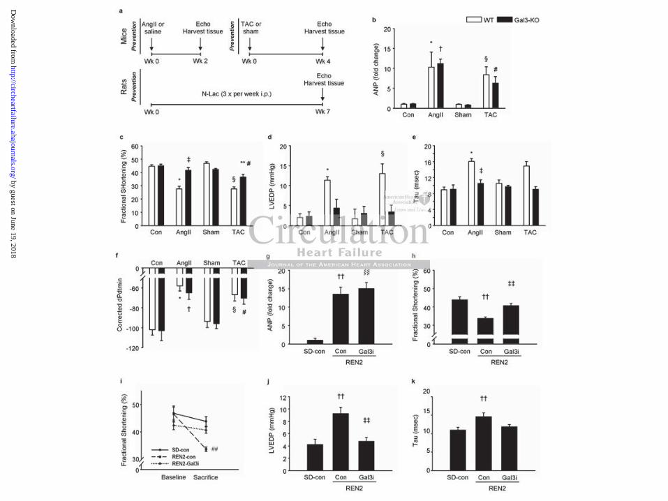
Galectin-3 knock-out mice are protected against left ventricular dysfunction
The intervention and treatment schemes of the WT and Gal3-KO mice are presented in
Figure 1a and the baseline characteristics and hemodynamic data at sacrifice are presented in
ppppppparararararararamamamamamamametetetetetetetererererererersssssss .... .. IfIfIfIfIfIfIf ttttttthhhhhhh
e waaaaaaayyyyyyy ANANANANANANANOVOVOVOVOVOVOVAAAAAAA w, y p y y
h s
quality of variances, statistical analyses were performed by Welc
w l
, y p y y
hohohohohoc tests (m(m(m(mmiciccceeeee grgrgrgrgrouououououppp pp A A A A A M=M=M=MM 66666 ttestttsss, mmmmmicicicicice eeee grrouououppppp BB M=M=M=M=M=66666 tetetetetestststststs,ssss rrraataaa s
quallalllititititityyy of vvvarariaiaiancnces, stststaata istititititicacacacacal lll annnallalllysysysseseseses wwwwwerrreee pepperfrfrffformememememeddd dd bybybbb WWWWelellllc
wwwelelll popopostststt h-hocococ tttesesesttt. CCCelelll exexexpepepepp ririmemementntntsss weweweeererere aaanananalylylyyyzezezedd wiwithth KKKrururuskskalalll
by guest on June 19, 2018http://circheartfailure.ahajournals.org/
Dow
nloaded from
Table 1. When compared with control or sham, LV galectin-3 expression was increased
almost 2-fold in WT mice treated with AngII or TAC while galectin-3 expression was absent
in Gal3-KO mice (mRNA and protein, Supplemental Figure S1 b-d). In the WT mice, both
interventions caused LV hypertrophy as evidenced by the increases in LV weight (normalized
to tibia length, Table 1), wall thicknesses (Table 1) and LV atrial natriuretic peptide (ANP)
expression (Figure 1b) along with a decrease in contractile function (fractional shortening,
FS, Figure 1c). Hemodynamic measurements revealed LV relaxation impairment in the WT
groups (Figure 1d-f) with increases in LVEDP and Tau (an isovolumetric relaxation constant
measured according to the Glantz method) along with decreases in dPdtmin (corrected for
peak systolic pressure).
As shown in Table 1, both Gal3-KO and WT mice subjected to AngII infusion or TAC had a
similar degree of LV hypertrophy. However, and irrespective of the perturbation, Gal3-KO
mice had preserved FS (Figure 1c). Hemodynamic measurements revealed that Gal3-KO
mice were protected against LV relaxation impairment following AngII infusion or TAC
(Figures 1d-f), which did not result in changes in LVEDP and Tau (P=NS vs. respective
controls). The only exception was the corrected dPdtmin which was significantly decreased
in the Gal3-KO-AngII group.
Inhibition of galectin-3 with N-acetyllactosamine prevents left ventricular dysfunction
in failure-prone REN2 rats
The intervention and treatment schemes of the SD and REN2 rats are presented in Figure 1a.
Table 2 shows the baseline characteristics and hemodynamic data at sacrifice. As expected,
LV weight (adjusted for tibia length) was significantly increased in the untreated REN2 rats.
Treatment with N-Lac did not prevent the development of LV hypertrophy or decrease LV
ANP levels (Table 2, Figure 1g). Fractional shortening (FS) progressively declined in the
untreated REN2 rats (Figure 1i) but was preserved in the Gal3i-treated rats (Figure 1h).
AnggggggIIIIIIIIIIIIII iiiiiiinfnfnfnfnfnfnfusususususususioioioioioioionnnnnnn oro, j g
f
ved FS (Figure 1c). Hemodynamic measurements revealed that G
cted against LV relaxation impairment following AngII infusio
, j g
f LLLVLL hyperrrrtrttrtt opopppphyhyhyhyhy. HoHoHoHoHowewewewewevevevev r, anaana d irrreeeeespspsss ececececectiiiiivevee ooooff fff ththhe e e e e pepepeeertrtrtrtrturururururbababababatititititiooonoo
veddddd FFFFFS SS (F(F(F(F(Figggurureee 1cc1c).) HHHHHemememodddddynynynynynamamaamiciciii mmmeaeaeaeae susuuuurememementntss revevevevevealededddd tttthahahatt GGG
ccctetetedd agagagaiaiainsnsnsttt LVLVLV rrrelelaxaxaxatatatioion n n imimpapapairirmemementntnttt ffolollolowiwiww ngngnggg AAAngngngggIIIIII iinfnfusususioionnn
by guest on June 19, 2018http://circheartfailure.ahajournals.org/
Dow
nloaded from
Hemodynamic measurements revealed an increased LVEDP in the untreated REN2 rats as
compared with the SD rats (Figure 1j) as well as Tau (Figure 1k), associated with increased
lung weights (Table 2), all suggestive of developing HF. Treatment with Gal3i reduced
LVEDP in REN2, and lung weigh, but not Tau. Finally, accelerated cardiac remodeling in
untreated REN2 rats was associated with poorer survival than Gal3i-treated REN2 rats
(Supplemental Figure S2).
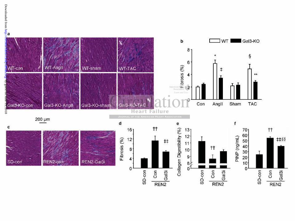
Galectin-3 disruption or inhibition attenuates the formation of fibrosis in the heart
To determine if galectin-3 is actively involved in the formation of fibrosis, we analyzed the
presence of myocardial fibrosis. Figures 2a and 2b show representative pictures of fibrotic
tissue and the fibrosis score in mouse hearts. The hearts of control and sham-operated WT
mice had very little fibrosis ( 2%). A significantly higher percentage of fibrosis was evident
in the WT-AngII and WT-TAC mice (Figure 2b). However, neither AngII infusion nor TAC
resulted in increased fibrosis in Gal3-KO animals. Similar results were also observed in rat
hearts. Compared with SD rats, REN2 rats exhibited a high percentage of fibrotic tissue and
treatment with Gal3i significantly reduced the percentage of fibrosis in the REN2 rats (Figure
2c, 2d).
We also evaluated collagen digestibility (% collagen released by proteolytic enzymes) as a
measure of the extent of collagen cross-linking. In our assay, the higher the numbers of cross-
links, the lower the amount of released collagen.19,20 Compared with SD rats, collagen
digestibility was significantly reduced in the hearts of REN2 rats (Figure 2e). As collagen
fibers in fibrotic lesions display a higher level of pyridinoline cross-links making them more
resistant to the enzymatic action of proteinases,20 our results indicate the presence of more
cross-linked collagen in the hearts from untreated REN2 rats (Figure 2e). Treatment of REN2
rats with Gal3i resulted in a collagen digestibility similar to that of SD rats (Figure 2e). We
also measured the plasma levels of PINP, a marker of collagen cleavage, with an ELISA
rrololololololol aaaaaandndndndndndnd ssssssshahahahahahaham-m-m-m-m-m-m-opopopopopopopeeeeeee
ntagegeeeeee ooooooofffffff fififififififibrbrbrbrbrbrbrosososososososisisisisisii w( ) g y g p g
I o
ased fibrosis in Gal3-KO animals. Similar results were also obse
d c
( ) g y g p g
I annnnd WT-TTTTTACACACCC mmmmmicicicicice e eee (F(FF(FFigigigigigurururu e 2b2b22 ). HHowowowowoweveveveveverr, neeeititititi heheerr rrr AnAnAnAnAngIgIgIgIg I II II inininninfufufufufusissss o
asedddd d fifififf brbrbrb osssisisiii iiin nn GGGaG l33333 KKK-KKOOO OO anananannimimimimimalalalss. SSSSSimimimimimilililililararararar resesesululullltsts werereere eeeee alalsoso ooobsbbsbb ee
dd wwwitithh SDSDSDS rrratatats,s,s,, RRRENENEN222 rararatststs eeexhxhiibibitetetedd a a a hihighghggg pppppererercececentntntagagagggeee ofof ffibibrororotiticcc
by guest on June 19, 2018http://circheartfailure.ahajournals.org/
Dow
nloaded from
assay. In REN2 rats, PINP was significantly increased compared with SD rats and inhibition
of galectin-3 resulted in a significant reduction of PINP concentration (Figure 2f).
Collectively, these results suggest that Gal3i can reduce pathological fibrosis through a
reduction of collagen deposition and synthesis along with increased collagen digestibility.
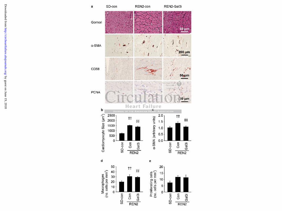
Galectin-3 inhibition reduces the number of -SMA positive cells in the heart
Figure 3 shows typical examples of Gomori, -SMA, CD68 (a macrophage marker) and
PCNA staining in rat hearts with their respective quantification. Cardiomyocyte size and -
SMA expression were significantly increased in REN2 rats. Treatment with Gal3i did not
decrease cardiomyocyte size (Figure 3a, 3b) but it normalized -SMA expression indicating a
lower number of myofibroblasts in these groups (Figure 3a, 3c). The number of macrophages
was also increased in both REN2 groups (Figure 3a, 3d), as was the number of proliferating
cells, although this was only significant in the untreated REN2 group (Figure 3a, 3e).
Immunohistochemical analysis revealed that galectin-3 immunoreactivity was predominantly
observed in the interstitial space, but not in cardiomyocytes (Supplemental Figure S3). In an
effort to determine the localization of cardiac galectin-3 and its source, we conducted further
studies. We observed that galectin-3 co-localized with macrophages (Supplemental Figure S4
a-d) and at sites of collagen deposition (Supplemental Figure S4, e-h). Furthermore, galectin-
3 protein expression was highest in macrophages. Galectin-3 protein expression was also
clearly detectable in fibroblast, but was not detectable in cardiomyocytes (Supplemental
Figure S5).
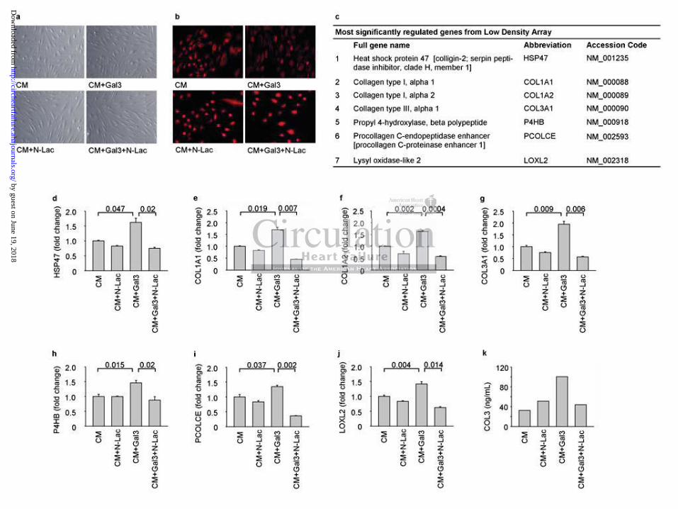
Galectin-3 inhibition prevents galectin-3-mediated effects in human dermal fibroblasts
Stimulation of human dermal fibroblasts with recombinant galectin-3, with or without Gal3i,
did not lead to visible differences in fibroblast appearance (Figure 4a). Fibroblast phenotype
was confirmed by staining for the mesenchymal marker vimentin (Figure 4b). Since our
preliminary results demonstrated that the mRNA level of COL1A1 in the presence of
ThThThThThThThe e e e e e e nununununununumbmbmbmbmbmbmbererererererer ooooooof f ff fff mmmmmmm
the nununununununumbmbmbmbmbmbmberererererere ooooooofffffff prppg p ( g , ), p
his was onl si ificant in the untreated REN2 ou (Figure 3a,
emical analysis revealed that galectin-3 immunoreactivity was pr
n r
g p ( g , ), p
hiisiii was onllllyyyyy siiiiigngngngngnififififificicicicicananananant tttt ininininin ttthehee uuntttreeatttttededededed RRRRRENENENENE 22 22 grgrg ouuuuupp ppp (F(F(F(F(Figigigigigururururureeee e 33a33a3 ,
emiiiiicacacacaal ll aaanalllysysisisis rreveveaaaleleleddd dd thhhhhatatatatat gggg lallleceectititittinnn-3333 imimimimimmumumunonnoreereactitittitivivivivivityy wwasasa ppr
nntetetersrsrstitititialala ssspapapapp cecece, bubuttt nononottt ininnnn cacacardrdioiomymymyyocococytytytyy eseses ((((SuSuSupppppppppplelememementntntalal FFFigigururur
by guest on June 19, 2018http://circheartfailure.ahajournals.org/
Dow
nloaded from
galectin-3 peaked 72 hours post-stimulation (data not shown), the expression of various
fibrillar collagens and proteins involved in their processing was measured at the 72h time
point with a low density array (overview in Supplemental Table S4; results are shown in
Figure 4). The expression of COL1A1, COL1A2, COL3A1 and the collagen-modifying
proteins encoded by P4HB, HSP47, PCOLCE and LOXL2 were all upregulated by galectin-3
(Figure 4, d-j) and inhibited by Gal3i co-treatment. The modulation of COL3A1 transcript
expression by galectin-3 and Gal3i was also reflected in collagen type III levels in the culture
medium measured with an ELISA (Figure 4k).
Genes involved in myocardial fibrogenesis
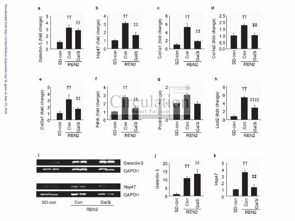
The expression of the above-mentioned collagens and fibrosis-associated proteins was
subsequently analyzed in the LV tissue of SD and REN2 rats by RT-qPCR (Figure 5, a-h;
primers are listed in Supplemental Table S2). These results confirm our in vitro observations
with human fibroblasts and show that Gal3i reduces the expression of pro-fibrotic genes also
in vivo. We also measured the LV protein expression of galectin-3 and Hsp47 in lysates from
SD and REN2 rat hearts (Figure 5, i-k). Galectin-3 and Hsp47 protein levels were increased
in untreated REN2 rats. Treatment with the Gal3i resulted in decreased Hsp47 (Figure 5k) but
not galectin-3 (Figure 5j) protein levels, which reflects the changes in mRNA expression
(Figure 5a-b). Finally, we analyzed the transcript levels of matrix metalloproteinases (MMP)
2 and 9 and tissue inhibitors of matrix proteases (TIMP) 1 and 2 in LV tissue of SD and
REN2 rats. Mmp-9, Timp-1, and Timp-2 levels were increased in untreated REN2 rats and
reduced by treatment with a Gal3i (Supplemental Figure S6).
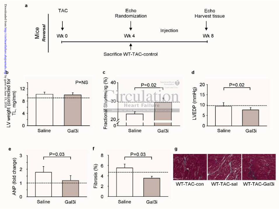
Galectin-3 inhibition prevents further progression of established left ventricular
remodeling
We provoked LV remodeling with TAC surgery followed by an observational period of 28
days without intervention (Figure 6a). We then treated the mice with N-Lac or saline for
ssssssssssssssococococococociaiaiaiaiaiaiateteteteteteted d d d d d d prprprprprprprotototototototeieieieieieieinnnnnnn
RT qqqqqqqPCPCPCPCPCPCPCRRRRRRR (F(F(F(F(F(F(Figigigigigigiguy y q ( g
d o
o i
measured the LV protein expression of galectin-3 and Hsp47 in
y y q ( g
ddddd innn n Suppleeeeememememementntntntntalalalalal TTTTTaaaaablblblblblee eee S2S2S2SS ). TTheeesse rrrrreeseee ululululults ccconnnnnfififff rmmmmm ooururururur a inininin vvvvvititititi rororororo o
obllllasasasasstststst andndndd sshohohohh ww thatatat GGGGGal3i3i3i3i3i rrrededededducuuceses ttttthehehehehe eeeeexpxpxpxprereresssssiooion ofofofofof ppproo-fffffibibiiibrorotitttt
mememeasasasurururedede ttthehe LLLVVV prprprpp otototeieinnn exexexprprprp esesessisiononon oooooff gagagagg lelectctctinin 33-3 aaandnd HHHspspsp474747 iinnn
by guest on June 19, 2018http://circheartfailure.ahajournals.org/
Dow
nloaded from
another 28 days. At the end of the follow-up period, no increase in LV weight (Figure 6b)
was observed. Treatment with Gal3i for 28 days had no effect on LV weight, compared with
the LV weights after the 28-day observational period (Figure 6b). However, a gradual
progression of LV remodeling was observed in the untreated mice as evidence by the further
decline in FS (Figure 6c) and by an increase in ANP levels (Figure 6e) and fibrosis (Figure
6f, g). In the Gal3i-treated mice, LVEDP was lower compared to untreated mice (Figure 6d).
Discussion
The current study provides several lines of evidence that galectin-3 is an active contributor in
the development of cardiac remodeling, myocardial fibrogenesis and HF. We have
demonstrated that inhibition of galectin-3 function by genetic disruption or pharmacological
intervention halts the progression of cardiac remodeling, attenuates myocardial fibrogenesis
and preserves LV function. These beneficial effects can be explained, at least in part, by the
lower number of myofibroblasts in combination with diminished collagen synthesis,
processing and cross-linking. Collectively, our results suggest that galectin-3 may be an
attractive target for the prevention and treatment of HF.
Disruption of galectin-3 attenuates cardiac remodeling and preserves cardiac function
To explore the hypothesis that galectin-3-targeted interventions may protect against
progressive cardiac remodeling and dysfunction, two experimental approaches were used in
well established mouse and rat models of cardiac remodeling: 1) complete genetic disruption
of galectin-3 and 2) pharmacological inhibition with an agent that specifically binds to the
CRD of galectin-3. Both the genetic disruption and pharmacological inhibition of galectin-3
resulted in considerable attenuation of cardiac remodeling and, specifically, to an almost
complete inhibition of cardiac fibrosis. Functionally, the inhibition of galectin-3 improved
diastolic dysfunction to a large extent (less increase in end-diastolic LV pressure and
ssssss aaaaaaandndndndndndnd HHHHHHHF.F.F.F.F.F.F. WWWWWWWe e e e e e e hahahahahahahavvvvvvv
sruptptptptptptptioioioioioioionnnnnnn ororororororor ppppppphahahahahahaharmrrrg y g p p
s f
V function. These beneficial effects can be explained, at least in p
f myofibroblasts in combination with diminished collagen synthe
g y g p p
sss thhehhh progrrrreseseee siiiiiononononon ooooof fff f cacacacacardrdrdrdrdiaiaiai ccc reeeemomooddeliiiiingngngngng,, attattetetenuuuuuaatatesssss mmmmmyyyyyocococococarararara didididdialalalalal f
V fffffununununnctctctioioioii n.nn TTTTTheheheseese benennefefeffficiaiaiaiaallll efefefefffefefeff ctctss cacacaaannn bebebebebe eeexpxpxplalalll ininiini ededededed, aaat lleaeaststst iinn p
f f mymymyofofibibbrororor blblasasastststs iinnn cococombmbininnnnatatatioionnn wiwiththhh dddddimimininisisssheheh dd cococollllagagagggenenen sssynynynththeee
by guest on June 19, 2018http://circheartfailure.ahajournals.org/
Dow
nloaded from
improved LV relaxation) in spite of the presence of significant LV hypertrophy. In this
respect, it is noteworthy that elevated levels of circulating galectin-3 have been shown to be
strong predictors of poor outcome in patients with “diastolic HF” or HF with preserved LV
ejection fraction.21
Although the animals in our experimental models were mainly characterized by impaired
diastolic function, mild (AngII, TAC-prevention) to moderate (REN2, TAC-reversal) systolic
dysfunction also developed over time. Inhibition of galectin-3 preserved systolic function, a
finding that was above all apparent in the reversal experiment where treatment was started
after four weeks of TAC. In the untreated mice, mild systolic dysfunction was present after
four weeks (Figure 6c) and progressed over another four weeks of follow-up to overt systolic
dysfunction (Figure 6c). However, when mice were treated with the galectin-3 inhibitor,
progression of systolic dysfunction was attenuated. These results suggest that galectin-3
inhibition might afford functional protection against developing and progressive cardiac
remodeling.
Additional evidence of the important role of galectin-3 in cardiac remodeling was obtained
by treating REN2 rats with N-acetyllactosamine, an established inhibitor that binds to the
galectin-3 CRD.14,22,23 The homozygous REN2 rat model is a well described model of rapidly
progressive cardiac remodeling driven by renin overexpression with changes typical for HF
such as increased sympathetic tone, LV hypertrophy, myocardial fibrosis and stress-related
pathways.4,10,12 Results of our study show that the typical course of HF development,
characterized by impaired LV relaxation and fast progression (within weeks) to overt HF,
was attenuated by galectin-3 inhibition. It remains to be determined if galectin-3 inhibition is
equally effective in other multifactorial models of HF such as the spontaneous hypertensive
rat or post-myocardial infarction HF, as a single treatment or in addition to established HF
therapy.
ofofofofofofof ffffffololololololollololololololow-w-w-w-w-w-w-upupupupupupup ttttttto oo o o o o ooooooo
the gagagagagagagalelelelelelelectctctctctctctininininininin 3333333 iiiin) , g
y l
) , g
yssstotototolic dysfsfsfsfsfuunu ctctctcctioioioioionnnnn wawwww ss sss atatatatattetetenuuuuaated.d.. TTTTThehehehehesesesesese ressulululllttstst suguguguguggeeeeeststststst ttttthahahahahatttt t gggagg l
affffffororororord ddd fuffff ncnnctitiiiionoonalalall ppproootetetectioooonn nnn agagagaa aiaiaiinsnst tt dededededeveveeeelolololol pipipiingnng aandndddnd ppppprrrrrogrgresessisisiiiveve
by guest on June 19, 2018http://circheartfailure.ahajournals.org/
Dow
nloaded from
Mechanisms underlying the cardioprotective effects of galectin-3 inhibition
Fibrosis is accepted as one of the main determinants of cardiac remodeling. Cessation of the
fibrotic process is one of the key targets to reverse cardiac remodeling and improve
prognosis. Fibroblasts, together with myofibroblasts and macrophages, have been identified
as key cells in the fibrotic process.24-26 The striking observation that myocardial fibrogenesis
was strongly inhibited when galectin-3 was genetically disrupted or pharmacologically
inhibited lead us to investigate the effect of galectin-3 on the fibrotic process. First, we
showed in REN2 rats that pharmacological inhibition of galectin-3 lead to a lower number of
myofibroblasts along with less collagen synthesis (lower PINP plasma levels) and deposition.
We substantiated these changes by showing that the stiffness of the fibrotic depositions was
also altered. In the collagen digestibility assay, the collagenase digested more collagens in
Gal3i-treated REN2 rats (Figure 2e) indicating less cross-linked fibrotic tissue. Studies on
fibrogenesis-related gene profiles in fibroblasts treated with recombinant galectin-3 revealed
changes in several genes relevant to the extracellular matrix processing. In vitro incubation of
human dermal fibroblasts with galectin-3 resulted in significant upregulation of genes coding
for various fibrillar collagens (COL1A1, COL1A2 and COL3A1) and genes involved in the
modification of (pro)collagens including prolyl-4-hydroxylase (P4HB), heat shock protein 47
(HSP47 or SERPINH1), the procollagen C-endopeptidase enhancer (PCOLCE) and the lysyl
oxidases (LOXL2). Inhibition of P4Hs has been shown to inhibit fibrosis and preserve
cardiac function in HF.27-29 Also, the antisense HSP47 was associated with less myocardial
fibrosis protecting hearts from post myocardial infarction remodeling.30 Importantly,
treatment with N-acetyllactosamine downregulated the expression of all these genes to
baseline levels. These results suggest that galectin-3 may affect several steps involved in
fibrogenesis, from enhanced synthesis of procollagen to the regulation of various enzymes
ttttttthehehehehehehe fffffffibibibibibibibrororororororotititititititiccccccc dededededededepopopopopopopo
digesssssssteteteteteteteddddddd momomomomomomorerererererere cccccolog g y y, g g
E S
a n
i
g g y y, g g
EN2N2N2N2N2 rats (FFFFigigigii ururururure e 2e2e2e2e2e) ) )) ) ininininindididididicaaaatttingggg lesssss crororororosssss-l-l-l-l- inininii kekekedd ddd fibrbrbrbrbr totottoticicicicic ttttisisisii sususususue.ee.ee S
ateddddd gggggenenene prpprofofofffililillleses in fififif brbrbrbb obbbbblalalalalaststststs trtrtreaeateeteteddd d wiwiwiwiwiththththth rrrecececommombibibibibinananananant ggalaleceectitittit n
alal gggenenenesesess rrrelelevevevanananttt tototo ttthehe eeextxtxtrararacececellllululararar mmmmmatatatririxxx prprprpp ocococesesessisingngnggg. InInIn vvvitititrororo ii
by guest on June 19, 2018http://circheartfailure.ahajournals.org/
Dow
nloaded from
involved in the processing of procollagen into mature intra and extracellular collagen. The
activation of these pathways is attenuated by galectin-3 inhibition (Figure 7).
As the effect of galectin-3 on fibroblasts provides an explanation as to why in vivo galectin-3
inhibition results in the cessation of the fibrotic process, we validated these findings in REN2
rats. The cardiac expression of Col1a1, Col1a2, Col3a1, P4hb, Hsp47 (Serpinh1), Pcolce and
Loxl2 genes was upregulated and treatment with N-acetyllactosamine normalized their
expression to levels similar to those of control SD rats. These observations were further
substantiated by showing, both in vitro and in vivo, decreased collagen levels in culture
medium (fibroblasts) and plasma (REN2 rats), increased collagen digestibility (Figure 2e),
regulation of MMPs and TIMPs (Supplemental Figure S6) and decreased myocardial fibrosis
upon Gal3i treatment (Figure 2d).
Overall, our results point towards a pivotal role of galectin-3 in cardiac fibrosis. The general
effect of galectin-3 on cardiac remodeling, due to myocardial fibrogenesis, appeared to be
similar between mice and rats. Furthermore, the same fibrogenic effects of galectin-3 (and its
inhibition by N-acetyllactosamine) were observed in primary human fibroblasts and in REN2
rat hearts.
Other observations
LV weight was increased in Gal3-KO mice and in REN2 rat treated with N-
acetyllactosamine. The mechanism(s) underlying this observation has not been elucidated as
we were not able to detect appreciable protein expression of galectin-3 in cardiomyocytes in
our preliminary experiments. It has also been reported that galectin-3 is mainly produced by
macrophages homing to sites of injury.4,8 We confirmed the influx of macrophages and the
co-localization of galectin-3 with macrophages in our REN2 models (Supplemental Figures
S4 and S5). However, anti-galectin-3 treatment did not reduce the number of macrophages
(Figure 3d). It has been suggested that the activation of macrophages is more pivotal than the
dededededeedecrcrcrcrcrcrcreaeaeaeaeaeaeasesesesesesesed d d d d d d mymymymymymymyocococococococaaaararara
( g )
ults oint towards a ivotal role of alectin-3 in cardiac fibrosis. T
n a
m t
( g )
ullllltssss point towowoowo ararararardsdsdsdsds aaaaa pppppivivivivi otototototaaal rololollee offf galalalalalececececectititititin--3333 ininnnn ccarrrrrdididididiaccccc fffffibibibbbrororororosisisisisisss.ss T
n-3 ooooonnn nn cccardddiaiaiii ccc reremmmodedededd liliiliing, dududududue eee tooo mmmyoyoyooocacacacaardrdrdrdrdiaiaiiai lll fifififiibrbrooogeneneneneneeesiss, apapppepea
mimicecece aaandndnd rrratatatsss. FFFurururththererermomomorerere,,, ththeee sasasamememe ffffibibrororogegegegg niniccc efeffefectctctsss ofof gggalalececectt
by guest on June 19, 2018http://circheartfailure.ahajournals.org/
Dow
nloaded from
number of macrophages in the development of cardiac remodeling. Usher and colleagues31
described mice with a deletion of the mineralocorticoid receptor in macrophages and showed
they were protected against cardiac remodeling. Our findings do not exclude a role for
macrophages in galectin-3-mediated HF but, from the data presented herein, we conclude that
Gal3i exerts its effects primarily via binding to the CRD of galectin-3, that prevents a pro-
fibrotic effect of activated galectin-3, and not through an increase or decrease in the number
of macrophages.
Clinical Perspectives
Clinical proof for a role of galectin-3 in HF comes from several studies reporting the value of
galectin-3 as a biomarker in HF.6,21,32-34 In the general population, it has been recently
observed that sustained elevation in galectin-3 levels may contribute to increased
cardiovascular risk, all cause mortality (PREVEND cohort)35 and new onset HF (Dr. J. Ho,
Framingham Heart Study, unpublished data, 2012). Our data lend further support to the role
of galectin-3 in cardiac remodeling as well as its potential role as a target for therapy. Future
studies are being designed to establish the role of galectin-3 inhibitory compounds in HF of
different etiology or on top of established HF therapy. Since high galectin-3 levels may
predispose for the development of HF, therapies targeted against galectin-3 may afford
protection. Interestingly, although speculative, high intake of dietary cereals rich in pectins
that inhibit galectin-3 has been associated with lower risk for new onset HF.36
Limitations
We studied only male animals and, given the established differences in cardiac remodeling
between sexes, our results cannot be extrapolated to female animals. Furthermore, results on
cardiac contractility and relaxation should be interpreted with caution as no load-independent
measures of LV function were reported (EES, EED, PRSW, etc). Because we only tested one
nnnnnnn,,,,,,, ititititititit hhhhhhhasasasasasasas bbbbbbbeeeeeeeeeeeeeen n n nn n n rerererererereccccccc
buteeeeeee tototototototo iiiiiiincncncncncncncrerererererereasasasasasasasedeeeeg y
i
art Study, unpublished data, 2012). Our data lend further suppo t
cardiac remodeling as well as its potential role as a target for th r
g y
isssssk,k,kkk all cauuuusesesss mmmmmororororortatatatatalililililitytytytyt (((((PRPRPRPRPREVEVEVVVENENNDD cococococohohohohohortrtrt))35555 aaandndnnn nnnnnewewewee ooooonsnsnsnsnsetetetetet HHHHHF FFFF
art SSSSStututut dydydydd , ununpuppublblblblishehehehh ddd dddad tatatataa, 202020202012122122).). OOOOOuuuur ddddd tattaa a lelelel ndndnddd fururururrththththther ssupupppoportr
cccararardidiacacac rrrremememe ododelelining g g gg asasas wwwelell l lll asasas iitststs pppppotototenenennntitialal rrrolollleee asasas aaa tttararargegegegg ttt foforrr ththererer
by guest on June 19, 2018http://circheartfailure.ahajournals.org/
Dow
nloaded from
dose of N-acetyllactosamine, dose-finding studies are warranted. Finally, not all fibrotic
genes responded to the same extent in our different models. This might be attributed to the
differences in etiology, model severity or to differences between mice and rats. Nevertheless,
and despite some gene-specific differences, the overall response to galectin-3 interference
(inhibition and knock-out) was similar.
Conclusions
Genetic disruption of galectin-3 and pharmacological inhibition of galectin-3 attenuated the
progression of cardiac remodeling in murine and rat models of HF. Inhibition of galectin-3
largely preserved systolic and diastolic function via the inhibition of myocardial fibrosis and
decreased collagen production, processing and cross-linking. Future, more in depth,
mechanistic studies would be needed to address the precise role of galectin-3 in HF
development. At later stages of remodeling, galectin-3 inhibition prevented further HF
progression. Taken together, our results strongly suggest a causal role of galectin-3 in the
development of cardiac remodeling and HF and we postulate that galectin-3-targeted therapy
may potentially be a useful addendum in the treatment of HF.
Acknowledgments
We thank Danielle Libersan, PhD for her assistance in preparing this manuscript. We thank
Inge Vreeswijk-Baudoin and Martin Dokter for excellent technical assistance.
Sources of Funding
Parts of these studies were funded by BG Medicine Inc. (Waltham, MA, USA) who provided
an unrestricted research grant to the department of Cardiology of the University Medical
Center Groningen, The Netherlands. Dr. de Boer is supported by the Innovational Research
ononnnnnn ooooooof f f f ff f mymymymymymymyocococococococararararararardididididididialalalalalalal
uturee mmmmmmmorororororororeeeeeee ininininininin dddddddeeepeep , p g g , p
dies would be needed to address the recise role of alectin-3 in H
t h
k n
p , p g g , p
diieiii sss s would bebbebb nnnnneeeeeedededededed ddd d tooooo aaaaadddddddd reeeesss thehhe ppppprrerrr cicicicicises rrrololleeeee ofoffff gggggalallllececececectititititin-n-n-n-n-33333 ininininin H
t lateteteteterrr rr ststtsttagesees ooof ffff reeremoodededelllill ngggg, gagagaalelelell ctctinininii -33333 iiiiinhnhnhnhnhibibibibibititittioioioi nn ppprevevevevveeeeenteedd fufufurtttrtthhh
kkenenen tttogogogetetettheher,r,r, oooururur rrresesesulultststs ssstrtrtronononglglg y y y yy sususuggggggg esesesee ttt aaa cacacausususalal rrrololeee ofof ggggalalececectitinnn
by guest on June 19, 2018http://circheartfailure.ahajournals.org/
Dow
nloaded from
Incentives Scheme program of the Netherlands Organization for Scientific Research (NWO
VENI, grant 916.10.117) and the Netherlands Heart Foundation (Grant 2007T046).
Disclosures
BG Medicine Inc. owns certain rights with respect to the use of galectin-3 as a biomarker. Dr.
de Boer and Dr. van Veldhuisen received honoraria from BG Medicine. BG Medicine
provided research grants to the department of Cardiology of the University Medical Center
Groningen, the Netherlands.
References
1. Yang RY, Rabinovich GA, Liu FT. Galectins: structure, function and therapeutic potential. Expert Rev Mol Med. 2008; 10: e17. 2. Henderson NC, Mackinnon AC, Farnworth SL, Kipari T, Haslett C, Iredale JP, Liu FT, Hughes J, Sethi T. Galectin-3 expression and secretion links macrophages to the promotion of renal fibrosis. Am J Pathol. 2008; 172: 288-298. 3. Kolatsi-Joannou M, Price KL, Winyard PJ, Long DA. Modified citrus pectin reduces galectin-3 expression and disease severity in experimental acute kidney injury. PLoS One. 2011; 6: e18683. 4. Sharma UC, Pokharel S, van Brakel TJ, van Berlo JH, Cleutjens JP, Schroen B, Andre S, Crijns HJ, Gabius HJ, Maessen J, Pinto YM. Galectin-3 marks activated macrophages in failure-prone hypertrophied hearts and contributes to cardiac dysfunction. Circulation. 2004; 110: 3121-3128. 5. Liu YH, D'Ambrosio M, Liao TD, Peng H, Rhaleb NE, Sharma U, Andre S, Gabius HJ, Carretero OA. N-acetyl-seryl-aspartyl-lysyl-proline prevents cardiac remodeling and dysfunction induced by galectin-3, a mammalian adhesion/growth-regulatory lectin. Am J Physiol Heart Circ Physiol. 2009; 296: H404-12. 6. de Boer RA, Voors AA, Muntendam P, van Gilst WH, van Veldhuisen DJ. Galectin-3: a novel mediator of heart failure development and progression. Eur J Heart Fail. 2009; 11: 811-817. 7. de Couto G, Ouzounian M, Liu PP. Early detection of myocardial dysfunction and heart failure. Nat Rev Cardiol. 2010; 7: 334-344. 8. Henderson NC, Mackinnon AC, Farnworth SL, Poirier F, Russo FP, Iredale JP, Haslett C, Simpson KJ, Sethi T. Galectin-3 regulates myofibroblast activation and hepatic fibrosis. ProcNatl Acad Sci U S A. 2006; 103: 5060-5065. 9. Hsu DK, Yang RY, Pan Z, Yu L, Salomon DR, Fung-Leung WP, Liu FT. Targeted disruption of the galectin-3 gene results in attenuated peritoneal inflammatory responses. AmJ Pathol. 2000; 156: 1073-1083. 10. de Boer RA, Pokharel S, Flesch M, van Kampen DA, Suurmeijer AJ, Boomsma F, van Gilst WH, van Veldhuisen DJ, Pinto YM. Extracellular signal regulated kinase and SMAD
tion anananananananddddddd thththththththerererererererapapapapapapapeeeuee, , pMC, Mackinnon AC, Farnworth SL, Kipari T, Haslett C, Iredale PT. Galectin-3 expression and secretion links macrophages to the
ou M, Price KL, Winyard PJ, Long DA. Modified citrus pectin rs P
, , pMMMMMededededed. 2020202020008000 ; ;;;; 111011 : e17. CC,C MMMMMackinnnnnnoon AAAAAC,C,C,C,C, FFFFFaaaraa nwnwnwnwnworororo thhhhh SSL,, KKipipipipiparrriiiii TTTT,T HHHaasasaa lletttttttttt CCCCC, IrIrIrIrIredededddalalalalale eeee JPJJJJT.T.T.T.T. GGGGGalectitittitin---33 exxpresesessessionnnnn aannnd seecrerretiononononon llinnnkks macaacrooooopphppp agggggeeeese tto ththhe Amm JJJJJ PPPPPathohohhh ll.lll 202020202 0800 ; 17177177222:2 222228888888888 2-22229889889 .
ou M, Price KL, WiWiWiWinynynynynyararararard dddd PJPJPJPJP ,, Loongngnggg DDDDDAAAA. MMMMododododdifififififieiii d citrus pectin rssssssioionnn ananand d d didiseseseasasaseee seseseveveveriritytytyy iinnn exexexpepepepp ririmememeentntntn alal aaacucucutetete kkididneneney y y yy ininjujuryryry. PPP
by guest on June 19, 2018http://circheartfailure.ahajournals.org/
Dow
nloaded from
signaling both mediate the angiotensin II driven progression towards overt heart failure in homozygous TGR(mRen2)27. J Mol Med (Berl). 2004; 82: 678-687. 11. Ruifrok WP, Qian C, Sillje HH, van Goor H, van Veldhuisen DJ, van Gilst WH, de Boer RA. Heart failure-associated anemia: bone marrow dysfunction and response to erythropoietin. J Mol Med (Berl). 2011; 89: 377-387. 12. Lee MA, Bohm M, Paul M, Bader M, Ganten U, Ganten D. Physiological characterization of the hypertensive transgenic rat TGR(mREN2)27. Am J Physiol. 1996; 270: E919-29. 13. Rockman HA, Wachhorst SP, Mao L, Ross J,Jr. ANG II receptor blockade prevents ventricular hypertrophy and ANF gene expression with pressure overload in mice. Am J Physiol. 1994; 266: H2468-75. 14. Demotte N, Wieers G, Van Der Smissen P, Moser M, Schmidt C, Thielemans K, Squifflet JL, Weynand B, Carrasco J, Lurquin C, Courtoy PJ, van der Bruggen P. A galectin-3 ligand corrects the impaired function of human CD4 and CD8 tumor-infiltrating lymphocytes and favors tumor rejection in mice. Cancer Res. 2010; 70: 7476-7488. 15. Kuipers I, Li J, Vreeswijk-Baudoin I, Koster J, van der Harst P, Sillje HH, Kuipers F, van Veldhuisen DJ, van Gilst WH, de Boer RA. Activation of liver X receptors with T0901317 attenuates cardiac hypertrophy in vivo. Eur J Heart Fail. 2010; 12: 1042-1050. 16. Pacher P, Nagayama T, Mukhopadhyay P, Batkai S, Kass DA. Measurement of cardiac function using pressure-volume conductance catheter technique in mice and rats. Nat Protoc. 2008; 3: 1422-1434. 17. Creemers LB, Jansen DC, van Veen-Reurings A, van den Bos T, Everts V. Microassay for the assessment of low levels of hydroxyproline. BioTechniques. 1997; 22: 656-658. 18. Kuipers I, van der Harst P, Kuipers F, van Genne L, Goris M, Lehtonen JY, van Veldhuisen DJ, van Gilst WH, de Boer RA. Activation of liver X receptor-alpha reduces activation of the renal and cardiac renin-angiotensin-aldosterone system. Lab Invest. 2010; 90: 630-636. 19. Badenhorst D, Maseko M, Tsotetsi OJ, Naidoo A, Brooksbank R, Norton GR, Woodiwiss AJ. Cross-linking influences the impact of quantitative changes in myocardial collagen on cardiac stiffness and remodelling in hypertension in rats. Cardiovasc Res. 2003; 57: 632-641. 20. van der Slot-Verhoeven AJ, van Dura EA, Attema J, Blauw B, Degroot J, Huizinga TW, Zuurmond AM, Bank RA. The type of collagen cross-link determines the reversibility of experimental skin fibrosis. Biochim Biophys Acta. 2005; 1740: 60-67. 21. de Boer RA, Lok DJ, Jaarsma T, van der Meer P, Voors AA, Hillege HL, van Veldhuisen DJ. Predictive value of plasma galectin-3 levels in heart failure with reduced and preserved ejection fraction. Ann Med. 2011; 43: 60-68. 22. Sorme P, Qian Y, Nyholm PG, Leffler H, Nilsson UJ. Low micromolar inhibitors of galectin-3 based on 3'-derivatization of N-acetyllactosamine. Chembiochem. 2002; 3: 183-189. 23. Umemoto K, Leffler H, Venot A, Valafar H, Prestegard JH. Conformational differences in liganded and unliganded states of Galectin-3. Biochemistry. 2003; 42: 3688-3695. 24. Berk BC, Fujiwara K, Lehoux S. ECM remodeling in hypertensive heart disease. J Clin Invest. 2007; 117: 568-575. 25. Kakkar R, Lee RT. Intramyocardial fibroblast myocyte communication. Circ Res. 2010; 106: 47-57. 26. Souders CA, Bowers SL, Baudino TA. Cardiac fibroblast: the renaissance cell. Circ Res. 2009; 105: 1164-1176. 27. Myllyharju J. Prolyl 4-hydroxylases, key enzymes in the synthesis of collagens and regulation of the response to hypoxia, and their roles as treatment targets. Ann Med. 2008; 40: 402-417.
A.A.A.A.A.A.A. MMMMMMMeaeaeaeaeaeaeasususususususurererererererememememememementntntntntntntin mmmmmmmiciciciciciciceee eeee ananananananandd ddddd rararararararatststststststs.
B, Jansen DC, van Veen-Reurings A, van den Bos T, Everts V. Mn
an der Harst P, Kuipers F, van Genne L, Goris M, Lehtonen JY, vv
v
D Maseko M Tsotetsi OJ Naidoo A Brooksbank R Norton GR
B, JJJJJananananansen nnnn DCDCDCDCDC, van Veen-Reuringssss s AA, van denenenenen BBBBBoosooo T, Everts V. Mnnnt of low leleleleevvelslslslsls oooooff fff hyhyhyhyhydrdrdrdrdroxoxoxoxo yyyproroorollinnne. BiBiBiBiBioToTToToTeeceee hnhnniqiqiqqqueu s.s..s.s. 19191919199797979797;;;; 2222222222:::: 666566
annnnn ddddder Harrrrrsttt PP, KuKK ipppppeeeree s F,FFFF vvvann GGennnneneeee LLLLL, GoGGorriis M,MM LLLLLeeheee toooonennnenenn JYY,, vvan GGGGGililillst WWWWWHHH,HH ddee Booererer RA.AAAA AAAAAccctititivavatititt onononno ooooofff ff liiiiivevever rr XXX XX rececececec ptpppp orr a-alpplphahahhhrenal and cardiac rerererenininininin-n-n-n-n-ananananangigigiggiotototennsisiiiin-n-n-n-n-alalalldodoststststerererrrononononone eee system. Lab Inv
DD MaMasesekokokokk MM TsTsototetetsisisii OOOJJ NNaiaiaiidododod oo AA BBBrorookokokksbsbsbbanankkkk RR NNorortotonn GRGR
by guest on June 19, 2018http://circheartfailure.ahajournals.org/
Dow
nloaded from
28. Fielitz J, Philipp S, Herda LR, Schuch E, Pilz B, Schubert C, Gunzler V, Willenbrock R, Regitz-Zagrosek V. Inhibition of prolyl 4-hydroxylase prevents left ventricular remodelling in rats with thoracic aortic banding. Eur J Heart Fail. 2007; 9: 336-342. 29. Nwogu JI, Geenen D, Bean M, Brenner MC, Huang X, Buttrick PM. Inhibition of collagen synthesis with prolyl 4-hydroxylase inhibitor improves left ventricular function and alters the pattern of left ventricular dilatation after myocardial infarction. Circulation. 2001; 104: 2216-2221. 30. Hagiwara S, Iwasaka H, Shingu C, Matumoto S, Hasegawa A, Noguchi T. Heat shock protein 47 (HSP47) antisense oligonucleotides reduce cardiac remodeling and improve cardiac function in a rat model of myocardial infarction. Thorac Cardiovasc Surg. 2011; 59: 386-392. 31. Usher MG, Duan SZ, Ivaschenko CY, Frieler RA, Berger S, Schutz G, Lumeng CN, Mortensen RM. Myeloid mineralocorticoid receptor controls macrophage polarization and cardiovascular hypertrophy and remodeling in mice. J Clin Invest. 2010; 120: 3350-3364. 32. van Kimmenade RR, Januzzi JL,Jr, Ellinor PT, Sharma UC, Bakker JA, Low AF, Martinez A, Crijns HJ, MacRae CA, Menheere PP, Pinto YM. Utility of amino-terminal pro-brain natriuretic peptide, galectin-3, and apelin for the evaluation of patients with acute heart failure. J Am Coll Cardiol. 2006; 48: 1217-1224. 33. Lok DJ, Van Der Meer P, de la Porte PW, Lipsic E, Van Wijngaarden J, Hillege HL, van Veldhuisen DJ. Prognostic value of galectin-3, a novel marker of fibrosis, in patients with chronic heart failure: data from the DEAL-HF study. Clin Res Cardiol. 2010; 99: 323-328. 34. Shah RV, Chen-Tournoux AA, Picard MH, van Kimmenade RR, Januzzi JL. Galectin-3, cardiac structure and function, and long-term mortality in patients with acutely decompensated heart failure. Eur J Heart Fail. 2010; 12: 826-832. 35. de Boer RA, van Veldhuisen DJ, Gansevoort RT, Muller Kobold AC, van Gilst WH, Hillege HL, Bakker SJ, van der Harst P. The fibrosis marker galectin-3 and outcome in the general population. J Intern Med. 2012; 272: 55-64. 36. Djousse L, Gaziano JM. Breakfast cereals and risk of heart failure in the physicians' health study I. Arch Intern Med. 2007; 167: 2080-2085.
jnjnjnjnjnjnjngagagagagagagaararararararardededededededen n n n n nn J,J,J,J,J,J,J, HHHHHHHililililililillelelelelelleof fibrbrbrbrbrbrbrosososososososisisisisisisis,, ininininininin pppppppatatatatatatatiiiiiiiCardidiiiiiiololololololol 2020202020202010101010101010;;;;;;; 999999999y ;
h .
hi
ker SJ, van der Harst P. The fibrosis marker galectin-3 and out oo
Gaziano JM Breakfast cereals and risk of heart failure in the phy
y ;heeeeen-n-n-n-n-ToToToToTourururururnononooouuuxuu AA, Picard MH, vaaaaannn Kimmennnnnadddddeeeee RR, Januzzi JL.
ananannd functiitiiononooo , anannanand dddd lololololongngngngng---t-- eerere mmm mmomm rrrtaalititititityy yyy ininininin ppatatatieientnntnn s wiwiwiwiwithththhh aaaaacucucucuutetetetetelylylylyly heeeeearararaart failuruuuu eee. Euurr J J HHHeHH arrrrrt FaFF ill. 2000110;;;;; 12121211 : 822666-832.2. van nnn VeVeVeVeV llldll huhuhhuisisisii enen DDDJ, GGGanseeeeevvvvvooooooortrtrt RRRRRT,TTT MMMMMululululullllell rr r KoKKoKoK bobobobb lddddd AAAAACCCC,C vvanann GGGGGiker SJ, van der Hararararstststt PPPPP. ThThThThTheeeee fifibrrosososssisisisisis mmmararkekekeer r rrr gagagagagalel ctin-3 and outcoononon. JJJ InInInteteteternrnrn MMMededed. 202020121212;;;; 2727272:2:2: 555555-646464.
GGazaziaiaianono JJJMM BBrereakakakkfafaffastst ccerereaealslslsl aandndndd rrisisisi kkkk ofofoff hhheaeartrt ffffaiaiaiilululul rere iiiinn thththee phphphyy
by guest on June 19, 2018http://circheartfailure.ahajournals.org/
Dow
nloaded from
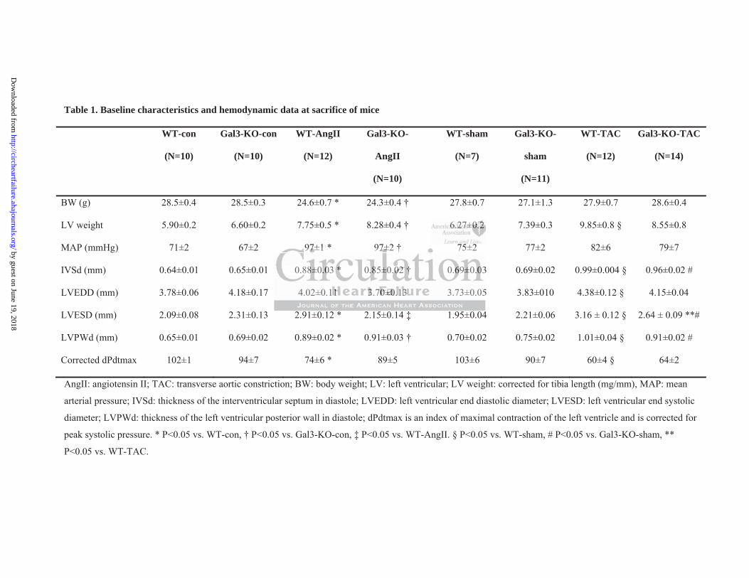
Table 1. Baseline characteristics and hemodynamic data at sacrifice of mice
WT-con
(N=10)
Gal3-KO-con
(N=10)
WT-AngII
(N=12)
Gal3-KO-
AngII
(N=10)
WT-sham
(N=7)
Gal3-KO-
sham
(N=11)
WT-TAC
(N=12)
Gal3-KO-TAC
(N=14)
BW (g) 28.5±0.4 28.5±0.3 24.6±0.7 * 24.3±0.4 † 27.8±0.7 27.1±1.3 27.9±0.7 28.6±0.4
LV weight 5.90±0.2 6.60±0.2 7.75±0.5 * 8.28±0.4 † 6.27±0.2 7.39±0.3 9.85±0.8 § 8.55±0.8
MAP (mmHg) 71±2 67±2 97±1 * 97±2 † 75±2 77±2 82±6 79±7
IVSd (mm) 0.64±0.01 0.65±0.01 0.88±0.03 * 0.85±0.02 † 0.69±0.03 0.69±0.02 0.99±0.004 § 0.96±0.02 #
LVEDD (mm) 3.78±0.06 4.18±0.17 4.02±0.11 3.70±0.13 3.73±0.05 3.83±010 4.38±0.12 § 4.15±0.04
LVESD (mm) 2.09±0.08 2.31±0.13 2.91±0.12 * 2.15±0.14 ‡ 1.95±0.04 2.21±0.06 3.16 ± 0.12 § 2.64 ± 0.09 **#
LVPWd (mm) 0.65±0.01 0.69±0.02 0.89±0.02 * 0.91±0.03 † 0.70±0.02 0.75±0.02 1.01±0.04 § 0.91±0.02 #
Corrected dPdtmax 102±1 94±7 74±6 * 89±5 103±6 90±7 60±4 § 64±2
AngII: angiotensin II; TAC: transverse aortic constriction; BW: body weight; LV: left ventricular; LV weight: corrected for tibia length (mg/mm), MAP: mean
arterial pressure; IVSd: thickness of the interventricular septum in diastole; LVEDD: left ventricular end diastolic diameter; LVESD: left ventricular end systolic
diameter; LVPWd: thickness of the left ventricular posterior wall in diastole; dPdtmax is an index of maximal contraction of the left ventricle and is corrected for
peak systolic pressure. * P<0.05 vs. WT-con, † P<0.05 vs. Gal3-KO-con, ‡ P<0.05 vs. WT-AngII. § P<0.05 vs. WT-sham, # P<0.05 vs. Gal3-KO-sham, **
P<0.05 vs. WT-TAC.
6666666.2.2.222227±7±7±7±7±7±7±0.0.0.0.0.0.0.2222222
97±1 * 97±2 † 75±2
0.88±0.03 * 0.85±0.02 0.69±0.03
4.02±0.11 3.70±0.13 3.73±0.05
97±7±7±7±7 1 * 977777±2±±±± † 75±2
0. 888±0± 00.03 *** ..0.858 ±0±± .000002 2222 † †† 0.666669±± .0.0300
4.02±0.11111 33333.70±0±0±±±0.0.0.0.0.131313133 3.73±0.05
by guest on June 19, 2018http://circheartfailure.ahajournals.org/
Dow
nloaded from
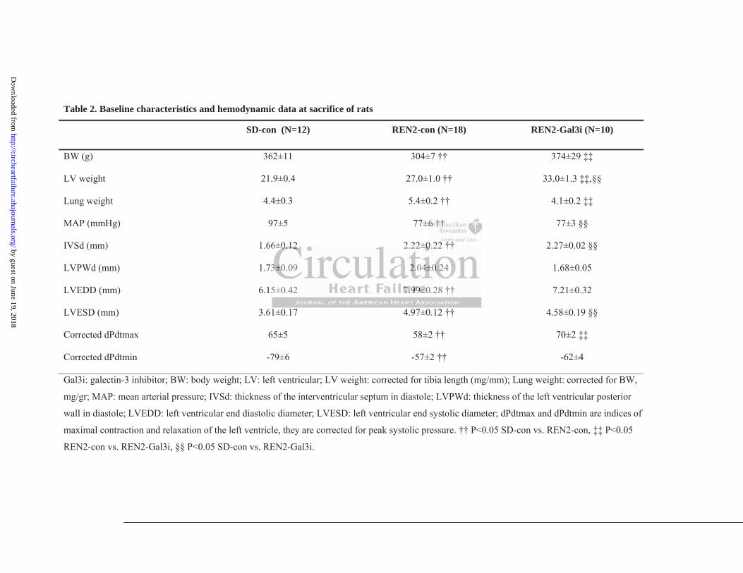
Table 2. Baseline characteristics and hemodynamic data at sacrifice of rats
SD-con (N=12) REN2-con (N=18) REN2-Gal3i (N=10)
BW (g) 362±11 304±7 †† 374±29 ‡‡
LV weight 21.9±0.4 27.0±1.0 †† 33.0±1.3 ‡‡,§§
Lung weight 4.4±0.3 5.4±0.2 †† 4.1±0.2 ‡‡
MAP (mmHg) 97±5 77±6 †† 77±3 §§
IVSd (mm) 1.66±0.12 2.22±0.22 †† 2.27±0.02 §§
LVPWd (mm) 1.73±0.09 2.04±0.24 1.68±0.05
LVEDD (mm) 6.15±0.42 7.99±0.28 †† 7.21±0.32
LVESD (mm) 3.61±0.17 4.97±0.12 †† 4.58±0.19 §§
Corrected dPdtmax 65±5 58±2 †† 70±2 ‡‡
Corrected dPdtmin -79±6 -57±2 †† -62±4
Gal3i: galectin-3 inhibitor; BW: body weight; LV: left ventricular; LV weight: corrected for tibia length (mg/mm); Lung weight: corrected for BW,
mg/gr; MAP: mean arterial pressure; IVSd: thickness of the interventricular septum in diastole; LVPWd: thickness of the left ventricular posterior
wall in diastole; LVEDD: left ventricular end diastolic diameter; LVESD: left ventricular end systolic diameter; dPdtmax and dPdtmin are indices of
maximal contraction and relaxation of the left ventricle, they are corrected for peak systolic pressure. †† P<0.05 SD-con vs. REN2-con, ‡‡ P<0.05
REN2-con vs. REN2-Gal3i, §§ P<0.05 SD-con vs. REN2-Gal3i.
±6±6±666±66 ††††††††††††††
6±0.12 2 22±0 22 ††
3
5
6±00000.1.11.122222 2.22±0±0±0±0±0.22 ††
33333±0±0±0±± .09 2..2. 404±0±0±000 222.224
5±0.42 7.7.7.7.7 9999999999±0±±±± .28 ††
by guest on June 19, 2018http://circheartfailure.ahajournals.org/
Dow
nloaded from
Figure Legends
Figure 1. Hemodynamic data in mice and rats at sacrifice. (a) Outline of the experimental
protocol of the prevention studies in mice and rats. (b) Expression of atrial natriuretic peptide
(ANP) mRNA in mouse hearts. (c) Fractional shortening in mouse hearts assessed with
echocardiography. (d) Left ventricular end diastolic pressure (LVEDP) in mouse hearts. (e)
Isovolumetric relaxation constant Tau in mouse hearts. (f) dPdtmin corrected for peak
systolic pressure in mouse hearts. (g) Expression of ANP mRNA in rat hearts. (h) Fractional
shortening in rat hearts assessed with echocardiography. (i) Change in fractional shortening
in rat hearts assessed with echocardiography at baseline and prior to sacrifice. (j) LVEDP in
rat hearts. (k) Tau in rat hearts. AngII: angiotensin II; TAC: transverse aortic constriction;
Wk: week; Gal3i: galectin-3 inhibitor (N-Lac: N-acetyllactosamine); con: control. N=5-12
per group. * P<0.05 vs. WT-con, † P<0.05 vs. Gal3-KO-con, ‡ P<0.05 vs. WT-AngII, §
P<0.05 vs. WT-sham, # P<0.05 vs. Gal3-KO-sham, ** P<0.05 vs. WT-TAC, †† P<0.05 SD-
con vs. REN2-con, ‡‡ P<0.05 REN2-con vs. REN2-Gal3i, §§ P<0.05 SD-con vs. REN2-
Gal3i, ## P<0.05 vs. all other groups at sacrifice.
Figure 2. Fibrosis in mouse and rat hearts. (a) Typical examples of fibrosis staining with
Masson’s trichrome staining in mouse hearts. (b) Percentage of fibrosis in mouse hearts. (c)
Typical examples of fibrosis staining in rat hearts. (d) Percentage of fibrosis in rat hearts. (e)
Collagen cross-linking represented as collagen digestibility in rat hearts. (f) Level of PINP
measured in rat blood plasma. Con: control; AngII: angiotensin II; TAC: transverse aortic
constriction; Gal3i: galectin-3 inhibitor. N=5-12 per group. * P<0.05 vs. WT-con, ‡ P<0.05
vs. WT-AngII, § P<0.05 vs. WT-sham, ** P<0.05 vs. WT-TAC, †† P<0.05 SD-con vs.
REN2-con, ‡‡ P<0.05 REN2-con vs. REN2-Gal3i, §§ P<0.05 SD-con vs. REN2-Gal3i.
orororororror ttttttto o o o o oo sasasasasasasacrcrcrcrcrcrcrififififififificicicicicicice.e.e.e.e.e.e. (((((((j)j)j)j)j)j)j)
nsversrsrsrsrsrsrseeeeeee aoaoaoaoaoaoaortrtrtrtrtrtrticicicicicicic cccccccoonooog g ;
i: alectin-3 inhibitor (N-Lac: N-acetyllactosamine); con: contro
0.05 vs. WT-con, † P<0.05 vs. Gal3-KO-con, ‡ P<0.05 vs. WT-A
sham, # P<0.05 vs. Gal3-KO-sham, ** P<0.05 vs. WT-TAC, ††
g g ;
i::::: ggggalectin-333 33 innnnnhihihihihibibibibibitototototorrr rr (N(N(N(N(N-L-L-LLLac:::: NN-accetttttylylylyly lalalalalacttctososammmmmiine)e)e)e)e);; cococococon:n:n:nn cccccononononontrtrtrtrro
0.05 5555 vsvsvvsv . WTWTWTWTWT c-conoon, † P<PP<P<P 000.00 055555 vvvvvsss. GGGGGalal3333-3 KOKOKOKOKO-ccccconnn, ‡‡‡ P<P<P<P<<0.0000 0505050505 vs.s WWWWWT-TTTT AAA
shshamamam, ### P<P<P<P 000.050505 vvvsss. GGGalal333-KOKOKO ss-shaham,m,m,,, **** P<P<P<000.050505 vvvsss. WWWTTT-TATATACCC, ††††
by guest on June 19, 2018http://circheartfailure.ahajournals.org/
Dow
nloaded from
Figure 3. Histological analysis of rat hearts. (a) Typical examples of Gomori staining for
cardiomyocyte size, -SMA staining for myofibroblasts, CD68 staining for macrophages and
PCNA staining for proliferating cells. (b) Quantification of cardiomyocyte size. (c)
Quantification of -SMA staining. (d) Quantification of macrophages per mm2. (e)
Quantification of proliferating cells per mm2. Con: control; Gal3i: galectin-3 inhibitor. N=5-8
per group. †† P<0.05 SD-con vs. REN2-con, ‡‡ P<0.05 REN2-con vs. REN2-Gal3i, §§
P<0.05 SD-con vs. REN2-Gal3i.
Figure 4. Effects of galectin-3 and Gal3i on human dermal fibroblasts (HDF) (a) Typical
examples of HDF appearance with or without treatment. (b) Confirmation of fibroblast
phenotype by vimentin staining. (c) Most significantly regulated genes from the Low Density
Array. (d-j) Confirmation of differential expression of genes identified in the low density
array by RT-qPCR: (d) HSP47, (e) COL1A1, (f) COL1A2, (g) COL3A1, (h) P4HB, (i)
PCOLCE, (j) LOXL2. (k) Amount of collagen 3 (COL3) in the tissue culture medium
measured with ELISA after 72h treatment. CM: culture medium; Gal3: galectin-3; N-Lac: N-
acetyllactosamine. N=3 per group (Kruskall-Wallis test).
Figure 5. Changes in gene expression of fibrotic genes in rat hearts. (a-h) mRNA expression
in rat hearts: (a) Galectin-3, (b) Hsp47, (c) Col1a1, (d) Col1a2, (e) Col3a1, (f) P4hb, (g)
Pcolce, (h) Loxl2, N=5-12 per group. (i) Western Blots for galectin-3, Hsp47 and GAPDH.
(j) Galectin-3 protein expression measured with Western Blot. (k) Hsp47 protein expression
measured with Western Blot. N=4 per group. Gal3i: galectin-3 inhibitor. †† P<0.05 SD-con
vs. REN2-con, ‡‡ P<0.05 REN2-con vs. REN2-Gal3i, §§ P<0.05 SD-con vs. REN2-Gal3i.
obobobobobboblalalalalalal stststststststsssssss (H(H(H(H(H(H(HDFDFDFDFDFDFDF))))))) (a(a(a(a(a(a(a)))))))
nfirmamamamamamamatititititititiononononononon ooooooofffffff fififififififibrbbbbpp ( )
mentin staini . (c) Most significantly r ulated enes from the
firmation of differential expression of genes identified in the low
C H
pp ( )
mmmmmeeene tin staiiiininninn ngngngngng. (c(c(c(c(c) ) ) )) MoMoMoMoMossstss sigigigninin fficaanntlylylylyly rrregegeege uulatatatededededed genenenenenesssss fffffrororororom mm mm thththththe eee
firmmmmataaatatioioioi n ofofo dddddifififffefefef rentntntiaiaialll ll expprprprpresesesessisisionon offf ff gegegegeennnenn sss idididdi enentitt fififififiededededed iiiiin ththhhthee lolloll ww
CRCRCR::: (d(d)) HSHSHSH P4P4P47,7,7,, ((((e)e)))) CCCOLOLOL1A1A1A111, ,, (f(f( ) ) ))) COCOCOL1L1L1A2A2A2, ,,,, (g(gggg) ) ))) COCOCOL3L3L3A1A1A1,,,,, (h(h)) P4P4P4HHH
by guest on June 19, 2018http://circheartfailure.ahajournals.org/
Dow
nloaded from
Figure 6. Gal3i inhibits progression of cardiac remodeling in mice. (a) Outline of the
experimental protocol. (b) Left ventricular (LV) weight corrected for tibia length (TL) at
sacrifice. (c) Fractional shortening assessed with echocardiography at sacrifice. (d) Left
ventricular end diastolic pressure (LVEDP). (e) Atrial natriuretic peptide (ANP) mRNA
expression in mouse hearts. (f) Amount of fibrosis in mouse hearts. (g) Typical examples of
Masson’s trichome staining in mouse hearts. TAC: transverse aortic constriction; Wk(s):
week(s); Gal3i: galectin-3 inhibitor; sal: saline, NS: non-significant. N=7-9 per group.
Differences between groups were compared at the eight weeks time point (t-test), baseline
four weeks is depicted as a dotted line.
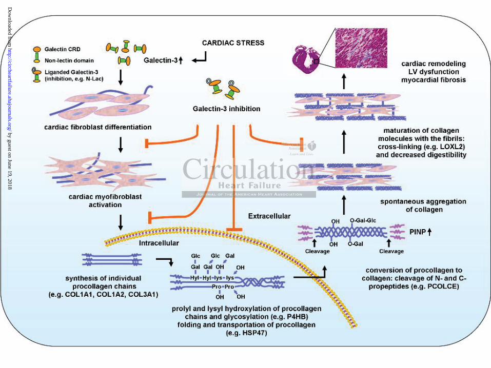
Figure 7. Schematic representation of myocardial fibrogenesis and the effects of galectin-3
and galectin-3 inhibition. Upon cardiac stressors (in our study: AngII, TAC, REN2, and rh-
Galectin-3 treatment), (cardiac) fibroblasts differentiate into myofibroblasts and collagen
production increases. Intracellularly, procollagen chains are processed by various gene
products and then secreted into the interstitium. Here, the procollagens are cleaved to become
collagens and further processed resulting in aggregation, maturation, and cross-linking.
Finally, the deposited cross-linked collagens in the myocardium add to myocardial fibrosis
and dysfunction. Along this process, galectin-3 exerts effects on genes involved in various
steps of fibrogenesis and galectin-3 inhibition with N-Lac inhibits these various steps.
and ttttttthehehehehehehe eeeeeeeffffffffffffffececececececectststststststs ooooofp y g
nhibition. U n cardiac stressors (in our stud A II, TAC, R N
ment), (cardiac) fibroblasts differentiate into myofibroblasts and
ases. Intracellularly, procollagen chains are processed by vario s
p y g
nhhhhhibbbbbition. UUUUUpponnn nn cacacacacardrdrdrdrdiaiaiaiaiac cccc ststsss rererer ssorooro s (((inn ououououour ssstss udududy: AAAAAngggggIIIIIIIIII, TATATATATAC,CC,C,C RRRRRENEEEE
mentntntntt)),))) ((((cardrdddrdiaiac)c)c))) ffffibiii rooblblblasts dddddififififi fefefeff rerrentntntiaiaaaatetetete iiiiinnntnn ooo mymmyofofofibbbbbrorororor blblblblb asststs aaanddnddd
asasaseseses. InInIntrtrtrtracacacelellululalarlrly,y,y,yy ppppprororocococollllagagagenenen ccchahaaainininnnsss ararareee prprprpp ocococesesessesesedd bybyyyy vvvarararioioususus
by guest on June 19, 2018http://circheartfailure.ahajournals.org/
Dow
nloaded from
by guest on June 19, 2018http://circheartfailure.ahajournals.org/
Dow
nloaded from
by guest on June 19, 2018http://circheartfailure.ahajournals.org/
Dow
nloaded from
by guest on June 19, 2018http://circheartfailure.ahajournals.org/
Dow
nloaded from
by guest on June 19, 2018http://circheartfailure.ahajournals.org/
Dow
nloaded from
by guest on June 19, 2018http://circheartfailure.ahajournals.org/
Dow
nloaded from
by guest on June 19, 2018http://circheartfailure.ahajournals.org/
Dow
nloaded from
by guest on June 19, 2018http://circheartfailure.ahajournals.org/
Dow
nloaded from
Bank, Wiek H. van Gilst, Herman H.W. Silljé and Rudolf A. de Boervan der Harst, Bertram Pitt, Irwin J. Goldstein, Jasper A. Koerts, Dirk J. van Veldhuisen, Ruud A.
Lili Yu, Willem P.T. Ruifrok, Maxi Meissner, Eelke M. Bos, Harry van Goor, Bahram Sanjabi, PimInterfering with Myocardial Fibrogenesis
Genetic and Pharmacological Inhibition of Galectin-3 Prevents Cardiac Remodeling by
Print ISSN: 1941-3289. Online ISSN: 1941-3297 Copyright © 2012 American Heart Association, Inc. All rights reserved.
is published by the American Heart Association, 7272 Greenville Avenue, Dallas, TX 75231Circulation: Heart Failure published online December 10, 2012;Circ Heart Fail.
http://circheartfailure.ahajournals.org/content/early/2012/12/10/CIRCHEARTFAILURE.112.971168World Wide Web at:
The online version of this article, along with updated information and services, is located on the
http://circheartfailure.ahajournals.org/content/suppl/2012/12/10/CIRCHEARTFAILURE.112.971168.DC1Data Supplement (unedited) at:
http://circheartfailure.ahajournals.org//subscriptions/
is online at: Circulation: Heart Failure Information about subscribing to Subscriptions:
http://www.lww.com/reprints Information about reprints can be found online at: Reprints:
document. Permissions and Rights Question and Answer process is available in the
click Request Permissions in the middle column of the Web page under Services. Further information about thisEditorial Office. Once the online version of the published article for which permission is being requested is located,
can be obtained via RightsLink, a service of the Copyright Clearance Center, not theCirculation: Heart Failure Requests for permissions to reproduce figures, tables, or portions of articles originally published inPermissions:
by guest on June 19, 2018http://circheartfailure.ahajournals.org/
Dow
nloaded from
1
SUPPLEMENTAL MATERIAL
Genetic and pharmacological inhibition of galectin-3 prevents cardiac remodeling by
interfering with myocardial fibrogenesis
Lili Yu 1,2 * MD; Willem P.T. Ruifrok 1 * MD, PhD; Maxi Meissner 1 PhD; Eelke M. Bos 3 MD;
Harry van Goor 3 PhD; Bahram Sanjabi 4 MSc; Pim van der Harst 1 MD, PhD; Bertram Pitt 5 MD;
Irwin J. Goldstein 6 PhD; Jasper A. Koerts 3 MSc; Dirk J. van Veldhuisen1 MD, PhD; Ruud A.
Bank 3 PhD, Wiek H. van Gilst 1 PhD; Herman H.W. Silljé 1 PhD; Rudolf A. de Boer 1 MD, PhD
1 Department of Cardiology, University Medical Center Groningen, University of Groningen,
Groningen, the Netherlands
2 Harbin Medical University, Harbin, P.R. China
3 Department of Pathology and Medical Biology, University Medical Center Groningen,
University of Groningen, Groningen, the Netherlands
4 Department of Genetics, University Medical Center Groningen, University of Groningen,
Groningen, the Netherlands
5 University of Michigan Medical School, Department of Medicine, Ann Arbor, MI, U.S.A.
6 University of Michigan Medical School, Department of Biological Chemistry, Ann Arbor, MI,
U.S.A.
* These authors contributed equally to this work
2
Supplemental methods
Animal models
We studied six- to ten-week old male mice deficient for the gene encoding galectin-3 (galectin-3
knock-out mice (Gal3-KO)). The Gal3-KO homozygous mice carrying a targeted mutation in
galectin-3 (a lectin, galactose binding soluble 3 or Lgals3) have been generated and are bred at
Jackson Laboratory (Bar Harbor, ME, USA) and shipped to Groningen, The Netherlands. A
targeting vector containing neomycin resistance and herpes simplex virus thymidine kinase genes
was used to disrupt 3.7kb of sequence that includes exons 2, 3 and 4. The construct was
electroporated into WW6 embryonic stem (ES) cells (derived from 129/Sv, C57BL/6 and SJL
mixed background mice). Correctly targeted ES cells were injected into outbred MF-1
blastocysts. The resulting chimeric male animals were crossed with 129 female mice and the
mutant strain was backcrossed with C57BL/6 for five generations. For an overview of the
generation of the Gal3-KO mice, see Supplemental Figure S1. Other researchers have generated
Gal3-KO mice as well.1-3 Transgene-negative male wild-type (WT) served as controls. For the
reversal experiment, we used six- to eight-week old male C56Bl6/J mice (Harlan, The
Netherlands).
We also studied six-week old, male, homozygous TGR(mREN2)27 rats (REN2). These rats
overexpress the mouse renin-2 gene (ren-2d) and have a phenotype of severe hypertension and
left ventricular (LV) hypertrophy which culminates into heart failure (HF) and low blood pressure
over the course of 12-16 weeks.4-7 The rats were bred at the Max Delbrück Center for Molecular
Medicine (Berlin Buch, Germany). Since Sprague Dawley (SD) rats represent the appropriate
control strain for REN2 rats, age-matched male SD rats were used as controls (Harlan, The
Netherlands).6
Animals were housed under standard condition. All animal studies were approved by the Animal
Ethical Committee of the University of Groningen, The Netherlands, and conducted in
accordance with existing guidelines for the care and use of laboratory animals.
Prevention study - mouse models
We induced cardiac remodeling by two interventions. First, cardiac remodeling was provoked by
subcutaneous administration of angiotensin II (AngII) via osmotic minipumps for 14 days.
Second, we induced pressure overload and cardiac remodeling by transverse aortic constriction
(TAC) for 28 days. The control group received saline infusion via osmotic minipumps for 14
days. In total, six groups were studied: WT-con (control, N=10), Gal3-KO-con (N=10), WT-
3
AngII (N=12), Gal3-KO-AngII (N=10), WT-sham (N=7), WT-TAC (N=12), Gal3-KO-sham
(N=11), and Gal3-KO-TAC (N=14).
AngII (Bachem AG, Bubendorf, Switzerland) was dissolved in 0.9% NaCl and injected into
osmotic minipumps (Alzet 2004, Durect Corporation, Cupertino, CA, USA). The dose delivered
by the osmotic minipumps was 2.5 µg/Kg/day. The minipumps were inserted subcutaneously on
the back of the mice.
The TAC is a well established model.8 In brief, mice were anesthetized with oxygen and
isoflurane (2%), intubated and ventilated. The thoracic cavity was opened between the second
and the third intercostal cavity. Then, a blunted needle (27G) was placed on the aortic arch
between both carotid arteries and, with an 8-0 nylon suture, the aorta was tied onto the needle.
Immediately after, the needle was removed creating a reproducible stenosis of the aorta of about
50%. Sham-operated mice served as controls. Mice were sacrificed after 28 days.
Reversal study - mouse model
The TAC model was used as described above. Mice were left untreated for 28 days. After 28
days, one third of all animals were sacrificed. The remainder were given intraperitoneal injections
of the galectin-3 inhibitor (Gal3i) N-acetyllactosamine (N-Lac) (Sigma-Aldrich, Zwijndrecht,
The Netherlands) that targets the carbohydrate recognition domain (CRD) of galectin-3 at a final
dose of 5 mg/Kg/day three times per week. N-Lac has been shown to effectively bind galectin-3.9
The treatment period lasted another 28 days and the total study duration was 56 days (eight
weeks). Control mice were injected with saline. In total, three groups were studied: WT-TAC-
control (N=9), WT-TAC-saline (N=9) and WT-TAC-Gal3i (N=9).
Rat model
To study the effect of galectin-3 binding protein on cardiac remodeling, we allocated SD and
REN2 rats to different treatment regimens. REN2 rats were treated with N-Lac at a final dose of 5
mg/Kg/day injected intraperitoneally three times per week. SD rats were used as controls for all
groups. Rats were sacrificed at week seven of the experimental protocol (when aged ~13 weeks).
In total, four groups were studied: SD-con (control, N=12), REN2-con (N=18), SD-Gal3i (N=8)
and REN2-Gal3i (N=10). Treatment with Gal3i did not exert any effects in control SD rats and,
therefore, the results shown are limited to the REN2 groups.
4
Echocardiography
Cardiac function was assessed by echocardiography at baseline and prior to sacrifice with Vivid 7
(GE Healthcare, Chalfont St Giles, UK) equipped with a 10-MHz (rats) and a 13-MHz (mice)
phase array linear transducer), as described previously.10 The echocardiographic measurements
were performed under general anesthesia with 2% isoflurane. Both 2-dimensional (2D) images in
parasternal long-axis and short-axis view and 2-D guided M-mode tracing were obtained.
Parasternal long-axis (PLAX) views were obtained in order to ensure that the mitral and aortic
valves and the apex were visualized. Left ventricular outflow tract (LVOT) diameter was
measured in PLAX. Short-axis views were recorded at the level of mid-papillary muscles. A total
of three loops was recorded and used for calculations. LV end-systolic diameter (LVESD) and
LV end-diastolic diameter (LVEDD) were measured from M-mode tracings. LV fractional
shortening was calculated as (LVEDD − LVESD)/LVEDD × 100%. Cardiac output was
calculated by echo Doppler measurements over the aortic valve (pulsed wave doppler) using the
LVOT diameter. To monitor the development of cardiac hypertrophy in both mice and rats,
echocardiography was performed at baseline and prior to sacrifice.
Measurement of hemodynamic function
Hemodynamic function was assessed invasively as described previously11 by introducing a 0.8-
French (for mice) and a 1.4 French (for rats) microtip pressure-volume transducer (Millar Instr.
Inc., Houston, TX, USA) via the right carotid artery into the aorta. A three minute period was
allowed for stabilization and then systolic and diastolic blood pressure and heart rate (HR) were
recorded in the aorta (average of 20 heart cycles). The catheter was then advanced into the LV.
Peak systolic pressure, LV end diastolic pressure (LVEDP), maximal indices of LV contraction
and relaxation (dPdtmax and dPdtmin) as well as the relaxation constant Tau were measured.
dPdtmax and dPdtmin were corrected for peak systolic pressure to correct for differences in
afterload, as described previously.4
Tissue procurement
After measuring hemodynamics, blood was drawn (PINP assay) and the hearts were rapidly
excised and weighed. Myocardial tissue was dissected transversally and processed for
immunohistochemistry or snap frozen for molecular analysis.
5
Immunohistochemistry
Hearts were isolated and fixed with buffered 3.7% formalin for 24 hours. Thereafter, tissue
sections were dehydrated and embedded in paraffin. Another slice was embedded in tissue-tec,
placed on a carton disk put on a petri-dish and immediately, but slowly, frozen above liquid
nitrogen. For immunohistochemistry, 3 μm-thick sections were cut on a microtome and mounted
on slides. Hearts were cut at the mid-papillary level.
To measure fibrosis, Masson’s trichrome staining was performed on paraffin sections for all
experimental animals. Whole stained sections were scanned (Nanozoomer 2.0-HT, Hamamatsu,
Japan) and fibrosis score for the entire section was calculated at 20x magnification for mice and
10x magnification for rats (ScanScope, Aperio Technologies, Vista, CA, USA). To measure
cardiomyocyte size, deparaffinized sections were stained with Gomori's silver staining.12
Cardiomyocyte size was measured as the cross-sectional area of transversally cut cardiomyocytes
at 40x magnification for rats. Myofibroblasts were stained with a monoclonal mouse anti-human
smooth muscle actin (SMA) antibody (#M0851, DAKO, Heverlee, Belgium). SMA in the entire
section was calculated at 10x magnification by the SanScope software for rats. To measure the
number of macrophages, macrophages were stained with a mouse anti-rat CD68 (ED1) antibody
(#MCA341R, AbD Serotec, Oxford, UK) for rats. The number of macrophages was calculated for
the entire section using the SanScope software at 40x magnification for rats. Proliferating cells
were stained with a mouse monoclonal anti-PCNA antibody [PC10]-proliferation marker (#ab29,
Abcam, Cambridge, UK). The number of proliferating cells in the entire section was calculated
by the SanScope software at 40x magnification for rats.
To stain galectin-3 in cardiac tissue, an anti-galectin-3 monoclonal antibody (Thermo Fisher
Scientific, Landsmeer, The Netherlands) was used. Briefly, paraffin sections were dewaxed and
subjected to an antigen retrieval procedure by incubating overnight at 80°C in 0.1M Tris/HCl, pH
9.0. After three washes with TBS, endogenous peroxidase was blocked and sections were
incubated with the primary antibody (anti-galectin-3 antibody, 1:100) in 1% BSA/PBS for 1 hour
at room temperature. The sections were then incubated with a secondary antibody (polyclonal
rabbit anti-mouse IgG/HRP, 1:100) diluted in 1% BSA/PBS buffer for two hours. The slides were
first stained with 3-animo-9-ethylcarbazole and counterstained with hematoxillin. For typical
examples of galectin-3 staining in cardiac tissue, see supplemental results (Supplemental Figures
S3 and S4).
6
Enzyme-linked immunosorbent assay (ELISA)
Procollagen type I N-terminal propeptide (PINP), a marker of collagen metabolism, was analyzed
in plasma using a commercial Enzyme Linked Immunosorbent Assay according to the
manufacturer’s instructions (rat procollagen I N-terminal propeptide ELISA kit #E90957Ra, Uscn
Life Science Inc, Wuhan, China). Human collagen type III (Human Collagen Type III (Col III)
ELISA Kit, Cat No. Hu9614, TSZ ELISA, Framingham, USA) was analyzed in human dermal
fibroblast supernatant (medium).
Collagen cross-linking assay
Collagen digestibility was measured following the digestion of tissue samples (for 6 hours at 37°
C) with Clostridium histolyticum collagenase (Sigma) at a final concentration of 5 μg enzyme/mg
tissue in a 50 mM Tris buffer containing 5 mM CaCl2 , 0.15 M NaCl, 1 μM ZnCl2, 0.02% (w/v)
NaN3 and 0.01% (v/v) Brij 35. After incubation, the supernatant and remaining tissue were
separated and hydrolyzed in 6 M HCl at 110°C for 20-24 hours. The relative amount of collagen
in the supernatant and remaining tissue was estimated by measuring the amount of
hydroxyproline in the hydrolysates of both the supernatant and the remaining tissue.13
Quantitative real-time PCR
cDNA synthesis was performed with 0.5 µg total RNA using a specific cDNA synthesis kit
according to the manufacturer’s protocol (Quantitect Rev. Transcriptase kit, Qiagen, Venlo, The
Netherlands), as described previously.10,14,15 Quantitative real-time PCR (RT-qPCR) was
performed using SYBR Green mix according to the manufacturer’s protocol (Absolute SYBR
Green ROX mix, Thermo Scientific, Breda, The Netherlands) on C1000 Thermal Cycler CFX384
Real-Time PCR Detection System (Bio-Rad Laboratories, Veenendaal, The Netherlands). All
targets were evaluated under the same experimental conditions (95°C for 15 minutes, then 36
cycles at 95° C for 15 seconds and 60°C for 30 seconds). Samples were analyzed by
quantification software (Bio-Rad CFX Manager 1.6). mRNA levels were expressed in relative
units based on a standard curve obtained with serial dilutions of a calibrator cDNA mixture. To
normalize expression data, reference genes were used. Reference genes were chosen with little
sample-to-sample variability (GAPDH and 36B4). See Supplemental Tables S1, S2, and S3 for a
list of primers used.
7
Western blotting
Rat hearts were homogenized in ice-cold RIPA (50 mM Tris pH 8.0, 1% nonidet P40, 0.5%
deoxycholate, 0.1% SDS, 150 mM NaCl) containing phosphatase inhibitor cocktail 1 (Sigma),
protease inhibitor (ROCHE), 1 mM phenylmethylsulfonyl fluoride (PMSF) and 15 mM
NaVanadate for 30 min. Equal amounts of protein (20 µg) were loaded on 12% polyacrylamide
gels. After electrophoresis, the gels were blotted onto nitrocellulose membranes. Membranes
were then incubated with anti-galectin-3 antibody (Thermo Fisher Scientific, Landsmeer, The
Netherlands), anti-Hsp47 antibody (#ab109117, Abcam, Cambridge, UK), anti-mac-2 antibody
(CL8942AP, Cedarlanelabs, USA), troponin T (T6277, Sigma, Netherlands), anti-vimentin
antibody (V5255, Sigma, Netherlands), anti-CD68 antibody (ab76308, Abcam, Cambridge, UK)
and anti-GAPDH antibody (10R-G109a, Fitzgerald, USA) overnight at 4° C. Membranes were
washed and incubated with the appropriate secondary HRP-conjugated antibody and signals were
visualized with ECL and analyzed with densitometry (Syngene, Cambridge, United
Kingdom).4,14,15
Human adult dermal fibroblast culture experiments
Human adult dermal fibroblasts (primary cells) were cultured for 72 hours in culture medium
(EMEM containing 1% Pen/Strep, 1% L-glutamine, 0.5% FCS and 0.173 mM ascorbid acid 2-
phosphate) with or without galectin-3 (10 μg/mL; #450-38, Preprotech, Rocky Hill, NY, USA),
N-Lac (10 mM) or a combination. The initial seeding density was 15,000 cells/cm2. Cells were
harvested and RNA was isolated. cDNA synthesis was conducted using RevertAid First Strand
cDNA Synthesis Kit (#K1622, Fermentas, St. Leon-Rot, Germany) and cDNA was hybridized to
a custom made low density array carrying probes for ~50 genes involved in fibrosis (collagen
production, collagen cross-linking, collagen processing and collagen degradation). The complete
list of gene is presented in Table S4 of the Supplemental data. Genes that were most significantly
expressed (LOXL2, HSP47, PCOLCE, COL1A1, COL1A2, COL3A1, and P4HB) were verified
using quantitative RT-qPCR and normalized against the reference gene GAPDH. In addition,
vimentin staining, a fibroblast marker, was carried out with anti-vimentin (C-20; Santa Cruz
Biotechnology, Santa Cruz, CA, USA) and protein levels of collagen type 3 were measured with
ELISA (human collagen 3 ELISA kit, #Hu9614, TSZ Scientific LLC, Framingham, MA, USA).
Collagen type 3 protein levels were measured in singulo, therefore error bars and statistics are not
provided.
8
Analyzes of galectin-3 expression in macrophages and in adult rat cardiac myocytes and
fibroblast
Adult rat cardiomyocytes and fibroblasts were isolated using a Langendorff perfusion system
with retrograde perfusion.16 The perfusion solution (10 mM HEPES (pH 7.4), 133.5 mM NaCl,
4.0 mM KCl, 1.2 mM NaH2PO4, 1.2 mM MgSO4, 20 M CaCl2) contained 0.2mg/ml Liberase
TM Research Grade (Roche) to digest the extracellular matrix. Cardiomyocytes were
subsequently separated from other cells by several rounds of velocity sedimentation in a
perfusion solution containing 1mg BSA/ml. Adult cardiomyocytes were directly frozen after
isolation. Cardiac fibroblasts were cultured for several days in DMEM medium supplemented
with 10% FCS and penicillin-streptomycin (100 IU/ml and 100ug/ml, respectively) to obtain
sufficient cells for analysis. A rat alveolar macrophage cell line17 was also cultured in the same
medium. Cell lysates were generated as described in the Western blot section and equal amounts
of proteins were subsequently loaded on a SDS-PAGE gel.
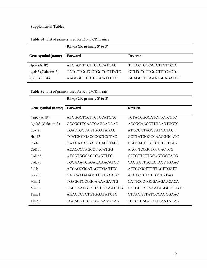
9
Supplemental Tables
Table S1. List of primers used for RT-qPCR in mice
RT-qPCR primer, 5’ to 3’
Gene symbol (name) Forward Reverse
Nppa (ANP) ATGGGCTCCTTCTCCATCAC TCTACCGGCATCTTCTCCTC
Lgals3 (Galectin-3) TATCCTGCTGCTGGCCCTTATG GTTTGCGTTGGGTTTCACTG
Rplp0 (36B4) AAGCGCGTCCTGGCATTGTC GCAGCCGCAAATGCAGATGG
Table S2. List of primers used for RT-qPCR in rats
RT-qPCR primer, 5’ to 3’
Gene symbol (name) Forward Reverse
Nppa (ANP) ATGGGCTCCTTCTCCATCAC TCTACCGGCATCTTCTCCTC
Lgals3 (Galectin-3) CCCGCTTCAATGAGAACAAC ACCGCAACCTTGAAGTGGTC
Loxl2 TGACTGCCAGTGGATAGAC ATGCGGTAGCCATCATAGC
Hsp47 TCATGGTGACCCGCTCCTAC GCTTATGGGCCAAGGGCATC
Pcolce GAAGAAAGGAGCCAGTTACC GGGCACTTTCTCTTGCTTAG
Col1a1 ACAGCGTAGCCTACATGG AAGTTCCGGTGTGACTCG
Col1a2 ATGGTGGCAGCCAGTTTG GCTGTTCTTGCAGTGGTAGG
Col3a1 TGGAAACCGGAGAAACATGC CAGGATTGCCATAGCTGAAC
P4hb ACCAGCGCATACTTGAGTTC ACTCCGGTTTGTACTTGGTC
Gapdh CATCAAGAAGGTGGTGAAGC ACCACCCTGTTGCTGTAG
Mmp2 TGAGCTCCCGGAAAAGATTG CATTCCCTGCGAAGAACACA
Mmp9 CGGGAACGTATCTGGAAATTCG CATGGCAGAAATAGGCCTTGTC
Timp1 AGAGCCTCTGTGGATATGTC CTCAGATTATGCCAGGGAAC
Timp2 TGGACGTTGGAGGAAAGAAG TGTCCCAGGGCACAATAAAG
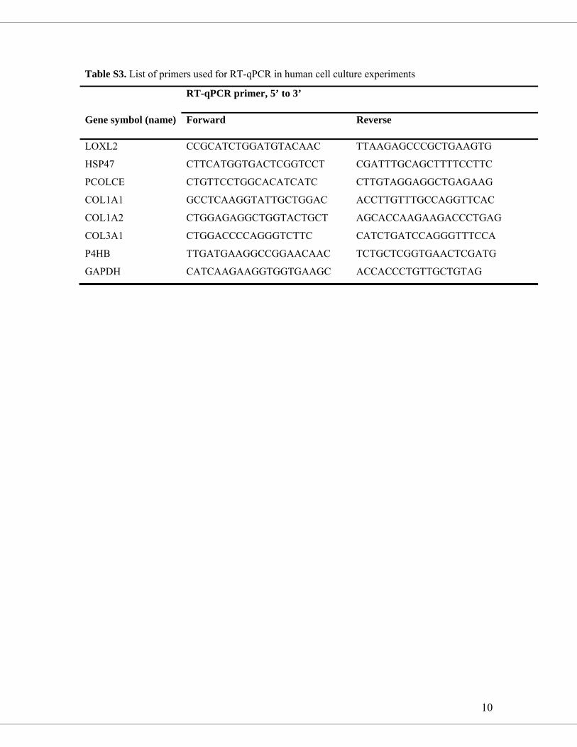
10
Table S3. List of primers used for RT-qPCR in human cell culture experiments
RT-qPCR primer, 5’ to 3’
Gene symbol (name) Forward Reverse
LOXL2 CCGCATCTGGATGTACAAC TTAAGAGCCCGCTGAAGTG
HSP47 CTTCATGGTGACTCGGTCCT CGATTTGCAGCTTTTCCTTC
PCOLCE CTGTTCCTGGCACATCATC CTTGTAGGAGGCTGAGAAG
COL1A1 GCCTCAAGGTATTGCTGGAC ACCTTGTTTGCCAGGTTCAC
COL1A2 CTGGAGAGGCTGGTACTGCT AGCACCAAGAAGACCCTGAG
COL3A1 CTGGACCCCAGGGTCTTC CATCTGATCCAGGGTTTCCA
P4HB TTGATGAAGGCCGGAACAAC TCTGCTCGGTGAACTCGATG
GAPDH CATCAAGAAGGTGGTGAAGC ACCACCCTGTTGCTGTAG
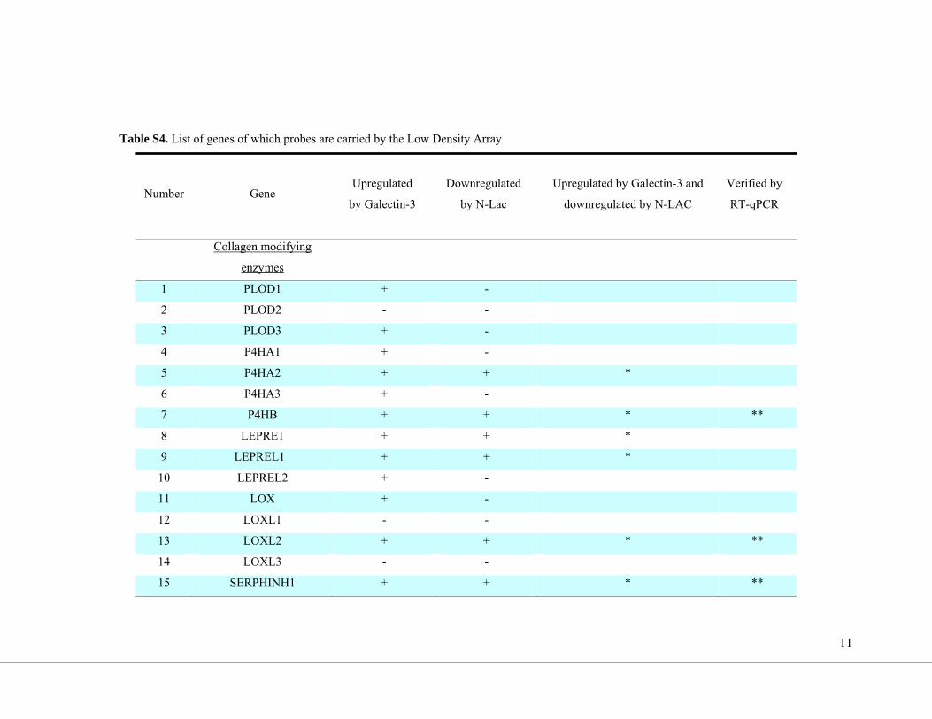
11
Table S4. List of genes of which probes are carried by the Low Density Array
Number Gene Upregulated
by Galectin-3
Downregulated
by N-Lac
Upregulated by Galectin-3 and
downregulated by N-LAC
Verified by
RT-qPCR
Collagen modifying
enzymes
1 PLOD1 + -
2 PLOD2 - -
3 PLOD3 + -
4 P4HA1 + -
5 P4HA2 + + *
6 P4HA3 + -
7 P4HB + + * **
8 LEPRE1 + + *
9 LEPREL1 + + *
10 LEPREL2 + -
11 LOX + -
12 LOXL1 - -
13 LOXL2 + + * **
14 LOXL3 - -
15 SERPHINH1 + + * **
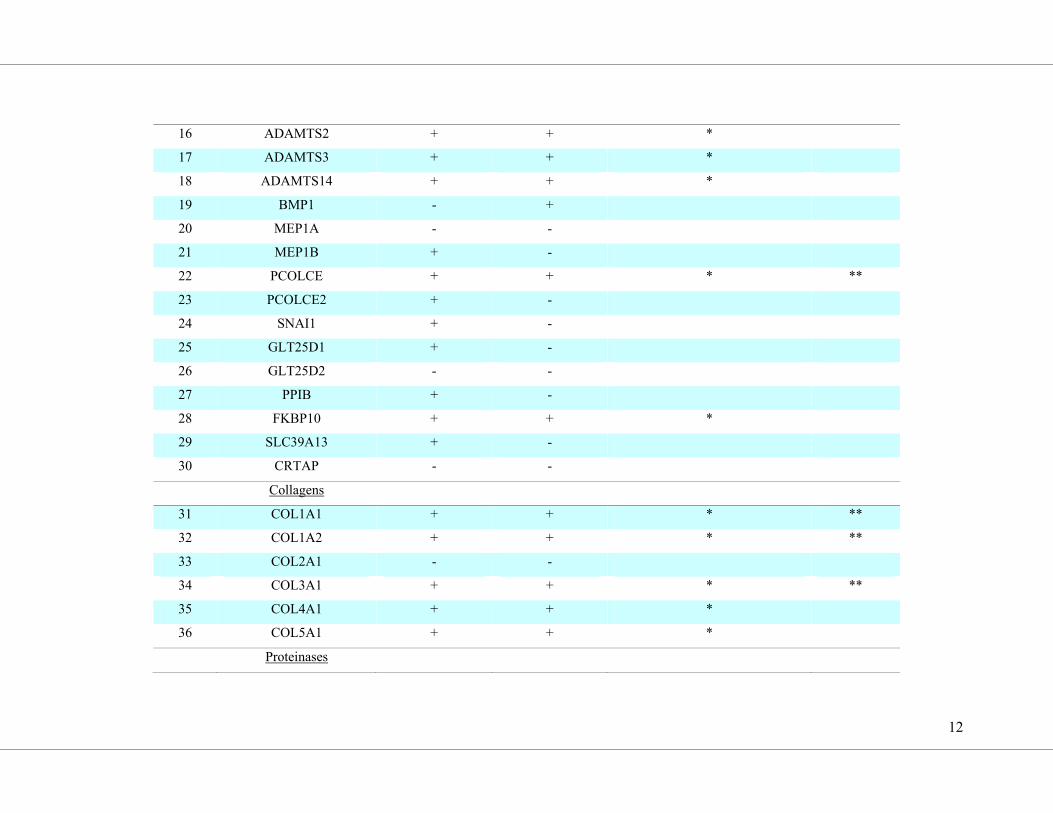
12
16 ADAMTS2 + + *
17 ADAMTS3 + + *
18 ADAMTS14 + + *
19 BMP1 - +
20 MEP1A - -
21 MEP1B + -
22 PCOLCE + + * **
23 PCOLCE2 + -
24 SNAI1 + -
25 GLT25D1 + -
26 GLT25D2 - -
27 PPIB + -
28 FKBP10 + + *
29 SLC39A13 + -
30 CRTAP - -
Collagens
31 COL1A1 + + * **
32 COL1A2 + + * **
33 COL2A1 - -
34 COL3A1 + + * **
35 COL4A1 + + *
36 COL5A1 + + *
Proteinases
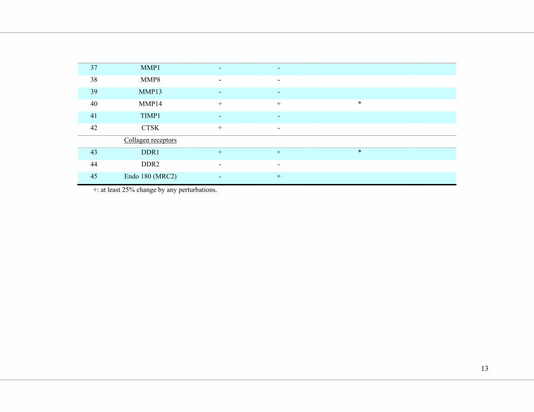
13
37 MMP1 - -
38 MMP8 - -
39 MMP13 - -
40 MMP14 + + *
41 TIMP1 - -
42 CTSK + -
Collagen receptors
43 DDR1 + + *
44 DDR2 - -
45 Endo 180 (MRC2) - +
+: at least 25% change by any perturbations.
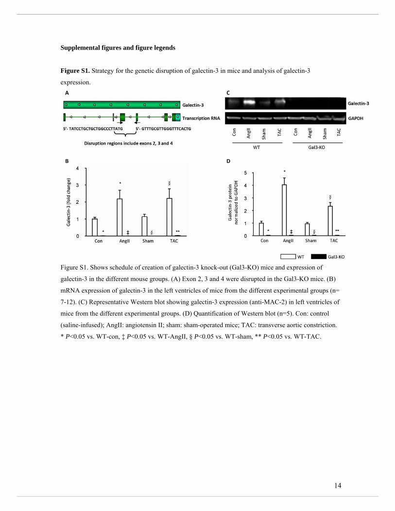
14
Supplemental figures and figure legends
Figure S1. Strategy for the genetic disruption of galectin-3 in mice and analysis of galectin-3
expression.
Figure S1. Shows schedule of creation of galectin-3 knock-out (Gal3-KO) mice and expression of
galectin-3 in the different mouse groups. (A) Exon 2, 3 and 4 were disrupted in the Gal3-KO mice. (B)
mRNA expression of galectin-3 in the left ventricles of mice from the different experimental groups (n=
7-12). (C) Representative Western blot showing galectin-3 expression (anti-MAC-2) in left ventricles of
mice from the different experimental groups. (D) Quantification of Western blot (n=5). Con: control
(saline-infused); AngII: angiotensin II; sham: sham-operated mice; TAC: transverse aortic constriction.
* P<0.05 vs. WT-con, ‡ P<0.05 vs. WT-AngII, § P<0.05 vs. WT-sham, ** P<0.05 vs. WT-TAC.
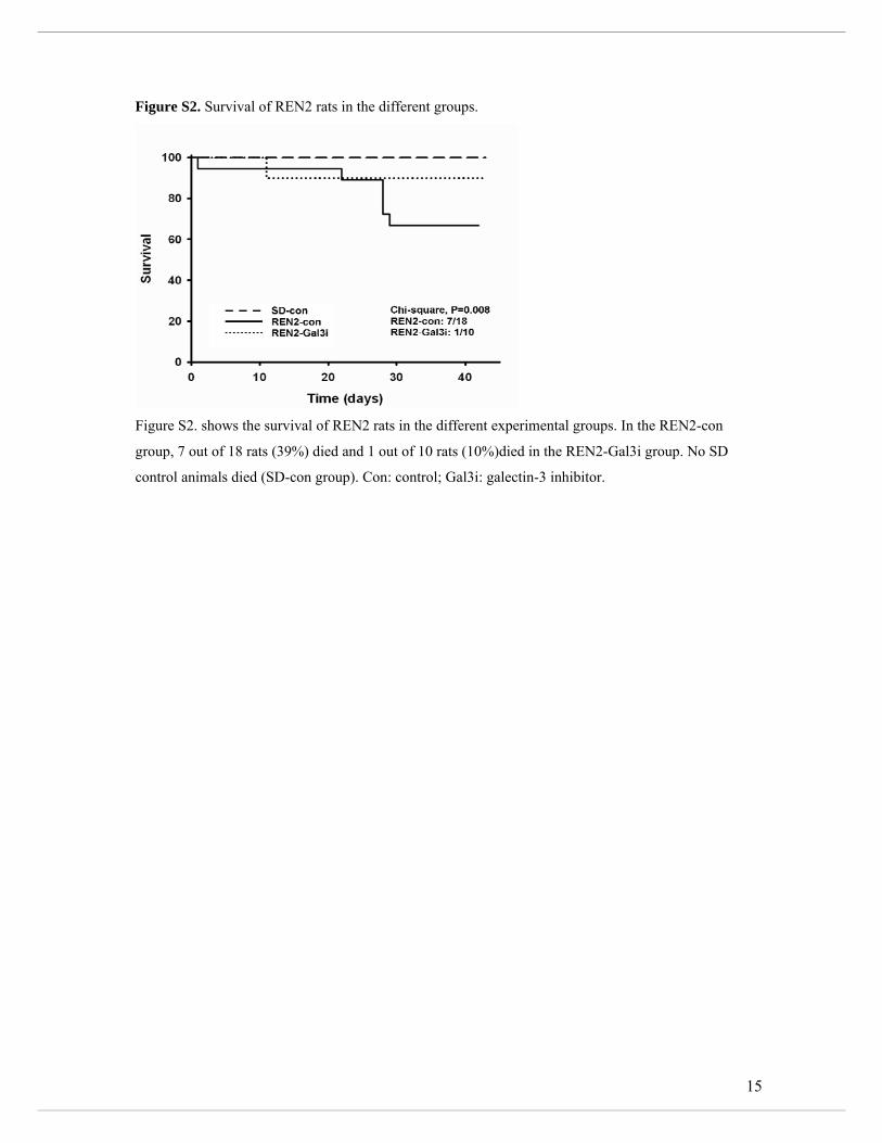
15
Figure S2. Survival of REN2 rats in the different groups.
Figure S2. shows the survival of REN2 rats in the different experimental groups. In the REN2-con
group, 7 out of 18 rats (39%) died and 1 out of 10 rats (10%)died in the REN2-Gal3i group. No SD
control animals died (SD-con group). Con: control; Gal3i: galectin-3 inhibitor.
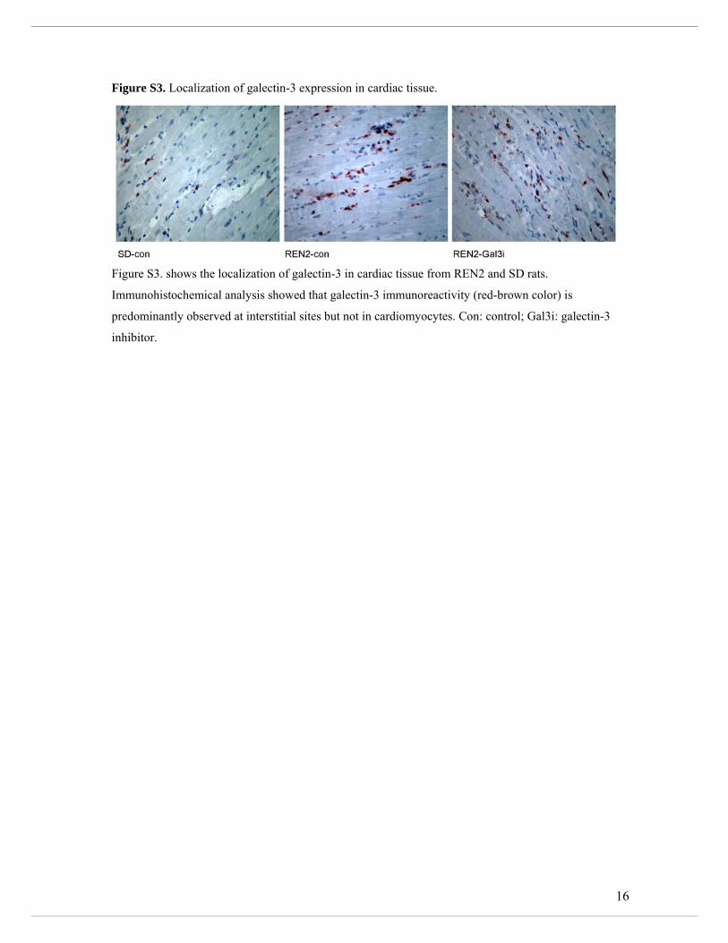
16
Figure S3. Localization of galectin-3 expression in cardiac tissue.
Figure S3. shows the localization of galectin-3 in cardiac tissue from REN2 and SD rats.
Immunohistochemical analysis showed that galectin-3 immunoreactivity (red-brown color) is
predominantly observed at interstitial sites but not in cardiomyocytes. Con: control; Gal3i: galectin-3
inhibitor.
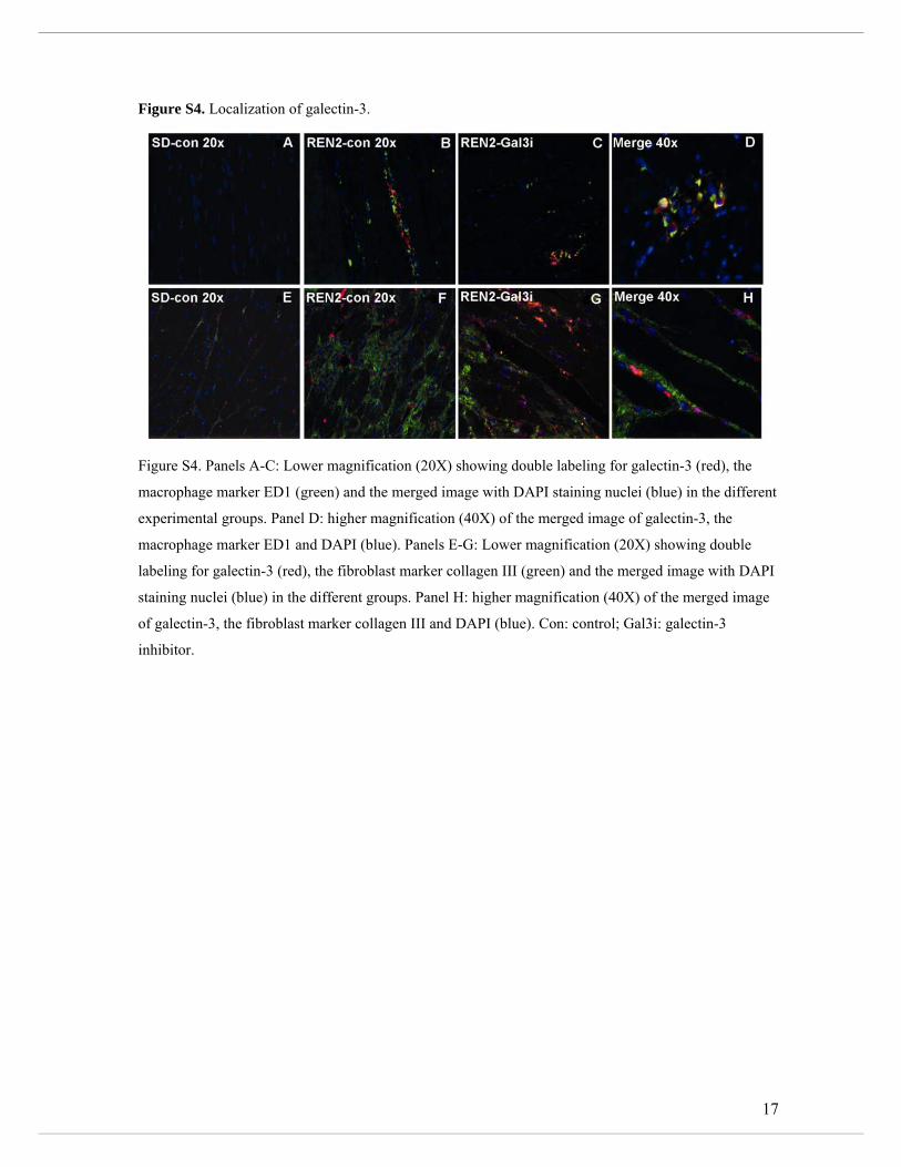
17
Figure S4. Localization of galectin-3.
Figure S4. Panels A-C: Lower magnification (20X) showing double labeling for galectin-3 (red), the
macrophage marker ED1 (green) and the merged image with DAPI staining nuclei (blue) in the different
experimental groups. Panel D: higher magnification (40X) of the merged image of galectin-3, the
macrophage marker ED1 and DAPI (blue). Panels E-G: Lower magnification (20X) showing double
labeling for galectin-3 (red), the fibroblast marker collagen III (green) and the merged image with DAPI
staining nuclei (blue) in the different groups. Panel H: higher magnification (40X) of the merged image
of galectin-3, the fibroblast marker collagen III and DAPI (blue). Con: control; Gal3i: galectin-3
inhibitor.
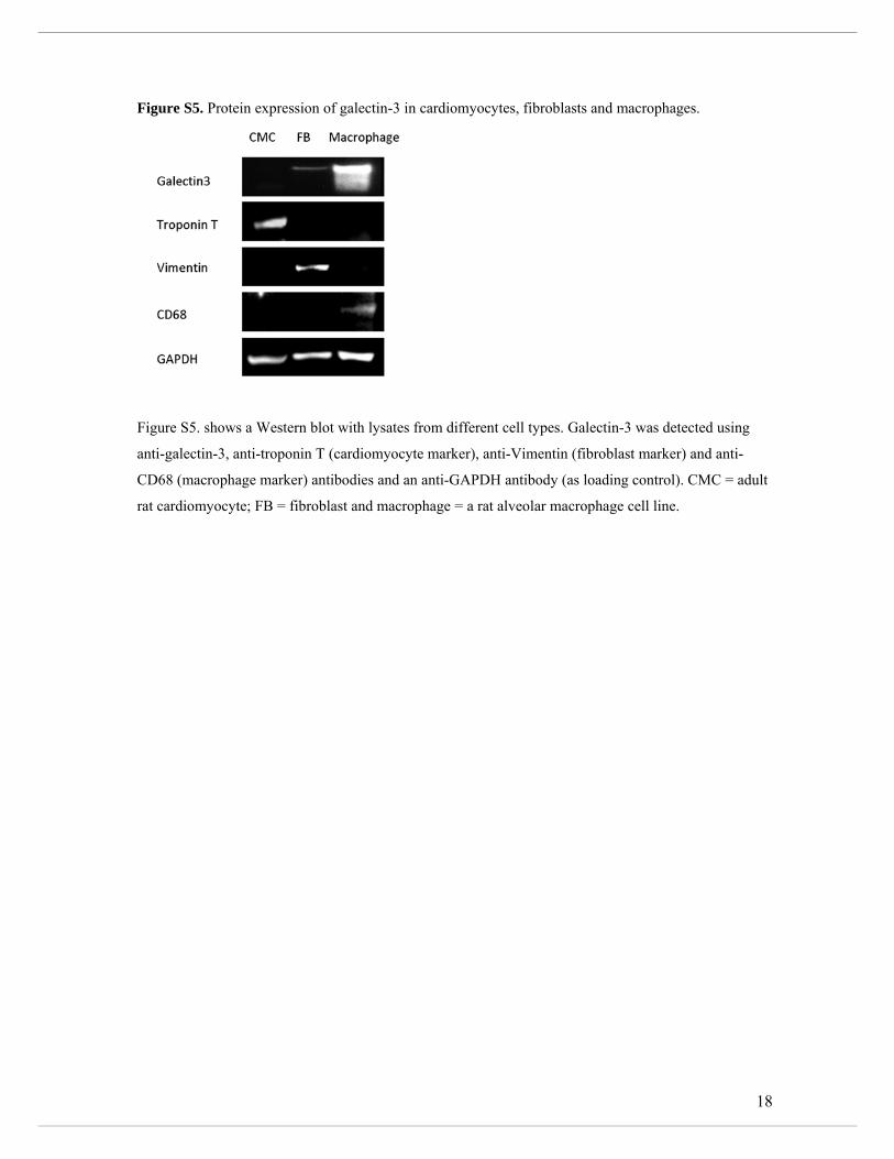
18
Figure S5. Protein expression of galectin-3 in cardiomyocytes, fibroblasts and macrophages.
Figure S5. shows a Western blot with lysates from different cell types. Galectin-3 was detected using
anti-galectin-3, anti-troponin T (cardiomyocyte marker), anti-Vimentin (fibroblast marker) and anti-
CD68 (macrophage marker) antibodies and an anti-GAPDH antibody (as loading control). CMC = adult
rat cardiomyocyte; FB = fibroblast and macrophage = a rat alveolar macrophage cell line.
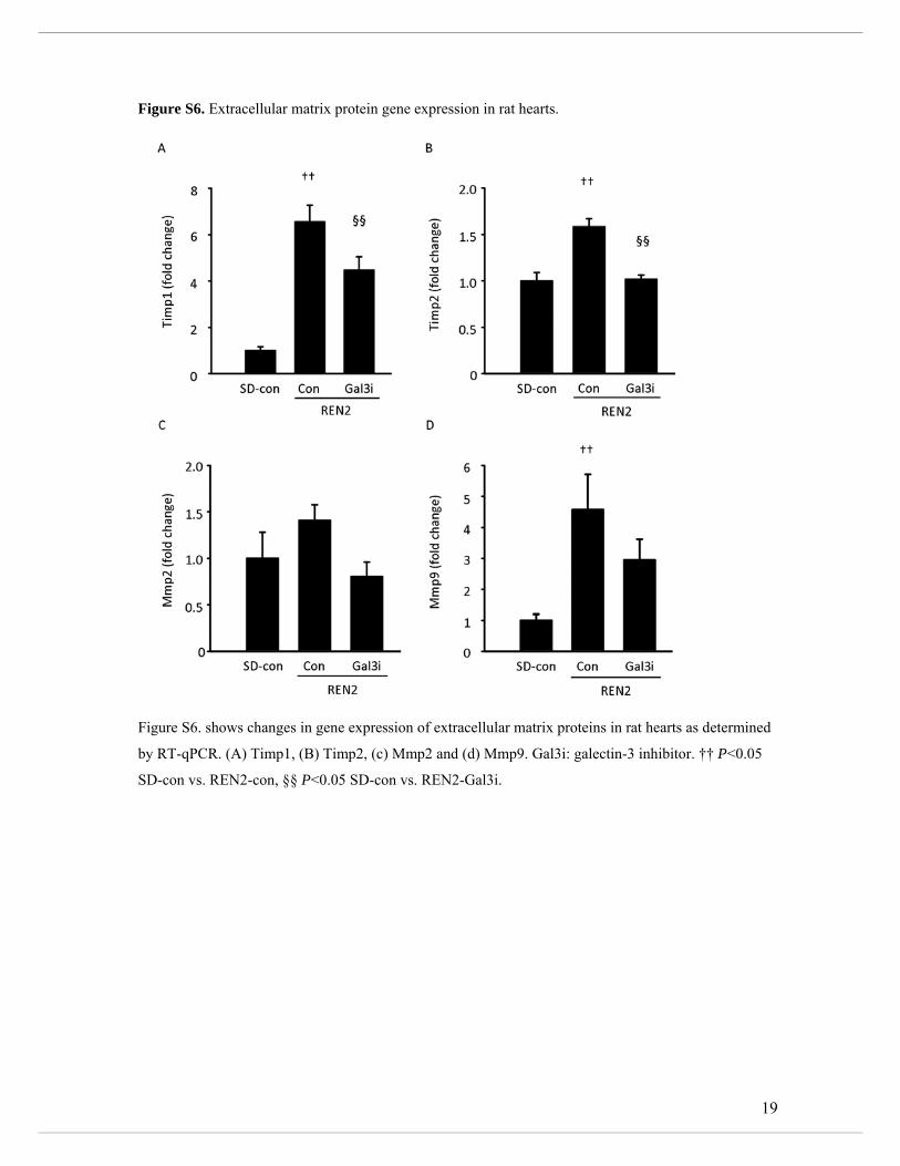
19
Figure S6. Extracellular matrix protein gene expression in rat hearts.
Figure S6. shows changes in gene expression of extracellular matrix proteins in rat hearts as determined
by RT-qPCR. (A) Timp1, (B) Timp2, (c) Mmp2 and (d) Mmp9. Gal3i: galectin-3 inhibitor. †† P<0.05
SD-con vs. REN2-con, §§ P<0.05 SD-con vs. REN2-Gal3i.
20
Supplemental References
1. Henderson NC, Mackinnon AC, Farnworth SL, Poirier F, Russo FP, Iredale JP, Haslett C, Simpson KJ, Sethi T. Galectin-3 regulates myofibroblast activation and hepatic fibrosis. Proc Natl Acad Sci U S A. 2006; 103: 5060-5065. 2. Henderson NC, Mackinnon AC, Farnworth SL, Kipari T, Haslett C, Iredale JP, Liu FT, Hughes J, Sethi T. Galectin-3 expression and secretion links macrophages to the promotion of renal fibrosis. Am J Pathol. 2008; 172: 288-298. 3. Hsu DK, Yang RY, Pan Z, Yu L, Salomon DR, Fung-Leung WP, Liu FT. Targeted disruption of the galectin-3 gene results in attenuated peritoneal inflammatory responses. Am J Pathol. 2000; 156: 1073-1083. 4. de Boer RA, Pokharel S, Flesch M, van Kampen DA, Suurmeijer AJ, Boomsma F, van Gilst WH, van Veldhuisen DJ, Pinto YM. Extracellular signal regulated kinase and SMAD signaling both mediate the angiotensin II driven progression towards overt heart failure in homozygous TGR(mRen2)27. J Mol Med (Berl). 2004; 82: 678-687. 5. de Borst MH, Navis G, de Boer RA, Huitema S, Vis LM, van Gilst WH, van Goor H. Specific MAP-kinase blockade protects against renal damage in homozygous TGR(mRen2)27 rats. Lab Invest. 2003; 83: 1761-1770. 6. Lee MA, Bohm M, Paul M, Bader M, Ganten U, Ganten D. Physiological characterization of the hypertensive transgenic rat TGR(mREN2)27. Am J Physiol. 1996; 270: E919-29. 7. Ruifrok WP, Qian C, Sillje HH, van Goor H, van Veldhuisen DJ, van Gilst WH, de Boer RA. Heart failure-associated anemia: bone marrow dysfunction and response to erythropoietin. J Mol Med (Berl). 2011; 89: 377-387. 8. Rockman HA, Wachhorst SP, Mao L, Ross J,Jr. ANG II receptor blockade prevents ventricular hypertrophy and ANF gene expression with pressure overload in mice. Am J Physiol. 1994; 266: H2468-75. 9. Demotte N, Wieers G, Van Der Smissen P, Moser M, Schmidt C, Thielemans K, Squifflet JL, Weynand B, Carrasco J, Lurquin C, Courtoy PJ, van der Bruggen P. A galectin-3 ligand corrects the impaired function of human CD4 and CD8 tumor-infiltrating lymphocytes and favors tumor rejection in mice. Cancer Res. 2010; 70: 7476-7488. 10. Kuipers I, Li J, Vreeswijk-Baudoin I, Koster J, van der Harst P, Sillje HH, Kuipers F, van Veldhuisen DJ, van Gilst WH, de Boer RA. Activation of liver X receptors with T0901317 attenuates cardiac hypertrophy in vivo. Eur J Heart Fail. 2010; 12: 1042-1050. 11. Pacher P, Nagayama T, Mukhopadhyay P, Batkai S, Kass DA. Measurement of cardiac function using pressure-volume conductance catheter technique in mice and rats. Nat Protoc. 2008; 3: 1422-1434. 12. van der Meer P, Lipsic E, Henning RH, Boddeus K, van der Velden J, Voors AA, van Veldhuisen DJ, van Gilst WH, Schoemaker RG. Erythropoietin induces neovascularization and improves cardiac function in rats with heart failure after myocardial infarction. J Am Coll Cardiol. 2005; 46: 125-133. 13. Creemers LB, Jansen DC, van Veen-Reurings A, van den Bos T, Everts V. Microassay for the assessment of low levels of hydroxyproline. BioTechniques. 1997; 22: 656-658. 14. Kuipers I, van der Harst P, Kuipers F, van Genne L, Goris M, Lehtonen JY, van Veldhuisen DJ, van Gilst WH, de Boer RA. Activation of liver X receptor-alpha reduces activation of the renal and cardiac renin-angiotensin-aldosterone system. Lab Invest. 2010; 90: 630-636. 15. Lu B, Tigchelaar W, Ruifrok WP, van Gilst WH, de Boer RA, Sillje HH. DHRS7c, a novel cardiomyocyte-expressed gene that is down-regulated by adrenergic stimulation and in heart failure. Eur J Heart Fail. 2012; 14: 5-13. 16. Louch WE, Sheehan KA, Wolska BM. Methods in cardiomyocyte isolation, culture, and gene transfer. J Mol Cell Cardiol. 2011; 51: 288-298. 17. Lane KB, Egan B, Vick S, Abdolrasulnia R, Shepherd VL. Characterization of a rat alveolar macrophage cell line that expresses a functional mannose receptor. J Leukoc Biol. 1998; 64: 345-350.


![Inhibition of colon carcinogenesis by a standardized ... · Inhibition of colon carcinogenesis by a standardized Cannabis sativa ... In several pharmacological ... experiments, [3H]CP55940](https://img.pdfslide.us/doc/110x75/5af53f647f8b9a8d1c8d4b2c/inhibition-of-colon-carcinogenesis-by-a-standardized-of-colon-carcinogenesis.jpg)