Embed Size (px)
Citation preview

Central Annals of Clinical Pathology
Cite this article: Abdel-Fattah Ismail SA (2015) Genetic and Immunological Studies on Spleen of Heat-Stressed Rats and the Protective Role of Propolis. Ann Clin Pathol 3(3): 1053.
*Corresponding author
Shimaa Ahmed Abdel-Fattah Ismail,Department of Clinical Pathology, Faculty of Veterinary Medicine, Zagazig University, 44511, Zagazig, Egypt; Tel:201-286-
Submitted: 30 July 2015
Accepted: 17 September 2015
Published: 21 September 2015
ISSN: 2373-9282
Copyright© 2015 Abdel-Fattah Ismail
OPEN ACCESS
Keywords•Heat shock protein 70•Heat stress•Humoral immunity•Total nucleic acid•Oxidative stress•Propolis
Research Article
Genetic and Immunological Studies on Spleen of Heat-Stressed Rats and the Protective Role of PropolisShimaa Ahmed Abdel-Fattah Ismail* Department of Clinical Pathology, Zagazig University, Egypt
Abstract
Heat stress was shown to cause oxidative damage, which can lead to cytotoxicity; thus, we carried out this study to investigate the genetic and immunological effects caused by heat stress in male wistar rats.This study was performedon40adultmalealbinorats,dividedintofourmaingroups;thefirstgroup A (10 rats) was left in a temperature controlled room (25±5°C). The second group B (10 rats), was fed a basal diet supplemented with 3 g (EEP) (Ethanolic extract 70% Dosic Imp. and Exp.Co., Ltd./China)/kg diet for 10 days, and the experimental group C (10 rats) was subjected to high temperature (40±1°C) for 12 hours and fed on a basal diet. In the same way, group D (10 rats) (EEP + heat stress) was fed a basal diet supplemented with 3 g EEP/kg diet for 10 days then subjected to high temperature (40±1°C) for 12 hours. During the time course of heat exposure rectal temperature was monitored using thermometer unit reached to 40°C. After application of the heat stress protocol, hematological analysis, quantitative changes in nucleic acid and protein contents, level of heat shock protein 70, activity of superoxide dismutase, and lipid peroxidation ,serum IgG and IgM were determined.The results showed that exposure to heat stress significantly stimulated theprocessof lipidperoxidation in spleencellsofmale rats. Further, it decreased total nucleic acid and protein contents and superoxide dismutase activities. In addition, it was observed that the level of IgG, IgM was decreased with appearance of leucopenia and lymphocytopenia.It was concluded that chronic heat exposure instigates oxidative stress, which can contribute to cellular dysfunction and immunological disturbance.
INTRODUCTIONWe are the witnesses of seasonal heat waves (temperature
of 32° C and above. lasting for more than three days) as a result of Earth global warming, which cause increased morbidity and mortality of population [1,2]. Therefore, the detailed examination of the elevated ambient temperature effects on many functions in the human organism is necessary. The actual model of homeostatic functioning is composed of neuro-endocrine system, immune system and environment. A single change within these systems has the influence on homeostasis, and induces changes in order to establish non pathologic equilibrium [3]. Emerging body of evidence has confirmed that the elevated ambient temperature affects the immune system. Changes are polymorphic and depend on the intensity and duration of the exposition, species, gender and aging [4,5]. The decreased body weight [6], decreased relative thymus mass, and leucopenia in the wistar rats have already been reported, as well as the increased spontaneous lymphoprolifreraiion [7,8].
Stress is a sudden environmental change that induces damage at the molecular, cellular and organismal level [9]. Macromolecules such as proteins are continuously exposed to potential damaging agents that can cause loss of molecular function and depletion of cell populations over the lifetime of essential organs. One of the key homeostatic responses involved in maintaining longevity is the induction of heat shock proteins (HSPs), a conserved reaction to damaged intracellular proteins [10]. Damage to macromolecules is characteristic of aging and degenerative diseases. Beyond DNA damage, protein damage may not only a consequence, but also a causal factor in cellular malfunction. Damage may induce misfolding, and the aggregating oligomeric species may gain a novel toxic property, severely compromising cellular function [11] .The relationship between thermal resistance and expression of inducible heat shock proteins, especially HSP70, depends on the species and temperature treatments [12]. The major stress of life is oxidative stress. Thus, it is not surprising that oxidized protein level increases with aging of all animal species [13].

Central
Abdel-Fattah Ismail (2015)Email:
Ann Clin Pathol 3(3): 1053 (2015) 2/8
External factors such as heat stress have been proposed to be responsible for stimulating ROS production because of similarities in the expression patterns of genes (including heat shock, oxidative stress proteins, or both), which has been observed following heat stress compared with oxidative stress [14]. Furthermore, acute and chronic heating of cells and tissues induces alterations in nuclear and cytoskeletal structures, decrease in mitotic figures in the epithelium and somites, disruption of neural and vascular basement membranes, increase in programmed cell death; that is apoptosis, and inhibition of natural cell-mediated immunity [15,16].
Antioxidant micronutrients are being widely studied for their alleged beneficial properties in the prevention of human diseases, cancer, arthritis and cardiovascular diseases [17]. Propolis is the substance that honeybees produce by mixing their own waxes with resins collected from plants. It has been used widely as a folk medicine from ancient times. Recently, it has gained popularity as a healthy food in various parts of the world because it promotes health and prevents diseases [18]. It has different biological activities such as antibacterial [19], anti-inflammatory [20,21], hepatoprotective effects [18] and antioxidant [22].
Therefore, the aim of the present study was to determine the effects of heat stress on the peroxidative damage on lipids, DNA and immune response with respect to the protective role of ethanolic extract of propolis.
MATERIALS AND METHODS
Animals
Forty adult male albino rats, aged 3 months, weighing 220±10 g were purchased from the Laboratory Animal breeding unit, Faculty of Veterinary Medicine, Zagazig University. Rats were acclimatized for one week prior to the beginning of the experimental study. The animals were housed in an insulated wooden box in a temperature controlled room (25±5°C) with relative humidity (50±10) and with 12 h. light/dark cycle. Rats were allowed to a standard commercial chow diet. All animals were treated in accordance with the guidelines of the National Institutes of Health (NIH) for the Care and Use of Laboratory Animals, and were comforted by Ethics of animal use in research committee (EAURC), Cairo University.
Heat stress protocol
The rats were placed in an insulated wooden box heated by a thermostatically controlled infra red electric lamp, model 250R 50/10. The box temperature was maintained at 40° Celsius and the test rats were exposed to this temperature for 12 h. The control rats were also placed in the same chamber to attain identical conditions for the same period of time, but without switching on the infra red lamp.
This study was performed on 40 adult male albino rats, divided into four equal groups; the first group A , was left in a temperature controlled room (25±5°C). The second group B, was fed a basal diet supplemented with 3 g (EEP) (Ethanolic extract 70% Dosic Imp. and Exp.Co., Ltd./China)/kg diet for 10 days, and the experimental group C, was subjected to high temperature (40±1°C) for 12 hours and fed on a basal diet. In
the same way, group D (EEP + heat stress) was fed a basal diet supplemented with 3 g EEP/kg diet for 10 days then subjected to high temperature (40±1°C) for 12 hours. During the time course of heat exposure rectal temperature was monitored using thermometer unit reached to 40°C.
Animals were decapitated after overnight fasting. The relative humidity of experimental group was 90%. Two blood samples were collected, the 1st sample was transferred to K2 EDTA tube and used for hematological analysis while, the 2nd sample was collected without anticoagulant for serum separation to measured serum IgG, IgM and HSP70. Spleen was collected from all animals in all groups and divided into 2 portions; the 1st was homogenized for measuring the nucleic acid and protein contents, activity of superoxide dismutase and lipid peroxidation. The 2nd portion used for histopathological examination
Body and spleen weights
The loss in body weight was recognized as a particular characteristic response to the stress [6]. Body and spleen weights were noted and compared with the respective control groups of rats of same age.
Hematological analysis
Baseline hematological parameters were determined at the end of the experiment. Leukogram was determined using automated hematology analyzer, Hospitex Hemascreen 18, Italy. Thin blood smears were prepared immediately after bleeding and stained using giemsa stain .The stained blood films were used to assess absolute differential WBC count [23], while the neutrophil-lymphocyte ratio was calculated.
Biochemical parameters related to stress and DNA damage
Determination of nucleic acid content: Quantitative changes in nucleic acid were determined by the method described by [24]. One gram of spleen was homogenized in 5% trichloroacteic acid, centrifuged and boiled in a mixture of absolute ethanol and ethanol/ether mixture 3:1. After centrifugation, trichloroacteic acid 5% was added. The supernatant was separated to be ready to be quantified using specific reagents for DNA (DPA reagent) and RNA (orcinol reagent).
Estimation of protein: Protein was estimated spectrophotometrically [25] with BSA as the standard.
Estimation of heat shock protein 70: HSP70 by ELISA kit from Stress Biotechnologies were investigated as described by [26].
Chromosomal aberrations: At the end of experimental period , two animals of each of the five groups were injected intraperitoneal (i.p.) with colchicine (4mg/kg) 1 hour prior to sacrifice by decapitation. The bone marrow cells were collected from the femora according to [27].Slides were stained with 10% phosphate buffered Giemsa (pH7.0) for 10 min. Metaphase plates were observed and chromosomal aberrations were scored using oil immersion (with 100x object lens) under a light microscope.
Measures of oxidative stress: Half gram of spleen tissue from each animal was homogenized in 5ml of phosphate buffer,
Central
Abdel-Fattah Ismail (2015)Email:
Ann Clin Pathol 3(3): 1053 (2015) 3/8
pH 7.4 on ice using Universal Laboratory Aid homogenizer, Type MPW-309 (Mechanika Precyzyjna, Warsaw, Poland). Homogenates were then centrifuged at 3000 rpm for 15min at 4 °C. The resulting supernatants were collected and preserved at −20 °C until measuring of superoxide dismutase activity
(SOD) and malondialdehyde (MDA) concentration according to methods described by [28,29], respectively
Detection of the serum IgG and IgM: Blood samples were obtained from the cervical vein into clean, dry and labelled tubes.
Figure 1 Changes in body and spleen weights in male rats at different experimental groups.
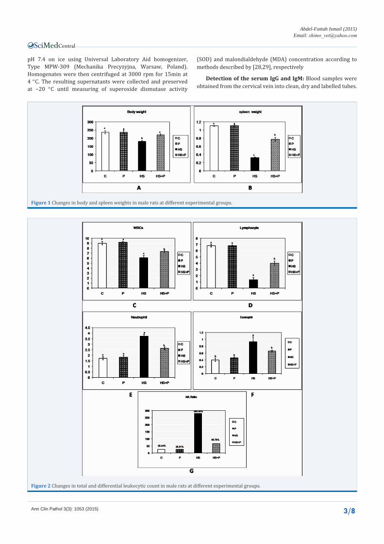
Figure 2 Changes in total and differential leukocytic count in male rats at different experimental groups.

Central
Abdel-Fattah Ismail (2015)Email:
Ann Clin Pathol 3(3): 1053 (2015) 4/8
Blood samples were left to coagulate and then centrifuged at 3000 rpm for 15 minutes. The clear non-hemolysed sera were stored in a deep freezer at -40°C until being used for biochemical evaluation.
Determination of serum immunoglobulin G (IgG) and immunoglobulin M (IgM) levels were done using methods described in the commercial IgG and IgM ELISA kits provided by My BioSource (San Diego, CA).
Histopathological Studies: For light microscopic examination, the treated animals and their controls were sacrificed by decapitation. The spleen were removed and fixed in 10% neutral formalin for 24 h. Tissues were rinsed three times in 70% ethanol, dehydrated using a graded ethanol series and then embedded in paraffin wax. Paraffin sections were cut into 5 micrometers thick slices and stained with haematoxylin and eosin and examined under light microscope.
Statistical analysis
All results are expressed as mean ± SEM. The statistical analysis was carried out with Duncan’s multiple range test. A P < 0.05 was considered the level of statistical significance.
RESULTS Table 1 summarizes the differences in body and spleen weight
values for control group, propolis supplemented, heat stressed rats and heat stressed treated with propolis. The body and spleen
weights during the heat stroke for rats without treatment were significantly (p<0.05) decreased (from the control group while, Resuscitation of their weights was occurred in heat stressed rats supplemented with propolis in their diet. There was no significant changes in these parameters during the whole course of experiment for all control and propolis supplemented animals.
Basic statistics for the leukogram for different experimental groups showed that the heat stressed group recorded a significant (p< 0.05) decrease in total WBC count with neutrophilia, eosinophilia. and lymphocytopenia, in addition to a significant(p< 0.05) increase in neutrophil/ lymphocyte ratio also was noticed in comparison with other groups . Propolis significantly improve the leukogram picture till became non significant with the control group. There was no significant changes in these parameters during the whole course of experiment for all control and propolis supplemented animals.
By chemical analysis of immunoglobulin (IgG and IgM),we found a significant decrease in their levels in heat stressed animals comparing with the control. Propolis significantly increase their levels near the control.
Concerning to the result of nucleic acid content, significant decrease in DNA,RNA and protein content in spleen tissue of rats exposed to heat strock and moderate improvement was found in these parameters following treatment with propolis. Figure 3 showed that male rats exposed to heat showed some chromosomal aberrations such as, D.:Deletion , C.A.:Centromeric
Figure 3 Photomicrographs of chromosomal aberrations in bone marrow cells of male rats exposed to heat. D: Deletion, C.A.: Centromeric attenuation, G: Gap, R.: Ring chromosome, F: Fragment.

Central
Abdel-Fattah Ismail (2015)Email:
Ann Clin Pathol 3(3): 1053 (2015) 5/8
Figure 4 Photomicrograph of H&E stained splenic sections of both control animals and propolis-treated group showing no difference in splenic architecture enclosing normal White (WP) and red pulps (RP) (A, B). Meanwhile group exposed to heat stress showed noticeable pathological changes among splenic parenchyma when compared to control group. These changes include lymphocytic necrosis and degeneration (D) especially at the periphery of lymphoid nodules with leukocytic infiltration (L) (C). Additionally, massive congested areas within the splenich red pulps (arrows) were noted (C). Cellular brownish pigments were seen along the congested blood vessels which give the evidence of hemosiderosis (arrow) (D). Heat stressed animals treated with propolis showed with significant difference from heat stress group including relative improvement of white pulp containing lymphatic nodules (LN) with mild to moderate congestion of splenic blood vessels along red pulp (arrows) (E).
Table 1: The effect of heat shock on selective immunological parameters, genetic and oxidative stress markers in male rats.
Control Propolis HS HS+PropolisIgG
(ng/ml) 1244.7± 4.333a 1246.0 ±5.033a 1065.0 ±27.838c 1160.0± 22.912b
IgM(ng/ml) 125.00 ±3.214a 128.00± 2.309a 97.33± 3.711b 107.33± 3.711b
DNA(mg/g) tissue .50± .017a .49± .029a .21± .020c .34± .023b
RNA(mg/g) tissue .30± .017a .30± .027a .15± .011b .21± .018b
Protein(mg/g) tissue 6.73± .370a 6.83±.166a 3.83± .440c 5.30± .321b
HSP70(ng/ml) 1.11± .060c 1.10±.040c 1.50± .040a 1.30± .054b
MDA(mol/g tissue) 8.57± .228c 8.53± .272c 17.18± .948a 12.38± .749b
SOD(U/g tissue) 7.64± .226a 7.70± .513a 4.58± .358c 5.93± .416b
Values are represented as Mean ± SE (n = 5). Means within the same row carrying different superscripts are significant at (p < 0.05). A Rats were orally treated with distilled water (Control), 3 g (EEP) (Ethanolic extract 70% Dosic Imp. and Exp.Co., Ltd./China)/kg diet for 10 days (propolis), subjected to high temperature (40±1°C) for 12 hours (HS), and fed a basal diet supplemented with 3 g EEP/kg diet for 10 days then subjected to high temperature (40±1°C) for 12 hours (HS+Proplis).

Central
Abdel-Fattah Ismail (2015)Email:
Ann Clin Pathol 3(3): 1053 (2015) 6/8
attenuation, G.: Gap, R.: Ring chromosome,F.:Fragment. While HSP70 was increased in heat stressed group and return to normal with propolis supplementation.
Regarding to the oxidative stress picture, significant increase in splenic MDA content with significant decrease in splenic SOD activity in heat shocked animal, while propolis relief all effects of heat and return their level to the control values.
Photomicrograph of H&E stained splenic sections of both control animals and propolis-treated group showing no difference in splenic architecture enclosing normal White (WP) and red pulps (RP) (A, B). Meanwhile group exposed to heat stress showed noticeable pathological changes among splenic parenchyma when compared to control group. These changes include lymphocytic necrosis and degeneration (D) especially at the periphery of lymphoid nodules with leukocytic infiltration (L) (C). Additionally, massive congested areas within the splenich red pulp (arrows) were noted (C). Cellular brownish pigments were seen along the congested blood vessels which give the evidence of hemosiderosis (arrow) (D). Heat stressed animals treated with propolis showed with significant difference from heat stress group including relative improvement of white pulp containing lymphatic nodules (LN) with mild to moderate congestion of splenic blood vessels along red pulp (arrows) (E).
DISCUSSIONEnvironmental heat is one of the well-known stressor to the
mankind. Although, the problems of heat-afflicted illness are receiving increased importance in view of the current estimates of global warming and its impact on biological systems [30].Heat stress significantly decreased the body weights of rats. It has already been reported that the growth axis is inhibited at many levels of stress [31].The present data showed a decrease in body weight in chronic heat stressed animals this may be a result of prolonged activation of stress system which has been reported to suppress the growth hormone secretion, leads to the decrease in the body weights [6].our results agreed with [30].
Leukocyte profiles are particularly useful in the field of conservation physiology because they are altered by stress and can be directly related to stress hormone levels. The present work showed a significant (p< 0.05) decrease in total WBC count with neutrophilia, eosinophilia. and lymphocytopenia, in addition to a significant(p< 0.05) increase in neutrophil/ lymphocyte ratio.These results may be due to the long-term effects of glucocorticoid hormones(stress hormones) on leukocyte populations [32,33] as it cause redistribution of lymphocytes from the blood to other body compartments inducing lymphocytopenia [34] also, adhere the circulating lymphocytes to the endothelial cells that line the walls of blood vessels, and subsequently undergo transmigration from circulation into other tissues [34,35,36]. In contrast, glucocorticoids also stimulate an influx of neutrophils into the blood from bone marrow and attenuate the egress of neutrophils from the blood to other compartments [37]. For the ecologist seeking a tool to assess stress, they result in an increase in N : L ratio that is proportional to the level of glucocorticoid release.
In the present study, heat stress induced a marked decrease in total nucleic acid content in spleen tissue of heat stressed animals compared with those of control. As DNA acts as a matrix for RNA
synthesis, any modifications in DNA will affect RNA synthesis [38]. As RNA is necessary for protein synthesis, its decrease will in turn reduce protein synthesis. This is relevant in the present study, in which a decline in total protein content was recorded in heat-stressed rat spleen as a result of a decrease in DNA and RNA contents. Similarly, [39,40] reported that hyperthermia has been shown to induce a number of effects in mammalian cells including inhibition of DNA, RNA, and protein synthesis. In fact, it was reported that heat induces base modifications such as oxidative base damage [41], basic DNA sites [42], deamination of cytosine [43], and other types of DNA damage through free radical species such as oxygen species [41] and nitrogen species [44]. In addition, a major effect of hyperthermia is the inhibition of DNA repair as a result of pleiotropic effects related to protein denaturation, inhibition of DNA polymerase activity in particularly that of polymerase b [45]. Finally, this may result in subsegment changes in mRNA, causing impairment in gene transcription that could inhibit protein synthesis [46].
Free radical production and oxidative stress are known to increase in response to environmental stress, including high temperature-induced stress, causing tissue injury [47]. In this regard, [48] have shown that tissue injuries in aged animals are associated with the increased production of ROS. Heat stress increased lipid peroxidation as a consequence of increased free radical generation and increases the MDA level in blood and tissues [49].In the present study,The increase of antioxidant enzyme activities such as SOD, CAT and GSH caused by propolis supplementation may be considered as a protective mechanism against heat-induced free radical production and lipid peroxidation . The protective role of propolis might be related to its antioxidant effect. Researchers suggest that propolis and especially propolis in dose supplemented 3 mg/kg diet might be considered to prevent oxidative stress in the broilers exposed to heat stress [49].
CONCLUSIONThe present study demonstrated that heat exposure markedly
increased DNA damage through increased levels of oxidative DNA damage. In addition, heat stress decreased total nucleic acid and protein contents, stimulated the process of LPO, and decreased SOD activity with increase in MDA content leading to cytotoxicity. Our study clearly demonstrated that propolis supplemented animals in their diet are less tolerant to environmental stress, such as heat stress, compared with ones not fed on propolis.
REFERENCES1. Epstein PR. Is global warming harmful to health? Sci Am. 2000; 283:
50-57.
2. Kalkstein LS, Smoyer KE. The impact of climate change on human health: some international implications. Experientia. 1993; 49: 969-979.
3. Wilckens T, De Rijk R. Glucocorticoids and immune function: unknown dimensions and new frontiers. Immunol Today. 1997; 18: 418-424.
4. Chayoth R, Christou NV, Nohr CW, Yale JF, Poussier P, Grose M, et al. Immunological responses to chronic heat exposure and food restriction in rats. Am J Clin Nutrss. 1988; 48: 361-367.
5. Ueda T, Yamauchi C. [Effects of environmental temperature on thymus

Central
Abdel-Fattah Ismail (2015)Email:
Ann Clin Pathol 3(3): 1053 (2015) 7/8
and spleen weights and lymphocytes in mice]. Jikken Dobutsu. 1986; 35: 479-483.
6. Cure M. Plasma corticosterone response in continuous versus discontinuous chronic heat exposure in rat.Physiol Behav. 1989; 45: 1117-1122.
7. Joseph IM, Suthanthirarajan N, Namasivayam A. Effect of acute heat stress on certain immunological parameters in albino rats.Indian J Physiol Pharmacol. 1991; 35: 269-271.
8. Lochmiller RL, Vestey MR, McMurray ST. Temporal variation in humoral and cell-mediated immune response in a Sigmodon hispid us population. Ecology. 1994; 75: 236-245.
9. Söti C, Csermely P. Protein stress and stress proteins: implications in aging and disease. J Biosci. 2007; 32: 511-515.
10. Calderwood SK, Murshid A, Prince T. The shock of aging: molecular chaperones and the heat shock response in longevity and aging--a mini-review. Gerontology. 2009; 55: 550-558.
11. Dobson CM. Protein folding and misfolding. Nature. 2003; 426: 884-890.
12. Bahrndorff S, Mariën J, Loeschcke V, Ellers J. The relationship between thermal resistance and expression of inducible heat shock proteins, especially HSP70, depends on the species and temperature treatments. Functional Ecol. 2009; 23: 233–239.
13. Stadtman C, Stadtman E R. Role of oxidant species in aging Post-translational modifications of proteins: implications for aging, antigen recognition, and autoimmunity. Curr Med Chem. 2004; 11: 1105–1112.
14. Salo DC, Donovan CM, Davies KJ. HSP70 and other possible heat shock or oxidative stress proteins are induced in skeletal muscle, heart, and liver during exercise. Free Radic Biol Med. 1991; 11: 239-246.
15. Lin PS, Ho KC, Sung SJ, Gladding J. Effect of tumour necrosis factor, heat, and radiation on the viability and microfilament organization in cultured endothelial cells.Int J Hyperthermia. 1992; 8: 667-677.
16. Katschinski DM, Boos K, Schindler SG, Fandrey J. Pivotal role of reactive oxygen species as intracellular mediators of hyperthermia-induced apoptosis. J Biol Chem. 2000; 275: 21094-21098.
17. Faure P, Barclay D, Joyeux-Faure M, Halimi S. Comparison of the effects of zinc alone and zinc associated with selenium and vitamin E on insulin sensitivity and oxidative stress in high-fructose-fed rats. J Trace Elem Med Biol. 2007; 21: 113-119.
18. Ricardo González, Diadelys Remirez, Sandra Rodriguez, Addys González, Odelsa Ancheta, Nelson Merino et al. Hepatoprotective effects of propolis extract on paracetamol-induced liver damage in mice. Phytotherapy Res.1994; 8: 229–232.
19. Sforcin JM, Fernandes A Jr, Lopes CA, Bankova V, Funari SR. Seasonal effect on Brazilian propolis antibacterial activity.J Ethnopharmacol. 2000; 73: 243-249.
20. Khayyal MT, el-Ghazaly MA, el-Khatib AS. Mechanisms involved in the antiinflammatory effect of propolis extract. Drugs Exp Clin Res. 1993; 19: 197-203.
21. Kanbur M1, Eraslan G, Silici S. Antioxidant effect of propolis against exposure to propetamphos in rats.Ecotoxicol Environ Saf. 2009; 72: 909-915.
22. Banskota AH, Tezuka Y, Adnyana IK, Midorikawa K, Matsushige K, Message D, Huertas AA. Cytotoxic, hepatoprotective and free radical scavenging effects of propolis from Brazil, Peru, the Netherlands and China.J Ethnopharmacol. 2000; 72: 239-246.
23. Jain N C. Schalm’s Veterinary Haematology, 4th edn. Lea and Febiger,
Philadelphia, USA 1986; 140-175.
24. Bregman AA. Laboratory investigations and cell biology. New York: John Willey and Sons 1983; 51–60.
25. LOWRY OH, ROSEBROUGH NJ, FARR AL, RANDALL RJ. Protein measurement with the Folin phenol reagent. J Biol Chem. 1951; 193: 265-275.
26. Amal I H, Abeer H A. Effect of Hyperthermia at Different Ages and Mode of Recovery on the Chromosomal Aberrations and Biological Parameters in Female Rats. Journal of American Science 2010; 6:153-166.
27. Khuda-Bukhsh AR, Chakrabarti J, Mallick P, Khuda-Bukhsh A, Mohanty KC, Biswas SJ. Cytogenetic effects of sonication on Spathosternum prasiniferum (grasshopper), Anabas testudineus (fish), and Mus musculus (mammal). Bull Environ Contam Toxicol. 2001; 66: 118-124.
28. Kakkar P, Das B, Viswanathan P. Adismutase. Indian J Biochem Biophys.1984; 21:131-132.
29. Esterbauer H, Cheeseman KH, Dianzani MU, Poli G, Slater TF. Separation and characterization of the aldehydic products of lipid peroxidation stimulated by ADP-Fe2+ in rat liver microsomes. Biochem J. 1982; 208: 129-140.
30. Sinha RK, Ray AK. Sleep-wake study in an animal model of acute and chronic heat stress. Physiol Behav. 2006; 89: 364-372.
31. Sinha RK. Study of changes in some pathophysiological stress markers in different age groups of an animal model of acute and chronic heat stress. Iran Biomed J. 2007; 11: 101-111.
32. Ottaway CA, Husband AJ. The influence of neuroendocrine pathways on lymphocyte migration. Immunol Today. 1994; 15: 511-517.
33. Brenner I, Shek PN, Zamecnik J, Shephard RJ. Stress hormones and the immunological responses to heat and exercise. Int J Sports Med. 1998; 19: 130-143.
34. Dhabhar FS. A hassle a day may keep the doctor away: stress and the augmentation of immune function. Integr Comp Biol. 2002; 42: 556-564.
35. Cohen JJ. Thymus-derived lymphocytes sequestered in the bone marrow of hydrocortisone-treated mice. J Immunol. 1972; 108: 841-844.
36. Fauci AS. Mechanisms of corticosteroid action on lymphocyte subpopulations. I. Redistribution of circulating T and b lymphocytes to the bone marrow. Immunology. 1975; 28: 669-680.
37. Bishop CR, Athens JW, Boggs DR, Warner HR, Cartwright GE, Wintrobe MM. Leukokinetic studies. 13. A non-steady-state kinetic evaluation of the mechanism of cortisone-induced granulocytosis. J Clin Invest. 1968; 47: 249-260.
38. Rao KS. Genomic damage and its repair in young and aging brain. Mol Neurobiol. 1993; 7: 23-48.
39. Banks S, King SA, Irvine DS, Saunders PT. Impact of a mild scrotal heat stress on DNA integrity in murine spermatozoa. Reproduction. 2005; 129: 505-514.
40. Mostafa SIM, Bayomy MFF, Hassan AI, Zahran NA-RM. Effects of experimental mild and severe whole body hyperthermia. Egypt J Med Lab Sci. 2009; 18:1–17.
41. Bruskov VI, Malakhova LV, Masalimov ZK, Chernikov AV. Heat-induced formation of reactive oxygen species and 8-oxoguanine, a biomarker of damage to DNA. Nucleic Acids Res. 2002; 30: 1354-1363.
42. Warters RL, Brizgys LM. Apurinic site induction in the DNA of cells heated at hyperthermic temperatures. J Cell Physiol. 1987; 133: 144-

Central
Abdel-Fattah Ismail (2015)Email:
Ann Clin Pathol 3(3): 1053 (2015) 8/8
Abdel-Fattah Ismail SA (2015) Genetic and Immunological Studies on Spleen of Heat-Stressed Rats and the Protective Role of Propolis. Ann Clin Pathol 3(3): 1053.
Cite this article
150.
43. Lindahl T, Nyberg B. Heat-induced deamination of cytosine residues in deoxyribonucleic acid. Biochemistry. 1974; 13: 3405-3410.
44. Hall DM, Buettner GR, Matthes RD, Gisolfi CV. Hyperthermia stimulates nitric oxide formation: electron paramagnetic resonance detection of NOheme in blood. J Appl Physiol. 1994; 77: 548–553.
45. Spiro IJ, Denman DL, Dewey WC. Effect of hyperthermia on CHO DNA polymerases alpha and beta. Radiat Res. 1982; 89: 134-149.
46. Lai H, Singh NP. Single- and double-strand DNA breaks in rat brain cells after acute exposure to radiofrequency electromagnetic radiation. Int
J Radiat Biol. 1996; 69: 513-521.
47. Katschinski DM, Boos K, Schindler SG, Fandrey J. Pivotal role of reactive oxygen species as intracellular mediators of hyperthermia-induced apoptosis. J Biol Chem. 2000; 275: 21094-21098.
48. Zhang HJ, Xu L, Drake VJ, Xie L, Oberley LW, Kregel KC. Heat-induced liver injury in old rats is associated with exaggerated oxidative stress and altered transcription factor activation. FASEB J. 2003; 17: 2293-2295.
49. Tatli Seven P, Yilmaz S, Seven I,Çerçi I H, Azman M A, Yilmaz M. The effect of propolis on selected blood indicators and antioxidant enzyme activities in broilers under heat stress. Acta Vet Brno 2009; 78:75-83.