1
GENES, NEURAL SYSTEMS and SOCIAL BEHAVIOR: Association between Cerebral Shape and Social Use of Language in Williams Syndrome Yvonne M. Searcy, 3 Doron Gothelf, 1,2 Judy Reilly, 4 Ursula Bellugi, 3 Tope Lanre-Amos, 7 Debra Mills, 3 Julie R Korenberg, 5 Albert Galaburda, 6 Allan L Reiss 7 1 Behavioral Neurogenetics Center, Department of Child Psychiatry, Schneider Children’s Medical Center of Israel, Petah Tiqwa, Israel; 2 Sackler Faculty of Medicine, Tel Aviv University, Ramat Aviv, Israel; 3 The Salk Institute for Biological Studies, La Jolla, CA; 4 San Diego State University and Université de Poitiers-CNRS, France; 5 Cedars-Sinai Medical Center, Medical Genetics Institute, Division of Neurogenetics and UCLA Departments of Human Genetics and Pediatrics; 6 Beth-Israel Deaconess Medical Center and Harvard Medical School, Boston, MA; 7 Center for Interdisciplinary Brain Sciences Research, Department of Psychiatry and Behavioral Sciences, Stanford University School of Medicine, Stanford, CA Williams syndrome (WS) A neurogenetic disorder resulting from a ~1.6Mb chromosomal microdeletion (~28 genes) at band 7q11.23 (1) A multisystem disorder characterized by aberrant development of the brain and a unique profile of cognitive and behavioral features (2) Cognitive & behavioral profile in WS ‘overly friendly’, increased interest in social interactions IQ typically in Mild to Moderate range of mental retardation major deficits in spatial processing relative strengths in language and face processing increased use of affective prosody and social language in narratives Neuroanatomical abnormalities in WS (3, 4) overall brain volume decreased by about 10%, particularly attributable to reductions in parietal and occipital lobe volumes regions with relatively increased gray matter volume: ventral prefrontal cortex, superior temporal gyrus, amygdala and posterior vermis of the cerebellum decreased parieto-occipital lobe volumes relative to frontal volumes, thought to lead to a more flat bending angle of the corpus callosum and cerebrum compared to typical Two key brain networks involved in regulating social behavior (5, 6) 1. Ventral circuit monitoring social cognition includes: amygdala, anterior cingulate, superior temporal gyrus and mediodorsal nucleus of the thalamus social information processed in the amygdala is transferred to the orbitofrontal cortex where social responses and behaviors are selected 2. Striatal-thalamocortical loop: prefrontal regions- the orbital and lateral cortex and anterior cingulate in charge of behavioral inhibition and includes prefrontal regions: the orbital and lateral cortex and anterior cingulate Purpose of the present study Employ signal detection method (QROC) to identify brain regions most strongly associated with WS vs typically developing controls (TD) (7) Evaluate the relationship between these brain regions, and a salient feature of the WS social phenotype: the atypically high use of social and affective language (social engagement devices, SED) in narratives (1) Bayes, M., Magano, L.F., Rivera, N., Flores, R. & Perez Jurado, L.A. (2003) Mutational mechanisms of Williams-Beuren syndrome deletions. Am J Hum Genet 73 131-151. (2) Bellugi U, Jarvinen-Pasley A, Doyle TF, Reilly J, Reiss AL, Korenberg JR (2007), Affect, social behavior and the brain in Williams syndrome. Current Directions in Psychological Science 16 99-104. (3) Meyer-Lindenberg, A., Kohn, P., Mervis, C.B., kippenhan, J.S., Olsen, R.K., Morris, C.A. & Berman K.F. (2004). Neural basis of genetically determined visuaospatial construction deficit in Williams syndrome. Neuron 43 623-631. (4) Reiss, A.L., Eckert, M.A., Rose, F.E., Karchemskiy, A., Kesler, S., Chang, M., Reynolds, M.F., Kwon, H. & Galaburda, A. (2004) An experiment of nature: brain anatomy parallels cognition and behavior in Williams syndrome. J Neurosci 24 5009-5015. (5) Adolphs, R. (2001) The neurobiology of social cognition. Curr Opin Neurobiol 11 231-239. (6) Bachevalier, J. & Loveland, K.A. (2006) The orbitofrontal-amygdala circuit and self regulation of social-emotional behavior in autism. Neurosci Biobehav Rev 30 97-117. (7) Kiernan, M., Kraemer, H.C., Winkleby, M.A., King, A.C. & Taylor, C.B. (2001) Do logistic regression and signal detection identify different subgroups at risk? Implications for the design of tailored interventions. Psychol Methods 6 35-48. (8) Reiss, A.L., Hennessey, J.G., Rubin, M., Beach, L., Abrams, M.T., Warsofsky, I.S., Liu, A.M. & Links, J.M. (1998) Reliability and validity of an algorithm for fuzzy tissue segmentation of MRI. J Comput Assist Tomogr 22 471-479. (9) Mayer, M. (1969). Frog, where are you? New York: Dial Books for Young Readers. (10) Reilly, J.S., Klima, E.S., & Bellugi, U. (1990). Once more with feeling: affect and language in atypical populations. Dev Psychopathol 2 367-391. (11) Schmitt JE, Eliez S, Bellugi U, Reiss AL (2001), Analysis of cerebral shape in Williams syndrome. Arch Neurol 58: 283-7. Stories were transcribed and coded for evaluative language (10) : elements within the story that convey the narrator’s perspective on events cognitive inferences: inferences about the characters and their motivations e.g. “The boy was happy to see his frog” social engagement devices (SED): phrases that add interest, serving to attract and maintain the listener’s attention: e.g. sound effects, intensifiers (very, so, “The boy was rea…lly sad”); and character speech (“The boy said, ‘Oh, little froooggie…”); “All of the sudden…” SED scores reflect the proportion of SED to the total use of evaluation devices Methods Introduction Results Discussion Selected References Brain regions that distinguish Williams syndrome We utilized a signal detection method (QROC) that has rarely been used in imaging studies. QROC was instrumental in the identification of specific brain measures that are most closely associated with WS. Using the QROC we identified the VAPFC, and consequently elucidate its association with the use of social language in WS. The VAPFC, as well as the bending angle of the corpus collusum, are strong distinguishing characteristics of individuals with WS. The relationship between brain and behavior The orbitofrontal cortex, which partially overlaps with the VAPFC region defined here, is a pivotal part of the ventral circuit that monitors social cognition and regulates emotional states and behavior. The abnormal morphology of the VAPFC may be associated with enhanced use of social language in individuals with WS. Thus our results associating the VAPFC with abnormal social use of language in WS provide support for the important role of prefrontal cortex abnormalities to the social phenotype in WS. Genes to Brain to Behavior Our findings support that aberrant neurodevelopment of the ventral anterior region of the prefrontal cortex is an important factor contributing to the unique cerebral morphology of individuals with WS, as a consequence of haploinsufficiency of genes from the deleted region. 696.8/P16 (10) Figure 2: 1. Mastery of morpho-syntax in narratives of children with WS and TD (1a), and use of social evaluation in narratives of children with WS and TD (1b). 1c. The specificity of evaluative language in WS. WS exhibit greater use of social language than TD, those with early focal lesions (FL), language impairment (LI), and high functioning autism (HFA). 1d. Excessive social affective evaluation in WS across languages and cultures, underscoring a genetic predisposition for use of social language in WS. Figure 1: a- subdivisions of the prefrontal cortex: D = dorsal, MS = middle-superior, MI = middle-inferior, VA = ventral-anterior b- The bending angle of the corpus callosum: the angle created by lines connecting the most anterior and posterior points of the corpus callosum with the midline Materials & Procedures MRI Participants were scanned with a General Electric 1.5 Tesla Signa Scanner. Sagittal brain images were acquired with a three-dimensional (3D) volumetric radio frequency spoiled gradient echo pulse sequence using the following scan parameters: repetition time, 24 ms; echo time, 6 ms; flip angle, 45°; slice thickness, 1.2 mm; field of view, 24 cm; and matrix size, 256 x 192 for 124 contiguous slices Data processing steps following a protocol including removal of non-brain tissues from the images, correction of equipment related image artifacts, and separation (segmentation) of tissue components (grey, white, CSF) (8) Figure 3: The QROC was applied to identify the brain regions that most sensitively and specifically distinguished WS from controls. Brain regions included in this analysis have been reported to be abnormal in subjects with WS . Gray matter volumes included in the QROC analysis: ventral and dorsal anterior prefrontal regions, dorsal-anterior cingulate, parietal and occipital lobes, superior temporal gyrus, cerebellum, amygdala, hippocampus, and thalamus. The bending angle of the corpus callosum also was included (11). The end result of the QROC partition yielded three groups: (1) VAPFC 22.76 cm 3 , 82.9% with WS; (2) VAPFC < 22.76 cm 3 and bending angle 109 0 , 60.0% with WS; (3) VAPFC < 22.76 cm 3 and bending angle < 109 0 , only 16.7% with WS. The combination of large or small VAPFC and large bending angle had 85.4% sensitivity and 75.0% specificity in distinguishing WS from controls. Figure 4: The WS subgroup with large VAPFC had significantly higher SED scores than the subgroup with small VAPFC (0.68 ± 0.16 vs. 0.51 ± 0.15, t = -02.5, P < 0.05). There was also a significant positive correlation between adjusted VAPFC volume and SED scores of the WS group (r = 0.50, P < .01). Linear regression indicated that only adjusted VAPFC volume significantly predicted SED scores (b = 0.52, r2 change = 0.28, P = 0.01), whereas amygdala volume (b = -0.19, P = 0.32) and STG volume (b = 0.08, P = 0.67) did not. There were no significant correlations between VAPC volume and other aspects of the WS phenotype such as VIQ (r = 0.10, P = 0.63) or PIQ (r = 0.33, P = 0.09). Association between social evaluation device scores and VAPFC size Social Language Participants were presented with the 24-page wordless picture book “Frog, where are you?” (9) , and asked to tell the story to the experimenter The extensive use of social devices to engage the listener is a consistent finding, and is a central feature of the unique social behavior of individuals with WS. The Quality Receiver Operating Characteristic Curve (QROC) analysis Acknowledgments This study was supported by the National Institute of Health (Grants MH01142, MH50047, HD31715, and HD40761 to ALR and the Program Project HD33113 “Williams syndrome: Linking Cognition, Brain and Genes” to UB, JRK, DM, AG, & ALR). Participants Evaluative Language Subset WS TD WS TD n = 41 n = 40 n = 27 n = 12 GENDER 19 M, 22 F 16 M, 24 F 15 M, 12 F 2 M, 10 F AGE years M (SD) 29.4 (9.0) 27.5 (7.4) 28.1 (10.0) 26.0 (7.0) Age range (years) 12.4 - 50.4 18.2 - 49.2 12.4 - 50.4 18.5 - 41.5 Verbal IQ 72.0 (7.5) ~100.0 (10.0) 72.7 (8.1) 102.4 (10.8) Performance IQ 67.5 (8.7) ~100.0 (10.0) 66.6 (9.4) 104.5 (13.2) Abbreviations used: PFC: prefrontal cortex QROC: quality receiver operating characteristic curve SED: social engagement devices STG: superior temporal gyrus TD: typically developing controls VAPFC: ventral anterior prefrontal cortex WS: Williams syndrome 1 2 3 More about “the frog story” and the use of social evaluation devices in WS:

GENES, NEURAL SYSTEMS and SOCIAL BEHAVIOR ...lcn.salk.edu/Posters/2002-2008/Searcy OFC_poster_SFN_07...GENES, NEURAL SYSTEMS and SOCIAL BEHAVIOR: Association between Cerebral Shape

Embed Size (px)

Citation preview

GENES, NEURAL SYSTEMS and SOCIAL BEHAVIOR:Association between Cerebral Shape and Social Use of Language in

Williams SyndromeYvonne M. Searcy,3 Doron Gothelf, 1,2 Judy Reilly,4 Ursula Bellugi,3 Tope Lanre-Amos,7 Debra Mills,3 Julie R Korenberg,5 Albert Galaburda,6 Allan L Reiss7

1 Behavioral Neurogenetics Center, Department of Child Psychiatry, Schneider Children’s Medical Center of Israel, Petah Tiqwa, Israel; 2 Sackler Faculty of Medicine, Tel Aviv University, Ramat Aviv, Israel; 3 The Salk Institute forBiological Studies, La Jolla, CA; 4 San Diego State University and Université de Poitiers-CNRS, France; 5 Cedars-Sinai Medical Center, Medical Genetics Institute, Division of Neurogenetics and UCLA Departments of Human Genetics

and Pediatrics; 6 Beth-Israel Deaconess Medical Center and Harvard Medical School, Boston, MA; 7 Center for Interdisciplinary Brain Sciences Research, Department of Psychiatry and Behavioral Sciences,Stanford University School of Medicine, Stanford, CA

Williams syndrome (WS)

• A neurogenetic disorder resulting from a ~1.6Mbchromosomal microdeletion (~28 genes) at band 7q11.23 (1)

• A multisystem disorder characterized by aberrantdevelopment of the brain and a unique profile of cognitiveand behavioral features (2)

Cognitive & behavioral profile in WS

‘overly friendly’, increased interest in social interactions IQ typically in Mild to Moderate range of mental retardation major deficits in spatial processing relative strengths in language and face processing increased use of affective prosody and social language innarratives

Neuroanatomical abnormalities in WS (3, 4)

overall brain volume decreased by about 10%, particularlyattributable to reductions in parietal and occipital lobevolumes

regions with relatively increased gray matter volume:ventral prefrontal cortex, superior temporal gyrus, amygdalaand posterior vermis of the cerebellum

decreased parieto-occipital lobe volumes relative to frontalvolumes, thought to lead to a more flat bending angle of thecorpus callosum and cerebrum compared to typical

Two key brain networks involved in regulating socialbehavior (5, 6)

1. Ventral circuit monitoring social cognition includes: amygdala, anterior cingulate, superior temporal gyrusand mediodorsal nucleus of the thalamus

social information processed in the amygdala istransferred to the orbitofrontal cortex where socialresponses and behaviors are selected

2. Striatal-thalamocortical loop: prefrontal regions- the orbital and lateral cortex andanterior cingulate

in charge of behavioral inhibition and includes prefrontalregions: the orbital and lateral cortex and anteriorcingulate

Purpose of the present study

Employ signal detection method (QROC) to identify brainregions most strongly associated with WS vs typicallydeveloping controls (TD) (7)

Evaluate the relationship between these brain regions, anda salient feature of the WS social phenotype: the atypicallyhigh use of social and affective language (social engagementdevices, SED) in narratives

(1) Bayes, M., Magano, L.F., Rivera, N., Flores, R. & Perez Jurado, L.A. (2003) Mutationalmechanisms of Williams-Beuren syndrome deletions. Am J Hum Genet 73 131-151.

(2) Bellugi U, Jarvinen-Pasley A, Doyle TF, Reilly J, Reiss AL, Korenberg JR (2007), Affect, socialbehavior and the brain in Williams syndrome. Current Directions in Psychological Science16 99-104.

(3) Meyer-Lindenberg, A., Kohn, P., Mervis, C.B., kippenhan, J.S., Olsen, R.K., Morris, C.A. & Berman K.F. (2004). Neural basis of genetically determined visuaospatial construction deficit in Williams syndrome. Neuron 43 623-631.

(4) Reiss, A.L., Eckert, M.A., Rose, F.E., Karchemskiy, A., Kesler, S., Chang, M., Reynolds, M.F.,Kwon, H. & Galaburda, A. (2004) An experiment of nature: brain anatomy parallels cognition and behavior in Williams syndrome. J Neurosci 24 5009-5015.

(5) Adolphs, R. (2001) The neurobiology of social cognition. Curr Opin Neurobiol 11 231-239.(6) Bachevalier, J. & Loveland, K.A. (2006) The orbitofrontal-amygdala circuit and self regulation

of social-emotional behavior in autism. Neurosci Biobehav Rev 30 97-117.(7) Kiernan, M., Kraemer, H.C., Winkleby, M.A., King, A.C. & Taylor, C.B. (2001) Do logistic

regression and signal detection identify different subgroups at risk? Implications for thedesign of tailored interventions. Psychol Methods 6 35-48.

(8) Reiss, A.L., Hennessey, J.G., Rubin, M., Beach, L., Abrams, M.T., Warsofsky, I.S., Liu, A.M. &Links, J.M. (1998) Reliability and validity of an algorithm for fuzzy tissue segmentation ofMRI. J Comput Assist Tomogr 22 471-479.

(9) Mayer, M. (1969). Frog, where are you? New York: Dial Books for Young Readers.(10) Reilly, J.S., Klima, E.S., & Bellugi, U. (1990). Once more with feeling: affect and language in

atypical populations. Dev Psychopathol 2 367-391.(11) Schmitt JE, Eliez S, Bellugi U, Reiss AL (2001), Analysis of cerebral shape in Williams

syndrome. Arch Neurol 58: 283-7.

• Stories were transcribed and coded for evaluative language (10):elements within the story that convey the narrator’s perspective on events

• cognitive inferences: inferences about the characters and theirmotivations e.g. “The boy was happy to see his frog”

• social engagement devices (SED): phrases that add interest,serving to attract and maintain the listener’s attention: e.g. soundeffects, intensifiers (very, so, “The boy was rea…lly sad”); and characterspeech (“The boy said, ‘Oh, little froooggie…”); “All of the sudden…”

• SED scores reflect the proportion of SED to the total use of evaluationdevices

MethodsIntroduction Results Discussion

Selected References

Brain regions that distinguish Williams syndrome

• We utilized a signal detection method (QROC) that has rarelybeen used in imaging studies. QROC was instrumental in theidentification of specific brain measures that are most closelyassociated with WS.

• Using the QROC we identified the VAPFC, and consequentlyelucidate its association with the use of social language in WS.

• The VAPFC, as well as the bending angle of the corpuscollusum, are strong distinguishing characteristics ofindividuals with WS.

The relationship between brain and behavior

• The orbitofrontal cortex, which partially overlaps with theVAPFC region defined here, is a pivotal part of the ventralcircuit that monitors social cognition and regulates emotionalstates and behavior.

• The abnormal morphology of the VAPFC may be associatedwith enhanced use of social language in individuals with WS.

• Thus our results associating the VAPFC with abnormal socialuse of language in WS provide support for the important roleof prefrontal cortex abnormalities to the social phenotype inWS.

Genes to Brain to Behavior

• Our findings support that aberrant neurodevelopment of theventral anterior region of the prefrontal cortex is an importantfactor contributing to the unique cerebral morphology ofindividuals with WS, as a consequence of haploinsufficiency ofgenes from the deleted region.

696.8/P16

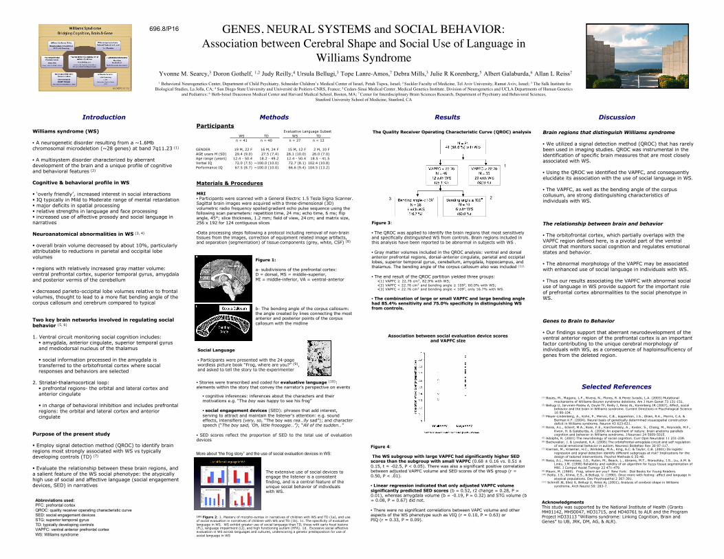
(10) Figure 2: 1. Mastery of morpho-syntax in narratives of children with WS and TD (1a), and useof social evaluation in narratives of children with WS and TD (1b). 1c. The specificity of evaluativelanguage in WS. WS exhibit greater use of social language than TD, those with early focal lesions(FL), language impairment (LI), and high functioning autism (HFA). 1d. Excessive social affectiveevaluation in WS across languages and cultures, underscoring a genetic predisposition for use ofsocial language in WS.

Figure 1:

a- subdivisions of the prefrontal cortex:D = dorsal, MS = middle-superior,MI = middle-inferior, VA = ventral-anterior

b- The bending angle of the corpus callosum:the angle created by lines connecting the mostanterior and posterior points of the corpuscallosum with the midline

Materials & Procedures

MRI• Participants were scanned with a General Electric 1.5 Tesla Signa Scanner.Sagittal brain images were acquired with a three-dimensional (3D)volumetric radio frequency spoiled gradient echo pulse sequence using thefollowing scan parameters: repetition time, 24 ms; echo time, 6 ms; flipangle, 45°; slice thickness, 1.2 mm; field of view, 24 cm; and matrix size,256 x 192 for 124 contiguous slices

•Data processing steps following a protocol including removal of non-braintissues from the images, correction of equipment related image artifacts,and separation (segmentation) of tissue components (grey, white, CSF) (8)

Figure 3:

• The QROC was applied to identify the brain regions that most sensitivelyand specifically distinguished WS from controls. Brain regions included inthis analysis have been reported to be abnormal in subjects with WS .

• Gray matter volumes included in the QROC analysis: ventral and dorsalanterior prefrontal regions, dorsal-anterior cingulate, parietal and occipitallobes, superior temporal gyrus, cerebellum, amygdala, hippocampus, andthalamus. The bending angle of the corpus callosum also was included (11).

• The end result of the QROC partition yielded three groups:•(1) VAPFC ≥ 22.76 cm3, 82.9% with WS;•(2) VAPFC < 22.76 cm3 and bending angle ≥ 1090, 60.0% with WS;•(3) VAPFC < 22.76 cm3 and bending angle < 1090, only 16.7% with WS.

• The combination of large or small VAPFC and large bending anglehad 85.4% sensitivity and 75.0% specificity in distinguishing WSfrom controls.

Figure 4:

The WS subgroup with large VAPFC had significantly higher SEDscores than the subgroup with small VAPFC (0.68 ± 0.16 vs. 0.51 ±0.15, t = -02.5, P < 0.05). There was also a significant positive correlationbetween adjusted VAPFC volume and SED scores of the WS group (r =0.50, P < .01).

• Linear regression indicated that only adjusted VAPFC volumesignificantly predicted SED scores (b = 0.52, r2 change = 0.28, P =0.01), whereas amygdala volume (b = -0.19, P = 0.32) and STG volume (b= 0.08, P = 0.67) did not.

• There were no significant correlations between VAPC volume and otheraspects of the WS phenotype such as VIQ (r = 0.10, P = 0.63) orPIQ (r = 0.33, P = 0.09).

Association between social evaluation device scoresand VAPFC size

Social Language

• Participants were presented with the 24-pagewordless picture book “Frog, where are you?” (9),and asked to tell the story to the experimenter

The extensive use of social devices toengage the listener is a consistentfinding, and is a central feature of theunique social behavior of individualswith WS.

The Quality Receiver Operating Characteristic Curve (QROC) analysis

AcknowledgmentsThis study was supported by the National Institute of Health (GrantsMH01142, MH50047, HD31715, and HD40761 to ALR and the ProgramProject HD33113 “Williams syndrome: Linking Cognition, Brain andGenes” to UB, JRK, DM, AG, & ALR).

ParticipantsEvaluative Language Subset

WS TD WS TD

n = 41 n = 40 n = 27 n = 12

GENDER 19 M, 22 F 16 M, 24 F 15 M, 12 F 2 M, 10 F

AGE years M (SD) 29.4 (9.0) 27.5 (7.4) 28.1 (10.0) 26.0 (7.0)

Age range (years) 12.4 - 50.4 18.2 - 49.2 12.4 - 50.4 18.5 - 41.5

Verbal IQ 72.0 (7.5) ~100.0 (10.0) 72.7 (8.1) 102.4 (10.8)

Performance IQ 67.5 (8.7) ~100.0 (10.0) 66.6 (9.4) 104.5 (13.2)

Abbreviations used:PFC: prefrontal cortexQROC: quality receiver operating characteristic curveSED: social engagement devicesSTG: superior temporal gyrusTD: typically developing controlsVAPFC: ventral anterior prefrontal cortexWS: Williams syndrome

1

23

More about “the frog story” and the use of social evaluation devices in WS: