Embed Size (px)
Citation preview

Generation of the configurational ensemble of anintrinsically disordered protein from unbiasedmolecular dynamics simulationUtsab R. Shresthaa, Puneet Junejab,1, Qiu Zhangb, Viswanathan Gurumoorthyc, Jose M. Borreguerob, Volker Urbanb,Xiaolin Chengd, Sai Venkatesh Pingalib, Jeremy C. Smitha,e, Hugh M. O’Neillb, and Loukas Petridisa,e,2
aUT/ORNL Center for Molecular Biophysics, Oak Ridge National Laboratory, Oak Ridge, TN 37831; bNeutron Scattering Division, Oak Ridge NationalLaboratory, Oak Ridge, TN 37831; cUT-ORNL Graduate School of Genome Science and Technology, University of Tennessee, Knoxville, TN 37996; dDivision ofMedicinal Chemistry and Pharmacognosy, College of Pharmacy, The Ohio State University, Columbus, OH 43210; and eDepartment of Biochemistry andCellular and Molecular Biology, University of Tennessee, Knoxville, TN 37996
Edited by Michael L. Klein, Temple University, Philadelphia, PA, and approved September 3, 2019 (received for review April 30, 2019)
Intrinsically disordered proteins (IDPs) are abundant in eukaryoticproteomes, play a major role in cell signaling, and are associatedwith human diseases. To understand IDP function it is critical todetermine their configurational ensemble, i.e., the collection of3-dimensional structures they adopt, and this remains an immensechallenge in structural biology. Attempts to determine this ensem-ble computationally have been hitherto hampered by the necessityof reweighting molecular dynamics (MD) results or biasing simula-tion in order to match ensemble-averaged experimental observ-ables, operations that reduce the precision of the generated modelbecause different structural ensembles may yield the same exper-imental observable. Here, by employing enhanced sampling MD wereproduce the experimental small-angle neutron and X-ray scatter-ing profiles and the NMR chemical shifts of the disordered Nterminal (SH4UD) of c-Src kinase without reweighting or constrain-ing the simulations. The unbiased simulation results reveal a weaklyfunneled and rugged free energy landscape of SH4UD, which givesrise to a heterogeneous ensemble of structures that cannot bedescribed by simple polymer theory. SH4UD adopts transienthelices, which are found away from known phosphorylation sitesand could play a key role in the stabilization of structural regionsnecessary for phosphorylation. Our findings indicate that ade-quately sampled molecular simulations can be performed to provideaccurate physical models of flexible biosystems, thus rationalizingtheir biological function.
intrinsically disordered protein | MD simulation | small-angle scattering |conformational ensemble | transient helices
Intrinsically disordered proteins (IDPs) are structurally flexibleand lack stable secondary structures in physiological conditions.
They play a critical role in many cellular functions, such as signaltransduction, transcriptional regulation, cell growth, binding, geneexpression, and homeostasis (1–5). IDPs are also associated withmultiple diseases, such as cancers (6, 7), amyloidosis, diabetes,cardiovascular problems, and neurodegenerative disorders (8, 9).To understand the disorder–function relationship IDPs pre-
sent and to elucidate the mechanism by which their pathologicalmutations lead to human diseases (2), we need to determinetheir configurational ensemble: the collection of 3-dimensional(3D) structures they adopt (10). Several experimental techniqueshave been applied to study the structures of IDPs, includingNMR spectroscopy (6, 11–18), single-molecule Förster resonanceenergy transfer (sm-FRET) (17, 19, 20), cryoelectron microscopy(21, 22), and small-angle X-ray and neutron scattering (SAXS andSANS) (6, 15, 17, 23–28). However, the information obtainablefrom these experiments is limited and they cannot provide a high-resolution description of IDP ensembles (29–34). Further, mosttechniques employed to study IDPs suffer from a conundrum: Theexperimental observables represent an average over the confor-mational ensemble, but the ensemble itself cannot be unequivo-
cally inferred from the experiments (10, 29, 35). For SAXS andSANS, unambiguous structural interpretation is made more dif-ficult by the inherent orientational averaging.Molecular dynamics (MD) simulation can, in principle, pro-
vide the structural ensemble of biomolecules. However, MDfaces 2 distinct, yet related, challenges when applied to IDPs: theaccuracy of the physics-based molecular mechanics force fieldsthat model the interactions in the system and the need for ad-equate sampling of the large configurational space of an IDP andits associated solvent. Recent progress has considerably improvedmolecular mechanics force fields for IDPs (36–43). Nevertheless,the accuracy of simulations, as assessed by comparison to experi-ments, was found to depend on the specific IDP, the employedforce field, and configurational sampling in a weakly funneledenergy landscape (36–38, 40, 42).The current state-of-the-art approach to structurally charac-
terizing IDPs at atomic detail requires either restraining the MDor reweighting (44–50) the MD-generated ensemble to matchthe experimental data (35, 51). This creates overfitting issues andmakes it difficult to judge the accuracy of the models because ofa degeneracy problem: Different ensembles may yield the same
Significance
A major challenge in biology is characterizing the structuralflexibility of intrinsically disordered proteins (IDPs). Ensemble-averaged experimental data do not provide the underlyingprotein structures. Here, we performed independently small-angle neutron and X-ray scattering experiments and unbiasedmolecular dynamics simulations to probe the solution structureof an IDP. We report that enhancing the sampling of the sim-ulations can generate an ensemble of IDP structures in quan-titative agreement with scattering and NMR, without the needfor biasing the simulation or reweighting the results. Thedemonstration of established simulation technology that pro-duces accurate physical models of flexible biosystems maypave the way to relating conformational flexibility to biologicalfunction.
Author contributions: U.R.S., V.U., X.C., J.C.S., H.M.O., and L.P. designed research; U.R.S.,P.J., Q.Z., V.G., S.V.P., and H.M.O. performed research; J.M.B. contributed new reagents/analytic tools; U.R.S., S.V.P., and L.P. analyzed data; and U.R.S., J.C.S., and L.P. wrotethe paper.
The authors declare no conflict of interest.
This article is a PNAS Direct Submission.
Published under the PNAS license.1Present address: School of Medicine, Emory University, Atlanta, GA 30322.2To whom correspondence may be addressed. Email: [email protected].
This article contains supporting information online at www.pnas.org/lookup/suppl/doi:10.1073/pnas.1907251116/-/DCSupplemental.
First published September 23, 2019.
20446–20452 | PNAS | October 8, 2019 | vol. 116 | no. 41 www.pnas.org/cgi/doi/10.1073/pnas.1907251116
Dow
nloa
ded
by g
uest
on
Oct
ober
12,
202
1
experimental observable (29, 52, 53). Despite recent successes ofBayesian methods when refining MD-derived structures againstSAXS data (54, 55), reproducing experiments without the needto modifying simulation results presents one of the most im-portant challenges in structural biology of IDPs. Addressing thischallenge would lead to increased confidence that the obtainedmodels accurately describe the physical behavior of the systems.Here, we combine MD simulation with SAXS, SANS, and
NMR experiments to elucidate the conformational ensemble ofthe 95-residue intrinsically disordered N terminus of the proto-oncogene nonreceptor human tyrosine kinase c-Src, which con-tains the Src homology 4 (SH4) and unique domains. c-Src playsa major role in cellular transduction pathways such as cellgrowth, differentiation, transcription, proliferation, adhesion,and survival (12, 56) and is associated with a variety of humancancers (57). SH4 regulates c-Src kinase localization in responseto cellular stimulation (56) by binding/anchoring to the cellmembrane, whereas the unique domain (UD) mediates the in-teraction of Src with lipids, specific receptor and protein targets(12, 13, 58).In the present work a heterogeneous ensemble of structures of
SH4UD was generated by enhancing the sampling of unbiasedatomistic simulations in an explicit solvent by using Hamiltonianreplica exchange MD (HREMD) (59–61). Critically, the SAXS,SANS, and NMR observables of SH4UD thus generated arefound to be in excellent agreement with the experimental datawithout the need for biasing or reweighting. The use of HREMDis critical in achieving this agreement. The simulations reveal aweakly funneled and rugged free energy landscape and predict3 transient helical regions that do not coincide with the knownphosphorylation sites but are proximal to a lipid-binding region.We hypothesize that such transient structures act as a molecularrecognition feature for phosphorylation-mediated regulator ofenzyme activity and for membrane binding.
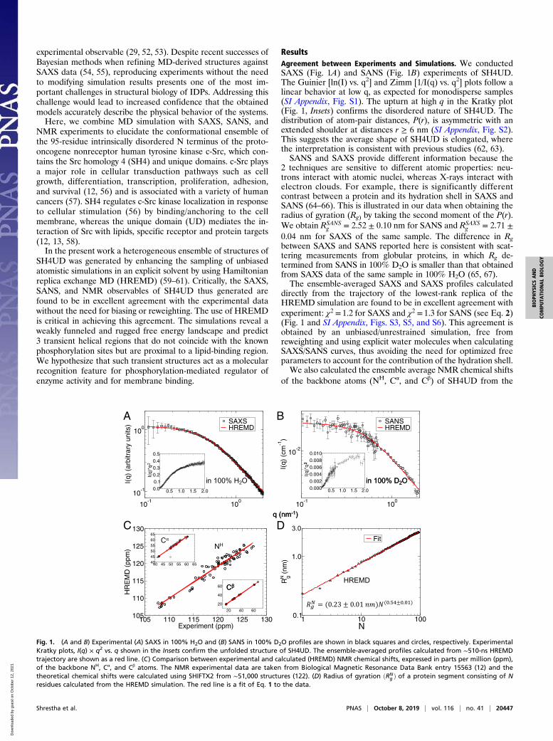
ResultsAgreement between Experiments and Simulations. We conductedSAXS (Fig. 1A) and SANS (Fig. 1B) experiments of SH4UD.The Guinier [ln(I) vs. q2] and Zimm [1/I(q) vs. q2] plots follow alinear behavior at low q, as expected for monodisperse samples(SI Appendix, Fig. S1). The upturn at high q in the Kratky plot(Fig. 1, Insets) confirms the disordered nature of SH4UD. Thedistribution of atom-pair distances, P(r), is asymmetric with anextended shoulder at distances r ≥ 6 nm (SI Appendix, Fig. S2).This suggests the average shape of SH4UD is elongated, wherethe interpretation is consistent with previous studies (62, 63).SANS and SAXS provide different information because the
2 techniques are sensitive to different atomic properties: neu-trons interact with atomic nuclei, whereas X-rays interact withelectron clouds. For example, there is significantly differentcontrast between a protein and its hydration shell in SAXS andSANS (64–66). This is illustrated in our data when obtaining theradius of gyration (Rg) by taking the second moment of the P(r).We obtain RSANS
g = 2.52 ± 0.10 nm for SANS and RSAXSg = 2.71 ±
0.04 nm for SAXS of the same sample. The difference in Rgbetween SAXS and SANS reported here is consistent with scat-tering measurements from globular proteins, in which Rg de-termined from SANS in 100% D2O is smaller than that obtainedfrom SAXS data of the same sample in 100% H2O (65, 67).The ensemble-averaged SAXS and SAXS profiles calculated
directly from the trajectory of the lowest-rank replica of theHREMD simulation are found to be in excellent agreement withexperiment: χ2 = 1.2 for SAXS and χ2 = 1.3 for SANS (see Eq. 2)(Fig. 1 and SI Appendix, Figs. S3, S5, and S6). This agreement isobtained by an unbiased/unrestrained simulation, free fromreweighting and using explicit water molecules when calculatingSAXS/SANS curves, thus avoiding the need for optimized freeparameters to account for the contribution of the hydration shell.We also calculated the ensemble average NMR chemical shifts
of the backbone atoms (NH, Cα, and Cβ) of SH4UD from the
!
"#$%&'()*+%
!
"#$%&'()*+%
1 10 100N
0.1
1.0
Rg (
nm)
Fit
10-1
100
10-1
100
I(q)
(ar
bitr
ary
units
) SAXSHREMD
0.5 1.0 1.5 2.00.00.10.20.30.40.5
I(q)
*q
10-1
100
10-2
I(q)
(cm
-1)
SANSHREMD
0.5 1.0 1.5 2.00.0000.0020.0040.0060.0080.010
I(q)
*q
105 110 115 120 125 130Experiment (ppm)
105
110
115
120
125
130
HR
EM
D (
ppm
)
40 45 50 55 60 65404550556065
20 40 60
20
40
60
A B
C D
Fig. 1. (A and B) Experimental (A) SAXS in 100% H2O and (B) SANS in 100% D2O profiles are shown in black squares and circles, respectively. ExperimentalKratky plots, I(q) × q2 vs. q shown in the Insets confirm the unfolded structure of SH4UD. The ensemble-averaged profiles calculated from ∼510-ns HREMDtrajectory are shown as a red line. (C) Comparison between experimental and calculated (HREMD) NMR chemical shifts, expressed in parts per million (ppm),of the backbone NH, Cα, and Cβ atoms. The NMR experimental data are taken from Biological Magnetic Resonance Data Bank entry 15563 (12) and thetheoretical chemical shifts were calculated using SHIFTX2 from ∼51,000 structures (122). (D) Radius of gyration ðRN
g Þ of a protein segment consisting of Nresidues calculated from the HREMD simulation. The red line is a fit of Eq. 1 to the data.
Shrestha et al. PNAS | October 8, 2019 | vol. 116 | no. 41 | 20447
BIOPH
YSICSAND
COMPU
TATIONALBIOLO
GY
Dow
nloa
ded
by g
uest
on
Oct
ober
12,
202
1

HREMD simulation and compared them to previously publishedexperimental values (12). The excellent agreement betweenNMR and HREMD is reflected by the regression coefficientsR2 > 0.93 (Fig. 1C and SI Appendix, Fig. S7).The unbiased HREMD simulations are thus consistent with
3 independent experimental probes of global and local proteinstructure: SAXS, SANS, and NMR. The underlying conforma-tional ensemble contains all of the structures from the ∼510-ns-long trajectory and has a broad distribution of Rg, as reflected inthe density plot of the theoretical I(q) (SI Appendix, Fig. S3). Theuse of HREMD is critical in obtaining good agreement withscattering and NMR experiments (SI Appendix, Figs. S3 and S7).The convergence of the calculations is shown in detail in SIAppendix, Figs. S5–S9.
Chain Statistics. The degree of compaction of a polypeptide chainin aqueous solution can be quantified by the Flory exponent, ν,which is determined by the relative strengths of the protein–solvent and intraprotein interactions (17, 24, 25, 68). Polymertheory predicts only 3 values of ν for polymers: When intraproteininteractions are favorable, ν = 0.333 and the protein adopts acollapsed conformation, for balanced interactions, ν = 0.5 and theprotein is a Gaussian random coil, whereas for favorable protein–solvent interactions ν = 0.588 and the protein adopts a self-avoiding random coil conformation (24).Here, the Flory exponent was calculated from the atomic co-
ordinates using the relation (24, 69, 70),
RNg = R0Nυ, [1]
where RNg is the radius of gyration of a protein segment consist-
ing of N residues and R0 is a constant. Fitting Eq. 1 to thesimulation data (Fig. 1D) gives ν = 0.54 ± 0.01, suggesting theoverall SH4UD–water interaction is favorable and SH4UD chainstatistics lie between Gaussian and self-avoiding random coilbehavior. Several studies have indicated similar chain statisticsof other IDPs (17, 24, 25, 68).
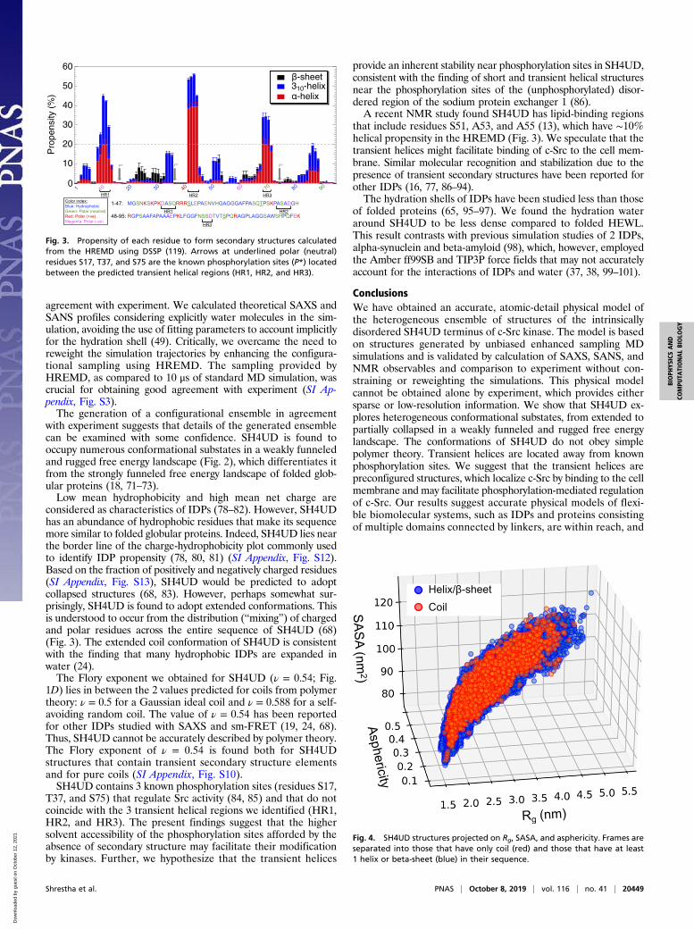
Weakly Funneled Free Energy Landscape. Rg and the solvent-accessible surface area (SASA) quantify important global proteincharacteristics: the overall size and solvent exposure, respectively.As such, they are used here as collective variables for describingthe conformations of SH4UD by projecting the free energy ontothem. SH4UD depicts a weakly funneled and rugged free en-ergy landscape (Fig. 2). We note that barrier heights derivedfrom 2D histogram analysis of HREMD simulations may carrylarge errors. The multiple shallow minima indicate the largenumber of conformational substates demonstrating a confor-mational heterogeneity. This structural heterogeneity is one ofthe distinctive characteristics of IDPs (25, 71–73).
Secondary Structure Propensity. The propensity of residues to formsecondary structure, calculated from the HREMD, indicates thatSH4UD mainly adopts coil structures with almost negligiblebeta-sheet content, in agreement with NMR experiments (12).However, 3 short sequence segments that display occasional he-lical structures were found, defined here as having helical pro-pensity higher than 20% (Fig. 3): helical region 1 (HR1), residuesD10-A11-S12-Q13; HR2, A42-S43-A44-D45; and HR3, S69-S70-D71.The conformations sampled by HREMD are of varying degree
of compactness and asphericity (70) (Fig. 4). The broad distri-bution of Rg, SASA, and asphericity indicate the continuous andheterogeneous nature of the structures comprising the ensemble.Coil conformations with no secondary structure represent only∼6% of the HREMD trajectory and are evenly distributed in the(Rg, asphericity, SASA) space (SI Appendix, Fig. S10). Thechain statistics (Eq. 1) of the coil structures are similar to those
of the entire ensemble and yield a ν = 0.54 ± 0.01 Flory exponent(SI Appendix, Fig. S10).
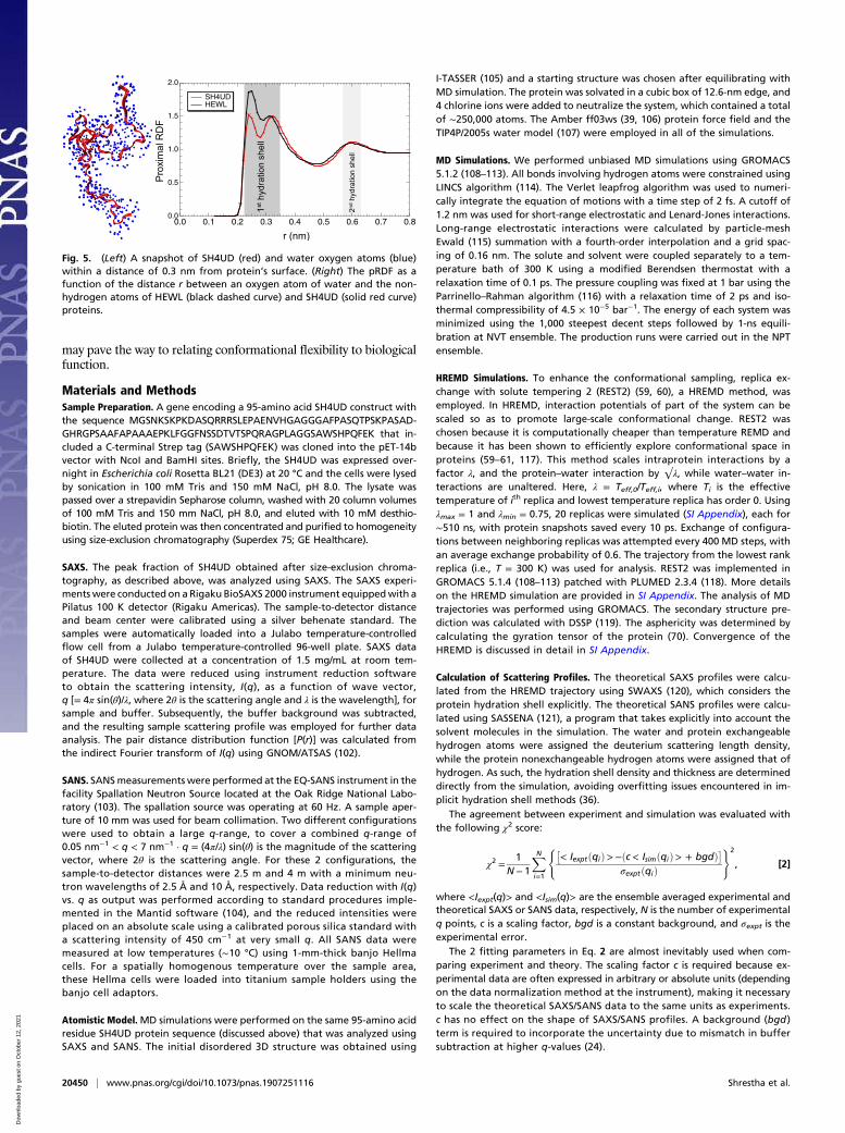
Hydration Shell Structure. The hydration shell structure wasquantified by calculating the proximal radial distribution func-tion (pRDF) (74, 75) of water oxygen atoms around the non-hydrogen atoms of protein (Fig. 5). The 2 peaks of the pRDF ata distance r ∼ 0.30 nm from the protein surface correspond to thefirst hydration shell, which is found here to have a higher densitythan the bulk. The peak at r ∼ 0.25 nm arises from correlationdistances involving protein oxygen and nitrogen atoms with wateroxygen atom, whereas the peak at r ∼ 0.30 nm corresponds tocarbon atoms (SI Appendix, Fig. S11).We compare the pRDFs of hydration water around SH4UD
and hen egg white lysozyme, HEWL, a globular folded protein.While the pRDFs are similar for distances >0.35 nm, the firsthydration shell is less dense for SH4UD than for HEWL,as shown by the lower peak heights, especially for the wateroxygen–protein nitrogen interactions (SI Appendix, Fig. S11).This result indicates that the IDP hydration shell is more similarto bulk than for folded proteins. A smaller fraction of hydro-phobic residues on the surface of HEWL compared to SH4UDwere found that might explain the smaller hydration shell densityof SH4UD (SI Appendix, Tables S1 and S2).
DiscussionThe biological significance of IDPs and their abundance in manygenomes have been increasingly realized (3). IDPs represent∼30% of the human genome, play important cellular functions,and are associated with many diseases. Understanding their bi-ological function in detail requires resolving the heterogeneous3D ensembles of their structures, which remains a challenge be-cause of the ensemble-averaged nature of most experimental data.To determine IDP ensembles MD simulation is frequently
combined with SAXS experiments (6, 24, 76, 77). However, it hashitherto been necessary to bias the populations of conformationsobtained by MD to produce agreement with experiment (35, 46).In the present work we conducted unbiased HREMD simulationsand obtained SAXS, SANS, and NMR observables in quantitative
Fig. 2. Free energy landscape of SH4UD as a function of its Rg and SASAfrom HREMD simulations showing a large number of the conformationalsubstates sampled in the simulation.
20448 | www.pnas.org/cgi/doi/10.1073/pnas.1907251116 Shrestha et al.
Dow
nloa
ded
by g
uest
on
Oct
ober
12,
202
1
agreement with experiment. We calculated theoretical SAXS andSANS profiles considering explicitly water molecules in the sim-ulation, avoiding the use of fitting parameters to account implicitlyfor the hydration shell (49). Critically, we overcame the need toreweight the simulation trajectories by enhancing the configura-tional sampling using HREMD. The sampling provided byHREMD, as compared to 10 μs of standard MD simulation, wascrucial for obtaining good agreement with experiment (SI Ap-pendix, Fig. S3).The generation of a configurational ensemble in agreement
with experiment suggests that details of the generated ensemblecan be examined with some confidence. SH4UD is found tooccupy numerous conformational substates in a weakly funneledand rugged free energy landscape (Fig. 2), which differentiates itfrom the strongly funneled free energy landscape of folded glob-ular proteins (18, 71–73).Low mean hydrophobicity and high mean net charge are
considered as characteristics of IDPs (78–82). However, SH4UDhas an abundance of hydrophobic residues that make its sequencemore similar to folded globular proteins. Indeed, SH4UD lies nearthe border line of the charge-hydrophobicity plot commonly usedto identify IDP propensity (78, 80, 81) (SI Appendix, Fig. S12).Based on the fraction of positively and negatively charged residues(SI Appendix, Fig. S13), SH4UD would be predicted to adoptcollapsed structures (68, 83). However, perhaps somewhat sur-prisingly, SH4UD is found to adopt extended conformations. Thisis understood to occur from the distribution (“mixing”) of chargedand polar residues across the entire sequence of SH4UD (68)(Fig. 3). The extended coil conformation of SH4UD is consistentwith the finding that many hydrophobic IDPs are expanded inwater (24).The Flory exponent we obtained for SH4UD (ν = 0.54; Fig.
1D) lies in between the 2 values predicted for coils from polymertheory: ν = 0.5 for a Gaussian ideal coil and ν = 0.588 for a self-avoiding random coil. The value of ν = 0.54 has been reportedfor other IDPs studied with SAXS and sm-FRET (19, 24, 68).Thus, SH4UD cannot be accurately described by polymer theory.The Flory exponent of ν = 0.54 is found both for SH4UDstructures that contain transient secondary structure elementsand for pure coils (SI Appendix, Fig. S10).SH4UD contains 3 known phosphorylation sites (residues S17,
T37, and S75) that regulate Src activity (84, 85) and that do notcoincide with the 3 transient helical regions we identified (HR1,HR2, and HR3). The present findings suggest that the highersolvent accessibility of the phosphorylation sites afforded by theabsence of secondary structure may facilitate their modificationby kinases. Further, we hypothesize that the transient helices
provide an inherent stability near phosphorylation sites in SH4UD,consistent with the finding of short and transient helical structuresnear the phosphorylation sites of the (unphosphorylated) disor-dered region of the sodium protein exchanger 1 (86).A recent NMR study found SH4UD has lipid-binding regions
that include residues S51, A53, and A55 (13), which have ∼10%helical propensity in the HREMD (Fig. 3). We speculate that thetransient helices might facilitate binding of c-Src to the cell mem-brane. Similar molecular recognition and stabilization due to thepresence of transient secondary structures have been reported forother IDPs (16, 77, 86–94).The hydration shells of IDPs have been studied less than those
of folded proteins (65, 95–97). We found the hydration wateraround SH4UD to be less dense compared to folded HEWL.This result contrasts with previous simulation studies of 2 IDPs,alpha-synuclein and beta-amyloid (98), which, however, employedthe Amber ff99SB and TIP3P force fields that may not accuratelyaccount for the interactions of IDPs and water (37, 38, 99–101).
ConclusionsWe have obtained an accurate, atomic-detail physical model ofthe heterogeneous ensemble of structures of the intrinsicallydisordered SH4UD terminus of c-Src kinase. The model is basedon structures generated by unbiased enhanced sampling MDsimulations and is validated by calculation of SAXS, SANS, andNMR observables and comparison to experiment without con-straining or reweighting the simulations. This physical modelcannot be obtained alone by experiment, which provides eithersparse or low-resolution information. We show that SH4UD ex-plores heterogeneous conformational substates, from extended topartially collapsed in a weakly funneled and rugged free energylandscape. The conformations of SH4UD do not obey simplepolymer theory. Transient helices are located away from knownphosphorylation sites. We suggest that the transient helices arepreconfigured structures, which localize c-Src by binding to the cellmembrane and may facilitate phosphorylation-mediated regulationof c-Src. Our results suggest accurate physical models of flexi-ble biomolecular systems, such as IDPs and proteins consistingof multiple domains connected by linkers, are within reach, and
Fig. 4. SH4UD structures projected on Rg, SASA, and asphericity. Frames areseparated into those that have only coil (red) and those that have at least1 helix or beta-sheet (blue) in their sequence.
60
50
40
30
20
10
Pro
pens
ity (
%)
-sheet 310-helix
-helix
Fig. 3. Propensity of each residue to form secondary structures calculatedfrom the HREMD using DSSP (119). Arrows at underlined polar (neutral)residues S17, T37, and S75 are the known phosphorylation sites (P*) locatedbetween the predicted transient helical regions (HR1, HR2, and HR3).
Shrestha et al. PNAS | October 8, 2019 | vol. 116 | no. 41 | 20449
BIOPH
YSICSAND
COMPU
TATIONALBIOLO
GY
Dow
nloa
ded
by g
uest
on
Oct
ober
12,
202
1
may pave the way to relating conformational flexibility to biologicalfunction.
Materials and MethodsSample Preparation. A gene encoding a 95-amino acid SH4UD construct withthe sequence MGSNKSKPKDASQRRRSLEPAENVHGAGGGAFPASQTPSKPASAD-GHRGPSAAFAPAAAEPKLFGGFNSSDTVTSPQRAGPLAGGSAWSHPQFEK that in-cluded a C-terminal Strep tag (SAWSHPQFEK) was cloned into the pET-14bvector with NcoI and BamHI sites. Briefly, the SH4UD was expressed over-night in Escherichia coli Rosetta BL21 (DE3) at 20 °C and the cells were lysedby sonication in 100 mM Tris and 150 mM NaCl, pH 8.0. The lysate waspassed over a strepavidin Sepharose column, washed with 20 column volumesof 100 mM Tris and 150 mm NaCl, pH 8.0, and eluted with 10 mM desthio-biotin. The eluted protein was then concentrated and purified to homogeneityusing size-exclusion chromatography (Superdex 75; GE Healthcare).
SAXS. The peak fraction of SH4UD obtained after size-exclusion chroma-tography, as described above, was analyzed using SAXS. The SAXS experi-mentswere conducted on a Rigaku BioSAXS 2000 instrument equippedwith aPilatus 100 K detector (Rigaku Americas). The sample-to-detector distanceand beam center were calibrated using a silver behenate standard. Thesamples were automatically loaded into a Julabo temperature-controlledflow cell from a Julabo temperature-controlled 96-well plate. SAXS dataof SH4UD were collected at a concentration of 1.5 mg/mL at room tem-perature. The data were reduced using instrument reduction softwareto obtain the scattering intensity, I(q), as a function of wave vector,q [= 4π sin(θ)/λ, where 2θ is the scattering angle and λ is the wavelength], forsample and buffer. Subsequently, the buffer background was subtracted,and the resulting sample scattering profile was employed for further dataanalysis. The pair distance distribution function [P(r)] was calculated fromthe indirect Fourier transform of I(q) using GNOM/ATSAS (102).
SANS. SANSmeasurements were performed at the EQ-SANS instrument in thefacility Spallation Neutron Source located at the Oak Ridge National Labo-ratory (103). The spallation source was operating at 60 Hz. A sample aper-ture of 10 mm was used for beam collimation. Two different configurationswere used to obtain a large q-range, to cover a combined q-range of0.05 nm−1 < q < 7 nm−1 · q = (4π/λ) sin(θ) is the magnitude of the scatteringvector, where 2θ is the scattering angle. For these 2 configurations, thesample-to-detector distances were 2.5 m and 4 m with a minimum neu-tron wavelengths of 2.5 Å and 10 Å, respectively. Data reduction with I(q)vs. q as output was performed according to standard procedures imple-mented in the Mantid software (104), and the reduced intensities wereplaced on an absolute scale using a calibrated porous silica standard witha scattering intensity of 450 cm−1 at very small q. All SANS data weremeasured at low temperatures (∼10 °C) using 1-mm-thick banjo Hellmacells. For a spatially homogenous temperature over the sample area,these Hellma cells were loaded into titanium sample holders using thebanjo cell adaptors.
Atomistic Model.MD simulations were performed on the same 95-amino acidresidue SH4UD protein sequence (discussed above) that was analyzed usingSAXS and SANS. The initial disordered 3D structure was obtained using
I-TASSER (105) and a starting structure was chosen after equilibrating withMD simulation. The protein was solvated in a cubic box of 12.6-nm edge, and4 chlorine ions were added to neutralize the system, which contained a totalof ∼250,000 atoms. The Amber ff03ws (39, 106) protein force field and theTIP4P/2005s water model (107) were employed in all of the simulations.
MD Simulations. We performed unbiased MD simulations using GROMACS5.1.2 (108–113). All bonds involving hydrogen atoms were constrained usingLINCS algorithm (114). The Verlet leapfrog algorithm was used to numeri-cally integrate the equation of motions with a time step of 2 fs. A cutoff of1.2 nm was used for short-range electrostatic and Lenard-Jones interactions.Long-range electrostatic interactions were calculated by particle-meshEwald (115) summation with a fourth-order interpolation and a grid spac-ing of 0.16 nm. The solute and solvent were coupled separately to a tem-perature bath of 300 K using a modified Berendsen thermostat with arelaxation time of 0.1 ps. The pressure coupling was fixed at 1 bar using theParrinello–Rahman algorithm (116) with a relaxation time of 2 ps and iso-thermal compressibility of 4.5 × 10⁻⁵ bar−1. The energy of each system wasminimized using the 1,000 steepest decent steps followed by 1-ns equili-bration at NVT ensemble. The production runs were carried out in the NPTensemble.
HREMD Simulations. To enhance the conformational sampling, replica ex-change with solute tempering 2 (REST2) (59, 60), a HREMD method, wasemployed. In HREMD, interaction potentials of part of the system can bescaled so as to promote large-scale conformational change. REST2 waschosen because it is computationally cheaper than temperature REMD andbecause it has been shown to efficiently explore conformational space inproteins (59–61, 117). This method scales intraprotein interactions by afactor λ, and the protein–water interaction by √λ, while water–water in-teractions are unaltered. Here, λ = Teff,0/Teff,i, where Ti is the effectivetemperature of ith replica and lowest temperature replica has order 0. Usingλmax = 1 and λmin = 0.75, 20 replicas were simulated (SI Appendix), each for∼510 ns, with protein snapshots saved every 10 ps. Exchange of configura-tions between neighboring replicas was attempted every 400 MD steps, withan average exchange probability of 0.6. The trajectory from the lowest rankreplica (i.e., T = 300 K) was used for analysis. REST2 was implemented inGROMACS 5.1.4 (108–113) patched with PLUMED 2.3.4 (118). More detailson the HREMD simulation are provided in SI Appendix. The analysis of MDtrajectories was performed using GROMACS. The secondary structure pre-diction was calculated with DSSP (119). The asphericity was determined bycalculating the gyration tensor of the protein (70). Convergence of theHREMD is discussed in detail in SI Appendix.
Calculation of Scattering Profiles. The theoretical SAXS profiles were calcu-lated from the HREMD trajectory using SWAXS (120), which considers theprotein hydration shell explicitly. The theoretical SANS profiles were calcu-lated using SASSENA (121), a program that takes explicitly into account thesolvent molecules in the simulation. The water and protein exchangeablehydrogen atoms were assigned the deuterium scattering length density,while the protein nonexchangeable hydrogen atoms were assigned that ofhydrogen. As such, the hydration shell density and thickness are determineddirectly from the simulation, avoiding overfitting issues encountered in im-plicit hydration shell methods (36).
The agreement between experiment and simulation was evaluated withthe following χ2 score:
χ2 =1
N− 1
XNi=1
(�< IexptðqiÞ>−ðc< IsimðqiÞ> + bgdÞ�
σexptðqiÞ
)2
, [2]
where <Iexpt(q)> and <Isim(q)> are the ensemble averaged experimental andtheoretical SAXS or SANS data, respectively, N is the number of experimentalq points, c is a scaling factor, bgd is a constant background, and σexpt is theexperimental error.
The 2 fitting parameters in Eq. 2 are almost inevitably used when com-paring experiment and theory. The scaling factor c is required because ex-perimental data are often expressed in arbitrary or absolute units (dependingon the data normalization method at the instrument), making it necessaryto scale the theoretical SAXS/SANS data to the same units as experiments.c has no effect on the shape of SAXS/SANS profiles. A background (bgd )term is required to incorporate the uncertainty due to mismatch in buffersubtraction at higher q-values (24).
2.0
1.5
1.0
0.5
0.00.80.70.60.50.40.30.20.10.0
SH4UD HEWL
Fig. 5. (Left) A snapshot of SH4UD (red) and water oxygen atoms (blue)within a distance of 0.3 nm from protein’s surface. (Right) The pRDF as afunction of the distance r between an oxygen atom of water and the non-hydrogen atoms of HEWL (black dashed curve) and SH4UD (solid red curve)proteins.
20450 | www.pnas.org/cgi/doi/10.1073/pnas.1907251116 Shrestha et al.
Dow
nloa
ded
by g
uest
on
Oct
ober
12,
202
1
ACKNOWLEDGMENTS. This work is supported by the Laboratory DirectedResearch and Development Program of Oak Ridge National Laboratory,managed by UT-Battelle LLC and by project ERKP300 funded by the Office ofBiological & Environmental Research in the Department of Energy (DOE)Office of Science (BER). Use of the Center for Structural Molecular Biologyresources is supported by BER. This research used the resources of 3 DOEuser facilities: The National Energy Research Scientific Computing Center(contract no. DE-AC02-05CH11231), the Oak Ridge Leadership ComputingFacility (contract no. DE-AC05-00OR22725), and the Spallation Neutron
Source. This manuscript has been authored by UT-Battelle, LLC, under con-tract DE-AC05-00OR22725 with the US DOE. The US government retains andthe publisher, by accepting the article for publication, acknowledges thatthe US government retains a nonexclusive, paid-up, irrevocable, worldwidelicense to publish or reproduce the published form of this manuscript, orallow others to do so, for US government purposes. DOE will provide publicaccess to these results of federally sponsored research in accordance withthe DOE Public Access Plan (https://www.energy.gov/downloads/doe-public-access-plan).
1. L. Mollica et al., Binding mechanisms of intrinsically disordered proteins: Theory,simulation, and experiment. Front. Mol. Biosci. 3, 52 (2016).
2. N. S. Latysheva, T. Flock, R. J. Weatheritt, S. Chavali, M. M. Babu, How do disorderedregions achieve comparable functions to structured domains? Protein Sci. 24, 909–922 (2015).
3. P. E. Wright, H. J. Dyson, Intrinsically disordered proteins in cellular signalling andregulation. Nat. Rev. Mol. Cell Biol. 16, 18–29 (2015).
4. J. Habchi, P. Tompa, S. Longhi, V. N. Uversky, Introducing protein intrinsic disorder.Chem. Rev. 114, 6561–6588 (2014).
5. N. D. Keul et al., The entropic force generated by intrinsically disordered segmentstunes protein function. Nature 563, 584–588 (2018).
6. M. Wells et al., Structure of tumor suppressor p53 and its intrinsically disorderedN-terminal transactivation domain. Proc. Natl. Acad. Sci. U.S.A. 105, 5762–5767 (2008).
7. V. N. Uversky, A. Roman, C. J. Oldfield, A. K. Dunker, Protein intrinsic disorder andhuman papillomaviruses: Increased amount of disorder in E6 and E7 oncoproteinsfrom high risk HPVs. J. Proteome Res. 5, 1829–1842 (2006).
8. T. P. J. Knowles, M. Vendruscolo, C. M. Dobson, The amyloid state and its associationwith protein misfolding diseases. Nat. Rev. Mol. Cell Biol. 15, 384–396 (2014).
9. V. N. Uversky, C. J. Oldfield, A. K. Dunker, Intrinsically disordered proteins in humandiseases: Introducing the D2 concept. Annu. Rev. Biophys. 37, 215–246 (2008).
10. P. Sormanni et al., Simultaneous quantification of protein order and disorder. Nat.Chem. Biol. 13, 339–342 (2017).
11. P. Robustelli, K. A. Stafford, A. G. Palmer, 3rd, Interpreting protein structural dy-namics from NMR chemical shifts. J. Am. Chem. Soc. 134, 6365–6374 (2012).
12. Y. Pérez, M. Gairí, M. Pons, P. Bernadó, Structural characterization of the nativelyunfolded N-terminal domain of human c-Src kinase: Insights into the role of phos-phorylation of the unique domain. J. Mol. Biol. 391, 136–148 (2009).
13. Y. Pérez et al., Lipid binding by the Unique and SH3 domains of c-Src suggests a newregulatory mechanism. Sci. Rep. 3, 1295 (2013).
14. C. Lee, D.-H. Kim, S.-H. Lee, J. Su, K.-H. Han, Structural investigation on the intrin-sically disordered N-terminal region of HPV16 E7 protein. BMB Rep. 49, 431–436(2016).
15. M. Arbesú et al., The unique domain forms a fuzzy intramolecular complex in srcfamily kinases. Structure 25, 630–640.e4 (2017).
16. M. Arai, K. Sugase, H. J. Dyson, P. E. Wright, Conformational propensities of in-trinsically disordered proteins influence the mechanism of binding and folding. Proc.Natl. Acad. Sci. U.S.A. 112, 9614–9619 (2015).
17. G. Fuertes et al., Decoupling of size and shape fluctuations in heteropolymeric se-quences reconciles discrepancies in SAXS vs. FRET measurements. Proc. Natl. Acad.Sci. U.S.A. 114, E6342–E6351 (2017).
18. M. R. Jensen, M. Zweckstetter, J. R. Huang, M. Blackledge, Exploring free-energylandscapes of intrinsically disordered proteins at atomic resolution using NMRspectroscopy. Chem. Rev. 114, 6632–6660 (2014).
19. H. Hofmann et al., Polymer scaling laws of unfolded and intrinsically disorderedproteins quantified with single-molecule spectroscopy. Proc. Natl. Acad. Sci. U.S.A.109, 16155–16160 (2012).
20. B. Schuler, A. Soranno, H. Hofmann, D. Nettels, Single-molecule FRET spectroscopyand the polymer physics of unfolded and intrinsically disordered proteins. Annu.Rev. Biophys. 45, 207–231 (2016).
21. D. J. Busch et al., Intrinsically disordered proteins drive membrane curvature. Nat.Commun. 6, 7875 (2015).
22. Z. A. Levine, L. Larini, N. E. LaPointe, S. C. Feinstein, J. E. Shea, Regulation and ag-gregation of intrinsically disordered peptides. Proc. Natl. Acad. Sci. U.S.A. 112, 2758–2763 (2015).
23. V. Receveur-Bréchot, D. Durand, How random are intrinsically disordered proteins?A small angle scattering perspective. Curr. Protein Pept. Sci. 13, 55–75 (2012).
24. J. A. Riback et al., Innovative scattering analysis shows that hydrophobic disorderedproteins are expanded in water. Science 358, 238–241 (2017).
25. S. Ruskamo et al., Juxtanodin is an intrinsically disordered F-actin-binding protein.Sci. Rep. 2, 899 (2012).
26. D. Johansen, C. M. J. Jeffries, B. Hammouda, J. Trewhella, D. P. Goldenberg, Effectsof macromolecular crowding on an intrinsically disordered protein characterized bysmall-angle neutron scattering with contrast matching. Biophys. J. 100, 1120–1128(2011).
27. D. P. Goldenberg, B. Argyle, Minimal effects of macromolecular crowding on anintrinsically disordered protein: A small-angle neutron scattering study. Biophys. J.106, 905–914 (2014).
28. A. Banks, S. Qin, K. L. Weiss, C. B. Stanley, H. X. Zhou, Intrinsically disordered proteinexhibits both compaction and expansion under macromolecular crowding. Biophys.J. 114, 1067–1079 (2018).
29. J. Trewhella, Small-angle scattering and 3D structure interpretation. Curr. Opin. Struct.Biol. 40, 1–7 (2016).
30. I. Drulyte et al., Approaches to altering particle distributions in cryo-electron mi-croscopy sample preparation. Acta Crystallogr. D Struct. Biol. 74, 560–571 (2018).
31. M. Hammel, Validation of macromolecular flexibility in solution by small-angle X-rayscattering (SAXS). Eur. Biophys. J. 41, 789–799 (2012).
32. J. A. Riback et al., Response to comment on “Innovative scattering analysis showsthat hydrophobic disordered proteins are expanded in water”. Science 361,eaar7949 (2018).
33. R. B. Best et al., Comment on “Innovative scattering analysis shows that hydrophobicdisordered proteins are expanded in water”. Science 361, eaar7101 (2018).
34. E. P. O’Brien, G. Morrison, B. R. Brooks, D. Thirumalai, How accurate are polymermodels in the analysis of Förster resonance energy transfer experiments on proteins?J. Chem. Phys. 130, 124903 (2009).
35. R. B. Best, Computational and theoretical advances in studies of intrinsically disor-dered proteins. Curr. Opin. Struct. Biol. 42, 147–154 (2017).
36. J. Henriques, L. Arleth, K. Lindorff-Larsen, M. Skepö, On the calculation of SAXSprofiles of folded and intrinsically disordered proteins from computer simulations. J.Mol. Biol. 430, 2521–2539 (2018).
37. J. Henriques, C. Cragnell, M. Skepö, Molecular dynamics simulations of intrinsicallydisordered proteins: Force field evaluation and comparison with experiment. J.Chem. Theory Comput. 11, 3420–3431 (2015).
38. J. Henriques, M. Skepö, Molecular dynamics simulations of intrinsically disorderedproteins: On the accuracy of the TIP4P-D water model and the representativeness ofprotein disorder models. J. Chem. Theory Comput. 12, 3407–3415 (2016).
39. R. B. Best, W. Zheng, J. Mittal, Balanced protein-water interactions improve prop-erties of disordered proteins and non-specific protein association. J. Chem. TheoryComput. 10, 5113–5124 (2014).
40. P. Robustelli, S. Piana, D. E. Shaw, Developing a molecular dynamics force field forboth folded and disordered protein states. Proc. Natl. Acad. Sci. U.S.A. 115, E4758–E4766 (2018).
41. J. Huang et al., CHARMM36m: An improved force field for folded and intrinsicallydisordered proteins. Nat. Methods 14, 71–73 (2017).
42. J. Huang, A. D. MacKerell, Jr, Force field development and simulations of intrinsicallydisordered proteins. Curr. Opin. Struct. Biol. 48, 40–48 (2018).
43. M. J. Robertson, J. Tirado-Rives, W. L. Jorgensen, Improved peptide and proteintorsional energetics with the OPLSAA force field. J. Chem. Theory Comput. 11, 3499–3509 (2015).
44. G. Hummer, J. Köfinger, Bayesian ensemble refinement by replica simulations andreweighting. J. Chem. Phys. 143, 243150 (2015).
45. P. Bernadó, D. I. Svergun, Structural analysis of intrinsically disordered proteins bysmall-angle X-ray scattering. Mol. Biosyst. 8, 151–167 (2012).
46. P. Bernadó, E. Mylonas, M. V. Petoukhov, M. Blackledge, D. I. Svergun, Structuralcharacterization of flexible proteins using small-angle X-ray scattering. J. Am. Chem.Soc. 129, 5656–5664 (2007).
47. B. Roux, J. Weare, On the statistical equivalence of restrained-ensemble simulationswith the maximum entropy method. J. Chem. Phys. 138, 084107 (2013).
48. C. Camilloni, M. Vendruscolo, Statistical mechanics of the denatured state of aprotein using replica-averaged metadynamics. J. Am. Chem. Soc. 136, 8982–8991(2014).
49. P. Cheng, J. Peng, Z. Zhang, SAXS-oriented ensemble refinement of flexible bio-molecules. Biophys. J. 112, 1295–1301 (2017).
50. S. Yang, L. Blachowicz, L. Makowski, B. Roux, Multidomain assembled states of Hcktyrosine kinase in solution. Proc. Natl. Acad. Sci. U.S.A. 107, 15757–15762 (2010).
51. S. Bottaro, K. Lindorff-Larsen, Biophysical experiments and biomolecular simula-tions: A perfect match? Science 361, 355–360 (2018).
52. W. Boomsma, J. Ferkinghoff-Borg, K. Lindorff-Larsen, Combining experiments andsimulations using the maximum entropy principle. PLoS Comput. Biol. 10, e1003406(2014).
53. W. Rieping, M. Habeck, M. Nilges, Inferential structure determination. Science 309,303–306 (2005).
54. R. Shevchuk, J. S. Hub, Bayesian refinement of protein structures and ensemblesagainst SAXS data using molecular dynamics. PLoS Comput. Biol. 13, e1005800(2017).
55. J. S. Hub, Interpreting solution X-ray scattering data using molecular simulations.Curr. Opin. Struct. Biol. 49, 18–26 (2018).
56. A. G. Tatosyan, O. A. Mizenina, Kinases of the Src family: Structure and functions.Biochemistry (Mosc.) 65, 49–58 (2000).
57. D. L. Wheeler, M. Iida, E. F. Dunn, The role of Src in solid tumors. Oncologist 14, 667–678 (2009).
58. J. J. Kathiriya et al., Presence and utility of intrinsically disordered regions in kinases.Mol. Biosyst. 10, 2876–2888 (2014).
59. G. Bussi, Hamiltonian replica exchange in GROMACS: A flexible implementation.Mol. Phys. 112, 379–384 (2013).
Shrestha et al. PNAS | October 8, 2019 | vol. 116 | no. 41 | 20451
BIOPH
YSICSAND
COMPU
TATIONALBIOLO
GY
Dow
nloa
ded
by g
uest
on
Oct
ober
12,
202
1

60. L. Wang, R. A. Friesner, B. J. Berne, Replica exchange with solute scaling: A moreefficient version of replica exchange with solute tempering (REST2). J. Phys. Chem. B115, 9431–9438 (2011).
61. Y. Sugita, Y. Okamoto, Replica-exchange molecular dynamics method for proteinfolding. Chem. Phys. Lett. 314, 141–151 (1999).
62. M. Nors Perdersen et al., Direct correlation between ligand-induced α-synucleinoligomers and amyloid-like fibril growth. Sci. Rep. 5, 10422 (2015).
63. D. Durand et al., Small-angle X-ray scattering reveals an extended organization forthe autoinhibitory resting state of the p47(phox) modular protein. Biochemistry 45,7185–7193 (2006).
64. D. I. Svergun et al., Protein hydration in solution: Experimental observation by x-rayand neutron scattering. Proc. Natl. Acad. Sci. U.S.A. 95, 2267–2272 (1998).
65. F. Merzel, J. C. Smith, Is the first hydration shell of lysozyme of higher density thanbulk water? Proc. Natl. Acad. Sci. U.S.A. 99, 5378–5383 (2002).
66. F. Zhang et al., Hydration and interactions in protein solutions containing concen-trated electrolytes studied by small-angle scattering. Phys. Chem. Chem. Phys. 14,2483–2493 (2012).
67. C. Stanley, S. Krueger, V. A. Parsegian, D. C. Rau, Protein structure and hydrationprobed by SANS and osmotic stress. Biophys. J. 94, 2777–2789 (2008).
68. R. K. Das, R. V. Pappu, Conformations of intrinsically disordered proteins are influ-enced by linear sequence distributions of oppositely charged residues. Proc. Natl.Acad. Sci. U.S.A. 110, 13392–13397 (2013).
69. J. E. Kohn et al., Random-coil behavior and the dimensions of chemically unfoldedproteins. Proc. Natl. Acad. Sci. U.S.A. 101, 12491–12496 (2004).
70. L. Petridis, R. Schulz, J. C. Smith, Simulation analysis of the temperature dependenceof lignin structure and dynamics. J. Am. Chem. Soc. 133, 20277–20287 (2011).
71. Y. Chebaro, A. J. Ballard, D. Chakraborty, D. J. Wales, Intrinsically disordered energylandscapes. Sci. Rep. 5, 10386 (2015).
72. G. H. Zerze, C. M. Miller, D. Granata, J. Mittal, Free energy surface of an intrinsicallydisordered protein: Comparison between temperature replica exchange moleculardynamics and bias-exchange metadynamics. J. Chem. Theory Comput. 11, 2776–2782(2015).
73. D. Granata et al., The inverted free energy landscape of an intrinsically disorderedpeptide by simulations and experiments. Sci. Rep. 5, 15449 (2015).
74. B. Lin, B. M. Pettitt, Note: On the universality of proximal radial distribution func-tions of proteins. J. Chem. Phys. 134, 106101 (2011).
75. B. L. Nguyen, B. M. Pettitt, Effects of acids, bases, and heteroatoms on proximalradial distribution functions for proteins. J. Chem. Theory Comput. 11, 1399–1409(2015).
76. T. Oroguchi, M. Ikeguchi, M. Sato, Towards the structural characterization of in-trinsically disordered proteins by SAXS and MD simulation. J. Phys. Conf. Ser. 272,012005 (2011).
77. A. Battisti, G. Ciasca, A. Tenenbaum, Transient tertiary structures in tau, an intrin-sically disordered protein. Mol. Simul. 39, 1084–1092 (2013).
78. V. N. Uversky, Intrinsically disordered proteins from A to Z. Int. J. Biochem. Cell Biol.43, 1090–1103 (2011).
79. R. van der Lee et al., Classification of intrinsically disordered regions and proteins.Chem. Rev. 114, 6589–6631 (2014).
80. V. N. Uversky, J. R. Gillespie, A. L. Fink, Why are “natively unfolded” proteins un-structured under physiologic conditions? Proteins 41, 415–427 (2000).
81. V. N. Uversky, Natively unfolded proteins: A point where biology waits for physics.Protein Sci. 11, 739–756 (2002).
82. S. Müller-Späth et al., From the cover: Charge interactions can dominate the di-mensions of intrinsically disordered proteins. Proc. Natl. Acad. Sci. U.S.A. 107, 14609–14614 (2010).
83. A. S. Holehouse, R. K. Das, J. N. Ahad, M. O. Richardson, R. V. Pappu, CIDER: Re-sources to analyze sequence-ensemble relationships of intrinsically disordered pro-teins. Biophys. J. 112, 16–21 (2017).
84. Y. Obara, K. Labudda, T. J. Dillon, P. J. Stork, PKA phosphorylation of Src mediatesRap1 activation in NGF and cAMP signaling in PC12 cells. J. Cell Sci. 117, 6085–6094(2004).
85. S. Shenoy, I. Chackalaparampil, S. Bagrodia, P. H. Lin, D. Shalloway, Role of p34cdc2-mediated phosphorylations in two-step activation of pp60c-src during mitosis. Proc.Natl. Acad. Sci. U.S.A. 89, 7237–7241 (1992).
86. R. Hendus-Altenburger et al., A phosphorylation-motif for tuneable helix stabilisa-tion in intrinsically disordered proteins–Lessons from the sodium proton exchanger 1(NHE1). Cell. Signal. 37, 40–51 (2017).
87. A. Battisti, G. Ciasca, A. Grottesi, A. Bianconi, A. Tenenbaum, Temporary secondarystructures in tau, an intrinsically disordered protein. Mol. Simul. 38, 525–533 (2012).
88. J. Rosenlöw, L. Isaksson, M. Mayzel, J. Lengqvist, V. Y. Orekhov, Tyrosine phos-phorylation within the intrinsically disordered cytosolic domains of the B-cell re-ceptor: An NMR-based structural analysis. PLoS One 9, e96199 (2014).
89. J. Song et al., Intrinsically disordered gamma-subunit of cGMP phosphodiesteraseencodes functionally relevant transient secondary and tertiary structure. Proc. Natl.Acad. Sci. U.S.A. 105, 1505–1510 (2008).
90. F. M. Ytreberg, W. Borcherds, H. Wu, G. W. Daughdrill, Using chemical shifts togenerate structural ensembles for intrinsically disordered proteins with convergeddistributions of secondary structure. Intrinsically Disord. Proteins 3, e984565 (2015).
91. Z. Sólyom et al., The disordered region of the HCV protein NS5A: Conformationaldynamics, SH3 binding, and phosphorylation. Biophys. J. 109, 1483–1496 (2015).
92. D. H. Kim, K. H. Han, Transient secondary structures as general target-binding motifsin intrinsically disordered proteins. Int. J. Mol. Sci. 19, E3614 (2018).
93. J. A. Kennedy, G. W. Daughdrill, K. H. Schmidt, A transient α-helical molecular rec-ognition element in the disordered N-terminus of the Sgs1 helicase is critical forchromosome stability and binding of Top3/Rmi1. Nucleic Acids Res. 41, 10215–10227(2013).
94. R. E. Ithuralde, A. G. Turjanski, Phosphorylation regulates the bound structure of anintrinsically disordered protein: The p53-TAZ2 case. PLoS One 11, e0144284 (2016).
95. K. Venu, L. A. Svensson, B. Halle, Orientational order and dynamics of hydrationwater in a single crystal of bovine pancreatic trypsin inhibitor. Biophys. J. 77, 1074–1085 (1999).
96. R. d. C. Barbosa, M. C. Barbosa, Hydration shell of the TS-Kappa protein: Higherdensity than bulk water. Physica A 439, 48–58 (2015).
97. D. Russo, J. Ollivier, J. Teixeira, Water hydrogen bond analysis on hydrophilic andhydrophobic biomolecule sites. Phys. Chem. Chem. Phys. 10, 4968–4974 (2008).
98. P. Rani, P. Biswas, Local structure and dynamics of hydration water in intrinsicallydisordered proteins. J. Phys. Chem. B 119, 10858–10867 (2015).
99. S.-H. Chong, P. Chatterjee, S. Ham, Computer simulations of intrinsically disorderedproteins. Annu. Rev. Phys. Chem. 68, 117–134 (2017).
100. S. Rauscher et al., Structural ensembles of intrinsically disordered proteins dependstrongly on force field: A comparison to experiment. J. Chem. Theory Comput. 11,5513–5524 (2015).
101. S. Piana, A. G. Donchev, P. Robustelli, D. E. Shaw, Water dispersion interactionsstrongly influence simulated structural properties of disordered protein states. J.Phys. Chem. B 119, 5113–5123 (2015).
102. D. Franke et al., ATSAS 2.8: A comprehensive data analysis suite for small-anglescattering from macromolecular solutions. J. Appl. Cryst. 50, 1212–1225 (2017).
103. J. K. Zhao, C. Y. Gao, D. Liu, The extended Q-range small-angle neutron scatteringdiffractometer at the SNS. J. Appl. Cryst. 43, 1068–1077 (2010).
104. O. Arnold et al., Mantid—Data analysis and visualization package for neutronscattering and SR experiments. Nucl. Instrum. Methods Phys. Res. Sect. A 764, 156–166 (2014).
105. A. Roy, A. Kucukural, Y. Zhang, I-TASSER: A unified platform for automated proteinstructure and function prediction. Nat. Protoc. 5, 725–738 (2010).
106. R. B. Best, J. Mittal, Protein simulations with an optimized water model: Cooperativehelix formation and temperature-induced unfolded state collapse. J. Phys. Chem. B114, 14916–14923 (2010).
107. J. L. Abascal, C. Vega, A general purpose model for the condensed phases of water:TIP4P/2005. J. Chem. Phys. 123, 234505 (2005).
108. H. J. C. Berendsen, D. van der Spoel, R. van Drunen, GROMACS: A message-passingparallel molecular dynamics implementation. Comput. Phys. Commun. 91, 43–56(1995).
109. E. Lindahl, B. Hess, D. van der Spoel, GROMACS 3.0: A package for molecular sim-ulation and trajectory analysis. J. Mol. Model. 7, 306–317 (2001).
110. D. Van Der Spoel et al., GROMACS: Fast, flexible, and free. J. Comput. Chem. 26,1701–1718 (2005).
111. R. Suardíaz, C. Pérez, R. Crespo-Otero, J. M. García de la Vega, J. S. Fabián, Influenceof density functionals and basis sets on one-bond carbon-carbon NMR spin-spincoupling constants. J. Chem. Theory Comput. 4, 448–456 (2008).
112. S. Pronk et al., GROMACS 4.5: A high-throughput and highly parallel open sourcemolecular simulation toolkit. Bioinformatics 29, 845–854 (2013).
113. M. J. Abraham et al., GROMACS: High performance molecular simulations throughmulti-level parallelism from laptops to supercomputers. SoftwareX 1–2, 19–25(2015).
114. B. Hess, H. Bekker, H. J. C. Berendsen, J. G. E. M. Fraaije, LINCS: A linear constraintsolver for molecular simulations. J. Comput. Chem. 18, 1463–1472 (1997).
115. T. Darden, D. York, L. Pedersen, Particle mesh Ewald: An N·log(N) method for Ewaldsums in large systems. J. Chem. Phys. 98, 10089–10092 (1993).
116. M. Parrinello, A. Rahman, Polymorphic transitions in single crystals: A new moleculardynamics method. J. Appl. Phys. 52, 7182–7190 (1981).
117. E. Peng, N. Todorova, I. Yarovsky, Effects of forcefield and sampling method in all-atom simulations of inherently disordered proteins: Application to conformationalpreferences of human amylin. PLoS One 12, e0186219 (2017).
118. M. Bonomi et al., PLUMED: A portable plugin for free-energy calculations withmolecular dynamics. Comput. Phys. Commun. 180, 1961–1972 (2009).
119. W. Kabsch, C. Sander, Dictionary of protein secondary structure: Pattern recognitionof hydrogen-bonded and geometrical features. Biopolymers 22, 2577–2637 (1983).
120. P. C. Chen, J. S. Hub, Validating solution ensembles from molecular dynamics sim-ulation by wide-angle X-ray scattering data. Biophys. J. 107, 435–447 (2014).
121. B. Lindner, J. C. Smith, Sassena - X-ray and neutron scattering calculated from mo-lecular dynamics trajectories using massively parallel computers. Comput. Phys.Commun. 183, 1491–1501 (2012).
122. B. Han, Y. Liu, S. W. Ginzinger, D. S. Wishart, SHIFTX2: Significantly improved proteinchemical shift prediction. J. Biomol. NMR 50, 43–57 (2011).
20452 | www.pnas.org/cgi/doi/10.1073/pnas.1907251116 Shrestha et al.
Dow
nloa
ded
by g
uest
on
Oct
ober
12,
202
1


![Ensemble Boosted Trees with Synthetic Features Generation ...tomczak/PDF/[MZSTJT].pdf · Ensemble Boosted Trees with Synthetic Features Generation in Application to Bankruptcy Prediction](https://img.pdfslide.us/doc/110x75/5ec229441ed38d58ed33ab70/ensemble-boosted-trees-with-synthetic-features-generation-tomczakpdfmzstjtpdf.jpg)