Embed Size (px)
Citation preview
Generation of heavy-chain-only antibodies in miceRick Janssens*, Sylvia Dekker*, Rudi W. Hendriks†, George Panayotou‡, Alexandra van Remoortere§, John Kong-a San*,Frank Grosveld*¶, and Dubravka Drabek*
*Departments of Cell Biology and †Immunology, ErasmusMC, P.O. Box 2040, 3000 CA, Rotterdam, The Netherlands; ‡Biomedical Sciences Research Center,Alexander Fleming, Varkiza 16602, Greece; and §Department of Parasitology, Leiden University Medical Center, Albinusdreef 2, 2333 ZA, Leiden,The Netherlands
Edited by Richard A. Flavell, Yale University School of Medicine, New Haven, CT, and approved August 15, 2006 (received for review February 9, 2006)
We have generated transgenic mice containing hybrid llama�human antibody loci that contain two llama variable regions andthe human D, J, and C� and�or C� constant regions. Such locirearrange productively and rescue B cell development efficientlywithout LC rearrangement. Heavy-chain-only antibodies (HCAb)are expressed at high levels, provided that the CH1 domain isdeleted from the constant regions. HCAb production does notrequire an IgM stage for effective pre-B cell signaling. Antigen-specific heavy-chain-only IgM or IgGs are produced upon immuni-zation. The IgG is dimeric, whereas IgM is multimeric. The chimericHCAb loci are subject to allelic exclusion, but several copies of thetransgenic locus can be rearranged and expressed successfully onthe same allele in the same cell. Such cells are not subject tonegative selection. The mice produce a full antibody repertoire andprovide a previously undescribed avenue to produce specific hu-man HCAb in the future.
immunoglobulin rearrangement � transgenic
Conventional antibodies contain two heavy and light chains (LC)coded for by heavy and LC loci. B cell development and
antibody production starts in the bone marrow (BM) by heavy chain(HC) VDJ recombination and expression of IgM associated with asurrogate LC on the cell surface. In a second round of recombi-nation, one of the LC rearranges in pre-B cells. If successful, the Bcells undergo selection, affinity maturation, and switching to dif-ferent HC constant regions to result in B cells, which expresstetrameric antibodies of different isotypes (IgA, IgG, and IgE).Normally absence of HC or LC expression leads to arrest of B celldevelopment. However, some species produce HC-only antibodies(HCAb) as part of their normal B cell development and repertoire.The best-known HCAb (i.e., no LC) are IgG2 and IgG3 in camelids(1). They undergo antigen-mediated selection and affinity matu-ration, and their variable domains are subject to somatic hypermu-tation (2, 3). HCAb are thought to recognize unusual epitopes, suchas clefts on the antigen surface (4). The first domain of the constantregion, CH1, is spliced out because of the loss of a consensus splicesignal (5, 6). CH1 exon loss also has been described in othermammals, albeit associated with disease, e.g., in mouse myelomas(7) and human HC disease (HCD) (8–10).
Camelid HCAbs contain a complete VDJ region. Its size, sta-bility, specificity, and solubility have generated considerable bio-technological interest. The antigen-binding site, a single-variabledomain (VHH), resembles VH of conventional Abs. However,differences in FR2 and CDR3 prevent VHH to pair with a variableLC, whereas hydrophilic amino acids provide solubility (11). HCAbof the IgM class have not been found in camelids, suggesting thatthe IgM� stage of HCAb formation is very transient and�orcircumvented.
Murine NSO myeloma cells can express a rearranged camelidVHH-�2a gene (12) and, recently, the same gene was expressed intransgenic mice (13). Here, we describe transgenic mice containingvarious nonrearranged chimeric HCAb loci and show they rear-range properly, result in allelic exclusion, efficiently rescue B celldevelopment, and undergo class switch recombination and affinitymaturation. They generate functional HCAbs after antigenic chal-lenge, providing a previously undescribed way of producing human
HCAb when the llama VHH regions are replaced with solublehuman VH.
ResultsThe CH1 Splice Mutation Is Insufficient for Exon Skipping in the HumanHC Locus. It is not known whether the generation of HCAb (IgG2and 3) in camelids needs an IgM� stage. Hence, we made twohybrid chimeric loci, one locus (MGS) with human C�, C�, C�2,and C�3 constant regions and one with only C�2 and C�3 (GS; Fig.7, which is published as supporting information on the PNAS website) in a �MT background (14). �MT animals do not producesurface IgM and have a block in B cell development at the pre-Bcell stage. The C� regions first were mutagenized to contain thecamelid CH1 splice mutation (5). GS was generated because of laterreports showing that �MT mice produce some IgG, IgA, and IgEin the absence of membrane IgM (15–17), suggesting some B cellsdevelop without IgM surface expression. Instead of mutatinghuman VH domains to improve solubility (18, 19), two llama VHHswere introduced. Camelid VHH contain characteristic amino acidsat positions 42, 49, 50, and 52 (20, 21). VHH1 contained these four,but VHH2 had a Q instead of an E at 49. The locus contained allof the human HC D and J regions and the locus control region(LCR) (Fig. 7). Surprisingly, the splice mutation gave incorrectCH1 exon skipping in mice and no chimeric Ig expression (Fig. 7).
Author contributions: R.J., F.G., and D.D. designed research; R.J., S.D., R.W.H., A.v.R., andD.D. performed research; G.P. and J.K.-a.S. contributed new reagents�analytic tools; R.J.,S.D., R.W.H., G.P., F.G., and D.D. analyzed data; and F.G. and D.D. wrote the paper.
The authors declare no conflict of interest.
This paper was submitted directly (Track II) to the PNAS office.
Freely available online through the PNAS open access option.
Abbreviations: BM, bone marrow; HC, heavy chain; HCAb, HC-only antibody; LC, light chain;sdAb, single-domain antibody.
¶To whom correspondence should be addressed. E-mail: [email protected].
© 2006 by The National Academy of Sciences of the USA
Fig. 1. The transgenic loci. Two llama VHH exons are linked to the human HCdiversity (D) and joining (J) regions, followed by the C�, C�, C�2, and C�3human genes and human HC Ig 3� LCR. The different constant region exons areshown in different colors (see Middle Right Inset). CH1 (red) was deleted fromC�2 and C�3 genes in constructs MG� and G� and also from C� in constructM�G�. LoxP sites (in red) enable removal of C� and C� genes by cre recom-bination. The Frt site (in green) enables the generation of a single copy froma multicopy array by Flp recombination.
15130–15135 � PNAS � October 10, 2006 � vol. 103 � no. 41 www.pnas.org�cgi�doi�10.1073�pnas.0601108103
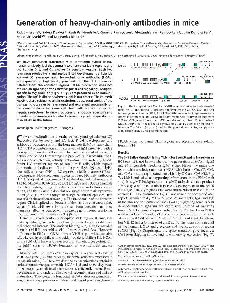
Chimeric Loci Lacking a Human CH1 Region. The CH1 splice problemwas solved by generating three new constructs (Fig. 1), all contain-ing C�2 and C�3 with CH1 deleted, one with C� and C� (MG�),one without C� and C� (G�), and one with CH1 deleted C�(M�G�). Three MG�, six G�, and four M�G� transgenic mouselines with one to five copies were obtained in a �MT background.Mice with different copy numbers gave the same results.G� and M�G� rescue B cell development. G� and M�G�, but notMG�, rescued B cell development in a �MT background. Therescue of B220�CD19 cells was between 30% and 100% in differentlymphoid compartments independent of copy number (Fig. 2A andTable 1). The M�G� mice contain human IgM-producing cells inthe BM absent in WT or �MT mice. Appropriately, they have notswitched class because chimeric IgG is absent. The G� mice containonly chimeric IgG� B cells. The MG� mice contain very few B cellsexpressing cell-surface chimeric Ig, but interestingly, 30% of theBM B220 cells express intracellular IgM, but not IgG (Fig. 2A). TheMG�, but not the M�G� and G� (data not shown), express mouseIg LC (see Fig. 5G). Thus, the C� and C� genes are expressed, andabsence of CH1 is crucial for surface-expressed HCAb.
HCAb replace mouse (pre-)BCR in the BM. During progression of largecycling into small resting pre-B cells, specific surface markers aredown-regulated in a pre-BCR-dependent manner (22). To testwhether chimeric HCAbs functionally replace the pre-BCR, variousmarkers were analyzed. Pro-B cells express high cytoplasmic SLC,IL-7R and CD43, which are down-regulated upon pre-BCR ex-pression and absent in mature B cells (Fig. 2B).
M�G���MT or G���MT chimeric Ig� B cells are SLC- and
Fig. 2. Flow cytometric analysis of B cells of wt, �MT, MG���MT, M�G���MT, and G���MT mice in BM. (A) Lymphoid cells were gated on forward and sidescatter. Surface expression of B220 and chimeric IgM or IgG is shown as dot plots. For MG���MT, the B220� fraction was gated and analyzed for the expressionof intracellular (ic) chimeric Ig � and � H chains, displayed as histogram overlays (red lines), with background stainings of B220� cells from �MT mice (black lines)as controls. The % of positive cells is indicated. (B) M�G� or G� transgenes rescue pre-BCR and BCR function. Shown are the expression profiles of the indicatedmarkers in total CD19� fractions from �MT mice (pro-B cells), in CD19� surface IgM� fractions (pro-B�pre-B cells), and CD19� surface IgM� fractions (B cells) fromWT, M�-G� �MT, and G� �MT mice. ic-Ig �, intracellular Ig � LC. Flow cytometric data are displayed as histograms representative of 3–8 animals examined ineach group. (C) Sequence alignment of BM cDNA showing VDJ recombination. Sequences are from G�. Green shows sequence identity.
Table 1. Percent of B220��CD19� cells in total population ofnucleated cells
Cell type WT G� (�5 copies) G� (single copy) M�G�
BM 10.80 � 2.09 5.94 � 1.44 4.93 � 1.79 6.06 � 1.53Spleen 41.80 � 6.05 32.14 � 9.46 28.70 � 8.70 33.95 � 3.24Blood 43.72 � 7.50 16.00 � 5.68 16.01 � 3.76 9.25 � 3.24Peritoneum 21.92 � 9.90 22.85 � 6.71* 22.30 � 7.29* 21.21 � 14.42
Mice were 14–20 weeks old. Numbers of mice analyzed are 5–11 per mouseline with the exception of two peritoneal cell measurements, where calcula-tions are based on two samples (marked by asterisks).
Janssens et al. PNAS � October 10, 2006 � vol. 103 � no. 41 � 15131
IMM
UN
OLO
GY
IL-7R-low, indicating that the chimeric HC IgG and IgM receptorsfunction as a pre-BCR in down-regulating SLC and IL-7R. CD43persists in M�G� (not in G�) mice, perhaps due to increased B-1B cell differentiation. CD2 and MHC class II are induced normally.The levels of the IL-2R�CD25, transiently present in pre-B cells, arevery low on mature M�G� or G���MT B cells as in WT (Fig. 2B).ic Ig� was absent in mature M�G� or G���MT B cells (Fig. 2B)and was not induced in BM cultures upon IL-7 withdrawal afterIL-7� culture (data not shown). Finally, the chimeric HCAb� B cellpopulations in M�G� or G� mice consisted of cells generated inthe BM (HSAhigh and AA4.1�CD93high) and cells matured in theperiphery that are recirculating (HSAlow and CD93low) as in wildtype.
Thus, chimeric HCAb IgG and IgM function as (pre-)BCR withrespect to developmentally regulated markers. IgL chain is notinduced (see below). Both VHHs are used for VDJ recombination,CH1 is absent and, importantly, CDR3 shows a large diversity(Fig. 2C).
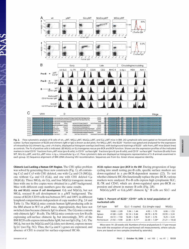
Multiple Rearrangements and Allelic Exclusion. M�G� and G�hybridomas were made after immunization. Particularly, the five-copy G� line1 could have more than one rearrangement. Of the fivedifferent five-copy hybridomas, one rearranged one in frame copy;two hybridomas had two rearrangements, each with one out offrame; one hybridoma had two in-frame rearrangements; and onehybridoma had four rearrangements, with two in frame.
Two express two productive mRNAs (mass spectrometry con-firmed the secreted HCAbs matching the cDNA; data not shown).We also carried out DNA fiber FISH on a hybridoma with onerearrangement and normal FISH on one with four rearrangementsby using an LCR probe detecting each copy and a probe betweenVHH and D detecting only nonrearranged copies (Fig. 3 A–E).Control cells showed five copies plus half a copy at each end (Fig.3A), in agreement with Southern blots (data not shown), whereasthe hybridomas show one and four rearranged copies, respectively(Fig. 3 B–E). Thus, multiple copies can rearrange successfully on thesame allele.
Moreover B220�CD19-positive BM cells of G� line1 transgenicmice in a WT background were analyzed for the expression of
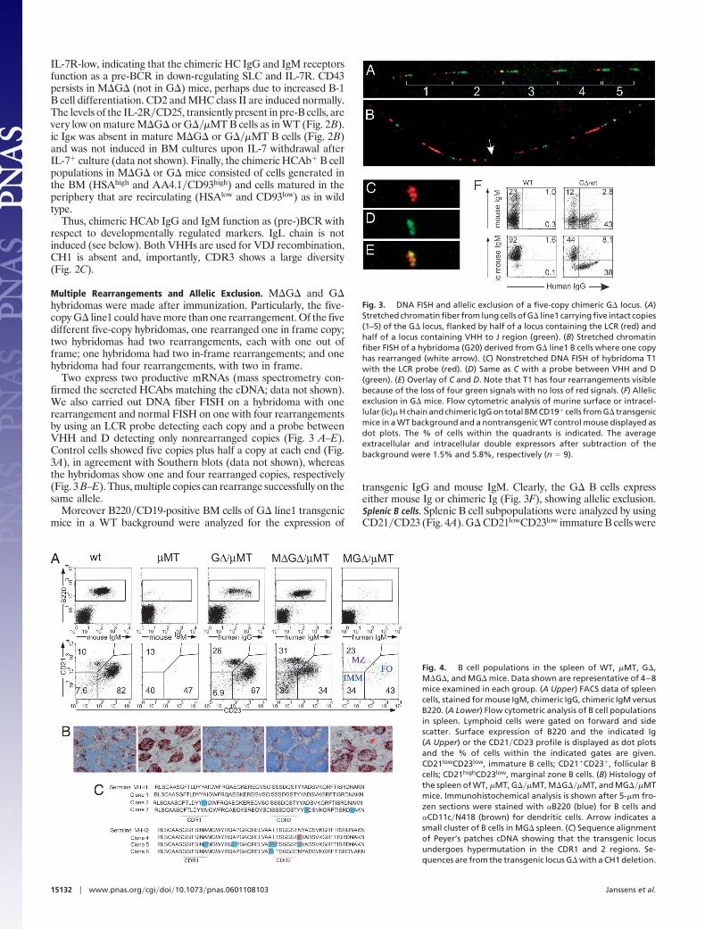
transgenic IgG and mouse IgM. Clearly, the G� B cells expresseither mouse Ig or chimeric Ig (Fig. 3F), showing allelic exclusion.Splenic B cells. Splenic B cell subpopulations were analyzed by usingCD21�CD23 (Fig. 4A). G� CD21lowCD23low immature B cells were
Fig. 4. B cell populations in the spleen of WT, �MT, G�,M�G�, and MG� mice. Data shown are representative of 4–8mice examined in each group. (A Upper) FACS data of spleencells, stained for mouse IgM, chimeric IgG, chimeric IgM versusB220. (A Lower) Flow cytometric analysis of B cell populationsin spleen. Lymphoid cells were gated on forward and sidescatter. Surface expression of B220 and the indicated Ig(A Upper) or the CD21�CD23 profile is displayed as dot plotsand the % of cells within the indicated gates are given.CD21lowCD23low, immature B cells; CD21�CD23�, follicular Bcells; CD21highCD23low, marginal zone B cells. (B) Histology ofthe spleen of WT, �MT, G���MT, M�G���MT, and MG���MTmice. Immunohistochemical analysis is shown after 5-�m fro-zen sections were stained with �B220 (blue) for B cells and�CD11c�N418 (brown) for dendritic cells. Arrow indicates asmall cluster of B cells in MG� spleen. (C) Sequence alignmentof Peyer’s patches cDNA showing that the transgenic locusundergoes hypermutation in the CDR1 and 2 regions. Se-quences are from the transgenic locus G� with a CH1 deletion.
Fig. 3. DNA FISH and allelic exclusion of a five-copy chimeric G� locus. (A)Stretched chromatin fiber from lung cells of G� line1 carrying five intact copies(1–5) of the G� locus, flanked by half of a locus containing the LCR (red) andhalf of a locus containing VHH to J region (green). (B) Stretched chromatinfiber FISH of a hybridoma (G20) derived from G� line1 B cells where one copyhas rearranged (white arrow). (C) Nonstretched DNA FISH of hybridoma T1with the LCR probe (red). (D) Same as C with a probe between VHH and D(green). (E) Overlay of C and D. Note that T1 has four rearrangements visiblebecause of the loss of four green signals with no loss of red signals. (F) Allelicexclusion in G� mice. Flow cytometric analysis of murine surface or intracel-lular (ic)� H chain and chimeric IgG on total BM CD19� cells from G� transgenicmice in a WT background and a nontransgenic WT control mouse displayed asdot plots. The % of cells within the quadrants is indicated. The averageextracellular and intracellular double expressors after subtraction of thebackground were 1.5% and 5.8%, respectively (n � 9).
15132 � www.pnas.org�cgi�doi�10.1073�pnas.0601108103 Janssens et al.
in normal ranges, and chimeric HC-IgG� cells differentiated intofollicular (FO; CD21�CD23�) and marginal zone (MZ;CD21highCD23low) B cells. In M�G�, the immature B cells wereincreased, i.e., differentiation of HC-IgM expressing cells into FOand MZ B cells appear somewhat impaired. CD23 reduction wasaccompanied by increased CD43 and CD5 (data not shown),indicative of differentiation into B-1 B cells. The few chimeric IgMexpressing B cells (also expressing mouse LC, see Fig. 5) in MG�mice had a FO�MZ distribution similar to M�G� mice.
Spleen architecture in M�G� and G�, but not MG� mice, isnormal (Fig. 4B). As in wild type, germinal centers in B cell folliclesare formed (data not shown) during T cell-dependent responsesthat in G� mice contain chimeric IgG� cells. We confirmedhypermutation of the HCAb by cDNA analysis from B cells presentin Peyer’s patches. (Fig. 4C). Both VHHs are used. Thus, in G� andM�G� mice, immature B cells migrating from BM differentiateinto spleen FO and MZ B cells and undergo somatic hypermutationupon antigen challenge.Single-copy loci rescue efficiently and CH1 absence is essential. The G�line1 mice (Table 1) had five copies and, hence, the efficient rescuewas related possibly to the copy number of the locus. A single-copyline generated from the G� line1 through breeding with a FlpeRline (23) gave the same B cell rescue (Table 1; Fig. 8, which ispublished as supporting information on the PNAS web site).
Confirmation that a single copy of the locus is sufficient forrescue and that a CH1 region is inhibitory was obtained bycre-mediated deletion of the C� and C� from MG� line 3, resultingin a single-copy G� line (Fig. 8). This locus now rescues B celldevelopment like the other G� lines. Thus, a CH1 region in C�inhibits B cell rescue, and copy number is not important.
Mouse Light Chains Do Not Rearrange in M�G� and G� Mice. MurineLC were absent in the M�G� and G� mice by Western blots (datanot shown, but see Fig. 2B and 5A) or FACS, suggesting that theLC genes do not rearrange as confirmed by comparing the Ig� locusgerm-line signals in sorted splenic B220� cells and liver DNA bySouthern blots (Fig. 9, which is published as supporting informationon the PNAS web site). Mouse LC remain in a germ-line config-uration. In contrast, LC are present in the few chimeric Ig� cells inthe MG���MT mice (see Fig. 5G).
Thus, the chimeric HCAb expression in early B cell developmentin BM fails to signal for LC rearrangement. In this respect, HCAblacking CH1 mimic a BCR rather than a pre-BCR, probablybecause of a failure to bind pseudo-LC (24).Serum analysis. Chimeric IgM was present in M�G� and chimericIgG in both M�G� and G� serum. In nonimmunized adults, thechimeric IgM (�50 �g�ml) and IgG (200–1,000 �g�ml) are present
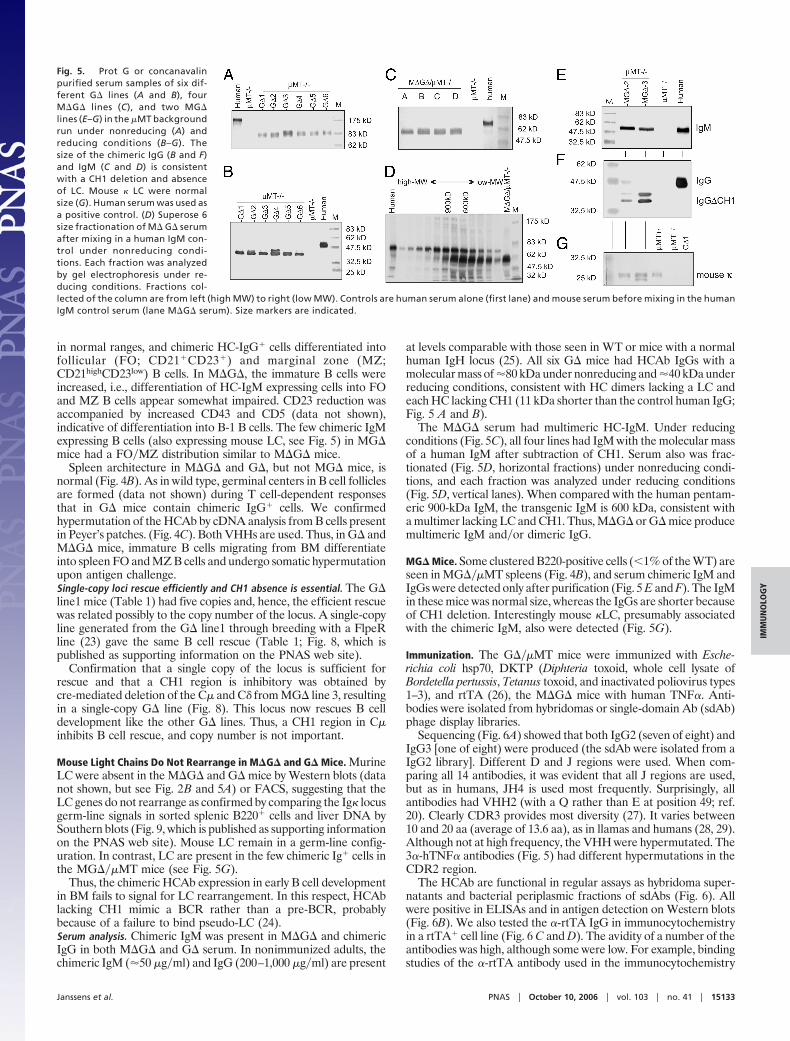
at levels comparable with those seen in WT or mice with a normalhuman IgH locus (25). All six G� mice had HCAb IgGs with amolecular mass of �80 kDa under nonreducing and �40 kDa underreducing conditions, consistent with HC dimers lacking a LC andeach HC lacking CH1 (11 kDa shorter than the control human IgG;Fig. 5 A and B).
The M�G� serum had multimeric HC-IgM. Under reducingconditions (Fig. 5C), all four lines had IgM with the molecular massof a human IgM after subtraction of CH1. Serum also was frac-tionated (Fig. 5D, horizontal fractions) under nonreducing condi-tions, and each fraction was analyzed under reducing conditions(Fig. 5D, vertical lanes). When compared with the human pentam-eric 900-kDa IgM, the transgenic IgM is 600 kDa, consistent witha multimer lacking LC and CH1. Thus, M�G� or G� mice producemultimeric IgM and�or dimeric IgG.
MG� Mice. Some clustered B220-positive cells (�1% of the WT) areseen in MG���MT spleens (Fig. 4B), and serum chimeric IgM andIgGs were detected only after purification (Fig. 5 E and F). The IgMin these mice was normal size, whereas the IgGs are shorter becauseof CH1 deletion. Interestingly mouse �LC, presumably associatedwith the chimeric IgM, also were detected (Fig. 5G).
Immunization. The G���MT mice were immunized with Esche-richia coli hsp70, DKTP (Diphteria toxoid, whole cell lysate ofBordetella pertussis, Tetanus toxoid, and inactivated poliovirus types1–3), and rtTA (26), the M�G� mice with human TNF�. Anti-bodies were isolated from hybridomas or single-domain Ab (sdAb)phage display libraries.
Sequencing (Fig. 6A) showed that both IgG2 (seven of eight) andIgG3 [one of eight) were produced (the sdAb were isolated from aIgG2 library]. Different D and J regions were used. When com-paring all 14 antibodies, it was evident that all J regions are used,but as in humans, JH4 is used most frequently. Surprisingly, allantibodies had VHH2 (with a Q rather than E at position 49; ref.20). Clearly CDR3 provides most diversity (27). It varies between10 and 20 aa (average of 13.6 aa), as in llamas and humans (28, 29).Although not at high frequency, the VHH were hypermutated. The3�-hTNF� antibodies (Fig. 5) had different hypermutations in theCDR2 region.
The HCAb are functional in regular assays as hybridoma super-natants and bacterial periplasmic fractions of sdAbs (Fig. 6). Allwere positive in ELISAs and in antigen detection on Western blots(Fig. 6B). We also tested the �-rtTA IgG in immunocytochemistryin a rtTA� cell line (Fig. 6 C and D). The avidity of a number of theantibodies was high, although some were low. For example, bindingstudies of the �-rtTA antibody used in the immunocytochemistry
Fig. 5. Prot G or concanavalinpurified serum samples of six dif-ferent G� lines (A and B), fourM�G� lines (C), and two MG�lines (E–G) in the �MT backgroundrun under nonreducing (A) andreducing conditions (B–G). Thesize of the chimeric IgG (B and F)and IgM (C and D) is consistentwith a CH1 deletion and absenceof LC. Mouse � LC were normalsize (G). Human serum was used asa positive control. (D) Superose 6size fractionation of M� G� serumafter mixing in a human IgM con-trol under nonreducing condi-tions. Each fraction was analyzedby gel electrophoresis under re-ducing conditions. Fractions col-lected of the column are from left (high MW) to right (low MW). Controls are human serum alone (first lane) and mouse serum before mixing in the humanIgM control serum (lane M�G� serum). Size markers are indicated.
Janssens et al. PNAS � October 10, 2006 � vol. 103 � no. 41 � 15133
IMM
UN
OLO
GY
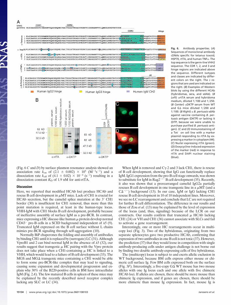
(Fig. 6 C and D) by surface plasmon resonance analysis showed anassociation rate kon of (2.1 � 0.002) 105 (M�1s�1) and adissociation rate koff of (4.1 � 0.02) 10�4 (s�1) resulting in adissociation constant KD of 1.9 nM for anti-rtTA.
DiscussionHere, we reported that modified HCAb loci produce HCAb andrescue B cell development in �MT mice. Lack of CH1 is crucial forHCAb secretion, but the camelid splice mutation at the 3� CH1border (30) is insufficient for CH1 removal, thus more than thispoint mutation is required, at least in the human-type locus.VHH-IgM with CH1 blocks B cell development, probably becauseof ineffective assembly of surface IgM as a pre-BCR. In contrast,mice expressing a HC disease-like human � protein develop normalCD43� pre-B cells in a SCID background independent of �5 (9).Truncated IgM expressed on the B cell surface without L chainsmimics pre-BCR signaling through self-aggregation (10).
Normally BiP chaperones the folding and assembly of antibodiesby binding CH1 until it is replaced by (surrogate) LC (31). AlthoughVpreB1 and 2 can bind normal IgM in the absence of �5 (32), ourresults suggest that transgenic � HC pairing with the Vpre proteindoes not take place when a CH1-containing � HC is linked to aVHH, which would lead to a failure of B cell development (33). TheMGS and MG� transgenic mice containing a CH1 would be ableto form some pre-BCR-like complex that may lead to signaling,causing some expansion and developmental progression and ex-plain why 30% of the B220-positive cells in BM have intracellularIgM (Fig. 2A). The few matured B cells in spleen of these mice maybe explained by the recently described novel receptor complexlacking any SLC or LC (34).
When IgM is removed and C� 2 and 3 lack CH1, there is rescueof B cell development, showing that IgG can functionally replaceIgM. IgG1 expression from the pro-B cell stage onwards, was shownto substitute for IgM in Rag2�/� B cell development (35). Recently,it also was shown that a prerearranged camelid IgG2a partiallyrescues B cell development in one transgenic line in a �MT (and aC��/�) background (13). In our case, IgM or IgG lacking CH1rescue B cell development in 10 of 10 independent lines. Moreover,we see no LC rearrangement and conclude that LC are not requiredfor further B cell differentiation. The difference in our results andthose of Zou et al. (13) may be explained by the level of expressionof the locus (and, thus, signaling) because of the LCR on ourconstructs. Our results confirm that truncated � HCAb lackingCH1 (24) or VH and CH1 (36) cannot associate with SLCs and failto activate � gene rearrangement.
Interestingly, one or more HC rearrangements occur in multi-copy loci (Fig. 3). Two of the hybridomas, originating from twoseparate splenocytes gave two productive HCAb, confirming thatexpression of two antibodies in one B cell is not toxic (37). However,the prediction (37) that they would loose in competition with singleantibody-producing cells under antigen challenge is not borne outby finding two double antibody-expressing cells of five hybridomas.
The (multicopy) locus is subject to and exerts allelic exclusion inWT background, because BM cells express either mouse or chi-meric cell surface Ig. Few BM cells expressed both on the surface(Fig. 3F). Interestingly, a five-copy G� WT mouse has two mousealleles with one Ig locus each and one allele with five chimericHCAb loci. If alleles are chosen, there should be more mouse thanchimeric Ig expression, and if genes are chosen, there should bemore chimeric than mouse Ig expression. In fact, mouse Ig is
Fig. 6. Antibody properties. (A)Sequences of monoclonal antibodycDNAs specific for tetanus toxoid;HSP70, rtTA, and human TNF�. Thetop sequence is the germ-line VHH2sequence. The CDR 1, 2, and 3 andhinge regions are indicated abovethe sequence. Different isotypesand classes are indicated by differ-ent colors on the right. The J re-gions that are used are indicated onthe right. (B) Examples of Westernblots by using the different HCAb(hybridomas, sera, and sdAb). (BLeft) �rtTA serum and hybridomamedium, diluted 1�100 and 1�250.(B Center) �DKTP serum from WTand G� mice diluted 1�200 and1�100. (B Right) � B. pertussis sdAbagainst vaccine containing B. per-tussis antigen (DKTP) or lacking it(DTP, because we were unable topurchase purified B. pertussis anti-gen). (C and D) Immunostaining ofa Tet� on cell line with a markerplasmid responding to rtTA by ex-pressing a marker in cytoplasm (44).(C) Nuclei expressing rtTA (green).(D) Doxycycline-induced expressionof the marker (red) in response tortTA and DAPI nuclear staining(blue).
15134 � www.pnas.org�cgi�doi�10.1073�pnas.0601108103 Janssens et al.
expressed more often (44�38; Fig. 3F). Ignoring possible deviationsfrom the random V use and a possible position effect on thetransgenic locus, suggesting that the first choice is one of alleles.
Normally, a productive rearrangement down-regulates recombi-nation to prevent rearrangement of the other allele. However, themultiple transgenic copies, when rearranged, exclude the mouseendogenous locus, but fail to exclude further rearrangement on thesame open locus before RAG down-regulation. This process mayinvolve a spatial component (‘‘compartment’’), in that the timebefore the RAGs are down-regulated would be sufficient to rear-range another gene in the locus because it would be in closeproximity. The observation that other species with multiple loci onthe same chromosome have more cells expressing two Abs (38)supports this argument. Alternatively multiple rearrangements maytake place at the same time.
Importantly, we show that HCAb loci can be expressed success-fully in mice. Antigen challenge results in antigen-specific chimericHCAb of different classes (dependent on locus composition)expressed at levels comparable with WT or conventional humanIgH transgenic mice (25). Only two VHHs were used, yet antibodieswith diverse specificity were isolated successfully to almost all of thetotally unrelated proteins we tested, demonstrating the efficiencyand efficacy of diversity generated by CDR3 (27). Thus, havingV(D)J recombination and in vivo selection provides an advantageover antibodies of fragments thereof from synthetic libraries. Hy-bridomas containing HCAb with a human effector function aregenerated easily. They can be used also for direct cloning andexpression of sdAb, which can alternatively also be derived by phagedisplay.
Thus, these mice open up new possibilities to produce humanHCAb for clinical or other purposes, particularly in light of theevidence (4) that HCAbs may recognize ‘‘difficult’’ epitopes such asenzyme active sites. The restricted number of VH may explain whynot all antigens were recognized; the polio and Diphteria proteinsgave no response in G� mice, whereas WT control mice did (datanot shown). Surprisingly, all antibodies had VHH2 lacking aconserved amino acid (39) at position 49 in contrast to VHH1 thathas one and should be more soluble. Perhaps, VHH1 expressionresults in negative selection.
The addition of more VHs should lead to an even broaderrepertoire. Whilst it is preferable to avoid multiple copies on a singleallele, it would be advantageous to have multiple alleles with a singlecopy of different VH regions to increase diversity. In such new loci,
one can use either normally occurring (human) VH or VH engi-neered for increased solubility (18).
In conclusion, we show that antigen-specific HCAb of potentiallyany class can be produced in mice. By introducing soluble humanVH domains in the locus, this technology allows the production offully human HCAb of any class or fragments thereof in response toantigen challenge for use as therapeutic agents in man. By usingdifferent vertebrate loci, our technology also allows for productionof antibodies from any vertebrate for use as reagents, diagnostics,or for the treatment of animals.
Materials and MethodsA standard genomic cosmid library was made from Lama glamablood. Two germ-line VHHs were chosen with hydrophilic aminoacid codons at positions 42, 50, and 52 according to ImMunoGe-neTics numbering (40), one with and one without a hydrophilicamino acid at 49. One is identical to IGHV1S1 (GenBank accessionno. AF305944), and the other has 94% identity with IGHV1S3(GenBank accession no. AF305946). PAC clone 1065 N8 containedhuman HC D and J regions, C� and C�, and clone 1115 N15contained C �3 (BACPAC Resource Center, Oakland, CA). Bacclone 11771 (Incyte Genomics, Palo Alto, CA) was used to obtainC�2 and the HC-LCR (41). C�3 and C�2 were subcloned separatelyinto pFastBac (Invitrogen, Carlsbad, CA). The point mutation (Gto A) (5) or deletion of CH1 was done by recombination (42).Similarly, frt and lox P sites were introduced 5� to the C� switchregion, and a second lox P site was placed 5� to the C�2 switchregion, resulting in MGS or MG�.
GS or G� were generated from MGS or MG� (Figs. 1 and7) by cre recombination (43). M�G� was obtained from MG�by deletion of the C� CH1 region through homologousrecombination. The generation of transgenic mice, breeding,and genotyping, RT-PCR, f low cytometry, Ig gene arrange-ment, DNA FISH analysis, immunization and hybridomaproduction, sdAB library production and screening, immuno-cytochemistry, Western blots, gel filtration, and BIAcoremeasurements are described in Supporting Methods, which ispublished as supporting information on the PNAS web site.
We thank I. Lede, K. Bezstarosti, J. Demmers, T. Nikolic, G. Dingjan,R. van Haperen, P. Rodriguez, and M. van de Corput (all fromErasmusMC) for materials and technical help during various stages ofthe project.
1. Hamers-Casterman C, Atarhouch T, Muyldermans S, Robinson G, Hamers C, Songa EB,Bendhman N, Hamers R (1993) Nature 363:446–448.
2. Nguyen VK, Hamers R, Wyns L, Muyldermans S (2000) EMBO J 19:921–930.3. Nguyen VK, Desmyter A & Muyldermans S (2001) Adv Immunol 79:261–296.4. Lauwereys M, Arbabi Ghahroudi M, Desmiter A, Kinne J, Holzar W, De Genst E, Wyns
L, Muyldermans S (1998) EMBO J 17:3512–3520.5. Nguyen VK, Hamers R, Wyns L, Muyldermans S (1999) Mol Immunol 36:515–524.6. Woolven BP, Frenken LG, van der Logt P, Nicholls PJ (1999) Immunogenetics 50:98–101.7. Brandt CR, Morrison SL, Birshtein BK, Milcarec C (1984) Mol Cell Biol 4:1270–1277.8. Fermand JP, Brouet JC (1999) Hematol Oncol Clin North Am 13:1281–1294.9. Corcos D, Iglesias A, Dunda O, Bucchini D, Jami J (1991) Eur J Immunol 21:2711–2716.
10. Corcos D, Dunda O, Butor C, Cesbron JY, Lores P, Bucchini D, Jami J (1995) Curr Biol5:1140–1148.
11. Muyldermans S, Cambillau C, Wyns L (2001) Trends Biochem Sci 26:230–235.12. Nguyen V, Zou X, Lauwereys M, Brys L, Bruggemann M, Muyldermans S (2003)
Immunology 109:93–101.13. Zou X, Smith JA, Nguyen VK, Ren L, Luyten K, Muylderman S, Bruggeman M (2005)
J Immunol 175:3769–3779.14. Kitamura D, Roes J, Kuhn R, Rajewsky K (1991) Nature 350:423–426.15. Macpherson AJ, Lamarre A, McCoy K, Harriman GR, Odermatt B, Dougan G, Hengartner
H, Zinkernagel RM (2001) Nat Immunol 2:625–631.16. Orinska Z, Osiak A, Lohler J, Bulanova E, Budagian V, Horak I, Bulfone-Paus S (2002) Eur
J Immunol 32:3472–3480.17. Hasan M, Polic B, Bralic M, Jonjic S, Rajewsky K (2002) Eur J Immunol 32:3463–3471.18. Davies J, Riechmann L (1996) Protein Eng 9:531–537.19. Riechmann L, Muyldermans S (1999) J Immunol Methods 231:25–38.20. De Genst E, Silence K, Ghahroudi M, Decanniere K, Loris R, Kinne J, Wyns L,
Muyldermans S (2005) J Biol Chem 280:14114–14121.21. Conrath K, Vincke C, Stijlemans B, Schymkowitz J, Decanniere K, Wyns L, Muyldermans
S, Loris R (2005) J Mol Biol 350:112–125.22. Middendorp S, Dingjan GM, Hendriks RW (2002) J Immunol 168:2695–2703.
23. Farley FW, Soriano P, Steffen LS, Dymecki SM (2000) Genesis 28:106–110.24. Iglesias A, Kopf M, Williams GS, Buhler B, Kohler G (1991) EMBO J 10:2147–2155.25. Wagner SD, Gross G, Cook GP, Davies SL, Neuberger MS (1996) Genomics 35:405–414.26. Urlinger S, Baron U, Thellmann M, Hasan MT, Bujard H, Hillen W (2000) Proc Natl Acad
Sci USA 97:7963–7968.27. Xu JL, Davis MM (2000) Immunity 13:37–45.28. Vu KB, Ghahroudi MA, Wyns L, Muyldermans S (1997) Mol Immunol 34:1121–1131.29. Zemlin M, Klinger M, Link J, Zemlin C, Bauer K, Engler JA, Schroeder HW, Jr, Kirkham
PM (2003) J Mol Biol 334:733–749.30. Conrath KE, Wernery U, Muyldermans S, Nguyen VK (2003) Dev Comp Immunol
27:87–103.31. Knarr G, Modrow S, Todd A, Gething MJ, Buchner J (1999) J Biol Chem 274:29850–29857.32. Seidl T, Rolink A, Melchers F (2001) Eur J Immunol 31:1999–2006.33. Mundt C, Licence S, Shimizu T, Melchers F, Martensson IL (2001) J Exp Med 193:435–445.34. Su YW, Flemming A, Wossning T, Hobeika E, Reth M, Jumaa H (2003) J Exp Med
198:1699–1706.35. Pogue SL, Goodnow CC (2000) J Exp Med 191:1031–1044.36. Shaffer AL, Schlissel MS (1997) J Immunol 159:1265–1275.37. Sonoda E, Pewzner-Jung Y, Schwers S, Taki S, Jung S, Eolat D, Rajewsky K (1997) Immunity
6:225–233.38. Eason DD, Litman RT, Luer CA, Kerr W, Litman GW (2004) Eur J Immunol 34:2551–2558.39. De Genst E, Saerens D, Muyldermans S, Conrath K (2006) Dev Comp Immunol 30:187–198.40. Lefranc M, Giudicelli V, Ginestoux C, Bodmer J, Muller W, Bontrop R, Lemaitre M, Malik
A, Barbie V, Chaume D (1999) Nucleic Acids Res 127:209–212.41. Mills F, Harindranath N, Mitchell M, Max E (1997) J Exp Med 186:845–858.42. Imam A, Patrinos G, de Krom M, Bottardi S, Janssens R, Katsantoni E, Wai A, Sherratt
D, Grosveld F (2000) Nucleic Acids Res 15:E65.43. Buchholz F, Angrand PO, Stuart AF (1996) Nucleic Acids Res 24:3118–3119.44. Dekker S, Toussaint W, Panayotou G, de Wit T, Visser P, Grosveld F, Drabek D (2003)
J Virol 77:12132–12139.
Janssens et al. PNAS � October 10, 2006 � vol. 103 � no. 41 � 15135
IMM
UN
OLO
GY