Embed Size (px)
Citation preview

RESEARCH ARTICLE
Generation of a Conditional Allele of theTranscription Factor Atonal Homolog 8(Atoh8)Miriam Ejarque1,2,3, Joan Mir-Coll1,3, Ramon Gomis1,2,3, Michael S. German4, FrancisC. Lynn5,6, Rosa Gasa1,2*
1 Diabetes and Obesity Research Laboratory, Institut d’Investigacions Biomèdiques August Pi i Sunyer(IDIBAPS), Barcelona, Spain, 2 Centro de Investigación Biomédica en Red de Diabetes y EnfermedadesMetabólicas Asociadas, Barcelona, Spain, 3 University of Barcelona, Barcelona, Spain, 4 Department ofMedicine, Diabetes Center, University of California, San Francisco, United States of America, 5 DiabetesResearch Program, Child and Family Research Institute, Vancouver, Canada, 6 Department of Surgery,University of British Columbia, Vancouver, Canada
AbstractAtonal Homolog 8 (Atoh8) is a basic helix-loop-helix (bHLH) transcription factor that is highly
conserved across species and expressed in multiple tissues during embryogenesis. In the
developing pancreas, Atoh8 is expressed in endocrine progenitors but declines in hor-
mone-positive cells, suggesting a role during early stages of the endocrine differentiation
program. We previously generated a whole-body Atoh8 knockout but early lethality of null
embryos precluded assessment of Atoh8 functions during organ development. Here we
report the generation of a conditional Atoh8 knockout mouse strain by insertion of two loxP
sites flanking exon 1 of the Atoh8 gene. Pancreas-specific Atoh8 knockout (Atoh8 Δpanc)
mice were obtained by mating this strain with a Pdx1-Cre transgenic line. Atoh8 Δpanc mice
were born at the expected mendelian ratio and showed normal appearance and fertility.
Pancreas weight and gross pancreatic morphology were normal. All pancreatic cell lineages
were present, although endocrine δ (somatostatin) cells were modestly augmented in
Atoh8 Δpanc as compared to control neonates. This increase did not affect whole-body glu-
cose tolerance in adult knockout animals. Gene expression analysis in embryonic pancre-
ases at the time of the major endocrine differentiation wave revealed modest alterations in
several early endocrine differentiation markers. Together, these data argue that Atoh8 mod-
ulates activation of the endocrine program but it is not essential for pancreas formation or
endocrine differentiation in the mouse. Given the ubiquitous expression pattern of Atoh8,
the availability of a mouse strain carrying a conditional allele for this gene warrants further
studies using temporally regulated Cre transgenic lines to elucidate time or cell-autono-
mous functions of Atoh8 during development and in the adult.
PLOS ONE | DOI:10.1371/journal.pone.0146273 January 11, 2016 1 / 12
OPEN ACCESS
Citation: Ejarque M, Mir-Coll J, Gomis R, GermanMS, Lynn FC, Gasa R (2016) Generation of aConditional Allele of the Transcription Factor AtonalHomolog 8 (Atoh8). PLoS ONE 11(1): e0146273.doi:10.1371/journal.pone.0146273
Editor: Ilse Rooman, Vrije Universiteit Brussel,BELGIUM
Received: June 11, 2015
Accepted: December 15, 2015
Published: January 11, 2016
Copyright: © 2016 Ejarque et al. This is an openaccess article distributed under the terms of theCreative Commons Attribution License, which permitsunrestricted use, distribution, and reproduction in anymedium, provided the original author and source arecredited.
Data Availability Statement: All relevant data arewithin the paper and its Supporting Information files.
Funding: This work has been supported by theSpanish Ministerio de Ciencia e Innovación(BFU2008-02299/BMC to RGa), Ministerio deEconomía y Competitividad/Instituto de Salud CarlosIII (PI13/01500 to RGa) and Generalitat de Catalunya(2014 SGR659 to RGo). The funders had no role instudy design, data collection and analysis, decision topublish, or preparation of the manuscript.
Competing Interests: The authors have declaredthat no competing interests exist.

IntroductionAtonal Homolog 8 (Atoh8) is a basic helix-loop-helix (bHLH) transcription factor highly con-served across species and expressed in multiple organs during embryonic development [1–5].Atoh8 is thought to participate in differentiation programs even though its precise molecularfunctions remain largely unknown. The Atoh8 gene is upregulated by ectopic expression of sev-eral lineage-determining bHLH factors in cultured cells, indicating a potential general role ofAtoh8 in differentiation programs driven by bHLH proteins [3, 6–8]. Thus far Atoh8 has beeninvolved in the specification and differentiation of neuronal lineages, favoring neurogenesisover gliogenesis [4]. Furthermore, Atoh8 is expressed in several neuronal subtypes in the adultbrain, indicating a function in the maintenance of these cellular subtypes besides of its roles inthe determination of neuronal fate. In mice, Atoh8 has been involved in kidney and liver devel-opment [2, 9]. It has also been implicated in the development of retina and skeletal muscle inchicken and zebrafish [1, 5, 10]. In humans, ATOH8 has been shown to contribute to shearstress stimulated endothelial differentiation during embryonic endothelial development [11],to participate in muscle fiber regeneration [12] and to repress stem cell genes in hepatocellularcarcinoma cells [13].
Given the ubiquitous expression pattern and multiple roles ascribed to Atoh8, a conditionalgene knockout mouse model would be highly valuable to decipher its function in specific tis-sues and cell lineages at precise developmental stages. Here we describe the generation of a con-ditional Atoh8 knockout mouse line by flanking the Atoh8 exon1 with loxP sites (flox). Toverify that this allele can be deleted in vivo by the Cre recombinase in a tissue-specific manner,Atoh8 floxed mice were mated with a Pdx1-Cre line [14] to generate pancreas-specific Atoh8knockouts. We had previously identified Atoh8 as a component of the embryonic pancreastranscriptional network, but due to early lethality of knockout embryos, its function duringpancreatic development in vivo could not been addressed [3].
Methods
Construction of the targeting vectorThe region (~-8->~+3.5kb) surrounding the Atoh8 translational start site was gap repairedfrom a BAC containing the entire Atoh8 gene (RP22-157F13; 129S6/SvEvTac) into a PGK-TKcontaining Bluescript vector using homology arms bounded by the following primers (5’F-GCCACTCCTCCTGCATTTTCTGTTAC; 5’R-CCATCTCCTCATGCCCTGTCAG; 3’F-TCAGGTTGCATCATGACGTTATCCTC; 3’R- GCCAGAGTTCGATCCCCA AG). The PGK-TKwas placed downstream of the shorter 3’ homology arm. 5’ and 3’ LoxP sites were then intro-duced using recombineering and the pL452 and pL451 vectors respectively and the first selec-tion cassette was then removed using Cre-expressing EL350 E Coli as previously described[15]. The 5’ LoxP site was inserted in the Atoh8 upstream region as indicated: ATCAATTGTTATCATTCCCAGGAGGA-LoxP-AGGTGTGGTTGTGACCCCTATCCT. The 3’ LoxP sitewas inserted into intron 1 as indicated CAGATGACAGAGGGCAGGGAGTTG-LoxP-CCTGTATATCTGCTTTGCTTGTGGTG. The final construct contained an FRT flanked PGK-pro-moter driven NeoR gene, that is part of pL451, for selection and subsequent excision usingFLPe.
Generation of the Atoh8 floxed alleleThe NotI linearized targeting vector was electroporated into E14 mouse embryonic stem cellsby the UCSF transgenic core. After standard selection with G418 and ganciclovir (Ganc), cor-rectly targeted ES cell clones were screened by PCR and then Southern blot analysis using
Conditional Allele for Atoh8
PLOS ONE | DOI:10.1371/journal.pone.0146273 January 11, 2016 2 / 12

external probes. Chimera mice were generated from three correctly targeted ES clones bymicroinjection into C57BL/6J blastocysts. Germline transmission was achieved by breeding toC57BL/6J females and targeting confirmed using Southern blotting on EcoNI and XmnIdigested genomic DNA. The PGK-neo selection cassette was then removed by crossing withActin-Flpe mice (Jax #005703) and confirmed by PCR. Subsequent genotyping of the floxedAtoh8 mice was carried out by PCR to detect LoxP inclusion (primers in S1 Table). Pancreas-specific Atoh8 knockout mice (Atoh8 Δpanc) were generated by crossing floxed Atoh8 with thePdx1-Cre strain. Genomic PCRs were done on DNAs isolated from tail tissues.
MicePdx1-Cre mice were described elsewhere [14]. Mice carrying the Cre transgene and the condi-tional Atoh8 allele, generated at the University of California at San Francisco, were sent to IDI-BAPS for phenotypic characterization. All mice were bred and maintained at the barrieranimal facility of the University of Barcelona. Embryonic tissues were collected at indicatedtimes, considering the morning of the appearance of a vaginal plug as embryonic day (E)0.5.Principles of laboratory animal care were followed (European and local government guidelines)and animal procedures were approved by the Animal Research Committee of the University ofBarcelona. Animals were euthanized by cervical dislocation.
RNA extraction and qRT-PCRTotal RNA was isolated from embryonic pancreases using the RNeasy MicroKit (Qiagen, Hil-den, Germany) and quantified using a Nanodrop 1000 (Thermo Scientific, Wilmington, MA).RNA was reverse-transcribed using the Superscript III Reverse Transcriptase (Invitrogen) fol-lowing the manufacturer’s instructions. Real time PCR was carried out in ABI7900 cycler usinga Sybr green master mix (Express Greener, Invitrogen). mRNA expression levels were normal-ized to the expression of the Tata binding protein (tbp) for transcription factors and ghrelinand Ppy or of beta-actin (actb) for the other hormones. Results are expressed as fold relative tolevels in control pancreases (value of 1). Primer sequences are provided in S1 Table.
Immunofluorescence and morphometric analysisPancreases were harvested, fixed 5h in 4% PFA and paraffin-embedded. Immunofluorescencestaining was performed on 3 μm sections following standard procedures. Primary antibodieswere: guinea pig anti-insulin (1:1000 dilution, Dako, Glostrup, Denmark), mouse anti-gluca-gon (1:500 dilution, Dako) and rabbit anti-somatostatin (1:500, Dako). Cye3 anti-guinea pigand Cy2 anti-rabbit or anti-mouse labeled secondary antibodies (1:500 dilution, JacksonImmunoresearch, Suffolk, UK) were used. Hoescht (1:500 dilution, Sigma-Aldrich) wasemployed as nuclear marker. Images were taken with a Leica DMR HC epifluorescence micro-scope and analyzed using Image J software. For morphometric measurements, 13 to 15 non-consecutive 3 μm thick sections (45 μm apart) were analyzed per pancreas.
Intraperitoneal Glucose Tolerance Test (IPGTT)In order to assess whole-body glucose tolerance, intraperitoneal glucose tolerance tests(IPGTT) were performed after 6 h food deprivation in Atoh8 Δpanc and wild-type mice. TheIPGTT was performed by administration of an injection of D-glucose (2 g/Kg body weight),and blood samples were collected from tail vein at 0, 15, 30, 60 and 120 minutes after injection.Glycaemia was measured at the same time points using a clinical glucometer and Accu-Checktest strips (Roche Diagnostics, Switzerland). Plasma was obtained by blood centrifugation and
Conditional Allele for Atoh8
PLOS ONE | DOI:10.1371/journal.pone.0146273 January 11, 2016 3 / 12

kept at -80°C for insulin determination using a mouse insulin ELISA kit (Mercodia, Uppsala,Sweden).
Islet isolation and ex vivo insulin secretion assayIslets were isolated using collagenase digestion and Histopaque gradient (Sigma-Aldrich) puri-fication as described elsewhere [16]. Separate batches of 8 freshly isolated islets were used todetermine insulin secretion in static incubation assays as previously described [16]. Insulin wasmeasured using a mouse insulin ELISA kit (Mercodia).
Statistical analysisData are presented as mean ± standard error of the mean (SE). Statistical significance wastested using Student’s t-test.
Results and DiscussionAtoh8 is expressed in multiple tissues during embryonic development [1–5]. In the developingpancreas, it is initially found in the mesenchyme and later in differentiating epithelial (endo-crine and exocrine) cells [3]. Within the endocrine compartment, Atoh8 was suggested tomodulate activation of the endocrine differentiation cascade, although this conclusion wasbased on gain-of-function experiments in cultured cells [3]. Given the ubiquitous expression ofAtoh8, to address the autonomous role of Atoh8 in pancreatic cell differentiation in vivo, wesought to generate a pancreas-specific mouse knockout model for this gene. Hence, wedesigned a conditional allele to eliminate exon 1 that encodes 237 of 322 amino acids, includingthe Proline-rich region, the first helix and the basic domain of the bHLH domain of this tran-scription factor [17]. Despite the presence of Atoh8 transcripts that comprise an occult exon inintron 1 spliced to exons 2 and 3, no open reading frame has been identified that includes theAtoh8 amino acids encoded by exons 2 and 3 [18]. It should be noted there have beendescribed two germline Atoh8mouse models, which have yielded discrepant survival pheno-types. Thus, one model exhibited early embryonic lethality around gastrulation [3] whilst theother presented normal viability [18]. The reason for this inconsistency remains uncertain butit may be related to the different genetic strategies used to generate these lines, i.e. combineddeletion of exons 1+2 (which encode the complete protein except for the stop codon which iscoded by exon 3) [3] versus deletion of only exon 1 [18]. In view of these observations, ourpresent strategy is expected to circumvent the potential effects of undefined regulatorysequences within intron 1 of the Atoh8 locus.
The targeting vector was constructed as described in Material and Methods and depicted inFig 1A. Heterozygous (Flox/+) F1 mice were intercrossed to generate F2 offspring and the micewere genotyped by PCR of tail genomic DNA (Fig 1B). The floxed Atoh8mice were subjectedto further characterization by mating them with the pancreas-wide deleter Pdx1-Cre line togenerate Atoh8 Δpanc mice. The Pdx1-Cre line drives Cre recombinase in the pancreatic epithe-lium starting at embryonic day (E)8.5 [14], which results in the elimination of the floxed genein all pancreatic cell lineages, namely ductal, exocrine and endocrine. Tissue-specific recombi-nation was verified by PCR genotyping of genomic DNA isolated from embryonic pancreasand liver (Fig 1C). Given that Atoh8 is first detected in the pancreatic epithelium after embry-onic day (E)13.5, we confirmed that Atoh8mRNA levels were significantly reduced in the pan-creas at (E)14.5 by qRT-PCR (Fig 1D). Remaining Atoh8mRNA expression (39%) detected inknockouts likely results from incomplete Atoh8 elimination from the Pdx1+ expressiondomain (Pdx1-Cre is known to exhibit mosaic expression [19]) and from non-epithelial Atoh8expression [3]. In this regard, it is noteworthy that the relative proportion of mesenchyme to
Conditional Allele for Atoh8
PLOS ONE | DOI:10.1371/journal.pone.0146273 January 11, 2016 4 / 12
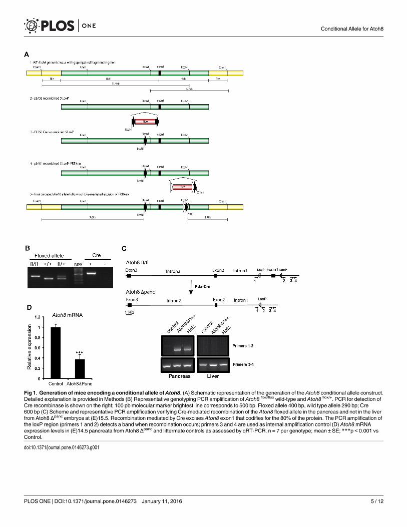
Fig 1. Generation of mice encoding a conditional allele of Atoh8. (A) Schematic representation of the generation of the Atoh8 conditional allele construct.Detailed explanation is provided in Methods (B) Representative genotyping PCR amplification of Atoh8 flox/flox wild-type and Atoh8 flox/+. PCR for detection ofCre recombinase is shown on the right; 100 pb molecular marker brightest line corresponds to 500 bp. Floxed allele 400 bp, wild type allele 290 bp; Cre600 bp (C) Scheme and representative PCR amplification verifying Cre-mediated recombination of the Atoh8 floxed allele in the pancreas and not in the liverfrom Atoh8 Δpanc embryos at (E)15.5. Recombination mediated by Cre excises Atoh8 exon1 that codifies for the 80% of the protein. The PCR amplification ofthe loxP region (primers 1 and 2) detects a band when recombination occurs; primers 3 and 4 are used as internal amplification control (D) Atoh8mRNAexpression levels in (E)14.5 pancreata from Atoh8 Δpanc and littermate controls as assessed by qRT-PCR. n = 7 per genotype; mean ± SE; ***p < 0.001 vsControl.
doi:10.1371/journal.pone.0146273.g001
Conditional Allele for Atoh8
PLOS ONE | DOI:10.1371/journal.pone.0146273 January 11, 2016 5 / 12
epithelium decreases as pancreas develops but it is still considerable at (E)14.5 [20]. Unfortu-nately, lack of working antibodies for immunostaining precluded confirmation of loss of Atoh8protein in these mutants.
Atoh8 Δpanc mice were born at the expected mendelian ratio (analysis of F2 mice showed24.5% of homozygous (flox/flox), 48% of heterozygous (flox/+) and 27.5% of wild-type (+/+)).Both flox/+ and flox/flox showed normal appearance and fertility. To assess the impact ofAtoh8 deficiency in pancreatic development, we examined Atoh8 Δpanc mice at postnatal day 1(P1). Knockout animals had similar body weight (control: 1.52±0.06 g, n = 10; Atoh8 Δpanc:1.58±0.06 g, n = 8) and glycemia (control: 46.6±2.5 mg/dL; Atoh8 Δpanc: 50.8±3.8 mg/dL) to lit-termate controls. Pancreas weight (control: 7.7±0.6 mg; Atoh8 Δpanc: 9.85±0.09 mg; p = 0.07)and gross morphology as assessed by H&E staining were comparable between knockout andcontrols (Fig 2A), revealing that Atoh8 is not essential for pancreas formation or growth. Wethen analyzed the pancreatic endocrine compartment by immunofluorescence staining of themajor islet hormones insulin (β-cells), glucagon (α-cells) and somatostatin (δ-cells). P1 knock-out pancreata stained positive for all three hormones and islet architecture was normal as com-pared to controls (Fig 2B). Morphometric quantification of the endocrine cell area revealedincreased δ-cell and comparable α- and β-cell areas between Atoh8 Δpanc and controls (Fig2C). An increase in δ-cell area was also observed using a different Pdx1-Cre transgenic line[21] (data not shown). Therefore, Atoh8 deficiency affects δ-cells but has no apparent effect onα-cell and β-cell number.
Next, we studied whether this developmental phenotype had functional consequences forglucose homeostasis in adult animals and assessed glucose tolerance in 10-wk- and 36-wk-oldAtoh8 Δpanc male mice after an intraperitoneal glucose challenge. Body weight (10-wk control:25.5±0.8 g; Atoh8 Δpanc: 26.1± 0.8 g and 36-wk control: 37.9±1 g; Atoh8 Δpanc: 35.4± 1 g) andglucose tolerance curves were indistinguishable between mutants and controls at both agesstudied (Fig 3A–3D). Atoh8 Δpanc females also displayed normal glucose tolerance (data notshown). Yet, we did observe a modest reduction in basal fasting glycemia and insulinemia in36-wk-old male Atoh8 Δpanc mice relative to controls (Fig 3E). We examined their pancreataand found that organ weight (control: 1.07±0.1 g vs Atoh8 Δpanc: 0.95±0.05 g) and gross mor-phology was similar to that of control animals of the same age (Fig 4A). Islet architecture wasalso comparable (Fig 4B). As endogenous islet somatostatin is thought to have a local effect onislet function acting as a paracrine tonic inhibitor of insulin and glucagon secretion, weassessed the ex vivo insulin secretory capacity of isolated islets using static incubation assaysand found equivalent glucose-induced insulin secretion by Atoh8 Δpanc and control islets (Fig4C). Therefore, Atoh8 deficiency in the fetal pancreas does not compromise whole body glu-cose tolerance or insulin secretion in adult islets, thus implying that the increment in δ-cellarea observed in Atoh8 Δpanc neonates appears to have no significant physiological impact inadult mice. In this regard, it is noteworthy that impaired δ-cell function in mice with pancreas-specific deletion of the homeodomain transcription factor Hhex does not compromise theirglucose tolerance, although plasma insulin concentrations at basal or after a glucose challengewere found elevated [22]. These and our findings are consistent with the ascribed minor role ofpancreatic somatostatin on the regulation of whole body glucose metabolism [23].
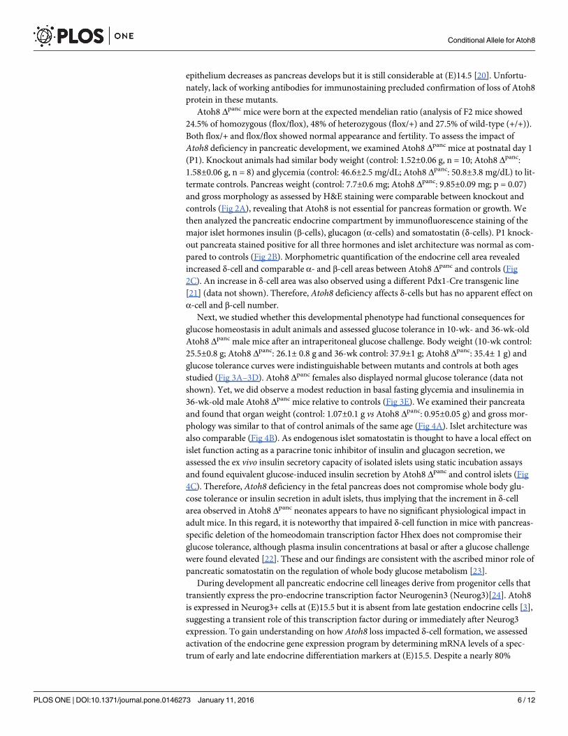
During development all pancreatic endocrine cell lineages derive from progenitor cells thattransiently express the pro-endocrine transcription factor Neurogenin3 (Neurog3)[24]. Atoh8is expressed in Neurog3+ cells at (E)15.5 but it is absent from late gestation endocrine cells [3],suggesting a transient role of this transcription factor during or immediately after Neurog3expression. To gain understanding on how Atoh8 loss impacted δ-cell formation, we assessedactivation of the endocrine gene expression program by determining mRNA levels of a spec-trum of early and late endocrine differentiation markers at (E)15.5. Despite a nearly 80%
Conditional Allele for Atoh8
PLOS ONE | DOI:10.1371/journal.pone.0146273 January 11, 2016 6 / 12

Fig 2. Pancreatic phenotype of Atoh8 Δpanc mice at postnatal day 1. (A) Hematoxilin-eosin staining of pancreatic sections revealed similar organstructure in P1 Atoh8 Δpanc and control littermates. Bars represent 50 μM (B) Representative immunofluorescence staining for the major islets cell types(insulin in red, glucagon in green and somatostatin in blue) on pancreas tissue sections from P1 Atoh8 Δpanc and control littermates. Bars represent 100 μM(C) Morphometric quantification of hormone immunoreactive areas relative to total pancreatic area. n = 3; mean ± SE; **p < 0.01 vs Control.
doi:10.1371/journal.pone.0146273.g002
Conditional Allele for Atoh8
PLOS ONE | DOI:10.1371/journal.pone.0146273 January 11, 2016 7 / 12

reduction of Atoh8 transcript at this stage, endocrine gene expression remained largely unmod-ified. Thus, no significant differences were found in islet hormone expression, although thesomatostatinmessage was slightly upregulated in mutants relative to controls (Fig 5A). On theother hand, mRNA levels for the endocrine differentiation transcription factors Neurog3, Pax4,NeuroD1 and Nkx6.1 were comparable, whereas transcripts for Pax6, Nkx2.2, Arx,Hhex andMnx1 were reduced in Atoh8 Δpanc relative to controls (Fig 5B). To date, Hhex is the onlygenetic factor shown to specify δ-cell fate during pancreatic development [22]. Yet, hhex tran-scripts were decreased by 20% in mutants, indicating that enhanced δ-cell formation did notcorrelate with upregulation of this transcription factor, at least at this developmental stage.Remarkably, it has been recently reported that inactivation ofMnx1 in endocrine progenitorsleads to increased δ-cell allocation to the detriment of β-cell differentiation [25]. Hence, ourfinding of reducedMxn1 transcript levels in Atoh8 Δpanc embryos would be compatible with
Fig 3. Glucose tolerance of adult Atoh8Δpanc mice. Intraperitoneal glucose tolerance tests were performed on 10-wk (A) and 36-wk-old (B) Atoh8 Δpanc
and control male mice fasted for 5 h. Area under the curve calculations for glucose tolerance tests in 10-wk-old (C) and 36-wk-old (D) mice. (E) Fastingglucose and insulin plasma levels of 36-wk-old control and Atoh8 Δpanc mice. n = 12 (controls) and n = 14 (Atoh8 Δpanc); mean ± SE; *p < 0.05 vs Control.
doi:10.1371/journal.pone.0146273.g003
Conditional Allele for Atoh8
PLOS ONE | DOI:10.1371/journal.pone.0146273 January 11, 2016 8 / 12

increased δ-cell differentiation in these mutants. Furthermore, it is interesting to note thatsimultaneous deletion of both Nkx2.2 and Arx in mice leads to the deregulation of somatostatinexpression [26, 27]. Thus, it is plausible that combined changes in levels of multiple transcrip-tional regulators results in increased δ-cell numbers in Atoh8mutants. Nonetheless, these
Fig 4. Pancreas morphology and islet insulin secretion in 36-week old Atoh8Δpanc mice. (A) Hematoxilin-eosin staining showed no apparentdifferences in pancreas gross morphology between 36-wk-old Atoh8 Δpanc mice and controls. Scale bars 100 μM (B) Immunofluorescence staining for insulin(blue), glucagon (green) and somatostatin (red) on pancreas sections from 36-wk-old Atoh8 Δpanc mice and controls. Representative images demonstratesimilar islet cell organization between mutants and controls. Scale bars 50 μM (C) Glucose-induced insulin secretion in isolated islets from 36-wk-old Atoh8Δpanc mice and controls. n = 3 mice per genotype (3–4 independent islet batches per animal); mean ± SE.
doi:10.1371/journal.pone.0146273.g004
Conditional Allele for Atoh8
PLOS ONE | DOI:10.1371/journal.pone.0146273 January 11, 2016 9 / 12
changes appear to be insufficient to affect the formation of the more abundant islet cell types αand β. These data argue that Atoh8 contributes mildly to activation of endocrine differentiationin vivo and implies that this transcription factor likely acts as a modulator rather than a pro-moter of differentiation in the pancreas. Although we cannot rule out compensatory effects byother genes, our current in vivo findings are consistent with prior results in a cell-based endo-crine differentiation model demonstrating lack of pro-endocrine activity of Atoh8 [17].
Further investigations on Atoh8 at a molecular level may help shed light on the function ofthis transcription factor during activation of the pancreatic endocrine program. In any case, abetter comprehension of δ-cell differentiation is required to discern the molecular pathwaysmodified by Atoh8 ablation that are being affected in Atoh8 Δpanc mutants. Further, given that(i) Atoh8 is first detected in the pancreatic epithelium after (E)13.5 [3], (ii) Neurog3+ endocrineprogenitors acquire the capacity to differentiate to δ-cells from (E)14.5 [28] and (iii) δ-cells are
Fig 5. Gene expression levels of endocrine and exocrine differentiation markers in pancreases from Atoh8 Δpanc mice at (E)15.5. Total pancreaticRNA was prepared from Atoh8 Δpanc and control embryos at (E)15.5. Gene expression levels were assessed by qRT-PCR as described in Methods. Valuesare expressed relative to control pancreases, set at 1. Expression levels for Atoh8 and endocrine cell markers (A), endocrine differentiation transcriptionfactors (B) and exocrine genes (C). n = 10 per genotype from 4 independent litters; mean ± SE; * p< 0.05, ** p< 0.01;, *** p< 0.001 vs Control.
doi:10.1371/journal.pone.0146273.g005
Conditional Allele for Atoh8
PLOS ONE | DOI:10.1371/journal.pone.0146273 January 11, 2016 10 / 12

first immunodetected at (E)15.5 [29], one possibility is that the specific impact on δ-cell forma-tion is related to a timing effect. The availability of this floxed model warrants further studiesusing temporally regulated Cre transgenic lines to address this as well as other questions.Finally, as Atoh8 is also expressed in the differentiating exocrine compartment [29], we deter-mined expression levels for amylase and the acinar-specific transcription factors Ptf1a andMist1. In agreement with normal pancreatic morphology and size, these genes were similarlyexpressed in Atoh8 Δpanc and their littermate controls at (E)15.5 (Fig 5C).
The floxed Atoh8mice will provide a valuable tool to elucidate time or cell-specific func-tions of Atoh8 during development and in the adult. Given the multiple cellular contexts whereAtoh8 has been reported to participate, availability of this model guarantees future studiesaimed at investigating the role of this little-known transcription factor in multiple organs andtissues in vivo.
Supporting InformationS1 Table. List of oligonucleotides used for genotyping and qRT-PCR.(PDF)
AcknowledgmentsWe thank L. Sanchez and A. Garcia for excellent technical assistance.
Author ContributionsConceived and designed the experiments: ME FCL MSG R. Gasa. Performed the experiments:ME JMC FCL R. Gasa. Analyzed the data: ME R. Gomis R. Gasa. Contributed reagents/materi-als/analysis tools: MSG R. Gomis. Wrote the paper: ME R. Gasa.
References1. Yao J, Zhou J, Liu Q, Lu D, Wang L, Qiao X, et al. Atoh8, a bHLH transcription factor, is required for the
development of retina and skeletal muscle in zebrafish. PloS one. 2010; 5: e10945. doi: 10.1371/journal.pone.0010945 PMID: 20532172
2. Ross MD, Martinka S, Mukherjee A, Sedor JR, Vinson C, Bruggeman LA. Math6 expression during kid-ney development and altered expression in a mouse model of glomerulosclerosis. Developmentaldynamics: an official publication of the American Association of Anatomists. 2006; 235: 3102–3109.
3. Lynn FC, Sanchez L, Gomis R, German MS, Gasa R. Identification of the bHLH factor Math6 as a novelcomponent of the embryonic pancreas transcriptional network. PloS one. 2008; 3: e2430. doi: 10.1371/journal.pone.0002430 PMID: 18560595
4. Inoue C, Bae SK, Takatsuka K, Inoue T, Bessho Y, Kageyama R. Math6, a bHLH gene expressed inthe developing nervous system, regulates neuronal versus glial differentiation. Genes to cells: devotedto molecular & cellular mechanisms. 2001; 6: 977–986.
5. Balakrishnan-Renuka A, Morosan-Puopolo G, Yusuf F, Abduelmula A, Chen J, Zoidl G, et al. ATOH8,a regulator of skeletal myogenesis in the hypaxial myotome of the trunk. Histochemistry and cell biol-ogy. 2014; 141: 289–300. doi: 10.1007/s00418-013-1155-0 PMID: 24186058
6. Serafimidis I, Rakatzi I, Episkopou V, Gouti M, Gavalas A. Novel effectors of directed and Ngn3-medi-ated differentiation of mouse embryonic stem cells into endocrine pancreas progenitors. Stem cells.2008; 26: 3–16. PMID: 17932425
7. Seo S, Lim JW, Yellajoshyula D, Chang LW, Kroll KL. Neurogenin and NeuroD direct transcriptional tar-gets and their regulatory enhancers. The EMBO journal. 2007; 26: 5093–5108. PMID: 18007592
8. Gasa R, Mrejen C, Lynn FC, Skewes-Cox P, Sanchez L, Yang KY, et al. Induction of pancreatic isletcell differentiation by the neurogenin-neuroD cascade. Differentiation; research in biological diversity.2008; 76: 381–391. PMID: 17924961
9. Kautz L, Meynard D, Monnier A, Darnaud V, Bouvet R, Wang RH, et al. Iron regulates phosphorylationof Smad1/5/8 and gene expression of Bmp6, Smad7, Id1, and Atoh8 in the mouse liver. Blood. 2008;112: 1503–1509. doi: 10.1182/blood-2008-03-143354 PMID: 18539898
Conditional Allele for Atoh8
PLOS ONE | DOI:10.1371/journal.pone.0146273 January 11, 2016 11 / 12

10. Kubo F, Nakagawa S. Cath6, a bHLH atonal family proneural gene, negatively regulates neuronal dif-ferentiation in the retina. Developmental dynamics: an official publication of the American Associationof Anatomists. 2010; 239: 2492–2500.
11. Fang F, Wasserman SM, Torres-Vazquez J, Weinstein B, Cao F, Li Z, et al. The role of Hath6, a newlyidentified shear-stress-responsive transcription factor, in endothelial cell differentiation and function.Journal of cell science. 2014; 127: 1428–1440. doi: 10.1242/jcs.136358 PMID: 24463812
12. Guttsches AK, Balakrishnan-Renuka A, Kley RA, Tegenthoff M, Brand-Saberi B, Vorgerd M. ATOH8: anovel marker in human muscle fiber regeneration. Histochemistry and cell biology. 2015; 143: 443–452. doi: 10.1007/s00418-014-1299-6 PMID: 25514850
13. Song Y, Pan G, Chen L, Ma S, Zeng T, Man Chan TH, et al. Loss of ATOH8 Increases Stem Cell Fea-tures of Hepatocellular Carcinoma Cells. Gastroenterology. 2015; 149: 1068–1081 e5. doi: 10.1053/j.gastro.2015.06.010 PMID: 26099525
14. Gu G, Dubauskaite J, Melton DA. Direct evidence for the pancreatic lineage: NGN3+ cells are islet pro-genitors and are distinct from duct progenitors. Development. 2002; 129: 2447–2457. PMID: 11973276
15. Liu P, Jenkins NA, Copeland NG. A highly efficient recombineering-based method for generating condi-tional knockout mutations. Genome research. 2003; 13: 476–484. PMID: 12618378
16. Pardo FN, Altirriba J, Pradas-Juni M, Garcia A, Ahlgren U, Barbera A, et al. The role of Raf-1 kinaseinhibitor protein in the regulation of pancreatic beta cell proliferation in mice. Diabetologia. 2012; 55:3331–3340. doi: 10.1007/s00125-012-2696-9 PMID: 22926403
17. Ejarque M, Altirriba J, Gomis R, Gasa R. Characterization of the transcriptional activity of the basichelix-loop-helix (bHLH) transcription factor Atoh8. Biochimica et biophysica acta. 2013; 1829: 1175–1183. doi: 10.1016/j.bbagrm.2013.08.003 PMID: 23938248
18. Rawnsley DR, Xiao J, Lee JS, Liu X, Mericko-Ishizuka P, Kumar V, et al. The transcription factor Atonalhomolog 8 regulates Gata4 and Friend of Gata-2 during vertebrate development. The Journal of biologi-cal chemistry. 2013; 288: 24429–24440. doi: 10.1074/jbc.M113.463083 PMID: 23836893
19. Magnuson MA, Osipovich AB. Pancreas-specific Cre driver lines and considerations for their prudentuse. Cell metabolism. 2013; 18: 9–20. doi: 10.1016/j.cmet.2013.06.011 PMID: 23823474
20. Landsman L, Nijagal A, Whitchurch TJ, Vanderlaan RL, ZimmerWE, Mackenzie TC, et al. Pancreaticmesenchyme regulates epithelial organogenesis throughout development. PLoS biology. 2011; 9:e1001143. doi: 10.1371/journal.pbio.1001143 PMID: 21909240
21. Hingorani SR, Petricoin EF, Maitra A, Rajapakse V, King C, Jacobetz MA, et al. Preinvasive and inva-sive ductal pancreatic cancer and its early detection in the mouse. Cancer cell. 2003; 4: 437–450.PMID: 14706336
22. Zhang J, McKenna LB, Bogue CW, Kaestner KH. The diabetes gene Hhex maintains delta-cell differen-tiation and islet function. Genes & development. 2014; 28: 829–834.
23. Braun M. The somatostatin receptor in human pancreatic beta-cells. Vitamins and hormones. 2014; 95:165–193. doi: 10.1016/B978-0-12-800174-5.00007-7 PMID: 24559918
24. Rukstalis JM, Habener JF. Neurogenin3: a master regulator of pancreatic islet differentiation andregeneration. Islets. 2009; 1: 177–184. doi: 10.4161/isl.1.3.9877 PMID: 21099270
25. Pan FC, Brissova M, Powers AC, Pfaff S, Wright CV. Inactivating the permanent neonatal diabetesgene Mnx1 switches insulin-producing beta-cells to a delta-like fate and reveals a facultative prolifer-ative capacity in aged beta-cells. Development. 2015; 142: 3637–3648. doi: 10.1242/dev.126011PMID: 26534984
26. Mastracci TL, Wilcox CL, Arnes L, Panea C, Golden JA, May CL, et al. Nkx2.2 and Arx genetically inter-act to regulate pancreatic endocrine cell development and endocrine hormone expression. Develop-mental biology. 2011; 359: 1–11. doi: 10.1016/j.ydbio.2011.08.001 PMID: 21856296
27. Kordowich S, Collombat P, Mansouri A, Serup P. Arx and Nkx2.2 compound deficiency redirects pan-creatic alpha- and beta-cell differentiation to a somatostatin/ghrelin co-expressing cell lineage. BMCdevelopmental biology. 2011; 11: 52. doi: 10.1186/1471-213X-11-52 PMID: 21880149
28. Johansson KA, Dursun U, Jordan N, Gu G, Beermann F, Gradwohl G, et al. Temporal control of neuro-genin3 activity in pancreas progenitors reveals competence windows for the generation of differentendocrine cell types. Developmental cell. 2007; 12: 457–465. PMID: 17336910
29. Teitelman G, Alpert S, Polak JM, Martinez A, Hanahan D. Precursor cells of mouse endocrine pancreascoexpress insulin, glucagon and the neuronal proteins tyrosine hydroxylase and neuropeptide Y, butnot pancreatic polypeptide. Development. 1993; 118: 1031–1039. PMID: 7903631
Conditional Allele for Atoh8
PLOS ONE | DOI:10.1371/journal.pone.0146273 January 11, 2016 12 / 12