Embed Size (px)
Citation preview
Lab exercise 8: Fish and Amphibians General Zoology Laborarory . Matt Nelson
The jawless fish represent the most basal group of Craniata. At one time, the two classes of jawless fish were thought to be polyphyletic, but they have since been established as monophyletic by multiple comprehensive molecular studies.
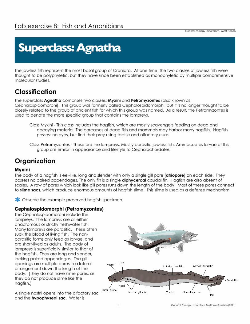
ClassificationThe superclass Agnatha comprises two classes: Myxini and Petromyzontes (also known as Cephalospidomorphi). This group was formerly called Cephalospidomorphi, but it is no longer thought to be closely related to the group of ancient fish for which this group was named. As a result, the Petromyzontes is used to denote the more specific group that contains the lampreys.
Class Myxini - This class includes the hagfish, which are mostly scavengers feeding on dead and decaying material. The carcasses of dead fish and mammals may harbor many hagfish. Hagfish possess no eyes, but find their prey using tactile and olfactory cues.
Class Petromyzontes - These are the lampreys. Mostly parasitic jawless fish. Ammocoetes larvae of this group are similar in appearance and lifestyle to Cephalochordates.
OrganizationMyxiniThe body of a hagfish is eel-like, long and slender with only a single gill pore (atriopore) on each side. They possess no paired appendages. The only fin is a single diphycercal caudal fin. Hagfish are also absent of scales. A row of pores which look like gill pores runs down the length of the body. Most of these pores connect to slime sacs, which produce enormous amounts of hagfish slime. This slime is used as a defense mechanism.
Observe the example preserved hagfish specimen.
Cephalospidomorphi (Petromyzontes)The Cephalospidomorphi include the lampreys. The lampreys are all either anadromous or strictly freshwater fish. Many lampreys are parasitic. These often suck the blood of living fish. The non-parasitic forms only feed as larvae, and are short-lived as adults. The body of lampreys is superficially similar to that of the hagfish. They are long and slender, lacking paired appendages. The gill openings are multiple pores in a lateral arrangement down the length of the body. (They do not have slime pores, as they do not produce slime like the hagfish.)
A single nostril opens into the olfactory sac and the hypophyseal sac. Water is
Superclass: Agnatha
1 General Zoology Laboratory. Matthew K Nelson (2011)
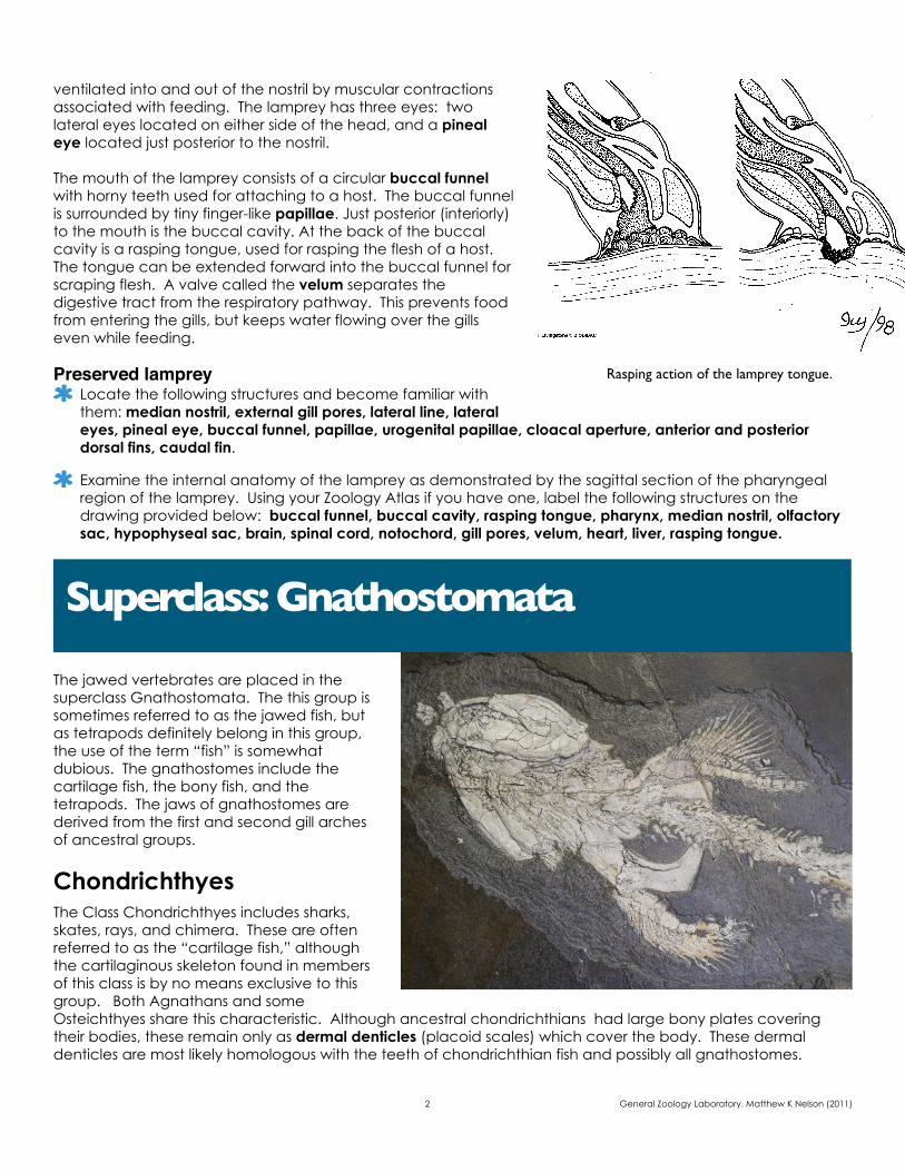
ventilated into and out of the nostril by muscular contractions associated with feeding. The lamprey has three eyes: two lateral eyes located on either side of the head, and a pineal eye located just posterior to the nostril.
The mouth of the lamprey consists of a circular buccal funnel with horny teeth used for attaching to a host. The buccal funnel is surrounded by tiny finger-like papillae. Just posterior (interiorly) to the mouth is the buccal cavity. At the back of the buccal cavity is a rasping tongue, used for rasping the flesh of a host. The tongue can be extended forward into the buccal funnel for scraping flesh. A valve called the velum separates the digestive tract from the respiratory pathway. This prevents food from entering the gills, but keeps water flowing over the gills even while feeding.
Preserved lamprey Locate the following structures and become familiar with them: median nostril, external gill pores, lateral line, lateral eyes, pineal eye, buccal funnel, papillae, urogenital papillae, cloacal aperture, anterior and posterior dorsal fins, caudal fin.
Examine the internal anatomy of the lamprey as demonstrated by the sagittal section of the pharyngeal region of the lamprey. Using your Zoology Atlas if you have one, label the following structures on the drawing provided below: buccal funnel, buccal cavity, rasping tongue, pharynx, median nostril, olfactory sac, hypophyseal sac, brain, spinal cord, notochord, gill pores, velum, heart, liver, rasping tongue.
The jawed vertebrates are placed in the superclass Gnathostomata. The this group is sometimes referred to as the jawed fish, but as tetrapods definitely belong in this group, the use of the term “fish” is somewhat dubious. The gnathostomes include the cartilage fish, the bony fish, and the tetrapods. The jaws of gnathostomes are derived from the first and second gill arches of ancestral groups.
ChondrichthyesThe Class Chondrichthyes includes sharks, skates, rays, and chimera. These are often referred to as the “cartilage fish,” although the cartilaginous skeleton found in members of this class is by no means exclusive to this group. Both Agnathans and some Osteichthyes share this characteristic. Although ancestral chondrichthians had large bony plates covering their bodies, these remain only as dermal denticles (placoid scales) which cover the body. These dermal denticles are most likely homologous with the teeth of chondrichthian fish and possibly all gnathostomes.
Superclass: Gnathostomata
2 General Zoology Laboratory. Matthew K Nelson (2011)
Rasping action of the lamprey tongue.
The jaws of chondrichthyes are connected to the chondrocranium (cartilaginous cranium) by the hyomandibular apparatus. This gives the lower jaw great flexibility and allows it to articulate forward and backward underneath the upper jaw.
The fins usually include a heterocercal tail, anterior dorsal fin, posterior dorsal fin, pectoral fins, pelvic fins, and an anal fin.
Reproduction in the Chondrichthyes is greatly varied, with examples of oviparity, ovoviviparity, and true viviparity (complete with a placenta-like structure). In viviparous chondrichthyes, fertilization is internal and the egg cases are sometimes referred to as mermaid’s purses. Males have modified pelvic fins referred to as claspers which are used to grasp the female and deliver sperm during copulation.
Observe the various examples of chondrichthians.
Subclass ElasmobranchiiThe sharks, skates and rays are in the subclass Elasmobranchii. This group has external gill slits (usually 5 per side) and one spiracle. The spiracle is a derived structure which allows the gills to be ventilated even if the mouth is closed. The elasmobranchs possess a well-developed lateral line system, as well as a related system of pores and canals referred to as the ampullae of Lorinzini.
Preserved dogfishExamine the specimen of Squalus the dogfish. Be sure you can identify the following external structures: rostrum, eyes, spiracle, gill slits, anterior dorsal fin, posterior dorsal fin, pectoral fins, pelvic fins, anal fin, caudal fin, lateral line, and the claspers if it is a male.
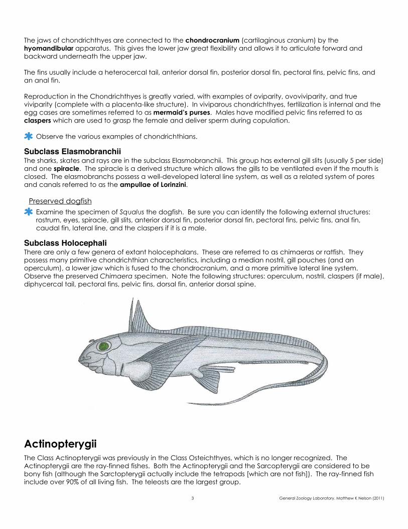
Subclass HolocephaliThere are only a few genera of extant holocephalans. These are referred to as chimaeras or ratfish. They possess many primitive chondrichthian characteristics, including a median nostril, gill pouches (and an operculum), a lower jaw which is fused to the chondrocranium, and a more primitive lateral line system. Observe the preserved Chimaera specimen. Note the following structures: operculum, nostril, claspers (if male), diphycercal tail, pectoral fins, pelvic fins, dorsal fin, anterior dorsal spine.
ActinopterygiiThe Class Actinopterygii was previously in the Class Osteichthyes, which is no longer recognized. The Actinopterygii are the ray-finned fishes. Both the Actinopterygii and the Sarcopterygii are considered to be bony fish (although the Sarctopterygii actually include the tetrapods [which are not fish]). The ray-finned fish include over 90% of all living fish. The teleosts are the largest group.
3 General Zoology Laboratory. Matthew K Nelson (2011)
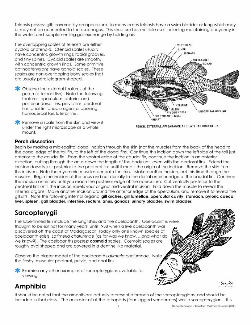
Teleosts possess gills covered by an operculum. In many cases teleosts have a swim bladder or lung which may or may not be connected to the esophagus. This structure has multiple uses including maintaining buoyancy in the water, and supplementing gas exchange by holding air.
The overlapping scales of teleosts are either cycloid or ctenoid. Ctenoid scales usually have concentric growth rings, radial grooves, and tiny spines. Cycloid scales are smooth, with concentric growth rings. Some primitive actinopterygians have ganoid scales. These scales are non-overlapping bony scales that are usually parallelogram-shaped.
Observe the external features of the perch (a teleost fish). Note the following features: operculum, anterior and posterior dorsal fins, pelvic fins, pectoral fins, anal fin, anus, urogenital opening, homocercal tail, lateral line.
Remove a scale from the skin and view it under the light microscope as a whole mount.
Perch dissectionBegin by making a mid-sagittal dorsal incision through the skin (not the muscle) from the back of the head to the dorsal edge of the tail fin, to the left of the dorsal fins. Continue this incision down the left side of the tail just anterior to the caudal fin. From the ventral edge of the caudal fin, continue the incision in an anterior direction, cutting through the anus down the length of the body until even with the pectoral fins. Extend the incision dorsally just posterior to the pectoral fins until it meets the origin of the incision. Remove the skin from this incision. Note the myomeric muscles beneath the skin. Make another incision, but this time through the muscles. Begin the incision at the anus and cut dorsally to the dorsal anterior edge of the caudal fin. Continue the incision anteriorly until you reach the posterior edge of the operculum. Cut ventrally posterior to the pectoral fins until the incision meets your original mid-ventral incision. Fold down the muscle to reveal the internal organs. Make another incision around the anterior edge of the operculum, and remove it to reveal the gill slits. Note the following internal organs: gill arches, gill lamellae, opercular cavity, stomach, pyloric caeca, liver, spleen, gall bladder, intestine, rectum, anus, gonads, urinary bladder, swim bladder.
SarcopterygiiThe lobe-finned fish include the lungfishes and the coelacanth. Coelacanths were thought to be extinct for many years, until 1938 when a live coelacanth was discovered off the coast of Madagascar. Today only one known species of coelacanth exists, Latimeria chalumnae (as far was we know, ...and what do we know?). The coelocanths possess cosmoid scales. Cosmoid scales are roughly oval shaped and are covered in a dentine like material.
Observe the plaster model of the coelacanth Latimeria chalumnae. Note the fleshy, muscular pectoral, pelvic, and anal fins.
Examine any other examples of sarcopterygians available for viewing.
AmphibiaIt should be noted that the amphibians actually represent a branch of the sarcopterygians, and should be included in that class. The ancestor of all the tetrapods (four-legged vertebrates) was a sarcopterygian. It is 4 General Zoology Laboratory. Matthew K Nelson (2011)
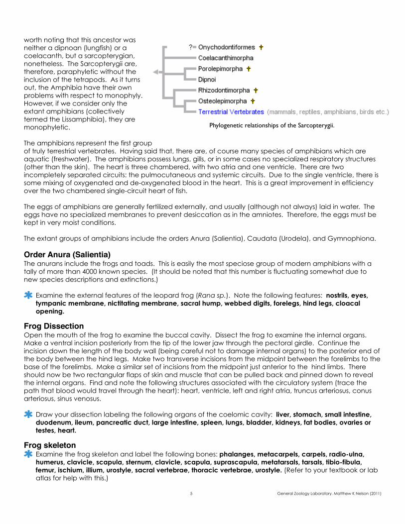
worth noting that this ancestor was neither a dipnoan (lungfish) or a coelacanth, but a sarcopterygian, nonetheless. The Sarcopterygii are, therefore, paraphyletic without the inclusion of the tetrapods. As it turns out, the Amphibia have their own problems with respect to monophyly. However, if we consider only the extant amphibians (collectively termed the Lissamphibia), they are monophyletic.
The amphibians represent the first group of truly terrestrial vertebrates. Having said that, there are, of course many species of amphibians which are aquatic (freshwater). The amphibians possess lungs, gills, or in some cases no specialized respiratory structures (other than the skin). The heart is three chambered, with two atria and one ventricle. There are two incompletely separated circuits: the pulmocutaneous and systemic circuits. Due to the single ventricle, there is some mixing of oxygenated and de-oxygenated blood in the heart. This is a great improvement in efficiency over the two chambered single-circuit heart of fish.
The eggs of amphibians are generally fertilized externally, and usually (although not always) laid in water. The eggs have no specialized membranes to prevent desiccation as in the amniotes. Therefore, the eggs must be kept in very moist conditions.
The extant groups of amphibians include the orders Anura (Salientia), Caudata (Urodela), and Gymnophiona.
Order Anura (Salientia)The anurans include the frogs and toads. This is easily the most speciose group of modern amphibians with a tally of more than 4000 known species. (It should be noted that this number is fluctuating somewhat due to new species descriptions and extinctions.)
Examine the external features of the leopard frog (Rana sp.). Note the following features: nostrils, eyes, tympanic membrane, nictitating membrane, sacral hump, webbed digits, forelegs, hind legs, cloacal opening.
Frog DissectionOpen the mouth of the frog to examine the buccal cavity. Dissect the frog to examine the internal organs. Make a ventral incision posteriorly from the tip of the lower jaw through the pectoral girdle. Continue the incision down the length of the body wall (being careful not to damage internal organs) to the posterior end of the body between the hind legs. Make two transverse incisions from the midpoint between the forelimbs to the base of the forelimbs. Make a similar set of incisions from the midpoint just anterior to the hind limbs. There should now be two rectangular flaps of skin and muscle that can be pulled back and pinned down to reveal the internal organs. Find and note the following structures associated with the circulatory system (trace the path that blood would travel through the heart): heart, ventricle, left and right atria, truncus arteriosus, conus arteriosus, sinus venosus.
Draw your dissection labeling the following organs of the coelomic cavity: liver, stomach, small intestine, duodenum, ileum, pancreatic duct, large intestine, spleen, lungs, bladder, kidneys, fat bodies, ovaries or testes, heart.
Frog skeletonExamine the frog skeleton and label the following bones: phalanges, metacarpels, carpels, radio-ulna, humerus, clavicle, scapula, sternum, clavicle, scapula, suprascapula, metatarsals, tarsals, tibio-fibula, femur, ischium, illium, urostyle, sacral vertebrae, thoracic vertebrae, urostyle. (Refer to your textbook or lab atlas for help with this.)
5 General Zoology Laboratory. Matthew K Nelson (2011)
Phylogenetic relationships of the Sarcopterygii.
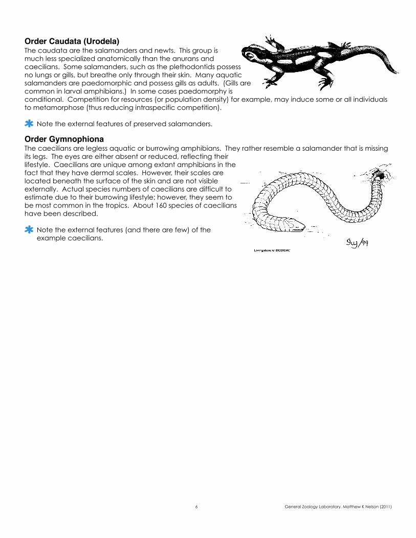
Order Caudata (Urodela)The caudata are the salamanders and newts. This group is much less specialized anatomically than the anurans and caecilians. Some salamanders, such as the plethodontids possess no lungs or gills, but breathe only through their skin. Many aquatic salamanders are paedomorphic and possess gills as adults. (Gills are common in larval amphibians.) In some cases paedomorphy is conditional. Competition for resources (or population density) for example, may induce some or all individuals to metamorphose (thus reducing intraspecific competition).
Note the external features of preserved salamanders.
Order GymnophionaThe caecilians are legless aquatic or burrowing amphibians. They rather resemble a salamander that is missing its legs. The eyes are either absent or reduced, reflecting their lifestyle. Caecilians are unique among extant amphibians in the fact that they have dermal scales. However, their scales are located beneath the surface of the skin and are not visible externally. Actual species numbers of caecilians are difficult to estimate due to their burrowing lifestyle; however, they seem to be most common in the tropics. About 160 species of caecilians have been described.
Note the external features (and there are few) of the example caecilians.
6 General Zoology Laboratory. Matthew K Nelson (2011)
NAME: ________________________ SECTION:______________ LAB EXERCISE 8
fish1. What sex was your perch?
2. What are the five scale types that are found in the chondrichthyes and osteichthyes?
3. What type of scales did your perch have?
amphibians1. Sarcopterygii are paraphyletic. What group would need to be included to make them monophyletic?
2. Do any amphibians have scales? If so, which amphibians?
questions
7 General Zoology Laboratory. Matthew K Nelson (2011)
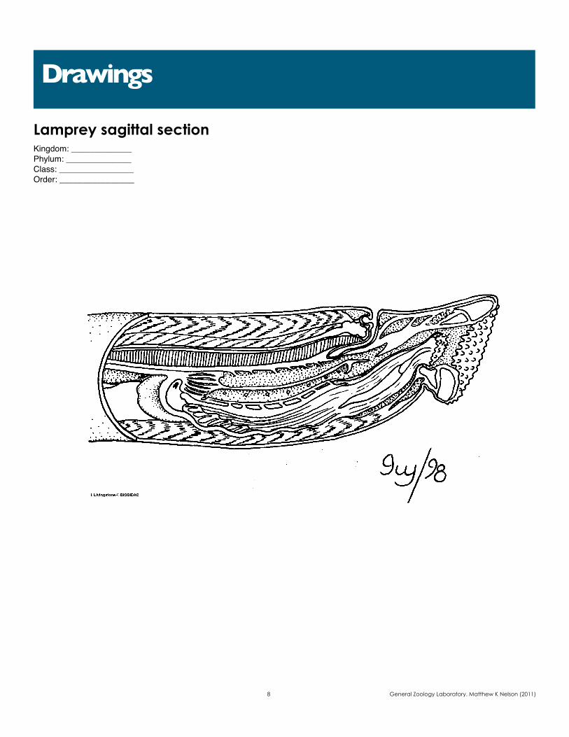
Lamprey sagittal sectionKingdom: _____________Phylum: ______________Class: ________________Order: ________________
Drawings
8 General Zoology Laboratory. Matthew K Nelson (2011)

Frog DissectionKingdom: _____________Phylum: ______________Class: ________________Order: ________________
Drawings
9 General Zoology Laboratory. Matthew K Nelson (2011)
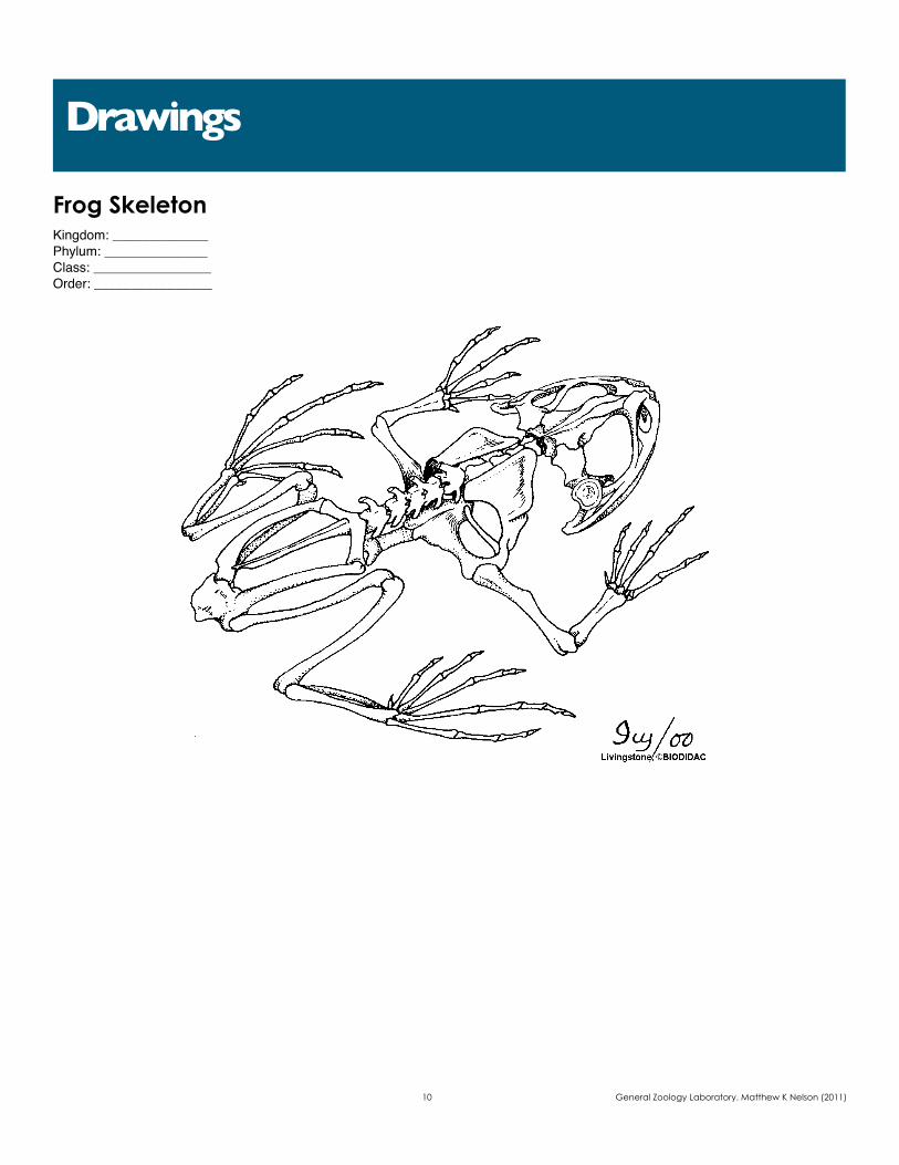
Frog SkeletonKingdom: _____________Phylum: ______________Class: ________________Order: ________________
Drawings
10 General Zoology Laboratory. Matthew K Nelson (2011)