Embed Size (px)
Citation preview

APPENDIX
Sanjay VB Patel February 17, 2016
Dr. M. Brackstone
General Surgery


BENIGN
APPENDIX
APPENDIX

Reginald Herbert Fitz
Shattuck Professor of Pathological
Anatomy Harvard
American
(1843 – 1913)
First to describe appendicitis
(The American Journal of the Medical
Sciences 1885)
“Perforating Inflammation of the Vermiform Appendix; With Special Reference to Its Early Diagnosis and Treatment”

Clinical Presentation of Acute Appendicitis
Epidemiology
• Most common cause of emergency surgery
worldwide
• Most commonly occurs in 10-30 years of age.
• More common in men (1.4 : 1)
• Life time incidence 6.7 – 8.6 %
APPENDIX

Clinical Presentation of Acute Appendicitis
History and Physical Examination
Classic constellation of symptoms
• Abdominal pain, anorexia, nausea and vomiting
Non-specific symptoms
• Indigestion, malaise, tenesmus, diarrhea,
dysuria, and atypical pain
Migratory abdominal pain – occurs in 50-60% of
patients
APPENDIX

Behind the caecum (ascending retrocaecal): 65% Inferior to the caecum (subcaecal): 31% Behind the caecum (transverse retrocaecal): 2% Anterior to the ileum (ascending paracaecal preileal): 1% Posterior to the ileum (ascending paracaecal retroileal): 0.5%

Clinical Presentation of Acute Appendicitis
History and Physical Examination
Tachycardia
Fever
Abdominal pain RLQ
Focal Peritonitis
Rebound tenderness
RLQ mass
Pelvic examination
Rectal examination
APPENDIX
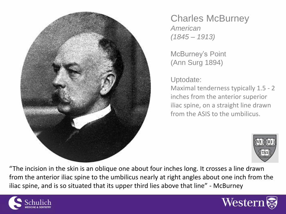
Charles McBurney American
(1845 – 1913)
McBurney’s Point
(Ann Surg 1894)
Uptodate: Maximal tenderness typically 1.5 - 2 inches from the anterior superior iliac spine, on a straight line drawn from the ASIS to the umbilicus.
“The incision in the skin is an oblique one about four inches long. It crosses a line drawn from the anterior iliac spine to the umbilicus nearly at right angles about one inch from the iliac spine, and is so situated that its upper third lies above that line” - McBurney

Niels Thorkild Rosving Danish
(1862 – 1927)
Rosving’s Sign
(Zentralblatt für Chirurgie1907)
Uptodate: Pain in the right lower quadrant when palpated in the left lower quadrant.
“Smerter i højre nedre kvadrant med palpering af den venstre nederste kvadrant” - Rosving

Sir Vincent Zachary Cope
English
(1881 – 1974)
Cope’s Psoas Sign
Cope’s Obturator Sign
(Cope’s Early Diagnosis of the Acute
Abdomen, 1921)
1
Psoas Sign - Right lower quadrant pain with passive right hip extension Obturator Sign - Right lower quadrant pain with passive flexion of the right hip and knee followed by internal rotation of the hip

Clinical Presentation of Acute Appendicitis
History and Physical Examination
APPENDIX
Sign Specificity Sensitivity Utility
McBurney’s Point Tenderness
50 – 94% 75 – 86% ✔✔✔
Rosving’s sign 22 – 68% 58 – 96% ✔✔
Cope’s Psoas Sign 13 – 48% 79 – 97% ✔
Cope’s Obturator Sign 8% 94% ✗

Clinical Presentation of Acute Appendicitis
Investigations
• CBC, electrolytes, CRE, bHCG
• Urine dip/culture
• WBC > 10,000 (sens 80%, spec 55%)
• Total Bilirubin >10 (sens 70%, spec 86%)
• Abdominal X-ray
• CT
• Ultrasound
APPENDIX

What are the CLASSIC findings on X-ray of appendicitis?

Imaging in Acute Appendicitis
Investigations – X ray
1. Fecalith
2. Dilated loops of bowel with air fluid levels
3. Scoliosis of the spine
4. Obliteration of the right lower psoas
shadow
5. Obliteration of the preperitoneal fat line
6. Paucity of gas in the RLQ
7. Small bowel obstruction
8. Air bubbles in the RLQ
9. Free air
APPENDIX

Imaging in Acute Appendicitis
Investigations - Ultrasound
• Dilated appendix ( >6 mm outer diameter) • Non-compressible • Appendicolith/Fecalith/Poopalith • Prominent echogenic pericaecal fat • Periappendiceal fluid collection • Target appearance (axial section)
APPENDIX

Imaging in Acute Appendicitis
Investigations – CT Scan
• Dilated appendix with distended lumen ( >6 mm diameter) • Thickened and enhancing wall • Thickening of the cecal apex (up to 80%):
• Cecal bar sign, arrowhead sign • Periappendiceal inflammation • Extraluminal fluid • Inflammatory phlegmon • Abscess formation • Appendicolith
APPENDIX

Impression Sensitivity Specificity
Diagnostic of appendicitis
72.5% 97.0%
Appendicitis cannot be out ruled
79.9% 84.0%

Modality N Diagnostic Sensitivity Specificity Negative Appendectomy
Ultrasound 1777 1101 99.1 91.7 5.2
CT 965 473 96.4 95.4 4.3
Physical Exam 303 218 99.0 76.1 12.2

Modality Sensitivity Specificity
Ultrasound only
77% 94%
CT 97% 91%
MRI 97% 83%
All patients had MRI Ultrasound first If US equivocal then CT was performed N=230


Clinical Scoring Systems - Alvarado
APPENDIX
Criteria Score
Migratory Abdominal Pain 2
Anorexia 1
Nausea AND Vomiting 1
Tenderness RLQ 1
Rebound tenderness 1
Elevated temp (>37.5) 1
Leukocytosis (>10,000) 2
Shift (Left shift/bands) 1
MANTRELS Highly Suspicious (7-10) -Operate -*Surgical consultation -Sensitivity 58-88% Intermediate (5-6) -Imaging with US or CT -*4-6 obtain CT Low (1-4) -Discharge from ER -* <3 No imaging indicated - Sensitivity 98% for non-acute appendicitis
*McKay, R. Ann Emerg Med 2007

• A cut point of 5 performs well as a rule out CPR in all patient groups with suspected appendicitis
• This CPR calibrates well in men, over-predicts in women and is inconclusive in children. • The Alvarado scoring system compares similarly to other CPR currently in use (Ottawa ankle) • Sensitivity (non-appendicitis) at a score of 5: 94-99%
• There is not enough evidence to support a decision to go to surgery at a cut-point of 7. • Sensitivity 88%, Specificity 81%, negative appendectomy rate 13.3-16.7%

Management of Acute Appendicitis
The goal of management is early diagnosis and prompt
surgical intervention
• Resuscitate the patient – IV access, fluid administration, urinary
catheter, correction of electrolytes and antibiotics.
• Antibiotics:
• Within 60 minute window of cutting skin
• Non-complicated:
• Cefazolin (1-2 gm IV) + metronidazole (500mg IV)
• Complicated (perforated):
• Piperacillin-Tazobactam or Ceftriaxone + metronidazole
• Within 24 – 72 hours of symptom onset
• Operation Open or Laparoscopic
APPENDIX

Open Appendectomy

Claudius Amyand French/British
(1680-1740)
First appendectomy
(Phil. Trans 1735)
-11yo boy with incarcerated hernia
-”Poppin” caused appendiceal perforation.
Amyand’s Hernia
-Inguinal hernia with the appendix within
the sac.
“This operation proved the most complicated and perplexing I ever met; with many unsuspected oddities and events concurring to make it as intricate as it proved laborious… ’Tis easy to conceive that this operation was a painful to the patient as laborious to me - it lasted nearly half an hour.”

APPENDIX


Sir Fredrick Treves British
(1853-1923)
Bloodless Fold of Treves
Friends with John Merrick
AKA Elephant man


Leonid Ivanovich Rogozov
Russian
(1934 - 2000)
First auto-appendectomy
(BMJ 2009)
-1960-61 Antarctic Expedition
-May 1961 2am
-27 year old first medical post
- OR time 1:45min

Laparoscopic
Appendectomy

Kurt Semm German
Obstetric and Gynecology
(1927 - 2003)
First laparoscopic appendectomy
(1980)
-Attempts to publish were rejected for
5 years – “unethical”
-German Surgical Society tried to
suspend his license.
-Muhe used Semm’s instruments to
perform first lap chole
“Both surgeons and gynecologists were angry with me, they were throwing stones at me. All my initial attempts to publish on laparoscopic appendectomy were refused, with the comment that such nonsense does not and will never belong to general surgery,”

SAGES - Appendectomy

Laparoscopic Versus Open:
Wound infections (OR 0.43, 0.34 – 0.54)
Less pain (p<0.05)
Return to normal activity 5 days (4 – 7days)
Intraabdominal infections (OR 1.77, 1.14 – 2.76)
Operative time 10 minutes longer
Return to work 2 (-2 to 4 days)
Actual cost ???
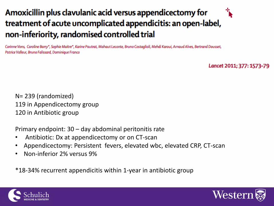
N= 239 (randomized) 119 in Appendicectomy group 120 in Antibiotic group Primary endpoint: 30 – day abdominal peritonitis rate • Antibiotic: Dx at appendicectomy or on CT-scan • Appendicectomy: Persistent fevers, elevated wbc, elevated CRP, CT-scan • Non-inferior 2% versus 9% *18-34% recurrent appendicitis within 1-year in antibiotic group

Included 4 RCT Results: • Peri-operative complications – RR 0.69 (RR 0.61) favoring antibiotic group • Length of stay – no difference • Treatment efficacy – Heterogenous
• Successful treatment 44 - 85% in the antibiotic group Conclusions: • Antibiotics can be used safely (no increased complications). • Perhaps acute uncomplicated appendicitis could be treated like uncomplicated
diverticulitis with an early trial of antibiotic therapy

Surgical outcomes are improving…
• Conversion from laparoscopic to open 1.2% (2.2%) • Intraoperative complications 0.7 % (3.1%) • Post op complications 1.9 % (6.1%)

Management of Acute Appendicitis Conclusion:
• Open and Laparoscopic approaches are
equivocal, however certain populations may
benefit from a laparoscopic approach.
• Diagnostic uncertainty – allows for assessment of other
Intraabdominal viscera if appendicitis is not the true diagnosis
• Female patients – Shown to have higher negative appendectomy
rates compared to men (~20%).
• Elderly patients – Shown to have lower morbidity in some studies
• Obese patients – Exposure can be difficult in the open approach
APPENDIX

Clinical Presentation of Acute Appendicitis
Management of Acute Appendicitis
• The goal of management is early diagnosis
and prompt surgical intervention.
• But….
• Patients presenting with >5 days of
symptoms
Or
• Mass in the RLQ with localizing pain
ie. walled off abscess
APPENDIX

Clinical Presentation of Acute Appendicitis
Management of Missed Appendicitis
Operating on missed appendicitis
• Risk of bowel and enterocutaneous fistula
• Right hemicolectomy or cecostomy
Non-operative management: • NPO
• Antibiotics
• Observation
• Interventional radiology – drains
• If patient does not respond then operative intervention is
indicated. Patient should be aware of the risk of bowel
resection.
APPENDIX

Clinical Presentation of Acute Appendicitis
Management of Missed Appendicitis
Interval Appendectomy
• Appendectomy performed electively 6-8
weeks after recovery.
• Colonoscopy should be performed in
patients >50 before surgery.
• Equipoise concerning the evidence to
support elective appendectomy versus
watchful waiting.
APPENDIX

32398 patient were diagnosed at 12 medical centers 1012 were treated non-operatively 148 were not treated with interval appendectomy 864 were treated with interval appendectomy Recurrence rates were 5% over a 4 year follow-up Length of hospital stay was 6 days for interval appendectomy Length of hospital stay was 4 days for recurrent appendicitis (P=0.006)

Results: • Risk of “enclosed inflammation” ie missed appendicitis was 3.8% (n= 59,448) • Failure rate of non-operative management 7.8% • Risk of recurrent appendicitis in non-operative groups 7.2% • Rate of malignant disease on follow-up 1.2% (n= 2775) • Rate of significant non-malignant disease at follow-up 0.7% (i.e crohn’s)
• Morbidity was compared, showed a significantly greater morbidity with surgical
invention compared to non-operative management with out interval appendectomy. • Significant heterogeneity in these outcomes

The first-year McGill student asked Houdini whether it was true that punches in the stomach did not hurt him. Houdini remarked rather unenthusiastically that his stomach could resist much, though he did not speak of it in superlative terms. Thereupon he gave Houdini some very hammer-like blows below the belt, first Securing Houdini's permission to strike him. Houdini was reclining at the time with his right side nearest Whitehead, and the said student was more or less bending over him. These blows fell on that part of the stomach to the right of the navel, and were struck on the side nearest to us, which was in fact Houdini's right side; I do not remember exactly how many blows were struck. I am certain, however, of at least four very hard and severe body blows, because at the end of the second or third blow I verbally protested against this Sudden onslaught on the part of this first-year student, using the words, "Hey there. You must be crazy, what are you doing?"
Jack Price’s description Author - Sir Arthur Conan Doyle
Joselyn Gordon Whitehead - McGill
Harry Houdini (1874 – 1926)


NEOPLASTIC
APPENDIX
APPENDIX

Classification of Appendiceal Neoplasms
• Appendiceal malignancy is rare
~1% of appendectomy specimens
• Mucocele (Benign)
• Adenocarcinomas
• Carcinoid Tumors
APPENDIX

Appendiceal Neoplasms: Mucoceles
• Appendiceal mucocele is a lesion
characterized by a distended mucus-filled
appendix
• Rare – 0.3% of appendix specimens
APPENDIX
Mucocele
Mucosal Hyperplasia
(52%)
Simple Retention Cyst (20%)
Mucinous Cystadenoma (18%)
Mucinous Cystadenocarcinoma
(10%)
* Relative frequency

Appendiceal Neoplasms: Mucocele
Clinical Presentation:
• Asymptomatic (Most common)
• RLQ abdominal pain
• Colicky abdominal pain
• GI bleeding – intussusception
• Bowel obstruction
• Hydroureter
• Acute abdomen if ruptures
Laboratory Findings:
• CA 19-9
• CEA
• ESR
*Evidence is not great to support these for diagnostic purposes
APPENDIX

Appendiceal Neoplasms:
Mucocele
Imaging:
• Ultrasound
• CT
Findings to suggest mucocele:
• Calcification in the
appendiceal wall
• Cystic lesion
• Irregular wall, but normal
wall thickness
• Hypodense spots in the
peritoneum
Endoscopy
APPENDIX

Appendiceal Neoplasms: Mucocele
Management:
• Early diagnosis and surgical resection
• Appendectomy – staple across the base of the cecum
• Right hemicolectomy – if malignant features
(cystadenocarcinoma), or obvious invasion into the terminal
ileum, cecum or adjacent mesentery at the time of operation.
• Rule out synchronous cancer – 20%
• Check colon, ovaries, endometrium, breast and kidney
APPENDIX

Pseudomyxoma Peritonei
“Jelly Belly bit with a big fat bite…” – Denis Lee

Pseudomyxoma Peritonei
Originally used to describe Intraabdominal spread of a
cystadenoma of the appendix.
Cystadenoma of the appendix ruptures, spreading mucous
producing cells throughout the peritoneum.
Mucous accumulates in the abdomen (Jelly Belly) until it causes
obstruction, which has no curative surgical treatment.
Some also include peritoneal carcinomatosis from malignant
mucous producing tumors of the appendix, small and large bowel,
lung, pancreas, stomach, breast and ovaries in the definition
Prognosis is very different for indolent cystadenoma type versus
all others.

Clinical Presentation of Pseudomyxoma Peritonei
Presenting Symptoms
Non-specific Symptoms
• Increasing abdominal girth
• Inguinal hernia in men
• Ovarian mass on pelvic examination

Typical CT findings: Mucin same density as water Calcifications are common Scalloping of the liver, spleen and peritoneum Spares the central portion of the abdomen (redistribution phenomenon)

Pseudomyxoma Peritonei: HIPEC and CRS Hyperthermic Intraperitoneal Chemotherapy and Cytoreductive Surgery
Theory behind the treatment: • Intraabdominal administration increases the effective
concentration 7-fold (mitomycin or 5-FU)
• Limits systemic effects
• Heating increases the depth of penetration
Patient Selection (Surgarbaker, PH)
1. Histopathological assessment
• Non-invasive neoplasms (better)
2. Radiological assessment
• No liver, lung mets or lymphadenopathy
• Segmental obstruction of small bowel (worse)
• Tumor deposit >5cm (worse)
3. Peritoneal Cancer index
4. Complete Cytoreduction

Pseudomyxoma Peritonei: HIPEC and CRS
http://www.surgicaloncology.com/gpmtitle.htm

Appendiceal Neoplasms: Adenocarcinoma
APPENDIX

Appendiceal Neoplasms: Adenocarcinoma
Histologic Subtypes
• Mucinous cystadenocarcinoma (Most
Common)
• Intestinal type adenocarcinoma
• Signet ring cell adenocarcinoma
APPENDIX
Histology Frequency 5 year Disease Specific Survival
Mucinous cystadenocarcinoma 37% 58%
Intestinal type adenocarcinoma 27% 55%
Signet Ring adenocarcinoma 6% 27%
Carcinoid Tumors 11% 93%

AJCC, 10th Edition, 2010


Appendiceal Neoplasms: Adenocarcinoma
Management
• Diagnosis made pre-op
• Right hemicolectomy
• However, some evidence to suggest survival is no
different for early lesions with simple appendectomy
• Found post-appendectomy
• Right hemi-colectomy
• No further operation if:
• Adenocarcinoma confined to the mucosa
• Well differentiated lesions no deeper then
submucosa
APPENDIX
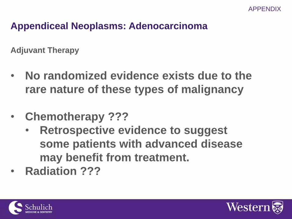
Appendiceal Neoplasms: Adenocarcinoma
Adjuvant Therapy
• No randomized evidence exists due to the
rare nature of these types of malignancy
• Chemotherapy ???
• Retrospective evidence to suggest
some patients with advanced disease
may benefit from treatment.
• Radiation ???
APPENDIX

Appendiceal Neoplasms:
Peritoneal Mucinous Carcinomatosis Adenocarcinoma
Role of HIPEC and CRS
APPENDIX

Only RCT looking at HIPEC and cytoreductive surgery
105 Patient with peritoneal carcinomatosis
• 87 with colon cancer
• 18 with appendiceal cancer
Control – Standard therapy
• 5-FU and leucovorin chemotherapy
• Palliative surgery if required
Experimental – HIPEC and debulking surgery
• Debulking to <2.5mm lesions
• HIPEC – 90 minutes of isotonic dialysis fluid with mitomycin at
41 degrees.
• 5-FU and leucovorin started 6 weeks post
Median Survival: 22.4 months versus 12.6 months

Cohort of 501 patients with mucinous adenocarcinoma of the appendix
• 17-year experience
• Compared appendectomy versus right hemi-colectomy
• All patients were treated with HIPEC and CRS
Overall results:
• 5 year survival 74% (SEER stage IV 22%)
• 10 year survival 52%

282 patients with peritoneal carcinomatosis undergoing HIPEC and CRS
Simplified peritoneal cancer index (SPCI) – pre-operative tumor load
Complete cytoreduction score (CC-score) – visible disease remaining
Lower SPCI showed better survival
Lower CC-score showed better survival
Survival 3 year – 67%
Survival 5 year – 52%

Appendiceal Neoplasms: Carcinoid Tumors

Appendiceal Neoplasm: Carcinoid Tumors
• Approximately 11% of appendiceal malignancies
• Age 40 – 50 years
• More common in women
• Occur most often in the distal third of the appendix
• Large tumors cause obstruction
Carcinoid Syndrome: Metastatic Disease
• Episodic Flushing (EtOH, emotional stress, eating, liver palpation and
anesthesia)
• Venous telangiectasia
• Bronchospasm
• Diarrhea
Carcinoid Crisis:
• Significant hemodynamic instability
• Octreotide Prophylaxis (300-500mcg IV), repeat PRN

Appendiceal Neoplasm: Carcinoid Tumors
Clinical Presentation
• Most patients are symptomatic at time of diagnosis
• Appendicitis (10% reside at base of appendix)
• Bowel obstruction
• Carcinoid Syndrome
• Incidental finding
Work-Up of Carcinoid Tumors
• CT abdomen
• CT chest
• Octreotide scan
• 24-Hour 5-HIAA (5-hydroxindolacetic acid)
• Chromogranin A


AJCC 7th edition, 2010

Appendiceal Neoplasm: Carcinoid Tumors
WHO Grade

Appendiceal Neoplasm: Carcinoid Tumors
Management:
NANETS
• <1cm tumors with no evidence of lymphovascular
invasion or invasion into the mesoappendix are
considered a cure.
• Right Hemicolectomy
• >1cm at the base of the appendix
• >2cm or size cannot be evaluated
• Grade 2 and 3 tumors
• Lymphovascular invasion
• Invasion of mesoappendix
• Positive margins

Appendiceal Neoplasm: Carcinoid Tumors
Management:
ENETS
• <2cm tumors with no evidence of lymphovascular
invasion or invasion into the mesoappendix are
considered a cure.
• Right Hemicolectomy (within 3 moths):
• >2cm or size cannot be evaluated
• Invasion of mesoappendix
• Positive margins
Appendiceal Neoplasm: Carcinoid Tumors
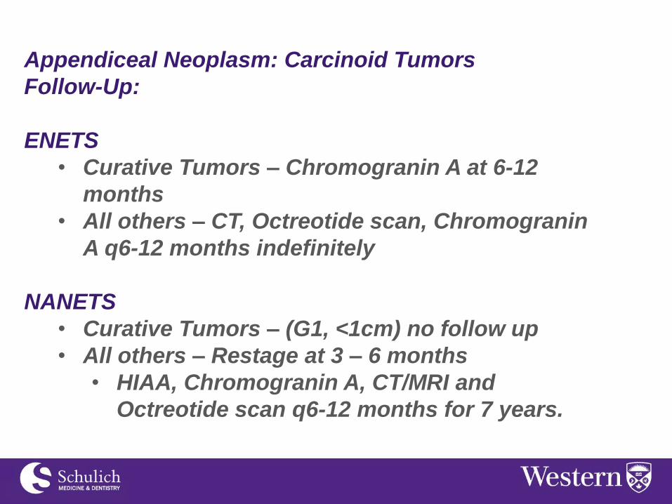
Follow-Up:
ENETS
• Curative Tumors – Chromogranin A at 6-12
months
• All others – CT, Octreotide scan, Chromogranin
A q6-12 months indefinitely
NANETS
• Curative Tumors – (G1, <1cm) no follow up
• All others – Restage at 3 – 6 months
• HIAA, Chromogranin A, CT/MRI and
Octreotide scan q6-12 months for 7 years.

- Appendix

Appendiceal Neoplasm: Carcinoid Tumors Advanced Disease:
Systemic Therapy • Octreotide alone
• Improved time to progression
• Survival advantage suspected
• Octreotide + INF alpha
• Cytotoxic Chemo (5FU – based)
• Everolimus/Sirolimus (mTOR)
• Bevacizumab (anti-VEGF)
Cytoreductive Surgery • Hepatic Resection
• Liver directed therapy
• Cryo, radio or microwave ablation
• Embolization (Bland, chemo or radioactive)
• Peptide receptor radiotherapy

“Idleness is the appendix to nobility…” - Robert Burton