Embed Size (px)
Citation preview
General stress proteins: Novel function and signals
for induction of stationary phase genes in E. coli
Örjan Persson
AKADEMISK AVHANDLING
För filosofie doktorsexamenen i mikrobiologi (examinator Thomas Nyström), som enligt fakultetssyrelsens beslut kommer att offentligt försvaras tisdagen den 25 maj
2010, kl. 10.00 i föreläsningssal Arvid Carlsson, Medicinaregatan 3, Göteborg
ISBN 978-91-628-8104-7
e-publication: http://hdl.handle.net/2077/22213

ii
To Gullborg

iii
iv
General stress proteins: Novel function and signals for induction of stationary phase genes in E.coli
Örjan Persson
ABSTRACT
Survival during conditions when nutrients become scarce requires adaptation and expression of genes for maintenance in order for the cell to survive. Among the numerous proteins involved in adaptation and regulation under these conditions, the stationary phase sigma factor, σS, and the Universal stress proteins contribute to survival and bestow the cell with general stress protective functions during growth arrest. In this work we found new mechanisms for the cell to prepare and sense the intracellular environment and respond accordingly.
The usp genes and the rpoS gene (encoding σS) were found to be positively regulated by metabolic intermediates of the glycolysis in the central metabolic pathway. Specifically, mutations and conditions resulting in fructose-6-phosphate accumulation elicit superinduction of these genes upon carbon starvation, whereas genetic manipulations reducing the pool size of fructose-6-phosphate have the opposite effect. Under carbon starvation, transcription of the usp and the rpoS genes require and are modulated by the alarmone ppGpp. The observed positive transcriptional regulation by fructose-6-phosphate is not via alterations of the levels of ppGpp. None of the known regulators examined were found to be required for the superinduction. We suggest a novel regulatory mechanism involving the phosphorylated intermediates as a signal molecule for monitoring and subsequent regulation of stress defense genes. Based on mutational studies we also suggest that this signaling mechanism secures accumulation of required survival proteins preceding the complete depletion of the external carbon source.
Entry into stationary phase promotes a dramatic stabilization on the sigma factor σS. Mistranslated and oxidized proteins were shown to contribute to elevated levels of σS and transcription of its regulon. Furthermore, ribosomal alleles with enhanced translational accuracy attenuate induction of the RpoS regulon and prevent stabilization of σS. Destabilization of σS is governed by the ClpXP protease, for which aberrant proteins also are substrates. Mechanistically, generation of mistranslated proteins by starvation, or other means, competes for the common enzyme for degradation, and thereby sequesters the pool in favor of σS stabilization.
A growing body of evidence shows that there is an intimate connection between proteins required for genome stability and stationary phase survival. We show that the integral membrane protein UspB, a member of the RpoS regulon, is required for proper DNA repair as mutants lacking uspB are sensitive to several DNA damaging conditions. Genetic and biochemical studies demonstrate that UspB acts in the RuvABC recombination repair pathway and removing uspB creates a phenocopy of the DNA resolvase mutant, ruvC, which includes a reduced efficiency in resolving Holliday junctions. Further, we show that the uspB mutant phenotype can be suppressed by ectopic overproduction of RuvC and that both ruvC and uspB mutants can be suppressed by inactivating recD. The fact that RuvABC-dependent repair requires UspB for proper activity suggests that the σS-regulon works together with DNA repair pathways under stress conditions to defend the cell against genotoxic stress.
v
List of papers
This thesis is based on the following papers, which are referred to by their Roman numerals:
I. Metabolic control of the Escherichia coli universal stress protein response through
fructose-6-phosphate. Persson Ö, Valadi A, Nyström T, Farewell A. Mol Microbiol. 2007 65(4):968-78.
II. The levels of fructose-6-phosphate regulate σS-dependent transcription upon entry to stationary phase in E. coli. Persson, Ö, Gummesson, B., Hallberg, E., Lilja, E., and Farewell, A. Manuscript
III. Decline in ribosomal fidelity contributes to the accumulation and stabilization of the master stress response regulator σS upon carbon starvation. Fredriksson Å, Ballesteros M, Peterson CN, Persson Ö, Silhavy TJ, Nyström T. Genes Dev. 2007 21(7):862-74.
IV. UspB, a member of the sigma-S regulon, is required for RuvC-dependent resolution of Holliday junctions. Persson, Ö, Nyström, T and Farewell, A. Under revision DNA Repair.
Other papers not included in this thesis the author published: Increased RNA polymerase availability directs resources towards growth at the expense of maintenance. Gummesson B, Magnusson LU, Lovmar M, Kvint K, Persson O, Ballesteros M, Farewell A, Nyström T. EMBO J. 2009 Jul 2;28(15):2209-2219.
vi
Table of contents
1. Introduction ............................................................................................................................................... 1
1.1. E. coli as a model organism...................................................................................................................... 1
2. Aim and findings at a glance ................................................................................................................... 2
3. Growth, Metabolism and Carbon source selection............................................................................... 3
4. Stationary phase........................................................................................................................................ 5
5. Adapting and optimization to different environments ......................................................................... 7
5.1. Sensing carbon metabolites...................................................................................................................... 7
5.2. Sensing starvation ..................................................................................................................................... 8
5.3. The stringent response and the alarmone ppGpp ................................................................................. 9
5.3.1. Regulation of stringent response ............................................................................................................. 9
5.3.2. The effect of the stringent response regulator, ppGpp, on RNAP .................................................... 11
5.3.3. Other effects of the stringent response regulator, ppGpp.................................................................. 12
5.4. Heat shock proteins, oxidative stress and translation fidelity ........................................................... 13
5.5. RpoS regulon and general stress defense ............................................................................................. 15
5.5.1. RpoS regulation....................................................................................................................................... 16
5.5.1.1. Transcriptional regulation ..................................................................................................................... 16
5.5.1.2. Translational regulation and sRNA’s ................................................................................................... 17
5.5.1.3. Protein stability ....................................................................................................................................... 18
6. Model genes and proteins....................................................................................................................... 20
6.1. UspA and the universal stress protein family ...................................................................................... 20
6.1.1. Regulation and function of UspA .......................................................................................................... 21
6.1.2. Regulation of uspA expression............................................................................................................... 22
6.2. UspB.......................................................................................................................................................... 23
6.2.1. Function and regulation ......................................................................................................................... 23
7. DNA damage............................................................................................................................................ 25
7.1. Replication, not that smooth .................................................................................................................. 25
7.2. DNA damage caused by UV irradiation............................................................................................... 27
vii
7.3. UV produced lesions are recognized and excised by the NER system.............................................. 29
7.4. SOS response; RecA and its function ................................................................................................... 29
7.5. Processing of DNA breaks (RecBCD and RecFOR) ........................................................................... 30
7.6. Branch migration and resolution by RuvABC and RecG.................................................................. 33
8. Results and discussion ............................................................................................................................ 36
8.1. Paper I: Accumulation of the glycolytic intermediate fructose-6-p serves as a positive regulatory signal to uspA transcription.................................................................................................................................. 36
8.2. Paper II: The levels of fructose-6-phosphate regulate σσσσS-dependent transcription upon entry to stationary phase in E. coli ..................................................................................................................................... 39
8.2.1. Speculations Paper I and II ................................................................................................................... 40
8.3. Paper III: Decline in ribosomal fidelity contributes to the accumulation and stabilization of the
master stress response regulator σσσσS upon carbon starvation ........................................................................... 43
8.3.1. Speculations paper III ............................................................................................................................ 44
8.4. Paper IV: Characterization of a uspB mutant indicates that the phenotype is linked to DNA damage 46
8.5. Speculations paper IV ............................................................................................................................ 48
9. Concluding remarks ............................................................................................................................... 49
10. Acknowledgments ................................................................................................................................... 51
11. References ................................................................................................................................................ 53
1
1. Introduction
1.1. E. coli as a model organism
In 1885, the German pediatrician Theodor Escherich, isolated and cultivated a rod-shaped
gram-negative bacterium; the “Bacterium coli” (later renamed Escherichia coli) (Lederberg
2004). Its natural habitat is the large intestine or colon of mammals and birds that it has a
remarkable capacity to colonize. In number, this microorganism does not dominate in the
abdomen, but is estimated to constitute only 1% of the microbial flora. However, it has been
found in every species examined and in almost every single individual of those (Neidhardt
1996).
Escherichia coli has emerged as a predominant model organism in labs that study bacteria.
Escherichia coli K-12, the dominant parental strain background in many laboratories
studying E.coli and considered wild type, was isolated at Stanford University, CA. In
contrast to its many derivatives, K-12 has not been treated with gamma or UV radiation or
other mutagenic agents. Despite this, during cultivation and selection, K-12 has lost O and K
antigen and is virtually unable to colonize a human gut (Smith 1975).
In its natural habitat, E. coli is constantly challenged with altered nutrient availability, partial
anaerobiosis, and changes in pH and osmotic stress, while the temperature is fairly constant,
at around 37°C. In addition, the bacteria must have capacity to sustain different
environmental assaults like H2O2, UV light and exposure to antibiotics. In order to proliferate
and survive during those conditions, cells have to adapt to the constantly changing
environment and throughout its evolution, E. coli has perfected its adaptive strategies to these
changes. Natural selection has thus equipped bacteria with systems for synchronously
altering many pathways, either by up- or down-regulating gene expression in response to new
environments. One mechanism of coordinate regulation is the structure of an operon, where
many genes are simultaneously transcribed due to a specific stimulus. Coordinate regulation
is also accomplished in E. coli by a variety of mechanisms including small messenger
molecules such as the alarmones ppGpp and cAMP, but also by alternative sigma factors and
regulons such as the SOS response that regulate sets of genes and their products. Among the
proteins essential for sustaining high viability during conditions when the nutrients are scarce
or lacking, the RpoS sigma factor (σS) and the Universal stress proteins are expressed and
play a role either directly or indirectly in the process of survival in a diversity of inhospitable
conditions. This thesis focuses on the regulation and function of these important proteins.
2
2. Aim and findings at a glance
The aim of this thesis was to find and characterize factors involved in regulation of the
universal stress proteins genes uspA, uspB and the stationary phase sigma factor RpoS as
well as characterize the function of UspB.
We found a new mechanism involved in the regulation of the uspA gene and other usp´s in
response to carbon stress. Accumulation of fructose-6-phosphate in the central metabolic
pathway, bestows a signal to the usp genes and positive regulation occurs (paper I). About
one generation before cells are depleted of carbon, uspA is normally induced. In mutants with
elevated levels of fructose-6-p the induction is stronger and superinduction occurs. This
regulation is not due to alteration of the known regulators of uspA. How this signal is
transmitted is unknown, but it implies that the cell is capable to sense and prepare
accordingly before food sources become exhausted.
In a similar manner, the well characterized general stress defense gene rpoS, encoding the
transcription factor σS, was also found to be regulated by metabolic accumulation of
fructose-6-p (paper II). Like the usp´s the regulation occurs at the level of transcription and
the protein levels correspond to the increased transcription of rpoS. Throughout growth an
increased level of σS can be observed. This results in superinduction of the RpoS-dependent
genes upon carbon starvation in cells with increased fructose-6-p.
On the regulation of σS we also found that during entry to carbon starvation, mistranslated or
oxidized proteins contribute to the elevation and stability of the sigma-S (σS) protein (paper
III). Aberrant proteins and sigma-S are both substrates for ClpP protease. Mechanistically,
via a titration of ClpXP, decreased translational fidelity of the ribosome stabilizes the RpoS
in stationary phase and elevated transcription of its regulon is observed.
Finally, the stationary phase inducible protein UspB is shown to be involved in DNA repair
(paper IV). A mutation in uspB reduces the survival significantly following DNA damage.
We showed by genetic and biochemical methods that a mutation in uspB affects the
resolution of Holliday junctions following DNA damage. A possible link to the resolvase
RuvC was found and a uspB deletion phenocopied a ruvC mutant under all tested conducted.
The results suggest that UspB is capable of modulating the effect of the resolvase RuvC
during repair of damage.
3
3. Growth, Metabolism and Carbon source selection
E. coli, like many other bacteria, divides by binary fission. Provided the growth conditions
are unrestricted (nutrients in excess and no accumulation of toxic byproducts) E. coli can
divide and give rise to a functional new cell in as little as 20 minutes. During these conditions
the cell is basically a protein factory with the majority of resources going to ribosomal
protein synthesis. To supply the demand for anabolic metabolites and energy requirements
the bacterium harbors an extensive and highly dynamic metabolic network for metabolizing
compounds found in the environment and converting them into different essential molecules
like precursor metabolites, reducing power (NADPH) and energy transfer molecules (NADH
and ATP). Different pathways are involved depending on the carbon source utilized. E.coli
can utilize an impressively broad range of different carbon sources, from high energy
carbohydrates, polyols, to two-carbon compounds or fatty acids as sole carbon and energy
source. In general, by relatively simple conversions of the molecules, like phosphorylation,
isomerisation and aldol cleavage etc. these different carbon sources can be fed into the
central metabolic pathways of glycolysis, the pentose pathway or the tricarboxylic acid
(TCA) cycle (Neidhardt 1996).
The preferred carbon source for E.coli cells is the monosaccharide glucose and when cells
are fed with glucose, no other carbon source is simultaneously utilized (Postma and Lengeler
1985; Postma et al. 1993). The presence of glucose inhibits the expression of enzymes
needed for uptake and metabolism of other carbon sources; these phenomenona are known as
inducer exclusion and carbon catabolite repression. Glucose exerts this repression of
utilization of other carbon sources till minute levels (µM) remain in the growth media
(Ferenci 1996). The uptake of glucose via the phosphoenolpyruvate:carbonhydrate
phosphotransferase (PTS) system together with cAMP-CRP mediate this response (Meadow
et al. 1990; Neidhardt 1996; Bruckner and Titgemeyer 2002). Uptake of glucose is an active
process via the PTS system, where glucose becomes phosphorylated by transfer of a
phosphate from Enzyme IIAGlc (EIIAGlc) and enters the glycolytic pathway as glucose-6-
phosphate. This results in the EIIAGlc protein becoming dephosphorylated and it then inhibits
uptake of alternative carbohydrates and fails to activate adenylate cyclase (AC) leading to
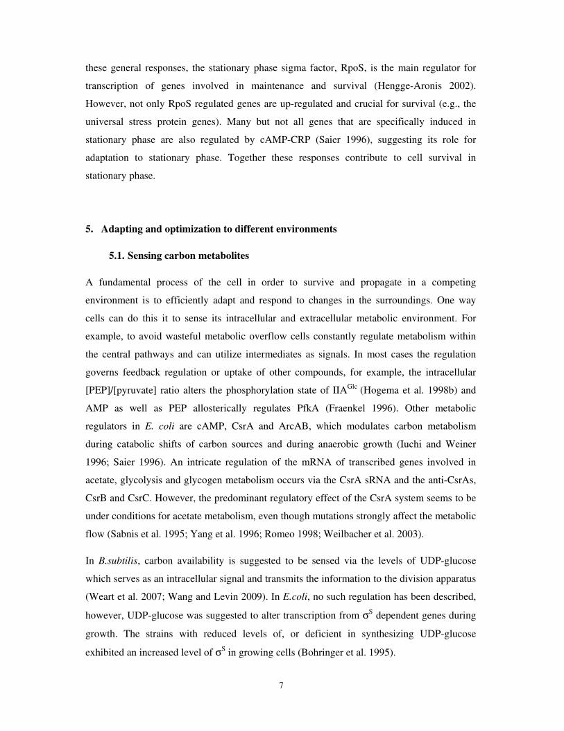
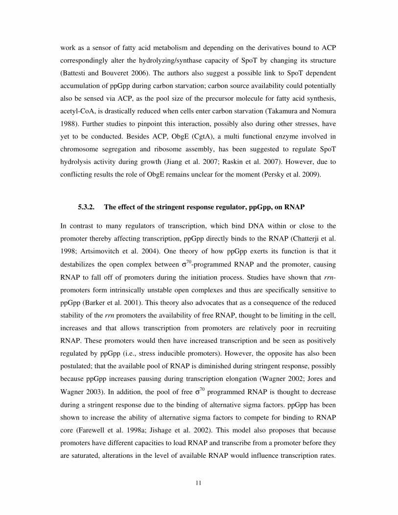
low cAMP levels (Ferenci 1996; Hogema et al. 1998) (Fig 1).

4
Figure 1. Mechanism of PTS-mediated catabolite repression and regulation. Glucose is rapidly phosphorylated by the PTS enzyme EIIBCGlc and fed into glycolysis. In this process, the protein IIAGlc becomes dephosphorylated. In its dephophorylated state it binds enzymes involved in the uptake of non-PTS carbon sources (like lactose and glycerol) and thereby inhibits the utilization and uptake of these carbon sources. The dephosphorylated state of IIAGlc also inhibits the synthesis of cAMP by adenylate cyclase (AC). For clarity not all steps are included. Adapted from (Hogema et al. 1998b).
In the absence of glucose, EIIAGlc remains phosphorylated and activates adenylate cyclase,
the product of the cya gene (Fig 1). Active adenylate cyclase synthesizes the alarmone
cAMP. cAMP with its cognate CRP (also known as CAP) protein binds operator sequences
with a specific cAMP-CRP recognition sequence and thereby affects transcription of about
100 promoters in a positive or negative way (Fic et al. 2009). For positive regulation, cAMP-
CRP binds the DNA and promotes transcription either by direct recruitment of RNAP to the
promoter region or by facilitating RNAP-promoter complex formation (reviewed in (Busby
and Ebright 1999)). In both cases the transcription is facilitated by protein-protein interaction
between RNAP and CAP. In some cases, additional activators besides CAP are needed for
full induction, for example the araBAD promoter also needs AraC and arabinose to become
activated (Lobell and Schleif 1991) or relief of inducer exclusion is needed for induction of
the permease lacY of the lactose uptake system (Hogema et al. 1998a). cAMP-CRP can also
exert negative effect by directly occupying core promoter regions, like Pgal, (Spassky et al.
1984) or indirectly by stabilization of a repressor (Kristensen et al. 1997). By these
mechanisms cAMP-CAP affects transcription of many operons involved in uptake and
5
metabolism of non-PTS carbon sources as well as gene products involved in processes like
biofilm formation, flagellum formation, chemotaxis and nitrogen utilization (Saier 1996).
Generally, the cAMP-CRP modulon increases the cell’s ability to scavenge the environment
for alternative carbon sources.
Whereas glucose is most efficient at catabolite repression, glycerol is considered to be a
carbon source that leads to low catabolite repression (Hogema et al. 1998b; Bettenbrock et al.
2007). Uptake of glycerol is mediated via the proteins of the glp regulon consisting of several
operons with a complex genetic structure (Weissenborn et al. 1992; Yang and Larson 1998).
In contrast to other carbohydrates, glycerol is taken up by facilitated diffusion across the
cytoplasmic membrane and is phosphorylated by the GlpK kinase to glycerol-3-phosphate
and further dehydrogenised to the glycolytic intermediate DHAP of the central metabolic
pathway (Voegele et al. 1993; Lin 1996). The kinase is allosterically regulated not only by
EIIAGlc but also fructose1,6-bisphosphate, thus glycerol uptake is tightly regulated as long as
the cell is able to take up glucose (Lin 1996). During growth on glycerol as carbon source
almost all of EIIAGlc is in the phosphorylated state (Hogema et al. 1998), thus a complete
stimulatory effect of cAMP synthesis occurs during this condition.
4. Stationary phase
In its natural habitat low nutrients availability is the prevalent situation, setting the stage for
evolution under those conditions (Llorens et al.). Furthermore, in batch cultures some of the
essential nutrients will eventually become exhausted. Upon depletion, the growth rate slows
down and the cell reaches a phase where no net increase in cell number occurs and a
balanced state between division and death is reached, called stationary phase. However, as
pointed out by Groat (1986), stationary phase is only defined as a condition in which the
cessation of growth occurs due to nutrient deprivation and the physiological response of the
cells is different depending on the type of starvation, for example, starvation for carbon,
nitrogen, phosphate or other compounds. Entry to, and maintenance in, stationary phase is
not a passive process and this can clearly be exemplified by blocking protein synthesis of the
cell when entering stationary phase. Adding chloramphenicol to wild type carbon starved
cells reduced survival of the cultures. The earlier the protein synthesis was blocked on
starved cells, the more pronounced was its effect on survival (Reeve et al. 1984; Nystrom et
al. 1990). Thus, during entry to starvation the cell induces specific proteins in order to
6
continue divide and reproduce (e.g., to utilize an alternative nutrient source that may be
present), or, if this fails, the cells induce a general stationary phase response by redirecting
resources into maintenance metabolism and stress response gene expression for protection.
Further, cells adapt physiologically and morphologically to this state by a number of different
changes. Initiation of DNA replication is stopped, but ongoing replication and the cell cycle
is completed (Kolter et al. 1993). At the same time the cell size decreases (most likely as a
consequence a final division), the cells become almost coccoid and the membrane
composition becomes less fluid (Kolter et al. 1993). Furthermore, aberrant and oxidized
proteins accumulate as a consequence of decreased translational fidelity. Misincorporation of
erroneous amino acids, translational frameshifting and stop-codon readthrough all contribute
to the increased pool of mistranslated proteins (O'Farrell 1978; Barak et al. 1996; Wenthzel
et al. 1998; Ballesteros et al. 2001). However, unnecessary protein synthesis is turned off
quickly; in particular ribosome protein synthesis is inhibited. Cells start to scavenge for
nutrients in the media as well as degrading surplus endogenous material like proteins, RNA
and lipids. Degradation of these components is thought to ensure building blocks for essential
stationary phase proteins. For example, at the onset of starvation, the RNase activity
increases two- to eightfold, leading to extensive degradation of ribosomal RNA (rRNA) and
within the first 4 hours of starvation 20-30% of the total RNA is degraded (Matin et al.
1989). In contrast to rRNA, the half-life of mRNA increases more than two-fold, regardless
of whether the transcript is repressed or induced in stationary phase (Albertson and Nystrom
1994).
As a result of these changes in gene expression and physiology, stationary phase cells are
more resistant towards a number of different environmental assaults, for example cold/heat
shock, oxidative stress, antibiotics, osmotic stress, ethanol and UV ((Matin et al. 1989) and
references therein). During stationary phase, Dps protein binds to the nucleoid and arranges
the DNA into a tightly packed and condensed structure that is resistant to a broad range of
different assaults, like oxidative, thermal, acid and base stresses; UV and gamma irradiation;
and iron and copper toxicity (Frenkiel-Krispin et al. 2004; Kim et al. 2004; Nair and Finkel
2004). In combination with chromosome compaction undertaken by Dps, the function of Dps
as a metal chelator and ferroxidase further protects the DNA. Increased HN-S concentration
may also contribute to DNA condensation (Ueguchi et al. 1993).
Thus, although there are specific responses to specific starvation conditions, the general
stationary phase response plays a vital role in the cells survival in stationary phase. Among
7
these general responses, the stationary phase sigma factor, RpoS, is the main regulator for
transcription of genes involved in maintenance and survival (Hengge-Aronis 2002).
However, not only RpoS regulated genes are up-regulated and crucial for survival (e.g., the
universal stress protein genes). Many but not all genes that are specifically induced in
stationary phase are also regulated by cAMP-CRP (Saier 1996), suggesting its role for
adaptation to stationary phase. Together these responses contribute to cell survival in
stationary phase.
5. Adapting and optimization to different environments
5.1. Sensing carbon metabolites
A fundamental process of the cell in order to survive and propagate in a competing
environment is to efficiently adapt and respond to changes in the surroundings. One way
cells can do this it to sense its intracellular and extracellular metabolic environment. For
example, to avoid wasteful metabolic overflow cells constantly regulate metabolism within
the central pathways and can utilize intermediates as signals. In most cases the regulation
governs feedback regulation or uptake of other compounds, for example, the intracellular
[PEP]/[pyruvate] ratio alters the phosphorylation state of IIAGlc (Hogema et al. 1998b) and
AMP as well as PEP allosterically regulates PfkA (Fraenkel 1996). Other metabolic
regulators in E. coli are cAMP, CsrA and ArcAB, which modulates carbon metabolism
during catabolic shifts of carbon sources and during anaerobic growth (Iuchi and Weiner
1996; Saier 1996). An intricate regulation of the mRNA of transcribed genes involved in
acetate, glycolysis and glycogen metabolism occurs via the CsrA sRNA and the anti-CsrAs,
CsrB and CsrC. However, the predominant regulatory effect of the CsrA system seems to be
under conditions for acetate metabolism, even though mutations strongly affect the metabolic
flow (Sabnis et al. 1995; Yang et al. 1996; Romeo 1998; Weilbacher et al. 2003).
In B.subtilis, carbon availability is suggested to be sensed via the levels of UDP-glucose
which serves as an intracellular signal and transmits the information to the division apparatus
(Weart et al. 2007; Wang and Levin 2009). In E.coli, no such regulation has been described,
however, UDP-glucose was suggested to alter transcription from σS dependent genes during
growth. The strains with reduced levels of, or deficient in synthesizing UDP-glucose
exhibited an increased level of σS in growing cells (Bohringer et al. 1995).
8
Recently it has been discovered that the uptake of glucose itself is regulated by a feedback
mechanism acting on the major PTS glucose transporter, EIICBGlc (PtsG). Under conditions
where glucose transport exceeds the capacity of the cell to further metabolize the
phosphorylated sugar, glucose transport is inhibited. This regulation is post-transcriptional
and involves specific degradation of the ptsG mRNA, thereby reducing the de novo protein
synthesis of EIICBGlc and thus the uptake of glucose. Intracellular accumulation of glucose-
6-phosphate (glucose-6-p) or fructose-6-phosphate (fructose-6-p), the first metabolic
intermediates after the uptake of glucose, most likely triggers this response (Kimata et al.
2001; Morita et al. 2003). However, it was also shown that replenishing the pool of
metabolites downstream of the block could prevent the degradation of ptsG and restore its
stability (Kimata et al. 2001), indicating that glucose-6-p or fructose-6-p might not be the
sole signaling molecules. Further work has shown that a transcription factor, SgrR, senses the
accumulation or imbalance of phosphorylated sugars and induces the expression of the small
regulatory RNA, SgrS. SgrS mediates the cellular response by base-pairing with the ptsG
mRNA and presenting it for degradation by the RNA degradosome (Vanderpool and
Gottesman 2004). It was shown that the degradation in turn was dependent on RNase E and
enolase, but not PNPase or RhlB of the degradosome (Morita et al. 2004).
In this thesis, we show an additional function of fructose-6-p, and possibly also glucose-6-p,
besides regulating the specific glucose uptake pathway. These intermediates can also
function as signal molecules for positive regulation of genes involved in the general stress
response (paper I and II). This highlights the importance of continuously sensing and
responding to fluctuations of the upper part of the glycolysis.
5.2. Sensing starvation
There are at least three major regulatory networks responsible for regulation of the genes
involved in stress protection during growth arrest: the stringent response, the heat shock
regulon and the sigma-S regulon. Two of these are controlled by alternative sigma factors.
The heat shock regulon is regulated by σ32 (rpoH) and the sigma-S regulon by the σS (rpoS),
the master regulator of general stress response. Sigma factors bind RNA polymerase and
direct the RNAP to a particular class of promoters (Gruber and Gross 2003) . ppGpp, the
modulator of stringent response, is an epistatic factor also binding RNAP and thereby
altering its activity at promoters.
9
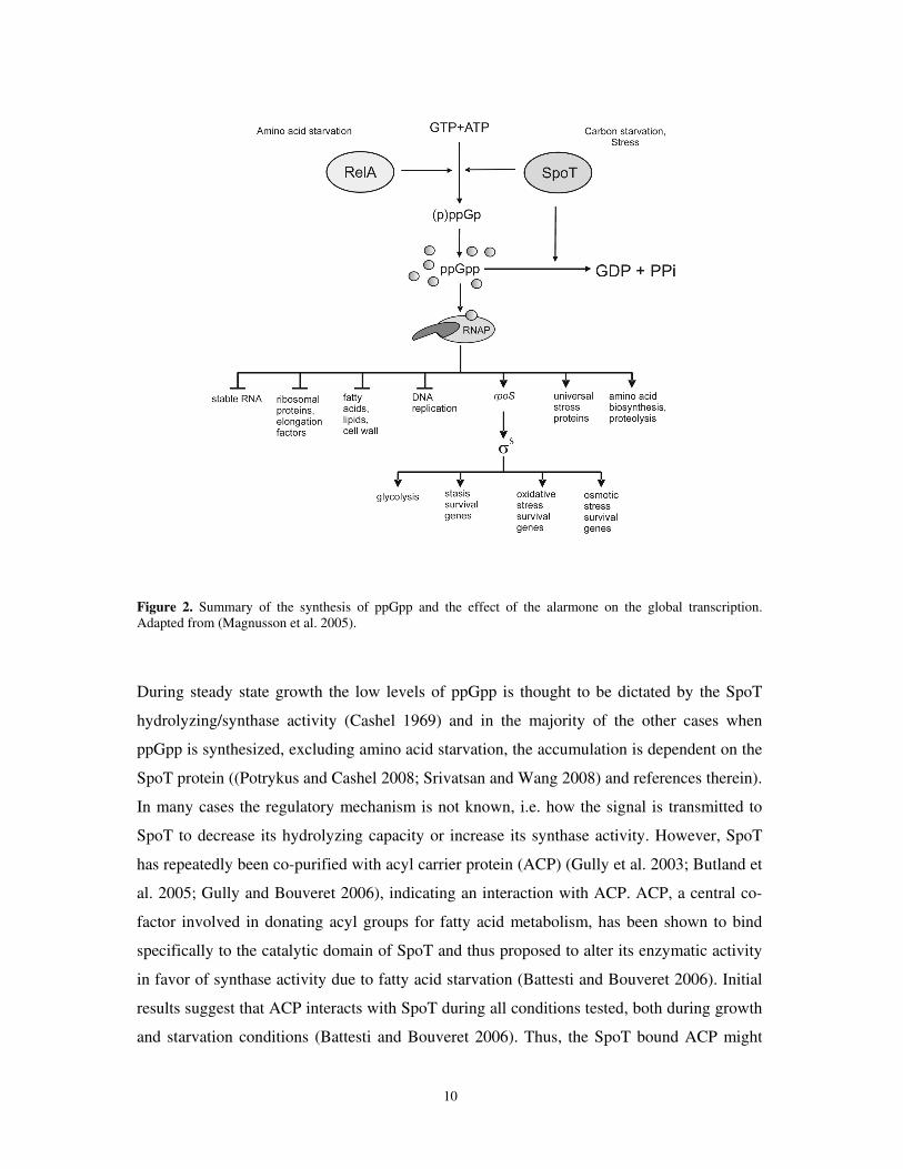
5.3. The stringent response and the alarmone ppGpp
Deprivation of an amino acid, carbon source, fatty acids, phosphate or iron in a bacterial cell
all results in a swift change in the major cellular overall metabolism (Xiao et al. 1991;
Seyfzadeh et al. 1993; Cashel 1996; Murray and Bremer 1996; Vinella et al. 2005; Bougdour
and Gottesman 2007). This pleiotropic response, initially identified in 1961 (Stent and
Brenner), is known as the stringent response. Guanosine tetraphosphate (ppGpp) and
guanosine pentaphosphate (pppGpp), collectively known as (p)ppGpp, is responsible for
mediating the response including the trademark of the stringent response, the abrupt
transcriptional cessation of the translational machinery components (rRNA and tRNA)
(Cashel 1996). In general, ppGpp down-regulates proliferation and growth promoting
processes and up-regulates genes involved in maintenance (see Fig 2). In addition, ppGpp
regulates a plethora of different physiological processes as starvation survival, replication,
secondary metabolism, virulence and biofilm formation (Magnusson et al. 2005; Potrykus
and Cashel 2008; Wu and Xie 2009). Among the positively regulated genes are the usp-
genes, as well as rpoS and many RpoS-dependent genes, like uspB and bolA (Gentry et al.
1993; Kvint et al. 2000a; Gustavsson et al. 2002).
5.3.1. Regulation of stringent response
In E.coli, synthesis of ppGpp is mediated by two pathways, dependent on the proteins RelA
and SpoT, SpoT is also essential for degrading ppGpp (Cashel 1996) (Fig 2). During amino
acid starvation, binding of uncharged tRNA in the ribosomal A site stimulates ppGpp
synthase by RelA (Haseltine and Block 1973; Wendrich et al. 2002). The ribosome
associated RelA protein catalyzes the phosphorylgroup transfer of phosphates from the ATP
donor to the GTP or GDP (Cashel 1969; Sy and Lipmann 1973), Uncharged tRNA has also
been implicated in inhibiting SpoT dependent hydrolysis of ppGpp (Richter 1980) as
exhaustion of the amino acid pool reduces the SpoT hydrolase activity (Murray and Bremer
1996) and thereby increasing the stringent response.
10
Figure 2. Summary of the synthesis of ppGpp and the effect of the alarmone on the global transcription. Adapted from (Magnusson et al. 2005).
During steady state growth the low levels of ppGpp is thought to be dictated by the SpoT
hydrolyzing/synthase activity (Cashel 1969) and in the majority of the other cases when
ppGpp is synthesized, excluding amino acid starvation, the accumulation is dependent on the
SpoT protein ((Potrykus and Cashel 2008; Srivatsan and Wang 2008) and references therein).
In many cases the regulatory mechanism is not known, i.e. how the signal is transmitted to
SpoT to decrease its hydrolyzing capacity or increase its synthase activity. However, SpoT
has repeatedly been co-purified with acyl carrier protein (ACP) (Gully et al. 2003; Butland et
al. 2005; Gully and Bouveret 2006), indicating an interaction with ACP. ACP, a central co-
factor involved in donating acyl groups for fatty acid metabolism, has been shown to bind
specifically to the catalytic domain of SpoT and thus proposed to alter its enzymatic activity
in favor of synthase activity due to fatty acid starvation (Battesti and Bouveret 2006). Initial
results suggest that ACP interacts with SpoT during all conditions tested, both during growth
and starvation conditions (Battesti and Bouveret 2006). Thus, the SpoT bound ACP might
11
work as a sensor of fatty acid metabolism and depending on the derivatives bound to ACP
correspondingly alter the hydrolyzing/synthase capacity of SpoT by changing its structure
(Battesti and Bouveret 2006). The authors also suggest a possible link to SpoT dependent
accumulation of ppGpp during carbon starvation; carbon source availability could potentially
also be sensed via ACP, as the pool size of the precursor molecule for fatty acid synthesis,
acetyl-CoA, is drastically reduced when cells enter carbon starvation (Takamura and Nomura
1988). Further studies to pinpoint this interaction, possibly also during other stresses, have
yet to be conducted. Besides ACP, ObgE (CgtA), a multi functional enzyme involved in
chromosome segregation and ribosome assembly, has been suggested to regulate SpoT
hydrolysis activity during growth (Jiang et al. 2007; Raskin et al. 2007). However, due to
conflicting results the role of ObgE remains unclear for the moment (Persky et al. 2009).
5.3.2. The effect of the stringent response regulator, ppGpp, on RNAP
In contrast to many regulators of transcription, which bind DNA within or close to the
promoter thereby affecting transcription, ppGpp directly binds to the RNAP (Chatterji et al.
1998; Artsimovitch et al. 2004). One theory of how ppGpp exerts its function is that it
destabilizes the open complex between σ70-programmed RNAP and the promoter, causing
RNAP to fall off of promoters during the initiation process. Studies have shown that rrn-
promoters form intrinsically unstable open complexes and thus are specifically sensitive to
ppGpp (Barker et al. 2001). This theory also advocates that as a consequence of the reduced
stability of the rrn promoters the availability of free RNAP, thought to be limiting in the cell,
increases and that allows transcription from promoters are relatively poor in recruiting
RNAP. These promoters would then have increased transcription and be seen as positively
regulated by ppGpp (i.e., stress inducible promoters). However, the opposite has also been
postulated; that the available pool of RNAP is diminished during stringent response, possibly
because ppGpp increases pausing during transcription elongation (Wagner 2002; Jores and
Wagner 2003). In addition, the pool of free σ70 programmed RNAP is thought to decrease
during a stringent response due to the binding of alternative sigma factors. ppGpp has been
shown to increase the ability of alternative sigma factors to compete for binding to RNAP
core (Farewell et al. 1998a; Jishage et al. 2002). This model also proposes that because
promoters have different capacities to load RNAP and transcribe from a promoter before they
are saturated, alterations in the level of available RNAP would influence transcription rates.
12
rrn promoters have been shown to have a high clearance rate and estimates suggest that these
promoters not are saturated under most growth conditions in vivo. In this model it is also
proposed that since ribosomal promoters are difficult to saturate they would be especially
sensitive to alterations in RNAP concentration. Experiments have shown that decreasing the
levels of σ70 programmed RNAP mimics a stringent response (Magnusson et al. 2003), and
increasing the levels gives the opposite response (Gummesson et al. 2009). These results
support but do not prove this second model of passive regulation via alterations in the levels
of RNAP during a stringent response.
The central regulatory role of ppGpp can easily been seen on proteomic 2-D gels or
following individual transcriptional fusions know to be part of the ppGpp regulon. A ppGpp0
strain entering stationary phase totally fails to down-regulate growth promoting gene
transcription (Magnusson et al. 2003) and positively regulated genes like uspA or uspB are
not induced when cells when enter stationary phase (Kvint et al. 2000a; Kvint et al. 2000b).
5.3.3. Other effects of the stringent response regulator, ppGpp
In addition to its role in regulating rrn and stress inducible promoters, ppGpp also modulates
DNA replication and cell division by decreasing transcription from the stringently controlled
dnaA promoter (Chiaramello and Zyskind 1990). The overall rate of replication is thought to
be determined by the initiator protein DnaA concentration (Lobner-Olesen et al. 1989) and
there is an inverse correlation between initiation of a new round of replication and the
concentration of ppGpp (Schreiber et al. 1995; Ferullo and Lovett 2008). In addition to this
role, in vitro data suggest that ppGpp inhibits DnaG primase activity in both E.coli and
B.subtilis (Wang et al. 2007; Maciag et al. 2010). Moreover, ObgE seems to work in concert
with ppGpp to control aspects of cell division and stringent response. One of the roles of the
ObgE GTPase involves control of replication in a G-Protein like fashion (Foti et al. 2005).
The finding that ObgE has a high affinity to (p)ppGpp and that an obgE mutant has an altered
pppGpp/ppGpp ratio (Persky et al. 2009), might imply that ObgE works as an effector of the
stringent response.
That ppGpp interacts with other molecules besides RNAP is also precedented by in vitro
studies where the GTPase translational factors IF2 and EF-Tu are affected by ppGpp either
by inactivating their function or increasing their accuracy (Pingoud and Block 1981; Dix and
13
Thompson 1986; Milon et al. 2006). This finding that ppGpp affects translation accuracy,
also results in a ppGpp0 strain producing more oxidized (carbonylated) proteins (Gummesson
et al. 2009), a measurement of translational error. Thus, the combined in vivo and in vitro
results suggest an increased accuracy of translation mediated by ppGpp during stringent
conditions. However, this effect could also be indirect since ppGpp facilitates alternative
sigma factors, like σS and σ32, to compete better with σ70 and redirect the transcription from
growth promoting genes to genes involved in maintenance function (Farewell et al. 1998a;
Jishage et al. 2002) and many of the genes being regulated by these σ-factors are known to
abrogate the cause of oxidized proteins (see below). Thus, the increased ribosomal accuracy
together with effect of stringently regulated RNAP has on transcription might be a way for
the cell to reduces synthesis of aberrant proteins.
5.4. Heat shock proteins, oxidative stress and translation fidelity
The heat shock response is important during both adverse conditions, like sudden
temperature shifts and exposure to organic chemicals, as well as under non-stress conditions
(Gross 1996; Hartl 1996). In response to protein misfolding in the cytoplasm, the heat shock
sigma factor σ32 becomes stabilized and directs transcription of proteins of its regulon (Straus
et al. 1987; Bukau 1993; Guisbert et al. 2004). The majority of these proteins are involved in
either protein folding (chaperones) or protein degradation (proteases). These processes
become increasingly important under conditions where damaged protein is produced.
However, proteins always have the potential to become damaged even under favorable
environmental conditions. Under aerobic conditions, the cell inevitably produces reactive
oxygen species (ROS), like hydrogen peroxide, superoxide anions and radicals, as a bi-
product of normal metabolic electron transport (Fridovich 1978). Oxidative damage by ROS
of proteins can produce carbonylated proteins via direct metal-catalyzed oxidation (MCO) on
amino acid side chains of specific amino acids, leading to potential loss of function and
denaturation of proteins (Stadtman and Levine 2000; Maisonneuve et al. 2009). Once
formed, a neighboring carbonylatable site is more prone to carbonylation (Maisonneuve et al.
2009), exacerbating the process of protein function degeneration. Being an irreversible
process, the carbonylation process is a threat to the cell. One way the cell can combat protein
carbonylation is by the means of synthesizing antioxidants, like catalase and superoxide
dismutase, thereby neutralizing ROS (Dukan and Nystrom 1999; Stadtman and Levine 2000;
14
paper III). Since misfolded proteins constitute a signal for the heat shock response, the
synthesized chaperones and proteases constitute a second way the cell can combat oxidative
modification of proteins. In order to prevent detrimental accumulation and possible
aggregation of denatured proteins a delicate balance between refolding and/or degrading of
the damaged proteins occurs. By their capacity to refold proteins, chaperones are essential for
maintaining the function of proteins (Hartl 1996). However, proteins can also be degraded by
proteases if the load of protein damage is high (Gottesman 1996).
Both chaperones and proteases are required for normal adaptation when cells enter stationary
phase, as both dnaK (a chaperone) and clpP (a protease) mutants display reduced survival
(Spence et al. 1990; Weichart et al. 2003). It has been suggested that proteolytic degradation
of damaged proteins may also be increasingly important during the entry into and during
stationary phase, because of the lack of dilution of components by protein synthesis and cell
division (Weichart et al. 2003). As a consequence of carbon starvation and amino acid down-
shift an imminent decline in the pool size of charged tRNAs occurs. This reduced availability
leads to reduced translational fidelity due to misincorporation of erroneous amino acids,
translational frameshifting and stop-codon readthrough (O'Farrell 1978; Barak et al. 1996;
Wenthzel et al. 1998; Ballesteros et al. 2001) as well as an induction of heat shock proteins
(Matin 1991; Ballesteros et al. 2001). However, despite an up-regulation of oxidative defense
proteins, those systems are not sufficient combat the levels of oxidation of damaged proteins
which increase during entry to stationary phase (Fredriksson et al. 2005; Diaz-Acosta et al.
2006).
Among the many proteins under the regulatory control of σ32, Clp and Lon are the major
proteases responsible for degradation of cytoplasmic proteins (Maurizi 1992). Lon is the
prime protease degrading misfolded and aberrant proteins and in its absence strong
aggregation of proteins occurs (Tomoyasu et al. 2001). The ClpP protease assisted by the
chaperones ClpA or ClpX degrades a variety of proteins, for example, proteins with
abnormal N-terminal amino acids (according to the N-end rule) and incompletely synthesized
proteins with SsrA-tag (Tobias et al. 1991; Gottesman et al. 1998). SsrA-tagging is a specific
modification of proteins adding a short polypeptide tag to the C-terminal end of a protein,
hence marking them for degradation (Tu et al. 1995; Keiler et al. 1996; Gottesman et al.
1998). ClpXP is also responsible for controlling the stability of RssB-bound RpoS, the
stationary phase sigma factor (See section 5.5.1.3 and paper III).
15
5.5. RpoS regulon and general stress defense
In contrast to many specific stress responses which are induced by a specific stress with the
capacity to repair or eliminate the immediate stress caused to the cell, the σS (RpoS)
dependent stress resistance has a general protective role in the cell. For example, cells deleted
for rpoS display reduced survival during carbon and nitrogen starvation as well as osmotic,
oxidative and heat stresses (Lange and Hengge-Aronis 1991; McCann et al. 1991), indicating
that RpoS governs the regulation of a large set of different stress responses.
Cessation of growth due to starvation of amino acids, phosphate, nitrogen or carbon,
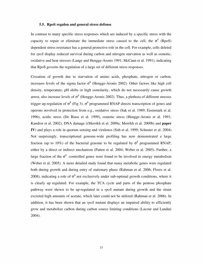
increases levels of the sigma factor σS (Hengge-Aronis 2002). Other factors like high cell
density, temperature, pH shifts or high osmolarity, which do not necessarily cause growth
arrest, also increase levels of σS (Hengge-Aronis 2002). Thus, a plethora of different stresses
trigger up-regulation of σS (Fig 3). σS programmed RNAP directs transcription of genes and
operons involved in protection from e.g., oxidative stress (Sak et al. 1989; Eisenstark et al.
1996), acidic stress (De Biase et al. 1999), osmotic stress (Hengge-Aronis et al. 1991;
Kandror et al. 2002), DNA damage ((Merrikh et al. 2009a; Merrikh et al. 2009b) and paper
IV) and plays a role in quorum sensing and virulence (Suh et al. 1999; Schuster et al. 2004)
Not surprisingly, transcriptional genome-wide profiling has now demonstrated a large
fraction (up to 10%) of the bacterial genome to be regulated by σS programmed RNAP,
either by a direct or indirect mechanism (Patten et al. 2004; Weber et al. 2005). Further, a
large fraction of the σS -controlled genes were found to be involved in energy metabolism
(Weber et al. 2005). A more detailed study found that many metabolic genes were regulated
both during growth and during entry of stationary phase (Rahman et al. 2006; Flores et al.
2008), indicating a role of σS not exclusively under sub-optimal growth conditions, where it
is clearly up regulated. For example, the TCA cycle and parts of the pentose phosphate
pathway were shown to be up-regulated in a rpoS mutant during growth and the strain
excreted high amounts of acetate, which later could not be utilized (Rahman et al. 2006). In
addition, it has been shown that an rpoS mutant displays an impaired ability to efficiently
grow and metabolize carbon during carbon source limiting conditions (Lacour and Landini
2004).
16
Figure 3. Summary of the regulation of σS. Multiple endogenous and exogenous stress conditions influence the cellular σS level. Elevated levels of σS can be obtained by increased transcription and/or translation or by inhibition of proteolysis. Adapted from (Hengge-Aronis 2002).
5.5.1. RpoS regulation
Regulation of the stationary phase sigma factor σS is complex and is executed at all levels:
transcription, rpoS mRNA translation and σS protein stability, controlled both by cis-
regulatory regions and trans-acting regulatory factors. Furthermore, it appears that different
environmental signals affect regulation differently (Fig 3). Despite the extensive regulation
control at all levels, altered regulation of stability of the protein is the major step affecting the
total level of σS (Zgurskaya et al. 1997).
5.5.1.1. Transcriptional regulation
During entry into stationary phase, a 2 to 3-fold increase of rpoS transcript can be observed,
transcribed from PrpoS (Lange et al. 1995; Zgurskaya et al. 1997). This promoter is located
within the nlpD gene and is growth phase regulated (McCann et al. 1993; Takayanagi et al.
1994; Lange et al. 1995). The resulting transcript has an unusually long untranslated 5´region
of 567nt, which is involved in hairpin formation and posttranscriptional regulation by
occluding and inhibiting access to the ribosomal binding site ((Hengge-Aronis 2002;
Majdalani et al. 2002) and references therein).
17
Negative control of rpoS transcription, exerted via the phosphorylation protein crr (IIAGlc)
suggests a possible link between rpoS and carbon metabolism (Ueguchi et al. 2001). This
mechanism is most likely not direct; instead the signal goes through cAMP-CRP regulation
of rpoS. Two putative cAMP-CRP sites are located in the rpoS promoter region. It has been
reported that strains unable to make cAMP (∆cya) are affected in transcription from PrpoS,
but with inconclusive results since both positive and negative regulation of rpoS was reported
(Lange and Hengge-Aronis 1991; McCann et al. 1993). However, in support of the negative
regulation, addition of external cAMP to a cya mutant repressed expression of rpoS (Lange
and Hengge-Aronis 1991b). Similarly, overproduction of CdpA leads to an increase of σS
levels in wild type cells (though not to ∆cya or ∆crp levels) (Barth et al. 2009). CpdA is a
phosphodiesterase which hydrolyses cAMP (Imamura et al. 1996). Addition of cAMP
significantly improved the growth rate compared to the severely affected cya mutant (Lange
and Hengge-Aronis 1991b). This poor growth rate in cya mutants complicates interpretation
of these results since it is known that RpoS levels is increased in slow growing cells
(Zgurskaya et al. 1997; Teich et al. 1999) perhaps via regulation by ppGpp.
The alarmone ppGpp acts as a positive signal in rpoS transcription (Gentry et al. 1993; Lange
et al. 1995) as well as transcription of σS dependent genes (Kvint et al. 2000a). Later it was
suggested that ppGpp does not stimulate transcription, instead the rate of translation is
positively affected by accumulation of ppGpp (Brown et al. 2002). Induction of RpoS was
also shown to be affected by DksA, a protein thought to enhance the effect of ppGpp (Brown
et al. 2002).
5.5.1.2. Translational regulation and sRNA’s
Small regulatory RNAs can modify the activity of proteins, affect the stability and regulate
transcription from mRNAs. At least three sRNAs, DsrA, RprA and OxyS, regulate RpoS
translation. Together with the RNA chaperone Hfq, these RNAs base pair with rpoS mRNA
thereby influencing the secondary structure, translation and stability. Deleting Hfq (HF-I)
severely reduces σS levels (Muffler et al. 1996). Under oxidative stress, the small RNA OxyS
represses RpoS translation. OxyS binds Hfq and might thereby alter its activity (Zhang et al.
1998). DsrA and RprA both basepair with a 5´upstream antisense element of rpoS RNA,
thereby facilitating ribosome binding and positively regulating translation (Sledjeski et al.
18
2001; Majdalani et al. 2002). Furthermore, the histone like protein, H-NS, is a nucleoid-
associated protein capable of binding rpoS mRNA and negatively regulating translation
efficiency and σS stability (Barth et al. 1995; Yamashino et al. 1995; Brescia et al. 2004).
5.5.1.3. Protein stability
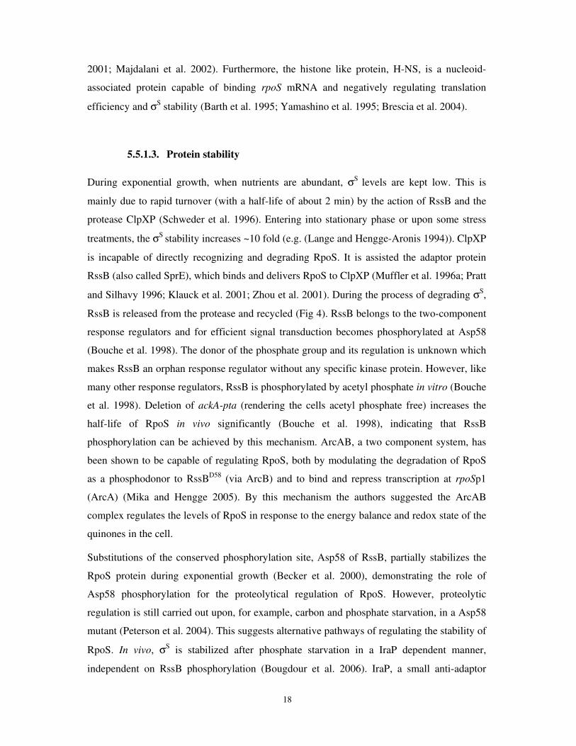
During exponential growth, when nutrients are abundant, σS levels are kept low. This is
mainly due to rapid turnover (with a half-life of about 2 min) by the action of RssB and the
protease ClpXP (Schweder et al. 1996). Entering into stationary phase or upon some stress
treatments, the σS stability increases ~10 fold (e.g. (Lange and Hengge-Aronis 1994)). ClpXP
is incapable of directly recognizing and degrading RpoS. It is assisted the adaptor protein
RssB (also called SprE), which binds and delivers RpoS to ClpXP (Muffler et al. 1996a; Pratt
and Silhavy 1996; Klauck et al. 2001; Zhou et al. 2001). During the process of degrading σS,
RssB is released from the protease and recycled (Fig 4). RssB belongs to the two-component
response regulators and for efficient signal transduction becomes phosphorylated at Asp58
(Bouche et al. 1998). The donor of the phosphate group and its regulation is unknown which
makes RssB an orphan response regulator without any specific kinase protein. However, like
many other response regulators, RssB is phosphorylated by acetyl phosphate in vitro (Bouche
et al. 1998). Deletion of ackA-pta (rendering the cells acetyl phosphate free) increases the
half-life of RpoS in vivo significantly (Bouche et al. 1998), indicating that RssB
phosphorylation can be achieved by this mechanism. ArcAB, a two component system, has
been shown to be capable of regulating RpoS, both by modulating the degradation of RpoS
as a phosphodonor to RssBD58 (via ArcB) and to bind and repress transcription at rpoSp1
(ArcA) (Mika and Hengge 2005). By this mechanism the authors suggested the ArcAB
complex regulates the levels of RpoS in response to the energy balance and redox state of the
quinones in the cell.
Substitutions of the conserved phosphorylation site, Asp58 of RssB, partially stabilizes the
RpoS protein during exponential growth (Becker et al. 2000), demonstrating the role of
Asp58 phosphorylation for the proteolytical regulation of RpoS. However, proteolytic
regulation is still carried out upon, for example, carbon and phosphate starvation, in a Asp58
mutant (Peterson et al. 2004). This suggests alternative pathways of regulating the stability of
RpoS. In vivo, σS is stabilized after phosphate starvation in a IraP dependent manner,
independent on RssB phosphorylation (Bougdour et al. 2006). IraP, a small anti-adaptor
19
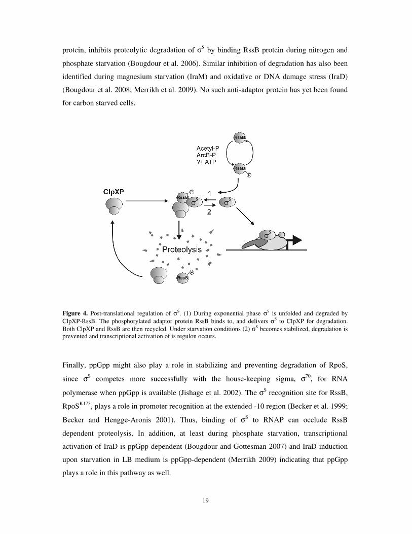
protein, inhibits proteolytic degradation of σS by binding RssB protein during nitrogen and
phosphate starvation (Bougdour et al. 2006). Similar inhibition of degradation has also been
identified during magnesium starvation (IraM) and oxidative or DNA damage stress (IraD)
(Bougdour et al. 2008; Merrikh et al. 2009). No such anti-adaptor protein has yet been found
for carbon starved cells.
Figure 4. Post-translational regulation of σS. (1) During exponential phase σS is unfolded and degraded by ClpXP-RssB. The phosphorylated adaptor protein RssB binds to, and delivers σS to ClpXP for degradation. Both ClpXP and RssB are then recycled. Under starvation conditions (2) σS becomes stabilized, degradation is prevented and transcriptional activation of is regulon occurs.
Finally, ppGpp might also play a role in stabilizing and preventing degradation of RpoS,
since σS competes more successfully with the house-keeping sigma, σ70, for RNA
polymerase when ppGpp is available (Jishage et al. 2002). The σS recognition site for RssB,
RpoSK173, plays a role in promoter recognition at the extended -10 region (Becker et al. 1999;
Becker and Hengge-Aronis 2001). Thus, binding of σS to RNAP can occlude RssB
dependent proteolysis. In addition, at least during phosphate starvation, transcriptional
activation of IraD is ppGpp dependent (Bougdour and Gottesman 2007) and IraD induction
upon starvation in LB medium is ppGpp-dependent (Merrikh 2009) indicating that ppGpp
plays a role in this pathway as well.
20
6. Model genes and proteins
When studying global gene responses which involve regulation of many genes and proteins
two complimentary approaches are often used. One is to use methods such as DNA
microarrays to visualize global responses and the second is to use model genes/proteins as
representatives of a class of gene/protein. This second approach allows one to study the
regulation of the model gene or protein in great detail and then later assess how general this
regulation is to other genes that respond to the same stimulus.
6.1. UspA and the universal stress protein family
UspA of E. coli is known as the paradigm protein of a superfamily that includes an ancient
and conserved group of proteins not only found in the genomes of bacteria, but also in
archaea, fungi, protozoa, and plants (Aravind et al. 2002; Kvint et al. 2003). The UspA
domain (Pfam accession number PF00582) has been found in more than 1,000 different
proteins. E. coli has six usp-family genes (uspA, C, D, E, F and G) and all are induced in
response to stasis and stress conditions. They are all regulated by the house-keeping sigma
factor, σ70, and require ppGpp for induction (Gustavsson et al. 2002). Structurally they can
be divided into four classes, which in most cases correspond to their function (Nachin et al.
2008). The Usp proteins of E. coli can form dimeric proteins and it is now evident that the
Usp proteins within a class form both homodimers and heterodimers (Nachin et al. 2008).
This may explain the heterogeneous functions of the individual Usp proteins (for UspA, see
below). Apart from being required for stasis survival and starvation-induced stress resistance,
including resistance to DNA damaging agents and oxidants (Nystrom and Neidhardt 1993;
Albertson and Nystrom 1994; Kvint et al. 2003; Nachin et al. 2005), some pathogenic
bacteria, e.g. Pseudomonas aeroginosa and Mycobacterium tuberculosis, require Usps to
combat anaerobic energy deficiency (O'Toole and Williams 2003; Boes et al. 2006) during
the persistent infections cystic fibrosis and tuberculosis. In line with the notion of Usps being
important for bacterial virulence, some of the E. coli Usps are involved in iron homeostasis,
motility and adhesion, essential features of bacterial pathogenesis (Nachin et al. 2005).
21
Recently, UspA-family proteins has also been shown to be required for biofilm formation
and virulence in pathogenic bacteria (Chen et al. 2006; Liu et al. 2007)
6.1.1. Regulation and function of UspA
When cells enter stationary phase a drastic increase of the UspA protein occurs, easily
detected on 2-D PAGE (Nystrom and Neidhardt 1992). The same pattern occurs during a
large number of stress conditions, including heat shock, heavy metal exposure, oxidative and
osmotic shock and starvation for carbon, DNA damage, nitrogen and phosphate (Nystrom
and Neidhardt 1992; Diez et al. 2000; Gustavsson et al. 2002) and these are all the result of
transcriptional activation of uspA (Nystrom and Neidhardt 1992). Thus, numerous conditions
elicit up-regulation of uspA, suggesting it plays a role during these diverse conditions.
Despite extensive investigations, however, no clear single function of the protein has been
found. Instead a mutant of uspA exhibits multiple phenotypes implying a broad range of
different functions including decreased survival during prolonged starvation for carbon
(Nystrom and Neidhardt 1994), exposure to DNA damaging conditions (Diez et al. 2000;
Gustavsson et al. 2002) and superoxide-generating compounds like H2O2 and PMS (Nystrom
and Neidhardt 1994; Diez et al. 2000; Nachin et al. 2005) as well as an inability to efficiently
respond to growth perturbation and altered utilization of carbon sources (see below).
Overproduction of UspA affects the cell physiology in several ways. Cells expressing
physiological stationary phase levels of UspA, from Ptac-uspA exhibit decreased growth rate
when grown in glucose minimal medium and a delay in outgrowth of stationary phase
cultures (Nystrom and Neidhardt 1996). As a consequence of this overproduction, a global
change in protein synthesis was observed and some proteins were shown to display altered an
isoelectric point on 2-D gels suggesting a change in protein modification. UspA itself is a
autophosphorylating serine and threonine phosphoprotein (Freestone et al. 1997), but it is not
known whether UspA has a kinase activity and is directly involved in post-translational
modification of proteins. Like overexpression of uspA, a mutant of uspA displays alterations
in the pattern of protein synthesis on 2D gels and a delayed regulatory response in the
synthesis of proteins during transition into stationary phase is observed when cells enter
stationary phase (Nystrom and Neidhardt 1994). Thus proper levels of UspA are crucial for
the cell to respond to changes in the environment. However, no further studies have been
22
conducted on the possible regulatory functions of UspA, even though it would have been
interesting.
Mutants of uspA show a diauxic type of growth when grown on glucose or gluconate as
carbon source (Nystrom and Neidhardt 1993). These mutants were shown to dissimilate
glucose to a higher extent during growth and due to overflow metabolism excrete abnormal
amounts of acetate into the media. After relief of catabolite repression (depletion of glucose),
cells were able to utilize acetate as a carbon source (Nystrom and Neidhardt 1993). It should
be noted, however, that this phenotype is most pronounced only in a specific strain
background (JM105). Nevertheless, the result suggests that uspA might somehow play a role
in the coupling of glucose and acetate co-metabolism. In line with this result, some of the
proteins which were up-regulated after overproduction of UspA were identified and found to
be involved in amino acid metabolism and the TCA cycle (Nystrom and Neidhardt 1996). In
Pseudomonas aeruginosa, two Usp-like (37% homology to uspA) proteins, PA3309 and
PA4352, were characterized and found to be essential for survival under anaerobic energy
metabolism conditions. Mutants of PA3309 and PA4352 displayed reduced survival
anearobically in the presence of pyruvate, or due to lack of an electron acceptor during shifts
to anaerobic environments (Boes et al. 2006; Schreiber et al. 2006). Perhaps one function of
the Usp proteins is in modulating the flow in central metabolic pathways. However, studies
on a uspA mutant could not detect any alterations in metabolic fluxes during growth on
glucose (Nanchen et al. 2008), excluding uspA as a general regulator under normal growth.
6.1.2. Regulation of uspA expression
Transcriptional expression of the uspA gene is positively regulated under conditions leading
to starvation and is dependent on the housekeeping sigma factor, σ70 (Nystrom and Neidhardt
1992). During entry to stationary phase, PuspA is positively regulated by the alarmone ppGpp
and a ppGpp0 strain (∆relA, ∆spoT) fails to induce uspA (Kvint et al. 2000b). Downstream of
the uspA promoter region two operator binding sites for FadR, a regulator of the fatty acid
metabolism, were identified and confirmed with footprinting analysis to be functional
(Farewell et al. 1996). Conditions where FadR is inactivated significantly derepresses
transcription from uspA in exponential phase, thus FadR exerts negative control on PuspA
transcription (Farewell et al. 1996). Later it was shown, however, that under conditions
eliciting stringent response, the alarmone ppGpp is capable of allowing RNAP to override the
23
repressive effect of FadR (Kvint et al. 2000b). Thus, FadR regulation most likely only plays a
role in setting uspA transcription levels in growing cells and when cells are using fatty acids
as a carbon source. uspA has also been shown to be regulated under some conditions in
response to DNA damage and/or cell division defects (Diez et al. 1997). A mutation in ftsK,
encoding a DNA translocase, exhibits superinduction from PuspA and this superinduction is
dependent on RecA (Diez et al. 2000). However, the positive regulation of uspA by RecA is
atypical in the sense that regulation is LexA independent and only occurs, at least under
conditions tested, in conjunction with the ftsK mutant (Diez et al. 2000). Finally, stability of
the uspA messenger RNA is affected by CspC and CspE, two cold shock proteins
constitutively expressed at normal temperatures. Overexpression of either of these proteins
stabilizes and deletions decrease the transcript (Phadtare and Inouye 2001; Phadtare et al.
2006).
Further characterization of the regulation of PuspA indicates that uspA is also positively
regulated by metabolic intermediates. Accumulation of fructose-6-p (and possibly also
glucose-6-p) intermediates at the start of glycolysis, increase the induction from PuspA upon
entry to stationary phase due to carbon starvation (paper I). This was shown most
dramatically in mutants that accumulate these intermediates but is also shown to play a role
in stationary phase induction in wild type cells.
6.2. UspB
6.2.1. Function and regulation
Located next to uspA on the E. coli chromosome, with promoter start sites separated by 135
bp, the uspB gene is transcribed divergently relative to the uspA gene. Transcription of the
uspB gene is induced by a large variety of stresses conditions (see below) and consequently it
was named universal stress protein B (Farewell et al. 1998b). However, based on sequence
homology, uspB is not part of the growing uspA-superfamily (Kvint et al. 2003). UspB
protein is only found in close relatives to E.coli. A BLAST-P search for orthologs of the
cytoplasmic domain show that UspB exists in Enterobacteriaceae, but also in Vibrio
(Gammaproteobacteria). Interestingly most of those bacteria also have a uspA family member
next to uspB and like E.coli divergently transcribed, suggesting that the two genes were
chromosomally linked in an ancestor of these species.
24
Transcription from the uspB promoter is regulated by the stationary phase sigma factor (σS)
and ppGpp (Farewell et al. 1998; Kvint et al. 2000a). ppGpp is required for induction of both
σS (Gentry et al. 1993; Lange et al. 1995; Hirsch and Elliott 2002) and transcription from σS -
dependent promoters, like uspB (Kvint et al. 2000a; Jishage et al. 2002). Consequently,
during exponential growth in rich media transcription of uspB is very low and is strongly
induced when the cells enter stationary phase (Farewell et al. 1998b). The same induction
pattern is seen when cells are starved for glucose, nitrogen, phosphate or exposed to osmotic
or oxidative stress. However, none of these conditions require functional UspB protein for
cell survival (Farewell et al. 1998b).
The uspB gene product is a small protein (111 aa) with a protein mass of about 14kDa. The
amino acid structure suggests that the protein harbors two membrane spanning sequences,
located in the very end of C and N termini of the protein. The membrane spanning domains
have now been confirmed and the final protein is anchored in the cytoplasmic membrane
with a 65 amino acid loop facing the cytoplasm (Daley et al. 2005). Functional
characterization of the protein has so far not been so successful but genetic studies have shed
light on the function of UspB. One phenotype is that UspB is needed for resistance to ethanol
during stationary phase (Farewell et al. 1998b). Further, during repetitive freeze thaw cycles
a beneficial effect of reduced uspB expression for culture survival was found (Sleight et al.
2008). However, why mutation in uspB is favored during these conditions remain unsolved.
Both ethanol and hypothermic stress lead to alterations in membrane composition toward
increased membrane fluidity (Sinensky 1974; Ingram and Vreeland 1980; Hazel 1995).
Ethanol, like thermal shock, also causes protein denaturation and induces the heat shock
response (Bukau 1993; Gross 1996; Feder and Hofmann 1999). Thus one possibility is that
uspB mutants not are able to properly alter their membrane composition when entering
stationary phase. However, no alteration in membrane composition could be detected in an
uspB mutant in either stationary phase or after repetitive freeze thaw cycles (Sleight et al.
2008). Similarly, no difference in survival could be observed following heat shock (Farewell
et al. 1998b). At the moment its unknown how the UspB works to increase the survival
following ethanol and freeze/thawing. Recent data indicates a novel functional role of UspB.
A uspB mutant displays sensitivity towards many agents known to affect the integrity of the
DNA and data suggests that the protein is taking part of the repair mechanism of such DNA
lesions (paper IV).
25
7. DNA damage
The maintenance of DNA integrity is vital for the viability and to avoid a high frequency of
alterations in the genome of the organism. The genome is constantly inflicted with DNA
damage from endogenous sources like metabolic byproducts, misincorporation of incorrect
bases during replication, and spontaneous deamination, depurination or depyrimidination, as
well as various environmental factors (Friedberg 2006). Cells have evolved a battery of
defense mechanisms in order to prevent maintenance of lesions leading alterations in the
blueprint. Examples of this are reversal of base damage, excision repair, translesion repair,
and strand break repair.
In many cases the DNA damage interferes with the ongoing replication of the DNA and a
cell has to overcome this situation. Thus the processes of DNA replication and DNA repair
must cooperate and work in synergy in order for the cell minimize genomic rearrangements
and avoid lethal situations. Moreover, following DNA damage, fast growing organisms with
multiple simultaneous rounds of replication like E.coli can take advantage of the multiple
chromosomes as a blueprint in order to repair the DNA by non-mutagenic mechanisms.
7.1. Replication, not that smooth
E. coli, like most other prokaryotic organisms, has one circular chromosome and in order for
the cell to divide and produce two daughter cells the genome must be duplicated. By
initiating a new round of replication before cells have divided, thus producing multiple
simultaneous rounds of replication , cells are able to divide faster than it takes for one round
of replication (Helmstetter 1996). Replication is highly controlled event and is regulated at
the level of initiation (Katayama and Sekimizu 1999). Initiation of replication occurs at a
fixed, specific site, oriC. DnaA and other specific initiator proteins, bind the oriC region and
melt a AT- rich sequence, form an open complex and prime the DNA for assembly of the
components of the DNA polymerase machinery (Bramhill and Kornberg 1988). Once bound,
the replisomes initiate the replication. In E.coli, replication is a bidirectional process, thus
two replisomes are actively synthesizing the new DNA in opposite directions from oriC.
Eventually those replisomes will meet at the terminus region (ter) (Messer et al. 2001).
The classical view of replication is that replication occurs in a semi-discontinuous way due to
the antiparallel structure of DNA. Catalyzed by DNA polymerase, de novo DNA is
26
synthesized in a 5’- 3’ direction; the leading strand is synthesized as a continuous chain and
the lagging strand discontinuously in shorter (1-2kb) Okazaki fragments. Over the last
decade, it has been established that the interruption of replication forks is a frequent event,
and even under conditions when cells are not exposed to genotoxic compounds replication
can be stalled or interrupted (Sandler and Marians 2000). In vivo studies where replication
restart proteins, for example, PriA and PriC, have been deleted, provide evidence that
replication restart is a frequent and essential mechanism for proper replication of the DNA
(Sandler 2000; Gregg et al. 2002; Rangarajan et al. 2002), as those mutants display a severe
decrease in replication accuracy or synthetic lethality in combination with mutant alleles of
replication proteins. Furthermore, those pri mutants rapidly acquired suppressor mutations in
the replisome assembly machinery (Sandler et al. 1996; Sandler et al. 1999), demonstrating
the fundamental process of replication reloading for survival and that replication is not a
continuous process from oriC till the ter site. Furthermore, various environmental
perturbations can introduce DNA lesions and further inhibit DNA replication. Whether these
hindrances lead to replisome stalling or collapse has been controversial and has led to many
models, including polymerase switching, replication restart, template switching, overriding
of the lesion and replisome reactivation (for ex (Gabbai and Marians 2010; Kowalczykowski
2000; Courcelle and Hanawalt 2003; Wang 2005; Lovett 2007; Michel et al. 2007)).
Further support of the importance of DNA repair during replication comes from findings
with mutants that forms dimeric chromosomes (Steiner and Kuempel 1998). Homologous
recombination during replication of the DNA, if causing an uneven number of cross-overs,
results in chromosomal dimerization, which can prevent proper segregation of the genome to
the daughter cells and is thought be a lethal event. To circumvent this potential demise, XerC
and XerD function as site specific resolvases and specifically resolve the dimerization near
the terminus of replication at a site called dif before cells can start divide and form the
septum. Mutants with defects in dif or xer display an 15% lethality and this number is
interpreted to directly correspond to dimeric genomes (Steiner and Kuempel 1998). These
experiments provide strong evidence that homologous recombination is a common event
during replication.
27
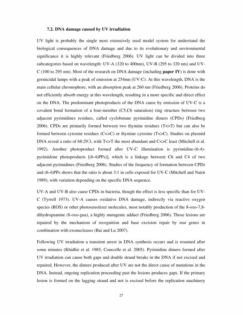
7.2. DNA damage caused by UV irradiation
UV light is probably the single most extensively used model system for understand the
biological consequences of DNA damage and due to its evolutionary and environmental
significance it is highly relevant (Friedberg 2006). UV light can be divided into three
subcategories based on wavelength: UV-A (320 to 400nm), UV-B (295 to 320 nm) and UV-
C (100 to 295 nm). Most of the research on DNA damage (including paper IV) is done with
germicidal lamps with a peak of emission at 254nm (UV-C). At this wavelength, DNA is the
main cellular chromophore, with an absorption peak at 260 nm (Friedberg 2006). Proteins do
not efficiently absorb energy at this wavelength, resulting in a more specific and direct effect
on the DNA. The predominant photoproducts of the DNA cause by emission of UV-C is a
covalent bond formation of a four-member (C5,C6 saturation) ring structure between two
adjacent pyrimidines residues, called cyclobutane pyrimidine dimers (CPDs) (Friedberg
2006). CPDs are primarily formed between two thymine residues (T<>T) but can also be
formed between cytosine residues (C<>C) or thymine cytosine (T<>C). Studies on plasmid
DNA reveal a ratio of 68:29:3, with T<>T the most abundant and C<>C least (Mitchell et al.
1992). Another photoproduct formed after UV-C illumination is pyrimidine-(6-4)-
pyrimidone photoproducts [(6-4)PPs)], which is a linkage between C6 and C4 of two
adjacent pyrimidines (Friedberg 2006). Studies of the frequency of formation between CPDs
and (6-4)PPs shows that the ratio is about 3:1 in cells exposed for UV-C (Mitchell and Nairn
1989), with variation depending on the specific DNA sequence.
UV-A and UV-B also cause CPDs in bacteria, though the effect is less specific than for UV-
C (Tyrrell 1973). UV-A causes oxidative DNA damage, indirectly via reactive oxygen
species (ROS) or other photosensitizer molecules, most notably production of the 8-oxo-7,8-
dihydroguanine (8-oxo-gua), a highly mutagenic adduct (Friedberg 2006). Those lesions are
repaired by the mechanism of recognition and base excision repair by mut genes in
combination with exonucleases (Bai and Lu 2007).
Following UV irradiation a transient arrest in DNA synthesis occurs and is resumed after
some minutes (Khidhir et al. 1985; Courcelle et al. 2005). Pyrimidine dimers formed after
UV irradiation can cause both gaps and double strand breaks in the DNA if not excised and
repaired. However, the dimers produced after UV are not the direct cause of mutations in the
DNA. Instead, ongoing replication proceeding past the lesions produces gaps. If the primary
lesion is formed on the lagging strand and not is excised before the replication machinery
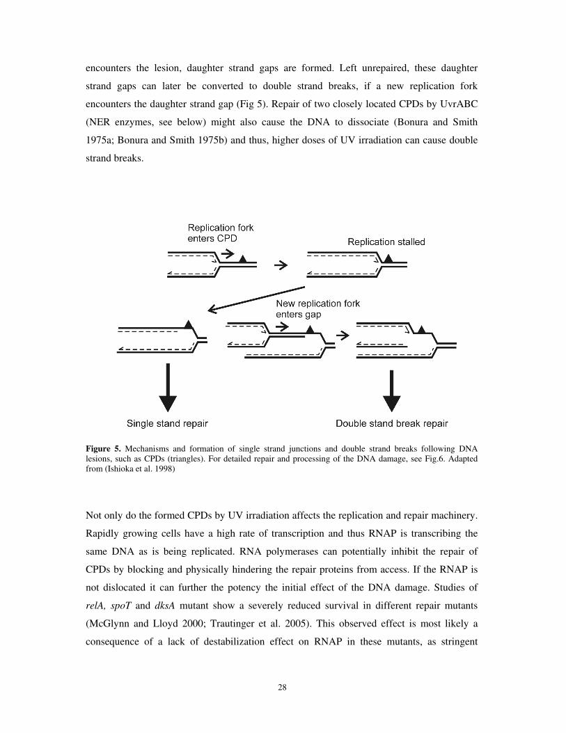
28
encounters the lesion, daughter strand gaps are formed. Left unrepaired, these daughter
strand gaps can later be converted to double strand breaks, if a new replication fork
encounters the daughter strand gap (Fig 5). Repair of two closely located CPDs by UvrABC
(NER enzymes, see below) might also cause the DNA to dissociate (Bonura and Smith
1975a; Bonura and Smith 1975b) and thus, higher doses of UV irradiation can cause double
strand breaks.
Figure 5. Mechanisms and formation of single strand junctions and double strand breaks following DNA lesions, such as CPDs (triangles). For detailed repair and processing of the DNA damage, see Fig.6. Adapted from (Ishioka et al. 1998)
Not only do the formed CPDs by UV irradiation affects the replication and repair machinery.
Rapidly growing cells have a high rate of transcription and thus RNAP is transcribing the
same DNA as is being replicated. RNA polymerases can potentially inhibit the repair of
CPDs by blocking and physically hindering the repair proteins from access. If the RNAP is
not dislocated it can further the potency the initial effect of the DNA damage. Studies of
relA, spoT and dksA mutant show a severely reduced survival in different repair mutants
(McGlynn and Lloyd 2000; Trautinger et al. 2005). This observed effect is most likely a
consequence of a lack of destabilization effect on RNAP in these mutants, as stringent
29
suppressor mutations in RNAP or a hydrolyzing deficient SpoT allele could partially restore
the survival (McGlynn and Lloyd 2000; Trautinger and Lloyd 2002; Trautinger et al. 2005).
7.3. UV produced lesions are recognized and excised by the NER system
In E.coli nucleotide excision repair is mediated by the UvrABC protein complex (Van
Houten et al. 2005). UvrABC has the capacity to recognize a lesion or structural distortion in
DNA like CPDs, (6-4)PPs and crosslinking with mitomycin C (Hanawalt and Haynes 1965;
Hanawalt 1993). Via a multistep process these proteins recognize the adduct, make contact
and make a dual incision in the DNA. Later the incised oligonucleotide is removed, DNA
polymerase I fills in the gap and ligase seals the newly synthesized DNA with the parental
strand (Van Houten et al. 2005; Truglio et al. 2006).
7.4. SOS response; RecA and its function
Following production of UV induced lesions, the cell repairs the DNA damage using the
nucleotide excision repair system. However if the imposed load of lesions exceeds the
capacity of NER, the ongoing replication fork will inevitable cause gaps or breaks in the
DNA. RecA protein monomers bind and form a filament on single stranded DNA (ssDNA)
and catalyze strand exchange with homologous duplex DNA (Cox 2007b; Cox 2007a). As
the nucleoprotein filament is formed, the multi-functional RecA also acts as a co-protease
facilitating auto-cleavage of the LexA repressor (Sassanfar and Roberts 1990; Little 1991) as
well as UmuD (Pham et al. 2002). LexA protein is inactivated and UmuD is activated by this
cleavage. This results in transcriptional activation of the approximately 40 SOS regulon
genes (Courcelle et al. 2001) as well as the transcriptional and post-translational activation of
the SOS mutagenesis translesion polymerase, DNA PolV (umuC of SOS regulon and
UmuD’) (Smith and Walker 1998; Friedberg 2006). RecA also plays a structural role by
maintaining the replication fork at lesions. Filamentation of RecA has been shown to
facilitate non-mutagenic repair and allows the resumption of replication (Chow and
Courcelle 2007). RecA can also facilitate the bypass of DNA lesions by PolV (Pham et al.
2002; Fujii et al. 2006; Schlacher et al. 2006). Accumulation of PolV is slow (Sommer et al.
1998; Opperman et al. 1999), providing time for non-mutagenic repair of the lesions.
30
However, if this fails the PolV can rescue a potentially lethal event by using translesion
synthesis to bypass the lesion and allowing replication to continue (Courcelle et al. 2005).
7.5. Processing of DNA breaks (RecBCD and RecFOR)
A battery of different repair mechanisms to combat the inevitable occurrence of DNA
damage has been developed in the cell. Most repair processes involve the DNA binding
protein RecA for recombination of the processed DNA. Cells with an inactivated RecA
protein display a hypersensitive phenotype following DNA damage with severe degradation
of the genome, illustrating its central role in DNA repair. Even though RecA can bind and
has high affinity towards ssDNA, in many cases the DNA must first be processed before the
RecA protein forms filaments and D-loops with daughter strands, thereby promoting
homologous recombination. In E.coli, the two major mechanisms for recombinational DNA
repair and processing of DNA are the RecBCD and RecFOR pathways. RecBCD is involved
in double strand break repair and RecFOR in the repair of single strand breaks and gaps.
However, RecFOR also has the capacity to repair DSBs in RecBC mutant carrying
suppressor mutations in sbcB and sbcC (or sbcD) genes (Kushner et al. 1971; Lloyd and
Buckman 1985) as well as under in vitro conditions (Handa et al. 2009). Thus, under some
conditions they have overlapping functions.
Biochemically, RecR interacts with both RecF and RecO and the resulting complex(es)
facilitates presynaptic formation by directing RecA nucleation onto gapped SSB-coated
ssDNA (Umezu et al. 1993; Umezu and Kolodner 1994). RecF(R) recognizes the 5´ end of
an ssDNA-dsDNA junction and RecRO assists the loading of RecA molecules onto ssDNA,
allowing 5´-3´ assembly and replacement the SSB proteins (Morimatsu and Kowalczykowski
2003; Sakai and Cox 2009). However, in vitro, RecO together with RecR seem to be
sufficient for mediating RecA filamentation and displacement of SSB-coated ssDNA is under
those conditions even inhibited by RecF (Umezu et al. 1993; Umezu and Kolodner 1994;
Sakai and Cox 2009). Moreover, overexpression of RecOR suppresses the reduced survival
of a recF mutant after UV stress (Sandler and Clark 1994), indicating that RecF is not always
essential.
However, studies conducted in vivo following a sub-lethal dose of UV irradiation show that
the replication fork fails to recover in single mutants of any of the recFOR genes (Rangarajan
31
et al. 2002; Chow and Courcelle 2004). Thus, under natural conditions all three proteins are
required for presynaptic formation as well as for protecting the DNA from degradation. In
contrast to processing of synthetically produced DNA molecules under in vitro experiments,
in in vivo conditions the RecFOR recombination pathway needs additional enzymes to
process the DNA into a presynaptic intermediate. Both the helicase RecQ and the
exonucleases RecJ are needed for processing the nascent DNA at blocked replication forks,
before loading of RecFOR (Courcelle and Hanawalt 1999). In wild type cells both RecJ and
RecQ are essential for non-mutagenic repair and by processing the nascent DNA are thought
to serve to lengthen the substrate that can be recognized by RecFOR and RecA (Courcelle et
al. 2006). In cells lacking any of the RecFOR proteins, the nascent lagging DNA strand is
extensively degraded by the RecQ and RecJ proteins (Chow and Courcelle 2004; Chow and
Courcelle 2007).
Genetic studies of the RecF pathway indicate that the gene products of recF, recO and recR
belong to the same epistasic group. Single and double mutants display the same reduced
survival after UV irradiation (Lloyd et al. 1988; Mahdi and Lloyd 1989) and single mutants
are neither defective in conjugation nor transduction (Kuzminov 1999). RecFOR is required
for proper SOS induction as a mutation in any of the genes causes a delayed response after
DNA damage, followed by SOS superinduction (Whitby and Lloyd 1995). During unstressed
conditions, these mutant strains display a chronic elevation of the SOS response (Whitby and
Lloyd 1995) and a recF mutant shows an increased mutation rate as well (Southworth and
Bridges 1984; Volkert 1989).
The RecBCD enzyme (Exonuclease V) is a heterotrimeric helicase/nuclease capable of
processing blunt or near blunt dsDNA ends into a substrate for homologous recombination in
DNA repair and during transduction as well as conjugational events (Willetts and Mount
1969; Lloyd et al. 1987). Once bound to the DNA substrate, the ATP-dependent RecBCD is
capable of unwinding and degrading the DNA at a remarkable speed (up to 2.000 bp/s),
making it the fastest helicase known. In vitro studies have shown that both RecB and RecD
possess helicase activity albeit with different polarity and translocation speed. The faster
RecD subunit translocates the DNA in the 5´-3´direction. The RecB protein has a 3´-5´ DNA
helicase activity and a C-terminal nuclease motif which possesses all endonuclease activity
of the holoenzyme (Yu et al. 1998a; Yu et al. 1998b; Dillingham et al. 2003; Taylor and
Smith 2003). When bound to DNA, the RecBCD complex subunits RecB and RecD unwind
and separate the duplex and the distally located nuclease motif degrades both of the DNA
32
strands. The endonuclease preferentially degrades the 3´-terminated end, but occasionally
also degrades the 5´ end. This seemingly contradictory event, to degrade both strands, is
attenuated when the complex encounters the recombinational hot spot sequence 5´-
GCTGGTGG-3´, called the Chi site (χ) (Stahl and Stahl 1977; Dixon and Kowalczykowski
1995). An occupied 3´ Chi site prevents degradation of this end and favors 5´degradation.
Further, Chi modified RecBCD now facilitates the loading of RecA on the 3´ ssDNA, which
polymerizes in the opposite direction of the growing 3´end further facilitates the degradation
of the 5´ end (Dixon and Kowalczykowski 1991; Dixon and Kowalczykowski 1995). The
RecA loaded ssDNA searches for homologous DNA and by forming D-loops promotes
recombinational repair.
Inactivating RecBCD function by mutations in recB or recC, causes cells to become
extremely sensitive to DNA damaging agents like UV light and gamma irradiation. These
mutants display poor viability and are highly reduced in homologous recombination, thus the
processing and recombinational event by this enzyme is crucial for efficient recovery of the
cell (Emmerson 1968; Willetts and Mount 1969; Capaldo et al. 1974). In contrast to recB or
recC mutants, inactivation of RecD does not create hypersensitivity to DNA damaging
agents (Lloyd et al. 1988; Lovett et al. 1988). Cells lacking the RecD subunit of the enzyme
are capable of processing DSBs, albeit with reduced kinetic activity (Korangy and Julin
1994). Further, it has been shown that the RecBC(D-) enzyme is devoid of nuclease activity,
does not recognize Chi-sites and is unable to regulate RecA loading on ssDNA (Masterson et
al. 1992; Churchill et al. 1999; Amundsen et al. 2000). Under some circumstances (or in
some assays) a recD mutant displays an increased hypermutability (Biek and Cohen 1986;
Thaler et al. 1989), possibly indicating the functional role the protein possess in the complex
besides the helicase activity.
Considering the fundamental functions of RecBCD, the extremely low amount of RecBCD
(approximately 10 molecules/cell) in the cell is somewhat surprising (Eichler and Lehman
1977; Taylor and Smith 1980). Moreover, expression of recBCD is not induced upon stress
and overexpression studies of RecBCD even show that too much RecBCD causes an increase
in genomic degradation and impaired DNA repair and homologous recombination (Dermic et
al. 2005). Thus, maintaining low levels of RecBCD seems essential to the cell.
33
7.6. Branch migration and resolution by RuvABC and RecG
Once DNA pairing and strand exchange with the sister chromosome is accomplished with the
assistance of RecA, processing of the unstable D-loop is critical. Branch migration can either
dissolve the structure and stabilize it. By moving the D-loop in opposite direction of the
growing RecA strand invasion, branch migration dissolves the structure. Alternatively, by
extending the region and forming so called Holliday junctions the structure is stabilized. The
directionality and how this is controlled is largely unknown, but in E.coli, RecG and RuvAB
helicases have been postulated to displace RecA, catalyze the branch migration and form
Holliday junctions (Sharples et al. 1999; van Gool et al. 1999; McGlynn and Lloyd 2002).
Depending on the DNA lesion and DNA repair mechanism involved, the DNA structure must
then either be resolved by an endonuclease enzyme or extended by forming a Holliday
junction and ligated (Fig 6). The biochemical formation of the RuvAB complex and the
resolution of the crystal structure have greatly facilitated the understanding of RuvAB branch
migration. In short, a tetrameric form of RuvA binds specifically to Holliday junctions and
makes contact with the phosphate backbone in the cross-over section, thereby serving as an
identifier of Holliday junctions (Iwasaki et al. 1992; Parsons and West 1993). RuvA also
targets RuvB to the junction (Parsons and West 1993). RuvB, harboring the helicase activity,
forms two hexameric rings on either side of RuvA and acts as a motor of branch migration.
Two divergently oriented RuvB rings translocate the DNA through RuvA by an ATP
dependent pump mechanism (Stasiak et al. 1994; Yamada et al. 2004). Assisted by the
RuvAB proteins, the endonuclease RuvC scans the DNA during branch migration and creates
dual incisions on two strands of the same polarity (Bennett and West 1995a; Bennett and
West 1995b). Endonucleatic cutting occurs most efficiently at the sequence 5´(A/T)TT(G/C)
(Shah et al. 1994). Depending on how it cuts the Holliday junction, RuvC resolution results
in either a patched or spliced product (Shah et al. 1994).
Mutants of any of the three genes of ruvABC display sensitivity and filamentous growth
following exposure of DNA damaging agents, like UV-, gamma radiation and mitomycin C
(Lloyd et al. 1984; Shurvinton et al. 1984; Ishioka et al. 1998). In general, double mutations
of ruv in combination with mutant in recF, recJ, recB or recC do not display an additive
effect following UV irradiation, whereas an addictive effect occurs in an uvrA, ruv mutant
(Lloyd et al. 1984; Ryder et al. 1994). Like many other DNA repair mutant, mutations in ruv
are only modestly affected in conjugational crosses. However, when combined with mutants
involved in processing of ssDNA or dsDNA a strong synergistic effect is observed (Lloyd et
34
al. 1984; Lloyd 1991; Zahradka et al. 2002). Thus, RuvABC play an important role in
processing of Holliday junctions following recombinational repair and homologous
recombination. As mentioned above, genetic studies following UV irradiation show that a
similar phenotype is caused by a ruvC mutation as a mutation in either ruvA or ruvB,
indicating a prerequisite for all three proteins in proper Holliday junction resolving activity
(Lloyd 1991). However, this does not mean that RuvAB and RuvC always work in concert
and several studies have found disparate results following inactivation of either RuvABC or
RuvC (for example (Lloyd 1991; Seigneur et al. 1998; Lovett 2006) and paper IV). In most
of those cases, branch migration by RuvAB can fulfill the requirements for repair of a lesion.
RecG is a dsDNA translocase capable of promoting branch migration of DNA structures.
Unlike the case with RuvAB, however, no associated resolvase for Holliday junctions has
been identified and RecG has no intrinsic ability to cleave junctions (Lloyd and Sharples
1993; Sharples et al. 1994). Thus, the function of RecG in DNA repair is still under debate.
Like in ∆ruvAB mutants, survival assays after DNA damage conducted on a ∆recG mutant
show a modest deficiency in survival and recombination (Lloyd 1991).
Double recG ruvAB mutant is severely affected compared to the individual mutants and
displays a synergistic effect with a hypersensitive phenotype, indicating an overlapping
function of the proteins and that branch migration is essential for efficient repair following
DNA damage (Lloyd 1991). Moreover, RecG can, like RuvAB, promote branch migration of
four way structures and D-loops in vitro (Lloyd and Sharples 1993b; Lloyd and Sharples
1993a). In addition, RecG has affinity for and is capable of migrating transcriptional
RNA/DNA hybrids, R-loops. However, it has been proposed that RecG, in contrast to
RuvAB, frequently aborts the strand exchange formed by RecA (Whitby et al. 1993). It was
recently shown that RecG inhibits PriA-dependent initiation of replication restart (iSDR) at
D- and R-loops after UV stress (Rudolph et al. 2009). As a result, a recG mutant accumulates
branched DNA structures derived from decontrolled re-initiation of replication (Rudolph et
al. 2009a; Rudolph et al. 2009b), and has multiple replication forks originating from non
oriC sites. This confirms previous findings of iSDR in recG thyA- strains and the dependence
on PriA in this condition(Asai and Kogoma 1994; Masai et al. 1994).
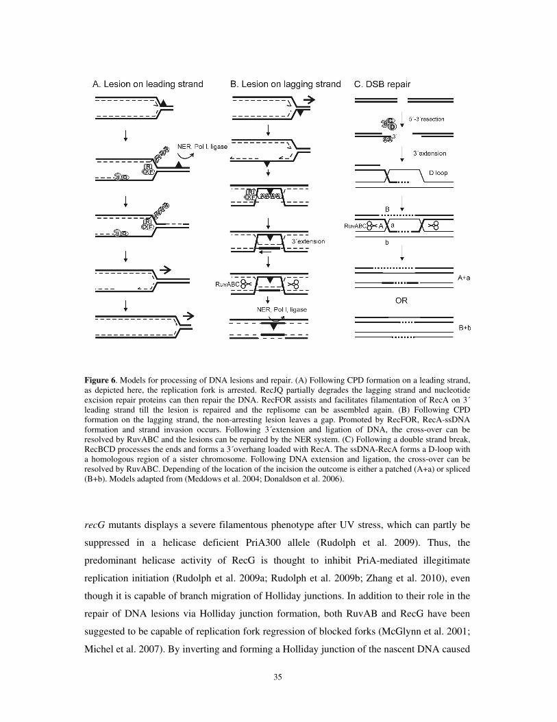
35
Figure 6. Models for processing of DNA lesions and repair. (A) Following CPD formation on a leading strand, as depicted here, the replication fork is arrested. RecJQ partially degrades the lagging strand and nucleotide excision repair proteins can then repair the DNA. RecFOR assists and facilitates filamentation of RecA on 3´ leading strand till the lesion is repaired and the replisome can be assembled again. (B) Following CPD formation on the lagging strand, the non-arresting lesion leaves a gap. Promoted by RecFOR, RecA-ssDNA formation and strand invasion occurs. Following 3´extension and ligation of DNA, the cross-over can be resolved by RuvABC and the lesions can be repaired by the NER system. (C) Following a double strand break, RecBCD processes the ends and forms a 3´overhang loaded with RecA. The ssDNA-RecA forms a D-loop with a homologous region of a sister chromosome. Following DNA extension and ligation, the cross-over can be resolved by RuvABC. Depending of the location of the incision the outcome is either a patched (A+a) or spliced (B+b). Models adapted from (Meddows et al. 2004; Donaldson et al. 2006).
recG mutants displays a severe filamentous phenotype after UV stress, which can partly be
suppressed in a helicase deficient PriA300 allele (Rudolph et al. 2009). Thus, the
predominant helicase activity of RecG is thought to inhibit PriA-mediated illegitimate
replication initiation (Rudolph et al. 2009a; Rudolph et al. 2009b; Zhang et al. 2010), even
though it is capable of branch migration of Holliday junctions. In addition to their role in the
repair of DNA lesions via Holliday junction formation, both RuvAB and RecG have been
suggested to be capable of replication fork regression of blocked forks (McGlynn et al. 2001;
Michel et al. 2007). By inverting and forming a Holliday junction of the nascent DNA caused
36
by a stalled replication fork, followed by processing by RuvABC or a RecBCD, the cell can
restore the fork via a PriA-dependent mechanism (Michel et al. 2007; Atkinson and McGlynn
2009). However, these mechanisms have been proposed to take place in various replication
mutants with increased replication fork collapse (Seigneur et al. 1998; Flores et al. 2001) and
might not occur under more common conditions, like UV irradiation of wild type cells
(Donaldson et al. 2004; Chow and Courcelle 2007).
8. Results and discussion
8.1. Paper I: Accumulation of the glycolytic intermediate fructose-6-p serves as a
positive regulatory signal to uspA transcription.
One of the goals of this work is to understand the regulation of stationary phase inducible
genes. To this end, we screened a transposon mutant library for mutants that altered
expression from our reporter construct PuspA-lacZ and found one mutant that showed higher
expression from PuspA. This mutant has a mutation in the gene encoding for the glycolytic
enzyme phosphoglucose isomerase (Pgi) and was shown to up-regulate the transcription of
uspA when grown on glucose. Pgi catalyzes the first enzymatic step in the glycolysis and
drive the reversible reaction glucose-6-p to fructose-6p, and is the only known enzyme doing
this reaction (Fig 7A). Cells inactivated in this enzymatic step accumulate high levels of
glucose-6-p when grown on glucose (Morita et al. 2003) and display a severe reduction in
growth rate compared to wild type cells (Hanson and Rose 1980; Kabir and Shimizu 2003).
pgi mutants grown on glucose are unable to feed glucose-6-phosphate into the glycolytic
pathway and must use the pentose phosphate pathway as the primary route for glucose
catabolism. However, the pentose phosphate pathway is incapable in converting the
intermediates fast enough to avoid accumulation of glucose-6-p and sole use of this pathway
also results in NADPH imbalance, both thought to affect growth rate of the cell (Canonaco et
al. 2001; Hua et al. 2003; Kabir and Shimizu 2003). In order to study the regulation of uspA
in a pgi mutant we first needed to find a carbon source which minimized the growth rate
effect since uspA is growth rate regulated (Nystrom and Neidhardt 1992; Tao et al. 1999).
Defined rich media with glycerol as carbon source was shown to fulfill our demands as a pgi
mutant displayed similar growth rate as wild type. A pgi mutant superinduces expression
from PuspA in this medium as it does in glucose medium, i.e., log phase expression is similar
to a wild type cell but the pgi mutant induces expression approximately two-fold more than
37
wild type upon entry to stationary phase (Fig 7B). We also observed that both normal
induction and superinduction of uspA occurred early in transition phase before the depletion
of carbon.
To understand how a pgi mutant modulates uspA transcription, we first examined known
regulators of uspA. Transcription of uspA is positively correlated with the levels of ppGpp
and strongly dependent on it (Kvint et al. 2000b). Carbon starvation stimulates synthesis of
ppGpp making this alarmone a strong candidate for the observed superinduction. However,
elevated transcription of uspA was still observed in a pgi mutant even in a ppGppO
background indicating that pgi regulation occurs independently of the stringent response.
Direct measurements of ppGpp verified this observation, and even showed that the levels are
lower in a pgi mutant compared to wild type. Thus, we concluded that the superinduction of
uspA does not depend on ppGpp in a pgi mutant. Likewise, we verified that superinduction
was independent of RecA/FtsK and FadR, both known to regulate uspA transcription
(Farewell et al. 1996; Diez et al. 2000). Thus, we concluded that the metabolic block in the
pgi mutant stimulates induction of uspA via a previously unknown mechanism.
A pgi mutant would be expected to have numerous alterations in the levels of glycolytic
intermediates and next we did a series of experiments aimed at testing whether we could
correlate uspA superinduction to any specific metabolite. A potential candidate for a
signaling molecule, UDP-glucose, was previously suggested to alter transcription from σS
dependent genes; strains with reduced levels, or deficient in synthesizing, of UPD-glucose
exhibits an increased level of σS in growing cells (Bohringer et al. 1995). Inactivation of
enzymes in the pathway for UDP-glucose synthesis, by single mutations in pgm, or galU,
which convert glucose-6-p to glucose-1-p and glucose-1-p to UDP-glucose respectively (see
Fig 7A) showed that the signal is unrelated to levels of UDP-glucose. By the same logic,
adding small amount of glucose (0.0004%) to a pgi mutant (increasing production of UDP-
glucose) did not alter the superinduction of PuspA, whereas this was previously shown to
repress σS dependent gene expression (Bohringer et al. 1995).
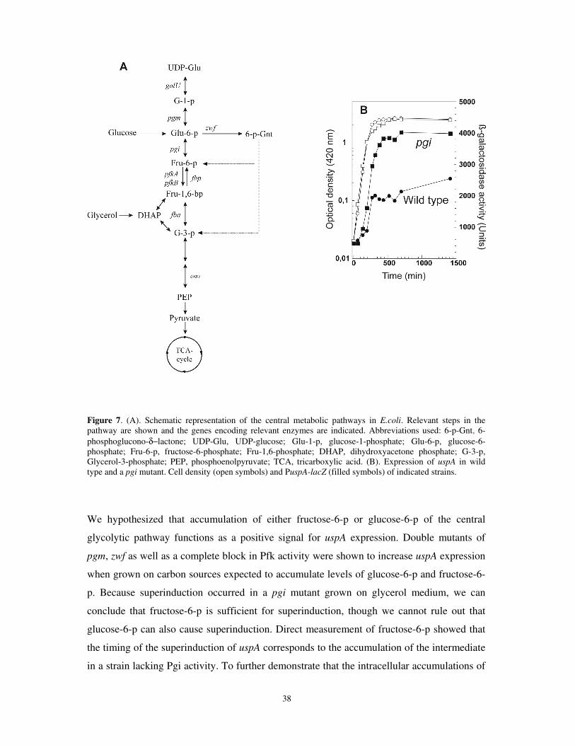
38
Figure 7. (A). Schematic representation of the central metabolic pathways in E.coli. Relevant steps in the pathway are shown and the genes encoding relevant enzymes are indicated. Abbreviations used: 6-p-Gnt, 6-phosphoglucono-δ−lactone; UDP-Glu, UDP-glucose; Glu-1-p, glucose-1-phosphate; Glu-6-p, glucose-6-phosphate; Fru-6-p, fructose-6-phosphate; Fru-1,6-phosphate; DHAP, dihydroxyacetone phosphate; G-3-p, Glycerol-3-phosphate; PEP, phosphoenolpyruvate; TCA, tricarboxylic acid. (B). Expression of uspA in wild type and a pgi mutant. Cell density (open symbols) and PuspA-lacZ (filled symbols) of indicated strains.
We hypothesized that accumulation of either fructose-6-p or glucose-6-p of the central
glycolytic pathway functions as a positive signal for uspA expression. Double mutants of
pgm, zwf as well as a complete block in Pfk activity were shown to increase uspA expression
when grown on carbon sources expected to accumulate levels of glucose-6-p and fructose-6-
p. Because superinduction occurred in a pgi mutant grown on glycerol medium, we can
conclude that fructose-6-p is sufficient for superinduction, though we cannot rule out that
glucose-6-p can also cause superinduction. Direct measurement of fructose-6-p showed that
the timing of the superinduction of uspA corresponds to the accumulation of the intermediate
in a strain lacking Pgi activity. To further demonstrate that the intracellular accumulations of
39
fructose-6-p in a pgi mutant stimulates transcription of uspA, we decided to ectopically lower
the intracellular levels of this intermediate. By overproducing the main enzyme converting
fructose-6-p into fructose-1,6-p, PfkA, in a pgi mutant the expression of uspA was suppressed
to the levels of wild type cells.
Finally, we also show that the effect of fructose-6-p is not specific to only uspA nor to
mutants in glycolysis. Besides uspA, three other σ70-dependent stationary phase inducible
usp´s exhibited superinduction in a pgi mutant. However, it should be noted that not every
usp gene responded this way: uspE did not. Further, indications suggest that in wild type
cells the intermediate plays a role in the normal induction of PuspA upon entry into stationary
phase, as overproduction of PfkA abolished normal induction in stationary phase. These
results indicate that fructose-6-p plays a signaling role in wild type cells upon entry to
stationary phase and that this mechanism may be widespread.
8.2. Paper II: The levels of fructose-6-phosphate regulate σσσσS-dependent
transcription upon entry to stationary phase in E. coli
We wished to expand our understanding of the regulation of stationary phase inducible genes
by fructose-6-P and chose to examine RpoS regulated genes since the regulation of this
regulon is well studied. We found, that similar to the σ70 dependent usp-genes, the σS
dependent genes uspB and katE were also shown to be superinduced in a pgi mutant. The
transcriptional up-regulation of those genes was, like uspA, shown to occur during entry of
stationary phase in a pgi mutant when grown of glycerol as a carbon source. Thus, we
hypothesized that those genes are positively regulated by accumulation of fructose-6-p.
Indeed, this was found to be correct. In all conditions tested when we expect accumulation of
fructose-6-p (and glucose-6-p in some cases) the uspB gene was shown to be superinduced.
Double mutations in zwf, pgm, but neither of these single mutants displayed superinduction
of uspB. So did the cells mutated in pfkAB when grown on glucose as carbon source. Finally,
titration of fructose-6-p, by overproduction of PfkA in a pgi background, reduced the
expression of uspB. Overexpression of PfkA in a wild type also could reduce uspB
expression in a wild type strain as well. These results strongly indicate that accumulation of
this intermediate do affect the transcription of σS-dependent genes, much like the positive
regulation of the σ70 regulated genes in paper I.
40
Since our model gene uspB is regulated by the alternative sigma factor σS (Farewell et al.
1998b), we decided to investigate if the transcriptional regulation of uspB was due to altered
levels of the σS protein. By using immuno-detection of the RpoS protein, we found that the
level of σS was elevated throughout growth in a pgi mutant. Intrigued by this result we
measured the levels of rpoS by different rpoS construct fused to lacZ. These fusions are
reported to reflect the different regulatory steps of rpoS. We found that the pgi mutant
displayed elevated expression of all the rpoS-lacZ fusions in transition and stationary phase.
Since the transcriptional fusion of PrpoS-lacZ was superinduced in a pgi mutant,
accumulation of fructose-6-p most likely stimulates transcription from PrpoS. This result was
confirmed by Northern blot where we found that the rpoS mRNA is elevated in a pgi mutant.
A more then two fold superinduction of rpoS is seen when cells enter transition phase. Thus
transcriptional activation of rpoS is positively regulated by accumulation of fructose-6-p.
8.2.1. Speculations Paper I and II
In paper I and II, we show that the general stress protein genes uspA and rpoS respond to the
levels of fructose-6-p. Increased intracellular levels positively regulate the transcription of
those genes, whereas diminished levels reduce the transcription. However, it is also possible
that glucose-6-p acts in a similar fashion; this could not be determined in our experiments. It
has been reported that both glucose-6-p and fructose-6-p, play a central role in the regulation
of the uptake system of glucose. The ptsG mRNA is specifically degraded when the capacity
of the cell to take up glucose exceeds the metabolic flow of central metabolic pathway
(Kimata et al. 2001; Morita et al. 2003), i.e., when accumulation of glucose-6-p or fructose-
6-p intermediates occurs. Contrary to these results, we observe an increased transcriptional
level of our genes studied, and unlike ptsG, our model genes are affected when cells are
grown on glyconeogenetic carbon sources (e.g., glycerol). Further, no degradation of uspA or
rpoS mRNA could be seen on Northern blots. Thus, this makes it unlikely that uspA and rpoS
are part of this same regulatory system. However, we cannot absolutely exclude a model
where a repressor of uspA and rpoS is under regulation of this system.
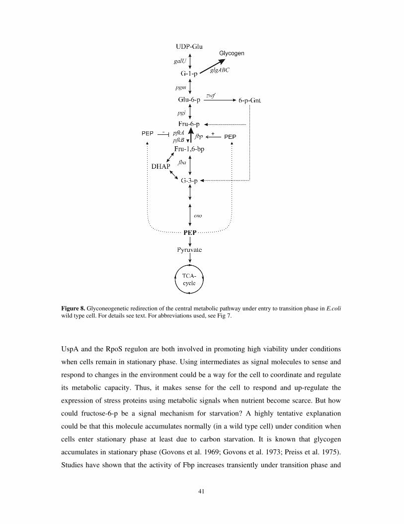
41
Figure 8. Glyconeogenetic redirection of the central metabolic pathway under entry to transition phase in E.coli wild type cell. For details see text. For abbreviations used, see Fig 7.
UspA and the RpoS regulon are both involved in promoting high viability under conditions
when cells remain in stationary phase. Using intermediates as signal molecules to sense and
respond to changes in the environment could be a way for the cell to coordinate and regulate
its metabolic capacity. Thus, it makes sense for the cell to respond and up-regulate the
expression of stress proteins using metabolic signals when nutrient become scarce. But how
could fructose-6-p be a signal mechanism for starvation? A highly tentative explanation
could be that this molecule accumulates normally (in a wild type cell) under condition when
cells enter stationary phase at least due to carbon starvation. It is known that glycogen
accumulates in stationary phase (Govons et al. 1969; Govons et al. 1973; Preiss et al. 1975).
Studies have shown that the activity of Fbp increases transiently under transition phase and
42
simultaneously PfkA activity deceases (Fraenkel and Horecker 1965; Sabnis et al. 1995),
thus a likely redirection of the central metabolism into glycogen production occurs (Fig 8).
This combined altered activity of Fbp and PfkA might then lead to a transient accumulation
of fructose-6-p. In support of this argument, we observed that overproduction of PfkA both in
pgi and wild type reduces the expression of uspA and rpoS. In addition, it is known that Fbp
is under positive, and PfkA under negative, allosteric regulation by PEP (Blangy et al. 1968;
Hines et al. 2007). In addition, interestingly, transcriptional profiling has demonstrated that
during growth on glucose in a pgi mutant, enolase (eno), the glycolytic enzyme converting 2-
phophoglycerate to PEP, is one of the most induced genes (Kabir and Shimizu 2003). This
might lead to an increase in PEP and also drive the accumulation of fructose-6-p under those
conditions (Fig 8). Under conditions when cells are growing on glyconeogenetic carbon
sources, like glycerol, the level of PEP is high, and possibly inhibits PfkA whereas Fbp is
stimulated (Blangy et al. 1968; Hines et al. 2007). Thus, fructose-6-p might be a highly
regulatable sensor for the cells metabolic status and thus a good choice as a regulatory signal
when cells experience a change in growth conditions such as are experienced upon entry to
stationary phase.
It should be noted that accumulation of the glycolytic intermediates, fructose-6-p and
glucose-6-p, is for unknown reasons toxic to the cell. However, we chose growth conditions
where we did not observe any growth retardation in the pgi mutant to avoid secondary
effects. We interpreted this lack of growth rate defect as the cells not accumulating high
enough levels of fructose-6-p to become toxic. Reducing sugars have the capacity to form
Maillard reactions and by nonenzymatic glycosylation of proteins produce advanced
glycosylation endproducts (AGE) and in vivo and in vitro studies have shown that reducing
sugars have an effect on mutation rates and results in insertions and deletions in the genome
(Lee and Cerami 1987; Lee and Cerami 1991; Levi and Werman 2001). Among the
phosphorylated sugars, fructose-6-p was shown to be most reactive (Levi and Werman 2001;
Levi and Werman 2003). Thus it is possible that observed elevated levels of uspA and RpoS
in the pgi mutant is a consequence of either oxidative protein or DNA damage, both
conditions know to cause transcriptional activation of uspA and stabilization of RpoS
((Nystrom and Neidhardt 1992; Diez et al. 2000; Merrikh et al. 2009a) and paper III),
caused by the reducing sugars. However, preliminary experiments could not detect any
alteration in carbonylation levels (a measure of oxidative damage) in crude extracts of total
protein of a pgi mutant following growth on glycerol (unpublished).
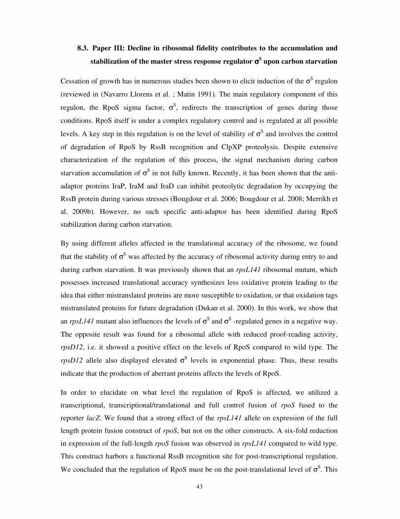
43
8.3. Paper III: Decline in ribosomal fidelity contributes to the accumulation and
stabilization of the master stress response regulator σσσσS upon carbon starvation
Cessation of growth has in numerous studies been shown to elicit induction of the σS regulon
(reviewed in (Navarro Llorens et al. ; Matin 1991). The main regulatory component of this
regulon, the RpoS sigma factor, σS, redirects the transcription of genes during those
conditions. RpoS itself is under a complex regulatory control and is regulated at all possible
levels. A key step in this regulation is on the level of stability of σS and involves the control
of degradation of RpoS by RssB recognition and ClpXP proteolysis. Despite extensive
characterization of the regulation of this process, the signal mechanism during carbon
starvation accumulation of σS in not fully known. Recently, it has been shown that the anti-
adaptor proteins IraP, IraM and IraD can inhibit proteolytic degradation by occupying the
RssB protein during various stresses (Bougdour et al. 2006; Bougdour et al. 2008; Merrikh et
al. 2009b). However, no such specific anti-adaptor has been identified during RpoS
stabilization during carbon starvation.
By using different alleles affected in the translational accuracy of the ribosome, we found
that the stability of σS was affected by the accuracy of ribosomal activity during entry to and
during carbon starvation. It was previously shown that an rpsL141 ribosomal mutant, which
possesses increased translational accuracy synthesizes less oxidative protein leading to the
idea that either mistranslated proteins are more susceptible to oxidation, or that oxidation tags
mistranslated proteins for future degradation (Dukan et al. 2000). In this work, we show that
an rpsL141 mutant also influences the levels of σS and σS -regulated genes in a negative way.
The opposite result was found for a ribosomal allele with reduced proof-reading activity,
rpsD12, i.e. it showed a positive effect on the levels of RpoS compared to wild type. The
rpsD12 allele also displayed elevated σS levels in exponential phase. Thus, these results
indicate that the production of aberrant proteins affects the levels of RpoS.
In order to elucidate on what level the regulation of RpoS is affected, we utilized a
transcriptional, transcriptional/translational and full control fusion of rpoS fused to the
reporter lacZ. We found that a strong effect of the rpsL141 allele on expression of the full
length protein fusion construct of rpoS, but not on the other constructs. A six-fold reduction
in expression of the full-length rpoS fusion was observed in rpsL141 compared to wild type.
This construct harbors a functional RssB recognition site for post-transcriptional regulation.
We concluded that the regulation of RpoS must be on the post-translational level of σS. This
44
was confirmed by measuring the stability of RpoS. The rpsL141 allele conferred an elevated
increase in RpoS degradation following carbon starvation.
Since the protease ClpXP is involved in the controlled degradation of RssB-bound RpoS as
well as the degradation of aberrant and ssrA-tagged proteins, we speculated that during
carbon starvation the ClpXP protease might be occupied in the degradation of aberrant
proteins. RpoS can thereby escape degradation by a titration mechanism, provided that some
of the components in this regulatory circuit are limiting. Experiments with chemically
induced aberrant protein synthesis supported this idea, since addition of canavanine (an
amino acid analog producing misfolded proteins) had a positive effect on RpoS stability.
Modulation of the levels of ClpX and ClpP also supported this theory. Inactivation of ClpX,
the chaperone facilitating aberrant protein degradation by ClpP, positively influenced the
stability of RpoS, both in wild type and rpsL141. Overproduction of the protease subunit
ClpP of ClpXP destabilized RpoS and was dependent on functional RssB for the degradation
of RpoS. In a similar manner, ectopic overproduction of ClpXP substrate, ssrA-tagged
proteins, was shown to stabilize RpoS in an otherwise wild type background. Taken together,
these results indicate that synthesis of aberrant proteins during carbon starvation may be a
way for the cell to modulate the stability of the RpoS protein and contributes to the
understanding of how RpoS is regulated under those conditions.
8.3.1. Speculations paper III
A fundamental property of the suggested titration model is that it is strongly dependent on
the assumption that heat shock proteins have a limited capacity to degrade their substrates
during the brief transition into stationary phase. Previous results have shown that the ClpX
and ClpP proteins are not induced in response to starvation for carbon when entering
stationary phase (Schweder et al. 1996; Mandel and Silhavy 2005). It has also been suggested
that RssB is a limiting factor in regulating the degradation of RpoS and a small increase in
transcription of rpoS could itself stabilize the RpoS protein (Pruteanu and Hengge-Aronis
2002; Hengge 2009). Thus, there are reasons to believe that any of those components could
be limiting for the cell. In addition, several studies on translation fidelity have observed a
reduced accuracy of protein synthesis when cells enter stationary phase (O'Farrell 1978;
Barak et al. 1996; Wenthzel et al. 1998; Ballesteros et al. 2001). Based on these observations,
the total load of aberrant proteins might then saturate the capacity of the protease machinery
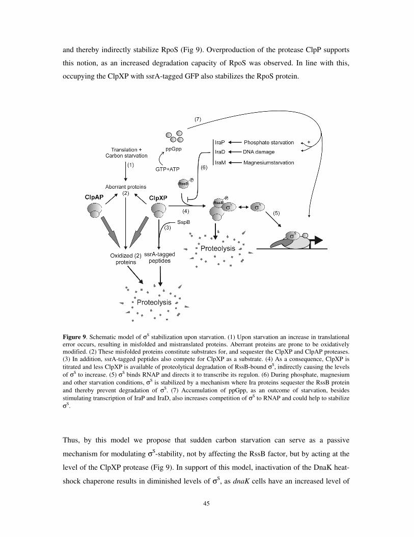
45
and thereby indirectly stabilize RpoS (Fig 9). Overproduction of the protease ClpP supports
this notion, as an increased degradation capacity of RpoS was observed. In line with this,
occupying the ClpXP with ssrA-tagged GFP also stabilizes the RpoS protein.
Figure 9. Schematic model of σS stabilization upon starvation. (1) Upon starvation an increase in translational error occurs, resulting in misfolded and mistranslated proteins. Aberrant proteins are prone to be oxidatively modified. (2) These misfolded proteins constitute substrates for, and sequester the ClpXP and ClpAP proteases. (3) In addition, ssrA-tagged peptides also compete for ClpXP as a substrate. (4) As a consequence, ClpXP is titrated and less ClpXP is available of proteolytical degradation of RssB-bound σS, indirectly causing the levels of σS to increase. (5) σS binds RNAP and directs it to transcribe its regulon. (6) During phosphate, magnesium and other starvation conditions, σS is stabilized by a mechanism where Ira proteins sequester the RssB protein and thereby prevent degradation of σS. (7) Accumulation of ppGpp, as an outcome of starvation, besides stimulating transcription of IraP and IraD, also increases competition of σS to RNAP and could help to stabilize σS.
Thus, by this model we propose that sudden carbon starvation can serve as a passive
mechanism for modulating σS-stability, not by affecting the RssB factor, but by acting at the
level of the ClpXP protease (Fig 9). In support of this model, inactivation of the DnaK heat-
shock chaperone results in diminished levels of σS, as dnaK cells have an increased level of
46
aberrant proteins which occupy the ClpXP protease (Rockabrand et al. 1998). Following the
stabilization of σS under these conditions, genes of its regulon like katE and dps are up-
regulated and are involved in resistance towards oxidative stress (Sak et al. 1989; Altuvia et
al. 1994). By up-regulating these genes before cells enter stationary phase, the organism can
prevent further oxidative attack in the cell. This might prevent cumulative oxidative protein
damage. Thereby, superfluous activities from the ATP-dependent ClpXP protease and
chaperones are prevented.
8.4. Paper IV: Characterization of a uspB mutant indicates that the phenotype is
linked to DNA damage
As previously mentioned, UspB is known to be involved in two processes. Firstly, it plays a
role in resistance to ethanol since a uspB mutant in stationary phase dies upon exposure to
ethanol. Secondly, UspB has a negative effect on survival after repeated freeze thawing. Both
these stresses affect the cytoplasmic membrane where UspB is anchored. Thus, we decided
to test if UspB was important for membrane structure and function. We compared survival of
a uspB mutant to an otherwise isogenic wild type strain during exposure to compounds
known to affect the membrane, but found no differences (unpublished). However, when we
exposed a uspB mutant to the DNA damaging agent mitomycin C a reduced survival was
found.
Further characterization following exposure of genotoxic agents showed that a uspB mutant
was sensitive to UV irradiation, ciprofloxacin and bleomycin. This phenotype indicates a
functional role of the protein in DNA repair or resistance towards such compounds. In order
to test if the phenotype observed following UV irradiation is specific to CPD formation in
DNA in the uspB mutant, we analyzed the cells for enzymatic photoreactivation and levels of
CPDs. A uspB mutant incubated in light showed an almost complete suppression of the
sensitive phenotype following UV irradiation. Thus, the enzyme DNA photolyase, which
utilizes visible light for reversion of the dimers (Friedberg 2006), is sufficient to reverse the
effect of UV on a uspB mutant and its’ sensitivity is specifically caused by CPDs and not
spurious effects like oxidative stress. A similar level of CPDs, measured with the TDH-2
antibodies, were found to be formed in uspB and wild type cells directly after UV treatment.
Following post-UV incubation both uspB and wild type repaired lesions with the same
kinetics indicating that NER is not affected by a uspB mutation.
47
The genotoxic compounds used for characterization of a uspB mutant all have the potential
effect of causing a single or double strand break as well obstruction of ongoing replication.
Bleomycin causes a direct break of DNA and the other agents cause breaks in an indirect
manner. We thus focused our attention on recombinational repair pathways and started by
comparing survival following UV irradiation of mutants of known repair pathways with uspB
and double mutants thereof. Inactivation of the RecF pathway genes recF and recR in
combination with a uspB mutation showed small additive effect with regards to survival. A
stronger additive effect was observed when the DNA processing proteins of the RecF
pathway, recJ an recQ was deleted, suggesting that UspB does not take part of this pathway.
However, mutants in the double strand break pathway, either recB or recC, in combination
with a uspB mutation showed no additive effect on sensitivity following UV irradiation. The
same results were found for mutants involved in Holliday junction resolvation, the ruvABC
genes, i.e. no additive phenotype in combination with a uspB mutant. These results
encouraged us to focus on these pathways. We found that a uspB mutant always showed the
same phenotype as a ruvC mutant following exposure to different genotoxic chemicals.
Moreover, a uspB mutant displayed a chronic two fold elevation of SOS response and
reduced efficiency in Hfr conjugation. Similar results have been reported for ruvC (Lloyd
1991; Asai and Kogoma 1994). Thus, several experiments indicate that UspB might work in
the same pathway as RuvC. Besides genetic evidence of that UspB is involved in the repair
of DNA lesion, we wished to demonstrate it in a more direct way by physical methods.
Results from the Courcelle laboratory has demonstrated that a ruvC mutant exhibits problems
in resolving Holliday junction intermediates following replication of UV irradiated cells
(Donaldson et al. 2006). By using the same two-dimensional agarose technique, we found
that a uspB mutant, much like ruvC, exhibits problems in resolving the intermediates. Cells
deleted for uspB were shown to display an increased level of unresolved intermediates
following UV irradiation and a delay in repair of those.
Finally, we showed that expression of an ectopic copy of ruvC could suppress the UV
sensitivity of a uspB mutant. The opposite was not true; uspB overexpression could not
complement a ruvC mutant. These results suggest that UspB functions upstream of RuvC.
Possibly UspB could either alter the transcription of RuvC or affect its function. By fusion
studies of ruvC and Q-PCR, we showed that UspB does not affect the transcriptional or
translational levels of ruvC. Thus, UspB might affect the function of RuvC at the protein
level. We also checked whether uspB itself is regulated by DNA damage and found that it is
48
not. During the course of studies we also found that a recD mutant suppressed the sensitive
phenotype of both a uspB and ruvC mutation. Further characterization demonstrated that it
was only those mutants which were suppressed by ∆recD; a double mutant of either ruvB or
ruvABC or a recG mutant in combination with recD was not suppressed.
8.5. Speculations paper IV
Our results in paper IV suggest that the UspB protein is required to maintain the integrity of
the DNA in both unstressed conditions as well as under conditions following DNA damage.
Several assays indicate that UspB has a functional role in DNA repair mechanisms when the
cellular pathways for recombinational crossover and the subsequent resolution of Holliday
junctions are active. The biochemical and genetic data further suggests that UspB is involved
in resolution of Holliday junctions. Still, it is hard to imagine how a membrane protein could
be directly involved in DNA repair mechanisms, especially since no single study has
demonstrated that the mechanism of DNA repair involves the cytoplasmic membrane. The
most plausible function for UspB is that it alters the activity of RuvC. However, several in
vitro assays demonstrates that the resolvase RuvC and its two associated proteins RuvA and
RuvB are sufficient for Holliday junction resolution (Eggleston et al. 1997; van Gool et al.
1998; Zerbib et al. 1998), making it hard to envisage a direct function of UspB for the
processes of branch migration and resolvation. Unlike ruvAB, the ruvC gene is not part of the
inducible SOS response regulon and is expressed at low levels ((Shinagawa et al. 1988;
Sharples and Lloyd 1991; West 1997) paper IV). Estimates are that there are about 50
dimers of RuvC per cell (West 1997), thus a small functional alteration of the protein could
influence its activity. One mechanism could be that UspB sequesters an unknown allosteric
repressor for RuvC and thereby alters its activity in vivo. Overexpression data of RuvC in a
uspB mutant background support this theory, since high concentrations of RuvC protein
suppresses the uspB phenotype. Alternatively, UspB could interact directly, or indirectly via
another protein, with RuvC and thereby facilitate its function.
uspB is part of the σS regulon and thus becomes induced when entering stationary phase.
However, the levels of UspB produced under exponential phase are crucial for proper
survival and repair of DNA damage under these conditions. The induction pattern, though,
indicates that under non-proliferating conditions, e.g. stationary phase, increased function of
UspB is desirable. Under these non-proliferative conditions about 20% of the genome is
49
broken down and resynthesized within a day (Tang et al. 1979) demonstrating that the
stationary phase not is a complete passive, dormant state on the DNA level. Several genes
involved in DNA repair and metabolism are under control of RpoS (Villarroya et al. 1998;
Saint-Ruf et al. 2004), and it has been shown that SOS response is up-regulated (Taddei et al.
1995) further indicating that DNA metabolism is active during stationary phase. However,
during stationary phase when the nutrients are scarce, repair of lethal lesions on DNA like
single stand and double strand breaks are fundamental for survival. UspB could under these
conditions, by its function in facilitating resolution by RuvC, thereby mitigate the stress.
During these conditions perfect restoration of the genome might not be crucial, exemplified
by the induction of the dinB gene, encoding the error prone DNA polymerase IV, during
stationary phase (Layton and Foster 2003). Translesion synthesis by Pol IV is capable of
bypassing lesions, exhibits reduced fidelity and thus causes increased mutagenesis in
stationary phase, at leased measured as F´lac reversions (Layton and Foster 2003; Galhardo
et al. 2009). During this mutagenic repair the processing of DNA is dependent on RecBCD
as well and RuvABC (Foster et al. 1996; Harris et al. 1996; He et al. 2006). UspB could
secure and increase the efficiency of recombinational repair of such mechanisms, especially
since the reversion is dependent on functional RuvABC.
9. Concluding remarks
To properly adapt to alterations in the environment is a fundamental process for the cell in
order to maintain viability and survive. In its natural habitat bacteria are seldom provided
with optimal nutritional conditions and non-differentiating bacteria like E.coli can respond to
nutrient deprivation by entering stationary phase. How cells sense and respond to the
impending nutrient deprivation in order to adapt to the new requirements is complex and
involves multiple signaling systems. Besides the effect of stringent control, by ppGpp, a
major effect on the molecular level is modulated by increasing levels of σS, the stationary
phase sigma factor. Both σS and ppGpp contribute to the redirection and expression of genes
involved in, for example, maintenance metabolism and general stress resistance, motility and
altered morphology (Lange and Hengge-Aronis 1991b; Lange and Hengge-Aronis 1991a;
Weber et al. 2005; Magnusson et al. 2007). However, the sensing-signal mechanism for
adapting to and the expression of genes during entry into stationary phase is less well
characterized. Sensing and responding to the intracellular metabolic intermediates, which

50
could reflect the extracellular environment contributes to the overall regulatory effect for
stationary phase genes, like the Universal stress protein genes, usp´s, and the σS regulon
(paper I and II). This mechanism appears to operate before the cells completely run out of
carbon and further work is need to determine how the cells sense this dwindling level of
carbon. In a similar sensing-signaling mechanism, the cell monitors the status of the
erroneous protein synthesis (due to e.g. nutrient deprivation) and is capable of transmitting
the information to σS (paper III). Together these two systems allow the cell to express
stationary phase genes and increase survival.
A novel function for UspB was discovered (paper IV). Observations indicate that the UspB
protein is involved in the late process of DNA damage repair during resolution of DNA
strand exchange. This DNA damage sensitive phenotype was observable in both stationary
phase conditions, where the UspB protein is highly expressed, and in exponential phase,
where UspB levels are low, indicating that the function of UspB is critical for proper DNA
repair under all growth conditions. The elevated levels of UspB and other stationary phase
inducible genes involved in DNA damage repair like DNA polymerase IV (dinB) (Layton
and Foster 2003), β subunit of DNA polymerase III (dnaN) (Villarroya et al. 1998) and Exo
III, abasic endonuclease and 3’ exonucleases (xth) (Sak et al. 1989) indicates that DNA
damage may be a common problem in stationary phase. Further studies will need to explore
and characterize the functional role of UspB under stationary phase conditions and determine
how UspB exerts its effect in the cell in more detail.
51
10. Acknowledgments
First of all I would like to express my gratitude towards my supervisor Anne, and to Thomas,
for an inspiring atmosphere and letting me know about the field of bacterial genetics. Your
vast knowledge about E.coli and molecular microbiology is truly impressive and stimulating.
I’m grateful for the opportunity to have had you, Anne, for my supervisor. From the very first
start, when attending your lectures, I was impressed the way you can explain difficult things
in an understandable way. A rare skill, but you possess it. Your patience in managing such a
stubborn creature as me is impressive. Your warm personality and capability to fix problems,
major or minor of mine, is also extraordinary.
Thomas, for bearing with me under all difficult times and never giving up faith. You kept me
running for the extra time I required to finish this thesis. Thank you so much, without your
generous help it would not have been possible.
I also want to thank present and past members of the group. Together you all make the lab
such great place to be. It’s a true joy spending time at this lab. My former office mate
Beidong, for your subtle humor and caring personality. And for taking command over the
drinking events, (it was you, right?). Laurence, my (almost exclusive) companion regarding
Facebook-matters. And for making me aware that growth curves is not the only way to study
bacterial genetics!! Bertil, for letting me defeat you and end up as the Bowling Champion of
the lab, at least once. Malin H, you need big improvements regarding the 9 ball-rules! Lisa
M, for (totally unaware?) introducing me to the world of MTB. This, by letting me haul a
container-sized trunk in the middle of the overcrowded streets of Manhattan. Micke, your
enthusiasm for everything and easy-going character is truly inspiring. Åsa, simply for you
being you (It’s positive!). Kristian, for your ability to make an ordinary conversation… more!
Antonio, I miss you and your great reluctant humor over here. Ana, for being such a warm
and fun person. I’ll miss the lunch conversations. Xin-Xin, for your excellent cooking and
nice warm personality. Veronica, your remarkable energy concerning everything. Junsheng,
your humble approach to everything. Manu, for making the lab a more interesting place.
David, the impressing elbow grease task in reporting about all bad movies seen during the
weekends. Alfredo, for ground breaking studies of how to replica plate!! Martin, the high
capacity man with a big portion of humor. Elin K-J, for being a positive and happy person
and the small chats in the corridor. Lisa L, for a friendly personality and nice pastries.
52
I also want to thank the numerous 10-90 point students, who have struggled and made the
initial studies on different projects.
Göteborgs Kex, for ruining my economy on cookies, but providing me with energy for
writing a thesis and “transfetter” enough for a life time.
My family, the Perssons; Mamma and Pappa for giving me the basic interest in biology from
the very beginning, like “Mulle-skolan”. Dad, I hope to see you more from now on. And my
brother Frank for the pioneering and pawing work for me in life!
LVG, for putting me on course again. Thank you all.
My second family, the Forsmarks, for the open and welcoming atmosphere.
Agneta for finding and sorting out the mysterious world of economy and invoice-matters.
Invaluable!
Administrational staff, especially Ann, Helena; you are always so helpful and professional.
And rarely did I leave your office without a smile.
Lars and Bruno for ctlr-alt-del related matter.
My Annabelle, for enriching my life much, much, much more. This, despite your cunning
habit of frequently opening (and almost finishing) the yummy box of cereals (which I’ve
been looking forward to for weeks) before finishing the plain, boring ones (which I
respectfully consume)! Your otherwise altruistic approach to humankind and to life, it rules.
53
11. References
Albertson, N.H. and Nystrom, T. 1994. Effects of starvation for exogenous carbon on functional
mRNA stability and rate of peptide chain elongation in Escherichia coli. FEMS Microbiol Lett
117(2): 181-187.
Altuvia, S., Almiron, M., Huisman, G., Kolter, R., and Storz, G. 1994. The dps promoter is activated by
OxyR during growth and by IHF and sigma S in stationary phase. Mol Microbiol 13(2): 265-
272.
Asai, T. and Kogoma, T. 1994. Roles of ruvA, ruvC and recG gene functions in normal and DNA
damage-inducible replication of the Escherichia coli chromosome. Genetics 137(4): 895-902.
Bai, H. and Lu, A.L. 2007. Physical and functional interactions between Escherichia coli MutY
glycosylase and mismatch repair protein MutS. J Bacteriol 189(3): 902-910.
Ballesteros, M., Fredriksson, A., Henriksson, J., and Nystrom, T. 2001. Bacterial senescence: protein
oxidation in non-proliferating cells is dictated by the accuracy of the ribosomes. EMBO J
20(18): 5280-5289.
Barak, Z., Gallant, J., Lindsley, D., Kwieciszewki, B., and Heidel, D. 1996. Enhanced ribosome
frameshifting in stationary phase cells. J Mol Biol 263(2): 140-148.
Barth, E., Gora, K.V., Gebendorfer, K.M., Settele, F., Jakob, U., and Winter, J. 2009. Interplay of
cellular cAMP levels, {sigma}S activity and oxidative stress resistance in Escherichia coli.
Microbiology 155(Pt 5): 1680-1689.
Battesti, A. and Bouveret, E. 2006. Acyl carrier protein/SpoT interaction, the switch linking SpoT-
dependent stress response to fatty acid metabolism. Mol Microbiol 62(4): 1048-1063.
Becker, G., Klauck, E., and Hengge-Aronis, R. 2000. The response regulator RssB, a recognition factor
for sigmaS proteolysis in Escherichia coli, can act like an anti-sigmaS factor. Mol Microbiol
35(3): 657-666.
Bettenbrock, K., Sauter, T., Jahreis, K., Kremling, A., Lengeler, J.W., and Gilles, E.D. 2007. Correlation
between growth rates, EIIACrr phosphorylation, and intracellular cyclic AMP levels in
Escherichia coli K-12. J Bacteriol 189(19): 6891-6900.
Biek, D.P. and Cohen, S.N. 1986. Identification and characterization of recD, a gene affecting plasmid
maintenance and recombination in Escherichia coli. J Bacteriol 167(2): 594-603.
Blangy, D., Buc, H., and Monod, J. 1968. Kinetics of the allosteric interactions of phosphofructokinase
from Escherichia coli. J Mol Biol 31(1): 13-35.
Bohringer, J., Fischer, D., Mosler, G., and Hengge-Aronis, R. 1995. UDP-glucose is a potential
intracellular signal molecule in the control of expression of sigma S and sigma S-dependent
genes in Escherichia coli. J Bacteriol 177(2): 413-422.
Bouche, S., Klauck, E., Fischer, D., Lucassen, M., Jung, K., and Hengge-Aronis, R. 1998. Regulation of
RssB-dependent proteolysis in Escherichia coli: a role for acetyl phosphate in a response
regulator-controlled process. Mol Microbiol 27(4): 787-795.
Bougdour, A., Cunning, C., Baptiste, P.J., Elliott, T., and Gottesman, S. 2008. Multiple pathways for
regulation of sigmaS (RpoS) stability in Escherichia coli via the action of multiple anti-
adaptors. Mol Microbiol 68(2): 298-313.
Bougdour, A. and Gottesman, S. 2007. ppGpp regulation of RpoS degradation via anti-adaptor
protein IraP. Proc Natl Acad Sci U S A 104(31): 12896-12901.
Bougdour, A., Wickner, S., and Gottesman, S. 2006. Modulating RssB activity: IraP, a novel regulator
of sigma(S) stability in Escherichia coli. Genes Dev 20(7): 884-897.
Bramhill, D. and Kornberg, A. 1988. A model for initiation at origins of DNA replication. Cell 54(7):
915-918.
Brown, L., Gentry, D., Elliott, T., and Cashel, M. 2002. DksA affects ppGpp induction of RpoS at a
translational level. J Bacteriol 184(16): 4455-4465.
54
Busby, S. and Ebright, R.H. 1999. Transcription activation by catabolite activator protein (CAP). J Mol
Biol 293(2): 199-213.
Canonaco, F., Hess, T.A., Heri, S., Wang, T., Szyperski, T., and Sauer, U. 2001. Metabolic flux response
to phosphoglucose isomerase knock-out in Escherichia coli and impact of overexpression of
the soluble transhydrogenase UdhA. FEMS Microbiology Letters 204(2): 247-252.
Cashel, M. 1969. The control of ribonucleic acid synthesis in Escherichia coli. IV. Relevance of
unusual phosphorylated compounds from amino acid-starved stringent strains. J Biol Chem
244(12): 3133-3141.
Cashel, M.G., D.R. Hernandes, V.J. Vinella D. 1996. The stringent response. in Escherichia and
Salmonella: Cellular and molecular biology (ed. F.C. Neidhardt), pp. 1458-1496. ASM Press.
Chiaramello, A.E. and Zyskind, J.W. 1990. Coupling of DNA replication to growth rate in Escherichia
coli: a possible role for guanosine tetraphosphate. J Bacteriol 172(4): 2013-2019.
Chow, K.H. and Courcelle, J. 2007. RecBCD and RecJ/RecQ initiate DNA degradation on distinct
substrates in UV-irradiated Escherichia coli. Radiat Res 168(4): 499-506.
Courcelle, C.T., Belle, J.J., and Courcelle, J. 2005. Nucleotide excision repair or polymerase V-
mediated lesion bypass can act to restore UV-arrested replication forks in Escherichia coli. J
Bacteriol 187(20): 6953-6961.
Courcelle, C.T., Chow, K.H., Casey, A., and Courcelle, J. 2006. Nascent DNA processing by RecJ favors
lesion repair over translesion synthesis at arrested replication forks in Escherichia coli. Proc
Natl Acad Sci U S A 103(24): 9154-9159.
Courcelle, J. and Hanawalt, P.C. 1999. RecQ and RecJ process blocked replication forks prior to the
resumption of replication in UV-irradiated Escherichia coli. Mol Gen Genet 262(3): 543-551.
Courcelle, J., Khodursky, A., Peter, B., Brown, P.O., and Hanawalt, P.C. 2001. Comparative gene
expression profiles following UV exposure in wild-type and SOS-deficient Escherichia coli.
Genetics 158(1): 41-64.
Dermic, D., Halupecki, E., Zahradka, D., and Petranovic, M. 2005. RecBCD enzyme overproduction
impairs DNA repair and homologous recombination in Escherichia coli. Res Microbiol 156(3):
304-311.
Diez, A., Gustavsson, N., and Nystrom, T. 2000. The universal stress protein A of Escherichia coli is
required for resistance to DNA damaging agents and is regulated by a RecA/FtsK-dependent
regulatory pathway. Mol Microbiol 36(6): 1494-1503.
Diez, A.A., Farewell, A., Nannmark, U., and Nystrom, T. 1997. A mutation in the ftsK gene of
Escherichia coli affects cell-cell separation, stationary-phase survival, stress adaptation, and
expression of the gene encoding the stress protein UspA. J Bacteriol 179(18): 5878-5883.
Dixon, D.A. and Kowalczykowski, S.C. 1991. Homologous pairing in vitro stimulated by the
recombination hotspot, Chi. Cell 66(2): 361-371.
-. 1995. Role of the Escherichia coli recombination hotspot, chi, in RecABCD-dependent homologous
pairing. J Biol Chem 270(27): 16360-16370.
Donaldson, J.R., Courcelle, C.T., and Courcelle, J. 2006. RuvABC is required to resolve holliday
junctions that accumulate following replication on damaged templates in Escherichia coli. J
Biol Chem 281(39): 28811-28821.
Dukan, S., Farewell, A., Ballesteros, M., Taddei, F., Radman, M., and Nystrom, T. 2000. Protein
oxidation in response to increased transcriptional or translational errors. Proc Natl Acad Sci
U S A 97(11): 5746-5749.
Eggleston, A.K., Mitchell, A.H., and West, S.C. 1997. In vitro reconstitution of the late steps of genetic
recombination in E. coli. Cell 89(4): 607-617.
Farewell, A., Diez, A.A., DiRusso, C.C., and Nystrom, T. 1996. Role of the Escherichia coli FadR
regulator in stasis survival and growth phase-dependent expression of the uspA, fad, and fab
genes. J Bacteriol 178(22): 6443-6450.
Farewell, A., Kvint, K., and Nystrom, T. 1998a. Negative regulation by RpoS: a case of sigma factor
competition. Mol Microbiol 29(4): 1039-1051.
55
-. 1998b. uspB, a new sigmaS-regulated gene in Escherichia coli which is required for stationary-
phase resistance to ethanol. J Bacteriol 180(23): 6140-6147.
Ferenci, T. 1996. Adaptation to life at micromolar nutrient levels: the regulation of Escherichia coli
glucose transport by endoinduction and cAMP. FEMS Microbiol Rev 18(4): 301-317.
Fic, E., Bonarek, P., Gorecki, A., Kedracka-Krok, S., Mikolajczak, J., Polit, A., Tworzydlo, M.,
Dziedzicka-Wasylewska, M., and Wasylewski, Z. 2009. cAMP receptor protein from
escherichia coli as a model of signal transduction in proteins--a review. J Mol Microbiol
Biotechnol 17(1): 1-11.
Flores, M.J., Bierne, H., Ehrlich, S.D., and Michel, B. 2001. Impairment of lagging strand synthesis
triggers the formation of a RuvABC substrate at replication forks. EMBO J 20(3): 619-629.
Foster, P.L., Trimarchi, J.M., and Maurer, R.A. 1996. Two enzymes, both of which process
recombination intermediates, have opposite effects on adaptive mutation in Escherichia
coli. Genetics 142(1): 25-37.
Fraenkel, D.G. 1996. Glycolysis. in Escherichia coli and Salmonella: Cellular and Molecular Biology
(ed. F.C. Neidhardt, Ingraham, J.L., Low, K.b., Magansanik, B., Schaechter, M., and Umberger,
H.E.), pp. 189-198. American Society for Microbiology Press., Washington,D.C.
Fraenkel, D.G. and Horecker, B.L. 1965. Fructose-1,6-Diphosphatase and Acid Hexose Phosphatase of
Escherichia coli. J Bacteriol 90(4): 837-842.
Freestone, P., Nystrom, T., Trinei, M., and Norris, V. 1997. The universal stress protein, UspA, of
Escherichia coli is phosphorylated in response to stasis. J Mol Biol 274(3): 318-324.
Fridovich, I. 1978. The biology of oxygen radicals. Science 201(4359): 875-880.
Friedberg, E. 2006. DNA repair and mutagenesis. ASM press.
Galhardo, R.S., Do, R., Yamada, M., Friedberg, E.C., Hastings, P.J., Nohmi, T., and Rosenberg, S.M.
2009. DinB upregulation is the sole role of the SOS response in stress-induced mutagenesis
in Escherichia coli. Genetics 182(1): 55-68.
Gentry, D.R., Hernandez, V.J., Nguyen, L.H., Jensen, D.B., and Cashel, M. 1993. Synthesis of the
stationary-phase sigma factor sigma s is positively regulated by ppGpp. J Bacteriol 175(24):
7982-7989.
Gottesman, S. 1996. Proteases and their targets in Escherichia coli. Annu Rev Genet 30: 465-506.
Gottesman, S., Roche, E., Zhou, Y., and Sauer, R.T. 1998. The ClpXP and ClpAP proteases degrade
proteins with carboxy-terminal peptide tails added by the SsrA-tagging system. Genes Dev
12(9): 1338-1347.
Govons, S., Gentner, N., Greenberg, E., and Preiss, J. 1973. Biosynthesis of bacterial glycogen. XI.
Kinetic characterization of an altered adenosine diphosphate-glucose synthase from a
"glycogen-excess" mutant of Escherichia coli B. J Biol Chem 248(5): 1731-1740.
Govons, S., Vinopal, R., Ingraham, J., and Preiss, J. 1969. Isolation of mutants of Escherichia coli B
altered in their ability to synthesize glycogen. J Bacteriol 97(2): 970-972.
Groat, R.G., Schultz, J.E., Zychlinsky, E., Bockman, A., and Matin, A. 1986. Starvation proteins in
Escherichia coli: kinetics of synthesis and role in starvation survival. J Bacteriol 168(2): 486-
493.
Gruber, T.M. and Gross, C.A. 2003. Multiple sigma subunits and the partitioning of bacterial
transcription space. Annu Rev Microbiol 57: 441-466.
Gummesson, B., Magnusson, L.U., Lovmar, M., Kvint, K., Persson, O., Ballesteros, M., Farewell, A.,
and Nystrom, T. 2009. Increased RNA polymerase availability directs resources towards
growth at the expense of maintenance. EMBO J.
Gustavsson, N., Diez, A., and Nystrom, T. 2002. The universal stress protein paralogues of Escherichia
coli are co-ordinately regulated and co-operate in the defence against DNA damage. Mol
Microbiol 43(1): 107-117.
Hanawalt, P.C. 1993. "Close-fitting sleeves"--recognition of structural defects in duplex DNA. Mutat
Res 289(1): 7-15.
Hanawalt, P.C. and Haynes, R.H. 1965. Repair Replication of DNA in Bacteria: Irrelevance of Chemical
Nature of Base Defect. Biochem Biophys Res Commun 19: 462-467.
56
Handa, N., Morimatsu, K., Lovett, S.T., and Kowalczykowski, S.C. 2009. Reconstitution of initial steps
of dsDNA break repair by the RecF pathway of E. coli. Genes Dev 23(10): 1234-1245.
Hanson, R.L. and Rose, C. 1980. Effects of an insertion mutation in a locus affecting pyridine
nucleotide transhydrogenase (pnt::Tn5) on the growth of Escherichia coli. J Bacteriol 141(1):
401-404.
Harris, R.S., Ross, K.J., and Rosenberg, S.M. 1996. Opposing roles of the holliday junction processing
systems of Escherichia coli in recombination-dependent adaptive mutation. Genetics 142(3):
681-691.
Hartl, F.U. 1996. Molecular chaperones in cellular protein folding. Nature 381(6583): 571-579.
He, A.S., Rohatgi, P.R., Hersh, M.N., and Rosenberg, S.M. 2006. Roles of E. coli double-strand-break-
repair proteins in stress-induced mutation. DNA Repair (Amst) 5(2): 258-273.
Helmstetter, C. 1996. Timing of synthetic activities in the cell cycle. . ASM press.
Hengge-Aronis, R. 2002. Signal transduction and regulatory mechanisms involved in control of the
sigma(S) (RpoS) subunit of RNA polymerase. Microbiol Mol Biol Rev 66(3): 373-395, table of
contents.
Hengge, R. 2009. Proteolysis of sigmaS (RpoS) and the general stress response in Escherichia coli. Res
Microbiol 160(9): 667-676.
Hines, J.K., Fromm, H.J., and Honzatko, R.B. 2007. Structures of activated fructose-1,6-
bisphosphatase from Escherichia coli. Coordinate regulation of bacterial metabolism and the
conservation of the R-state. J Biol Chem 282(16): 11696-11704.
Hogema, B.M., Arents, J.C., Bader, R., Eijkemans, K., Inada, T., Aiba, H., and Postma, P.W. 1998a.
Inducer exclusion by glucose 6-phosphate in Escherichia coli. Mol Microbiol 28(4): 755-765.
Hogema, B.M., Arents, J.C., Bader, R., Eijkemans, K., Yoshida, H., Takahashi, H., Aiba, H., and Postma,
P.W. 1998b. Inducer exclusion in Escherichia coli by non-PTS substrates: the role of the PEP
to pyruvate ratio in determining the phosphorylation state of enzyme IIAGlc. Mol Microbiol
30(3): 487-498.
Hua, Q., Yang, C., Baba, T., Mori, H., and Shimizu, K. 2003. Responses of the central metabolism in
Escherichia coli to phosphoglucose isomerase and glucose-6-phosphate dehydrogenase
knockouts. J Bacteriol 185(24): 7053-7067.
Imamura, R., Yamanaka, K., Ogura, T., Hiraga, S., Fujita, N., Ishihama, A., and Niki, H. 1996.
Identification of the cpdA gene encoding cyclic 3',5'-adenosine monophosphate
phosphodiesterase in Escherichia coli. J Biol Chem 271(41): 25423-25429.
Ishioka, K., Fukuoh, A., Iwasaki, H., Nakata, A., and Shinagawa, H. 1998. Abortive recombination in
Escherichia coli ruv mutants blocks chromosome partitioning. Genes Cells 3(4): 209-220.
Iuchi, S. and Weiner, L. 1996. Cellular and molecular physiology of Escherichia coli in the adaptation
to aerobic environments. J Biochem 120(6): 1055-1063.
Jiang, M., Sullivan, S.M., Wout, P.K., and Maddock, J.R. 2007. G-protein control of the ribosome-
associated stress response protein SpoT. J Bacteriol 189(17): 6140-6147.
Jishage, M., Kvint, K., Shingler, V., and Nystrom, T. 2002. Regulation of sigma factor competition by
the alarmone ppGpp. Genes Dev 16(10): 1260-1270.
Jores, L. and Wagner, R. 2003. Essential steps in the ppGpp-dependent regulation of bacterial
ribosomal RNA promoters can be explained by substrate competition. J Biol Chem 278(19):
16834-16843.
Kabir, M.M. and Shimizu, K. 2003. Gene expression patterns for metabolic pathway in pgi knockout
Escherichia coli with and without phb genes based on RT-PCR. J Biotechnol 105(1-2): 11-31.
Keiler, K.C., Waller, P.R., and Sauer, R.T. 1996. Role of a peptide tagging system in degradation of
proteins synthesized from damaged messenger RNA. Science 271(5251): 990-993.
Kimata, K., Tanaka, Y., Inada, T., and Aiba, H. 2001. Expression of the glucose transporter gene, ptsG,
is regulated at the mRNA degradation step in response to glycolytic flux in Escherichia coli.
EMBO J 20(13): 3587-3595.
Kolter, R., Siegele, D.A., and Tormo, A. 1993. The stationary phase of the bacterial life cycle. Annu
Rev Microbiol 47: 855-874.
57
Korangy, F. and Julin, D.A. 1994. Efficiency of ATP hydrolysis and DNA unwinding by the RecBC
enzyme from Escherichia coli. Biochemistry 33(32): 9552-9560.
Kristensen, H.-H., Valentin-Hansen, P., and Sogaard-Andersen, L. 1997. Design of CytR regulated,
cAMP-CRP dependent class II promoters in Escherichia coli: RNA polymerase-promoter
interactions modulate the efficiency of CytR repression. Journal of Molecular Biology 266(5):
866-876.
Kuzminov, A. 1999. Recombinational repair of DNA damage in Escherichia coli and bacteriophage
lambda. Microbiol Mol Biol Rev 63(4): 751-813, table of contents.
Kvint, K., Farewell, A., and Nystrom, T. 2000a. RpoS-dependent promoters require guanosine
tetraphosphate for induction even in the presence of high levels of sigma(s). J Biol Chem
275(20): 14795-14798.
Kvint, K., Hosbond, C., Farewell, A., Nybroe, O., and Nystrom, T. 2000b. Emergency derepression:
stringency allows RNA polymerase to override negative control by an active repressor. Mol
Microbiol 35(2): 435-443.
Kvint, K., Nachin, L., Diez, A., and Nystrom, T. 2003. The bacterial universal stress protein: function
and regulation. Curr Opin Microbiol 6(2): 140-145.
Lacour, S. and Landini, P. 2004. SigmaS-dependent gene expression at the onset of stationary phase
in Escherichia coli: function of sigmaS-dependent genes and identification of their promoter
sequences. J Bacteriol 186(21): 7186-7195.
Lange, R., Fischer, D., and Hengge-Aronis, R. 1995. Identification of transcriptional start sites and the
role of ppGpp in the expression of rpoS, the structural gene for the sigma S subunit of RNA
polymerase in Escherichia coli. J Bacteriol 177(16): 4676-4680.
Lange, R. and Hengge-Aronis, R. 1991a. Growth phase-regulated expression of bolA and morphology
of stationary-phase Escherichia coli cells are controlled by the novel sigma factor sigma S. J
Bacteriol 173(14): 4474-4481.
-. 1991b. Identification of a central regulator of stationary-phase gene expression in Escherichia coli.
Mol Microbiol 5(1): 49-59.
Layton, J.C. and Foster, P.L. 2003. Error-prone DNA polymerase IV is controlled by the stress-
response sigma factor, RpoS, in Escherichia coli. Mol Microbiol 50(2): 549-561.
Lederberg, J. 2004. E.coli K-12. Microbiology today 31.
Lin , E.C.C. 1996. Dissimilatory Pathways for Sugars, Polyols, and Carboxylates. in Escherichia coli and
Salmonella: Cellular and Molecular Biology (ed. F.C. Neidhardt, Ingraham, J.L., Low, K.b.,
Magansanik, B., Schaechter, M., and Umberger, H.E.), pp. 307-326. American Society for
Microbiology Press., Washington,D.C.
Llorens, J.M.N., Tormo, A., and Martínez-García, E. Stationary phase in gram-negative bacteria. FEMS
Microbiology Reviews 9999(9999).
Lloyd, R.G. 1991. Conjugational recombination in resolvase-deficient ruvC mutants of Escherichia coli
K-12 depends on recG. J Bacteriol 173(17): 5414-5418.
Lloyd, R.G., Benson, F.E., and Shurvinton, C.E. 1984. Effect of ruv mutations on recombination and
DNA repair in Escherichia coli K12. Mol Gen Genet 194(1-2): 303-309.
Lloyd, R.G., Buckman, C., and Benson, F.E. 1987. Genetic analysis of conjugational recombination in
Escherichia coli K12 strains deficient in RecBCD enzyme. J Gen Microbiol 133(9): 2531-2538.
Lloyd, R.G., Porton, M.C., and Buckman, C. 1988. Effect of recF, recJ, recN, recO and ruv mutations
on ultraviolet survival and genetic recombination in a recD strain of Escherichia coli K12. Mol
Gen Genet 212(2): 317-324.
Lobell, R.B. and Schleif, R.F. 1991. AraC-DNA looping: orientation and distance-dependent loop
breaking by the cyclic AMP receptor protein. J Mol Biol 218(1): 45-54.
Lobner-Olesen, A., Skarstad, K., Hansen, F.G., von Meyenburg, K., and Boye, E. 1989. The DnaA
protein determines the initiation mass of Escherichia coli K-12. Cell 57(5): 881-889.
Lovett, S.T. 2006. Replication arrest-stimulated recombination: Dependence on the RecA paralog,
RadA/Sms and translesion polymerase, DinB. DNA Repair (Amst) 5(12): 1421-1427.
58
Lovett, S.T., Luisi-DeLuca, C., and Kolodner, R.D. 1988. The genetic dependence of recombination in
recD mutants of Escherichia coli. Genetics 120(1): 37-45.
Magnusson, L.U., Farewell, A., and Nystrom, T. 2005. ppGpp: a global regulator in Escherichia coli.
Trends Microbiol 13(5): 236-242.
Magnusson, L.U., Gummesson, B., Joksimovic, P., Farewell, A., and Nystrom, T. 2007. Identical,
independent, and opposing roles of ppGpp and DksA in Escherichia coli. J Bacteriol 189(14):
5193-5202.
Magnusson, L.U., Nystrom, T., and Farewell, A. 2003. Underproduction of sigma 70 mimics a
stringent response. A proteome approach. J Biol Chem 278(2): 968-973.
Maisonneuve, E., Ducret, A., Khoueiry, P., Lignon, S., Longhi, S., Talla, E., and Dukan, S. 2009. Rules
governing selective protein carbonylation. PLoS One 4(10): e7269.
Mandel, M.J. and Silhavy, T.J. 2005. Starvation for different nutrients in Escherichia coli results in
differential modulation of RpoS levels and stability. J Bacteriol 187(2): 434-442.
Matin, A. 1991. The molecular basis of carbon-starvation-induced general resistance in Escherichia
coli. Mol Microbiol 5(1): 3-10.
Matin, A., Auger, E.A., Blum, P.H., and Schultz, J.E. 1989. Genetic basis of starvation survival in
nondifferentiating bacteria. Annu Rev Microbiol 43: 293-316.
Maurizi, M.R. 1992. Proteases and protein degradation in Escherichia coli. Experientia 48(2): 178-
201.
McGlynn, P. and Lloyd, R.G. 2000. Modulation of RNA polymerase by (p)ppGpp reveals a RecG-
dependent mechanism for replication fork progression. Cell 101(1): 35-45.
-. 2002. Genome stability and the processing of damaged replication forks by RecG. Trends Genet
18(8): 413-419.
Meddows, T.R., Savory, A.P., and Lloyd, R.G. 2004. RecG helicase promotes DNA double-strand break
repair. Mol Microbiol 52(1): 119-132.
Merrikh, H., Ferrazzoli, A.E., Bougdour, A., Olivier-Mason, A., and Lovett, S.T. 2009a. A DNA damage
response in Escherichia coli involving the alternative sigma factor, RpoS. Proc Natl Acad Sci U
S A 106(2): 611-616.
Merrikh, H., Ferrazzoli, A.E., and Lovett, S.T. 2009b. Growth phase and (p)ppGpp control of IraD, a
regulator of RpoS stability, in Escherichia coli. J Bacteriol.
Mitchell, D.L., Jen, J., and Cleaver, J.E. 1992. Sequence specificity of cyclobutane pyrimidine dimers
in DNA treated with solar (ultraviolet B) radiation. Nucleic Acids Res 20(2): 225-229.
Mitchell, D.L. and Nairn, R.S. 1989. THE BIOLOGY OF THE (6-4) PHOTOPRODUCT. Photochemistry and
Photobiology 49(6): 805-819.
Morita, T., El-Kazzaz, W., Tanaka, Y., Inada, T., and Aiba, H. 2003. Accumulation of glucose 6-
phosphate or fructose 6-phosphate is responsible for destabilization of glucose transporter
mRNA in Escherichia coli. J Biol Chem 278(18): 15608-15614.
Murray, K.D. and Bremer, H. 1996. Control of spoT-dependent ppGpp synthesis and degradation in
Escherichia coli. J Mol Biol 259(1): 41-57.
Nachin, L., Brive, L., Persson, K.C., Svensson, P., and Nystrom, T. 2008. Heterodimer formation within
universal stress protein classes revealed by an in silico and experimental approach. J Mol Biol
380(2): 340-350.
Nachin, L., Nannmark, U., and Nystrom, T. 2005. Differential roles of the universal stress proteins of
Escherichia coli in oxidative stress resistance, adhesion, and motility. J Bacteriol 187(18):
6265-6272.
Nanchen, A., Schicker, A., Revelles, O., and Sauer, U. 2008. Cyclic AMP-dependent catabolite
repression is the dominant control mechanism of metabolic fluxes under glucose limitation
in Escherichia coli. J Bacteriol 190(7): 2323-2330.
Navarro Llorens, J.M., Tormo, A., and Martinez-Garcia, E. Stationary phase in gram-negative
bacteria. FEMS Microbiol Rev.
Neidhardt, F.C. 1996. Escherichia coli and Salmonella; cellular and molecular biology. ASM,
Washington DC.
59
Nystrom, T., Flardh, K., and Kjelleberg, S. 1990. Responses to multiple-nutrient starvation in marine
Vibrio sp. strain CCUG 15956. J Bacteriol 172(12): 7085-7097.
Nystrom, T. and Neidhardt, F.C. 1992. Cloning, mapping and nucleotide sequencing of a gene
encoding a universal stress protein in Escherichia coli. Mol Microbiol 6(21): 3187-3198.
-. 1993. Isolation and properties of a mutant of Escherichia coli with an insertional inactivation of the
uspA gene, which encodes a universal stress protein. J Bacteriol 175(13): 3949-3956.
-. 1994. Expression and role of the universal stress protein, UspA, of Escherichia coli during growth
arrest. Mol Microbiol 11(3): 537-544.
-. 1996. Effects of overproducing the universal stress protein, UspA, in Escherichia coli K-12. J
Bacteriol 178(3): 927-930.
O'Farrell, P.H. 1978. The suppression of defective translation by ppGpp and its role in the stringent
response. Cell 14(3): 545-557.
Parsons, C.A. and West, S.C. 1993. Formation of a RuvAB-Holliday junction complex in vitro. J Mol
Biol 232(2): 397-405.
Persky, N.S., Ferullo, D.J., Cooper, D.L., Moore, H.R., and Lovett, S.T. 2009. The ObgE/CgtA GTPase
influences the stringent response to amino acid starvation in Escherichia coli. Mol Microbiol
73(2): 253-266.
Peterson, C.N., Ruiz, N., and Silhavy, T.J. 2004. RpoS proteolysis is regulated by a mechanism that
does not require the SprE (RssB) response regulator phosphorylation site. J Bacteriol
186(21): 7403-7410.
Pham, P., Seitz, E.M., Saveliev, S., Shen, X., Woodgate, R., Cox, M.M., and Goodman, M.F. 2002. Two
distinct modes of RecA action are required for DNA polymerase V-catalyzed translesion
synthesis. Proc Natl Acad Sci U S A 99(17): 11061-11066.
Postma, P.W. and Lengeler, J.W. 1985. Phosphoenolpyruvate:carbohydrate phosphotransferase
system of bacteria. Microbiol Rev 49(3): 232-269.
Postma, P.W., Lengeler, J.W., and Jacobson, G.R. 1993. Phosphoenolpyruvate:carbohydrate
phosphotransferase systems of bacteria. Microbiol Rev 57(3): 543-594.
Potrykus, K. and Cashel, M. 2008. (p)ppGpp: still magical? Annu Rev Microbiol 62: 35-51.
Preiss, J., Greenberg, E., and Sabraw, A. 1975. Biosynthesis of bacterial glycogen. Kinetic studies of a
glucose-1-phosphate adenylyltransferase (EC 2.7.7.27) from a glycogen-deficient mutant of
Escherichia coli B. J Biol Chem 250(19): 7631-7638.
Pruteanu, M. and Hengge-Aronis, R. 2002. The cellular level of the recognition factor RssB is rate-
limiting for sigmaS proteolysis: implications for RssB regulation and signal transduction in
sigmaS turnover in Escherichia coli. Mol Microbiol 45(6): 1701-1713.
Rahman, M., Hasan, M.R., Oba, T., and Shimizu, K. 2006. Effect of rpoS gene knockout on the
metabolism of Escherichia coli during exponential growth phase and early stationary phase
based on gene expressions, enzyme activities and intracellular metabolite concentrations.
Biotechnol Bioeng 94(3): 585-595.
Raskin, D.M., Judson, N., and Mekalanos, J.J. 2007. Regulation of the stringent response is the
essential function of the conserved bacterial G protein CgtA in Vibrio cholerae. Proc Natl
Acad Sci U S A 104(11): 4636-4641.
Reeve, C.A., Amy, P.S., and Matin, A. 1984. Role of protein synthesis in the survival of carbon-starved
Escherichia coli K-12. J Bacteriol 160(3): 1041-1046.
Richter, D. 1980. Uncharged tRNA inhibits guanosine 3',5'-bis (diphosphate) 3'-
pyrophosphohydrolase [ppGppase], the spoT gene product, from Escherichia coli. Mol Gen
Genet 178(2): 325-327.
Rockabrand, D., Livers, K., Austin, T., Kaiser, R., Jensen, D., Burgess, R., and Blum, P. 1998. Roles of
DnaK and RpoS in Starvation-Induced Thermotolerance of Escherichia coli. J Bacteriol 180(4):
846-854.
Romeo, T. 1998. Global regulation by the small RNA-binding protein CsrA and the non-coding RNA
molecule CsrB. Mol Microbiol 29(6): 1321-1330.
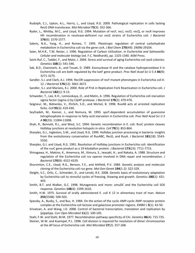
60
Rudolph, C.J., Upton, A.L., Harris, L., and Lloyd, R.G. 2009. Pathological replication in cells lacking
RecG DNA translocase. Mol Microbiol 73(3): 352-366.
Ryder, L., Whitby, M.C., and Lloyd, R.G. 1994. Mutation of recF, recJ, recO, recQ, or recR improves
Hfr recombination in resolvase-deficient ruv recG strains of Escherichia coli. J Bacteriol
176(6): 1570-1577.
Sabnis, N.A., Yang, H., and Romeo, T. 1995. Pleiotropic regulation of central carbohydrate
metabolism in Escherichia coli via the gene csrA. J Biol Chem 270(49): 29096-29104.
Saier, M.H.R., T.M. Reizer, J. 1996. Regulation of Carbon Utilization. in Escherichia and Salmonella:
Cellular and molecular biology (ed. F.C. Neidhardt), pp. 1325-1340. ASM Press.
Saint-Ruf, C., Taddei, F., and Matic, I. 2004. Stress and survival of aging Escherichia coli rpoS colonies.
Genetics 168(1): 541-546.
Sak, B.D., Eisenstark, A., and Touati, D. 1989. Exonuclease III and the catalase hydroperoxidase II in
Escherichia coli are both regulated by the katF gene product. Proc Natl Acad Sci U S A 86(9):
3271-3275.
Sandler, S.J. and Clark, A.J. 1994. RecOR suppression of recF mutant phenotypes in Escherichia coli K-
12. J Bacteriol 176(12): 3661-3672.
Sandler, S.J. and Marians, K.J. 2000. Role of PriA in Replication Fork Reactivation in Escherichia coli. J
Bacteriol 182(1): 9-13.
Schweder, T., Lee, K.H., Lomovskaya, O., and Matin, A. 1996. Regulation of Escherichia coli starvation
sigma factor (sigma s) by ClpXP protease. J Bacteriol 178(2): 470-476.
Seigneur, M., Bidnenko, V., Ehrlich, S.D., and Michel, B. 1998. RuvAB acts at arrested replication
forks. Cell 95(3): 419-430.
Seyfzadeh, M., Keener, J., and Nomura, M. 1993. spoT-dependent accumulation of guanosine
tetraphosphate in response to fatty acid starvation in Escherichia coli. Proc Natl Acad Sci U S
A 90(23): 11004-11008.
Shah, R., Bennett, R.J., and West, S.C. 1994. Genetic recombination in E. coli: RuvC protein cleaves
Holliday junctions at resolution hotspots in vitro. Cell 79(5): 853-864.
Sharples, G.J., Ingleston, S.M., and Lloyd, R.G. 1999. Holliday junction processing in bacteria: insights
from the evolutionary conservation of RuvABC, RecG, and RusA. J Bacteriol 181(18): 5543-
5550.
Sharples, G.J. and Lloyd, R.G. 1991. Resolution of Holliday junctions in Escherichia coli: identification
of the ruvC gene product as a 19-kilodalton protein. J Bacteriol 173(23): 7711-7715.
Shinagawa, H., Makino, K., Amemura, M., Kimura, S., Iwasaki, H., and Nakata, A. 1988. Structure and
regulation of the Escherichia coli ruv operon involved in DNA repair and recombination. J
Bacteriol 170(9): 4322-4329.
Shurvinton, C.E., Lloyd, R.G., Benson, F.E., and Attfield, P.V. 1984. Genetic analysis and molecular
cloning of the Escherichia coli ruv gene. Mol Gen Genet 194(1-2): 322-329.
Sleight, S.C., Orlic, C., Schneider, D., and Lenski, R.E. 2008. Genetic basis of evolutionary adaptation
by Escherichia coli to stressful cycles of freezing, thawing and growth. Genetics 180(1): 431-
443.
Smith, B.T. and Walker, G.C. 1998. Mutagenesis and more: umuDC and the Escherichia coli SOS
response. Genetics 148(4): 1599-1610.
Smith, H.W. 1975. Survival of orally administered E. coli K 12 in alimentary tract of man. Nature
255(5508): 500-502.
Spassky, A., Busby, S., and Buc, H. 1984. On the action of the cyclic AMP-cyclic AMP receptor protein
complex at the Escherichia coli lactose and galactose promoter regions. EMBO J 3(1): 43-50.
Srivatsan, A. and Wang, J.D. 2008. Control of bacterial transcription, translation and replication by
(p)ppGpp. Curr Opin Microbiol 11(2): 100-105.
Stahl, F.W. and Stahl, M.M. 1977. Recombination pathway specificity of Chi. Genetics 86(4): 715-725.
Steiner, W.W. and Kuempel, P.L. 1998. Cell division is required for resolution of dimer chromosomes
at the dif locus of Escherichia coli. Mol Microbiol 27(2): 257-268.
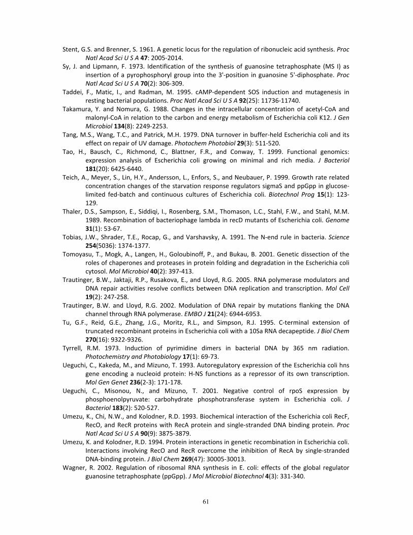
61
Stent, G.S. and Brenner, S. 1961. A genetic locus for the regulation of ribonucleic acid synthesis. Proc
Natl Acad Sci U S A 47: 2005-2014.
Sy, J. and Lipmann, F. 1973. Identification of the synthesis of guanosine tetraphosphate (MS I) as
insertion of a pyrophosphoryl group into the 3'-position in guanosine 5'-diphosphate. Proc
Natl Acad Sci U S A 70(2): 306-309.
Taddei, F., Matic, I., and Radman, M. 1995. cAMP-dependent SOS induction and mutagenesis in
resting bacterial populations. Proc Natl Acad Sci U S A 92(25): 11736-11740.
Takamura, Y. and Nomura, G. 1988. Changes in the intracellular concentration of acetyl-CoA and
malonyl-CoA in relation to the carbon and energy metabolism of Escherichia coli K12. J Gen
Microbiol 134(8): 2249-2253.
Tang, M.S., Wang, T.C., and Patrick, M.H. 1979. DNA turnover in buffer-held Escherichia coli and its
effect on repair of UV damage. Photochem Photobiol 29(3): 511-520.
Tao, H., Bausch, C., Richmond, C., Blattner, F.R., and Conway, T. 1999. Functional genomics:
expression analysis of Escherichia coli growing on minimal and rich media. J Bacteriol
181(20): 6425-6440.
Teich, A., Meyer, S., Lin, H.Y., Andersson, L., Enfors, S., and Neubauer, P. 1999. Growth rate related
concentration changes of the starvation response regulators sigmaS and ppGpp in glucose-
limited fed-batch and continuous cultures of Escherichia coli. Biotechnol Prog 15(1): 123-
129.
Thaler, D.S., Sampson, E., Siddiqi, I., Rosenberg, S.M., Thomason, L.C., Stahl, F.W., and Stahl, M.M.
1989. Recombination of bacteriophage lambda in recD mutants of Escherichia coli. Genome
31(1): 53-67.
Tobias, J.W., Shrader, T.E., Rocap, G., and Varshavsky, A. 1991. The N-end rule in bacteria. Science
254(5036): 1374-1377.
Tomoyasu, T., Mogk, A., Langen, H., Goloubinoff, P., and Bukau, B. 2001. Genetic dissection of the
roles of chaperones and proteases in protein folding and degradation in the Escherichia coli
cytosol. Mol Microbiol 40(2): 397-413.
Trautinger, B.W., Jaktaji, R.P., Rusakova, E., and Lloyd, R.G. 2005. RNA polymerase modulators and
DNA repair activities resolve conflicts between DNA replication and transcription. Mol Cell
19(2): 247-258.
Trautinger, B.W. and Lloyd, R.G. 2002. Modulation of DNA repair by mutations flanking the DNA
channel through RNA polymerase. EMBO J 21(24): 6944-6953.
Tu, G.F., Reid, G.E., Zhang, J.G., Moritz, R.L., and Simpson, R.J. 1995. C-terminal extension of
truncated recombinant proteins in Escherichia coli with a 10Sa RNA decapeptide. J Biol Chem
270(16): 9322-9326.
Tyrrell, R.M. 1973. Induction of pyrimidine dimers in bacterial DNA by 365 nm radiation.
Photochemistry and Photobiology 17(1): 69-73.
Ueguchi, C., Kakeda, M., and Mizuno, T. 1993. Autoregulatory expression of the Escherichia coli hns
gene encoding a nucleoid protein: H-NS functions as a repressor of its own transcription.
Mol Gen Genet 236(2-3): 171-178.
Ueguchi, C., Misonou, N., and Mizuno, T. 2001. Negative control of rpoS expression by
phosphoenolpyruvate: carbohydrate phosphotransferase system in Escherichia coli. J
Bacteriol 183(2): 520-527.
Umezu, K., Chi, N.W., and Kolodner, R.D. 1993. Biochemical interaction of the Escherichia coli RecF,
RecO, and RecR proteins with RecA protein and single-stranded DNA binding protein. Proc
Natl Acad Sci U S A 90(9): 3875-3879.
Umezu, K. and Kolodner, R.D. 1994. Protein interactions in genetic recombination in Escherichia coli.
Interactions involving RecO and RecR overcome the inhibition of RecA by single-stranded
DNA-binding protein. J Biol Chem 269(47): 30005-30013.
Wagner, R. 2002. Regulation of ribosomal RNA synthesis in E. coli: effects of the global regulator
guanosine tetraphosphate (ppGpp). J Mol Microbiol Biotechnol 4(3): 331-340.
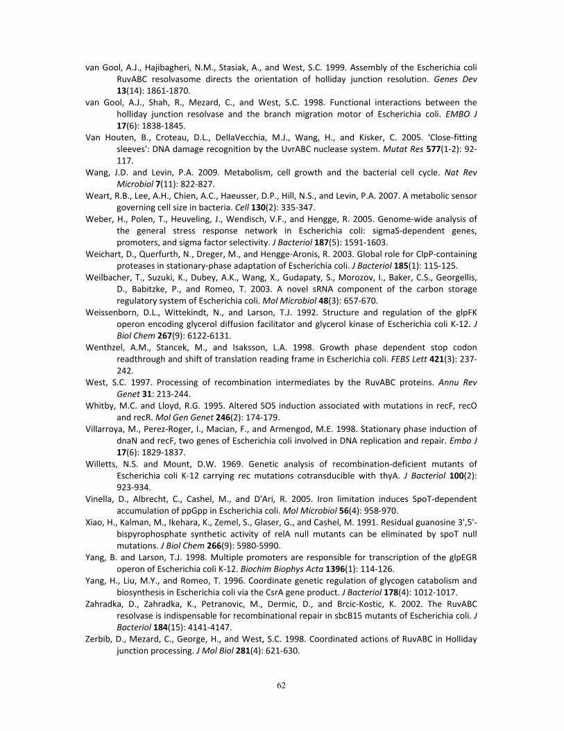
62
van Gool, A.J., Hajibagheri, N.M., Stasiak, A., and West, S.C. 1999. Assembly of the Escherichia coli
RuvABC resolvasome directs the orientation of holliday junction resolution. Genes Dev
13(14): 1861-1870.
van Gool, A.J., Shah, R., Mezard, C., and West, S.C. 1998. Functional interactions between the
holliday junction resolvase and the branch migration motor of Escherichia coli. EMBO J
17(6): 1838-1845.
Van Houten, B., Croteau, D.L., DellaVecchia, M.J., Wang, H., and Kisker, C. 2005. 'Close-fitting
sleeves': DNA damage recognition by the UvrABC nuclease system. Mutat Res 577(1-2): 92-
117.
Wang, J.D. and Levin, P.A. 2009. Metabolism, cell growth and the bacterial cell cycle. Nat Rev
Microbiol 7(11): 822-827.
Weart, R.B., Lee, A.H., Chien, A.C., Haeusser, D.P., Hill, N.S., and Levin, P.A. 2007. A metabolic sensor
governing cell size in bacteria. Cell 130(2): 335-347.
Weber, H., Polen, T., Heuveling, J., Wendisch, V.F., and Hengge, R. 2005. Genome-wide analysis of
the general stress response network in Escherichia coli: sigmaS-dependent genes,
promoters, and sigma factor selectivity. J Bacteriol 187(5): 1591-1603.
Weichart, D., Querfurth, N., Dreger, M., and Hengge-Aronis, R. 2003. Global role for ClpP-containing
proteases in stationary-phase adaptation of Escherichia coli. J Bacteriol 185(1): 115-125.
Weilbacher, T., Suzuki, K., Dubey, A.K., Wang, X., Gudapaty, S., Morozov, I., Baker, C.S., Georgellis,
D., Babitzke, P., and Romeo, T. 2003. A novel sRNA component of the carbon storage
regulatory system of Escherichia coli. Mol Microbiol 48(3): 657-670.
Weissenborn, D.L., Wittekindt, N., and Larson, T.J. 1992. Structure and regulation of the glpFK
operon encoding glycerol diffusion facilitator and glycerol kinase of Escherichia coli K-12. J
Biol Chem 267(9): 6122-6131.
Wenthzel, A.M., Stancek, M., and Isaksson, L.A. 1998. Growth phase dependent stop codon
readthrough and shift of translation reading frame in Escherichia coli. FEBS Lett 421(3): 237-
242.
West, S.C. 1997. Processing of recombination intermediates by the RuvABC proteins. Annu Rev
Genet 31: 213-244.
Whitby, M.C. and Lloyd, R.G. 1995. Altered SOS induction associated with mutations in recF, recO
and recR. Mol Gen Genet 246(2): 174-179.
Villarroya, M., Perez-Roger, I., Macian, F., and Armengod, M.E. 1998. Stationary phase induction of
dnaN and recF, two genes of Escherichia coli involved in DNA replication and repair. Embo J
17(6): 1829-1837.
Willetts, N.S. and Mount, D.W. 1969. Genetic analysis of recombination-deficient mutants of
Escherichia coli K-12 carrying rec mutations cotransducible with thyA. J Bacteriol 100(2):
923-934.
Vinella, D., Albrecht, C., Cashel, M., and D'Ari, R. 2005. Iron limitation induces SpoT-dependent
accumulation of ppGpp in Escherichia coli. Mol Microbiol 56(4): 958-970.
Xiao, H., Kalman, M., Ikehara, K., Zemel, S., Glaser, G., and Cashel, M. 1991. Residual guanosine 3',5'-
bispyrophosphate synthetic activity of relA null mutants can be eliminated by spoT null
mutations. J Biol Chem 266(9): 5980-5990.
Yang, B. and Larson, T.J. 1998. Multiple promoters are responsible for transcription of the glpEGR
operon of Escherichia coli K-12. Biochim Biophys Acta 1396(1): 114-126.
Yang, H., Liu, M.Y., and Romeo, T. 1996. Coordinate genetic regulation of glycogen catabolism and
biosynthesis in Escherichia coli via the CsrA gene product. J Bacteriol 178(4): 1012-1017.
Zahradka, D., Zahradka, K., Petranovic, M., Dermic, D., and Brcic-Kostic, K. 2002. The RuvABC
resolvase is indispensable for recombinational repair in sbcB15 mutants of Escherichia coli. J
Bacteriol 184(15): 4141-4147.
Zerbib, D., Mezard, C., George, H., and West, S.C. 1998. Coordinated actions of RuvABC in Holliday
junction processing. J Mol Biol 281(4): 621-630.

63
Zgurskaya, H.I., Keyhan, M., and Matin, A. 1997. The sigma S level in starving Escherichia coli cells
increases solely as a result of its increased stability, despite decreased synthesis. Mol
Microbiol 24(3): 643-651.