Embed Size (px)
Citation preview
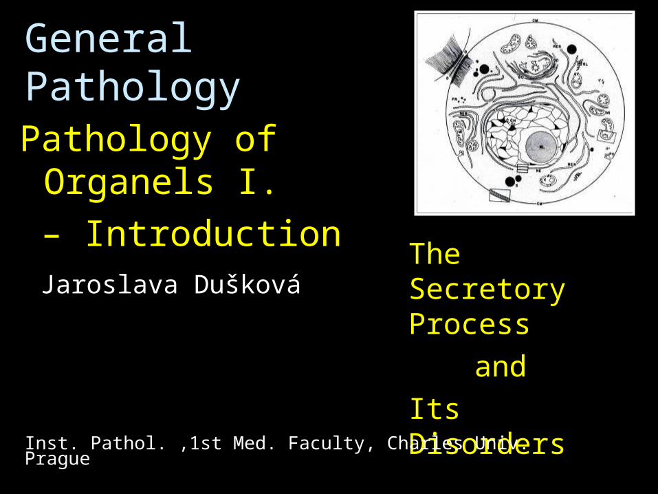
General Pathology
Pathology of Organels I.
– Introduction
The Secretory Process
and
Its Disorders
Inst. Pathol. ,1st Med. Faculty, Charles Univ. Prague
Jaroslava Dušková
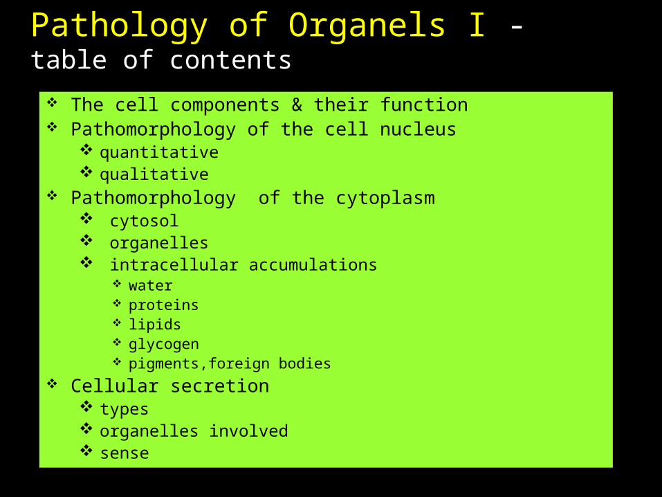
Pathology of Organels I - table of contents
The cell components & their function Pathomorphology of the cell nucleus
quantitative qualitative
Pathomorphology of the cytoplasm cytosol organelles intracellular accumulations
water proteins lipids glycogen pigments,foreign bodies
Cellular secretion types organelles involved sense
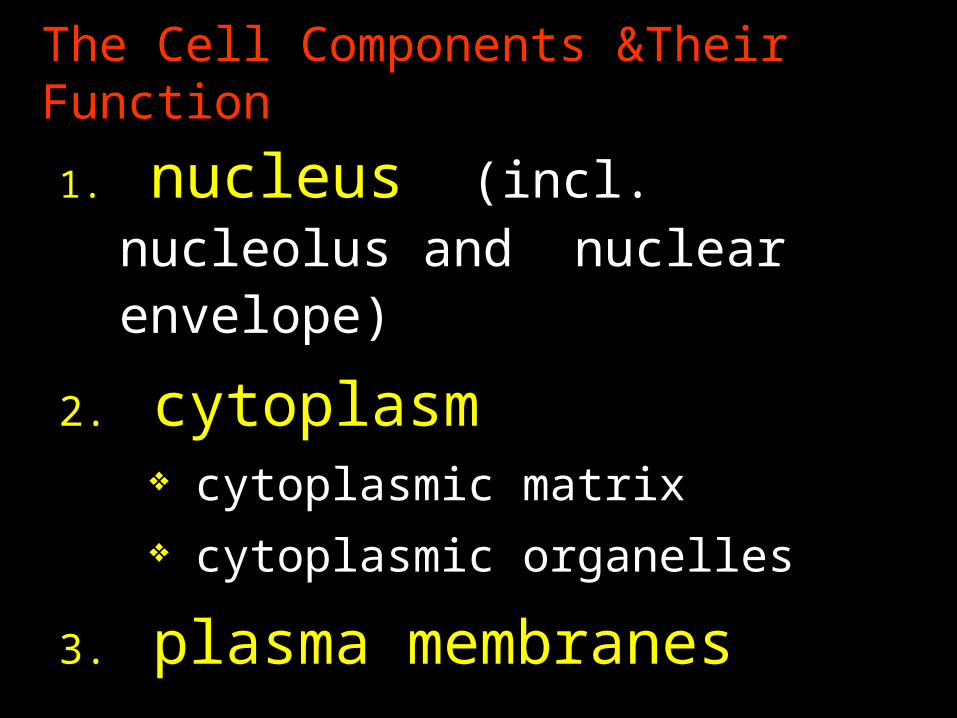
The Cell Components &Their Function
1. nucleus (incl. nucleolus and nuclear envelope)
2. cytoplasm cytoplasmic matrix cytoplasmic organelles
3. plasma membranes
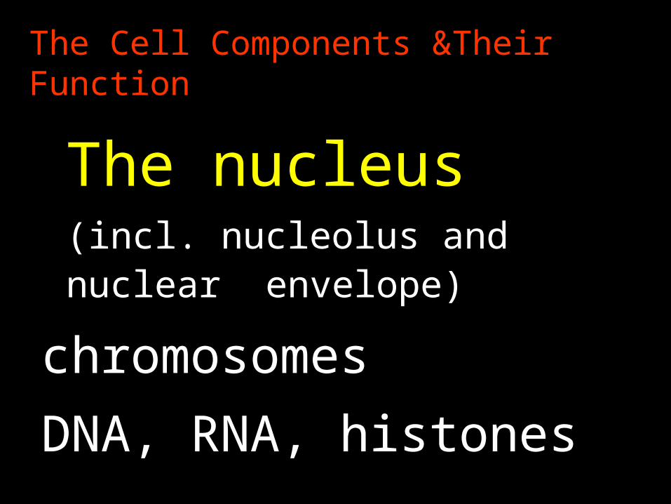
The Cell Components &Their Function
The nucleus
(incl. nucleolus and nuclear envelope)
chromosomes
DNA, RNA, histones
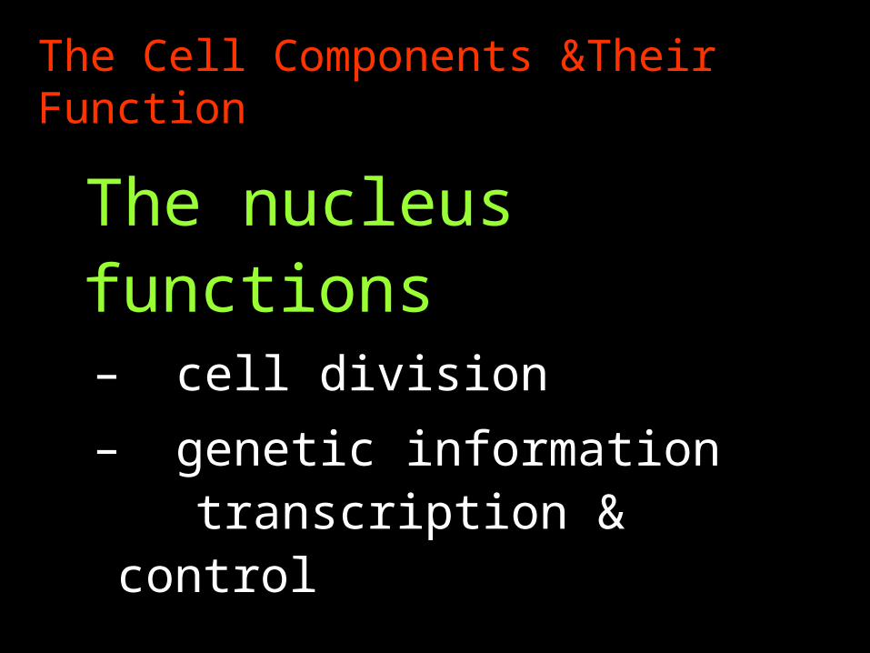
The Cell Components &Their Function
The nucleus functions – cell division
– genetic information transcription &
control
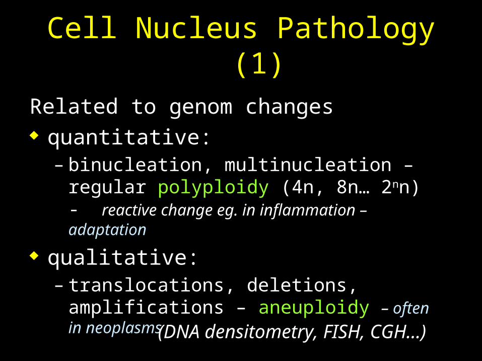
Cell Nucleus Pathology (1)
Related to genom changes quantitative:
– binucleation, multinucleation – regular polyploidy (4n, 8n… 2nn) - reactive change eg. in inflammation –adaptation
qualitative:– translocations, deletions, amplifications –
aneuploidy – often in neoplasms
(DNA densitometry, FISH, CGH…)
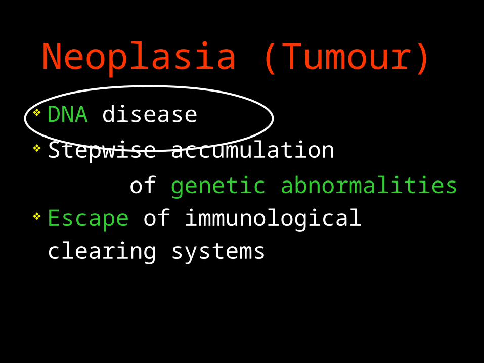
Neoplasia (Tumour)
DNA disease Stepwise accumulation
of genetic abnormalities Escape of immunological
clearing systems
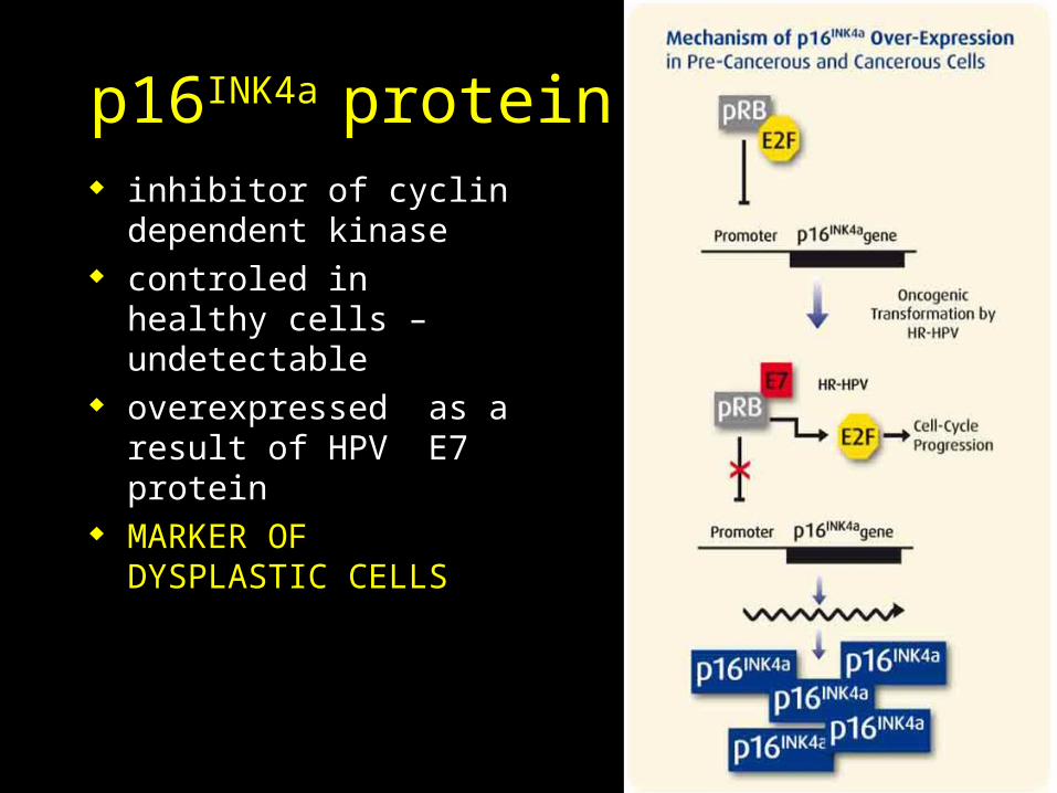
p16INK4a protein inhibitor of cyclin
dependent kinase controled in healthy
cells – undetectable overexpressed as a
result of HPV E7 protein
MARKER OF DYSPLASTIC CELLS
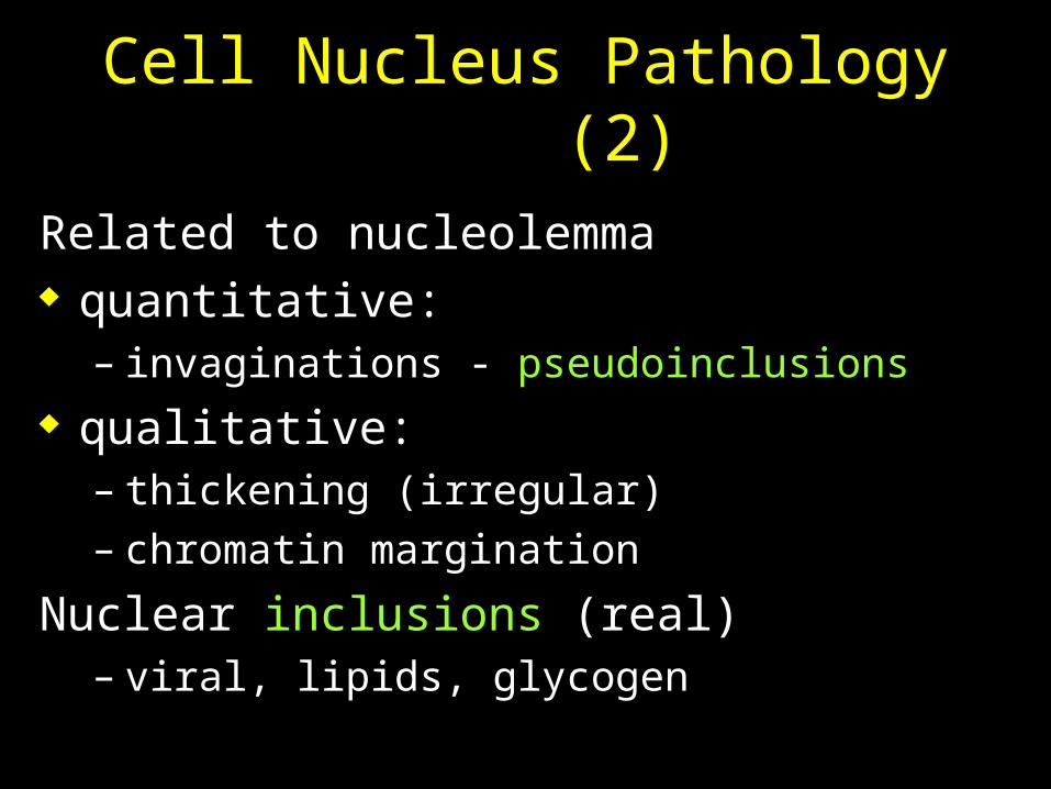
Cell Nucleus Pathology (2)
Related to nucleolemma quantitative:
– invaginations - pseudoinclusions qualitative:
– thickening (irregular)– chromatin margination
Nuclear inclusions (real) – viral, lipids, glycogen
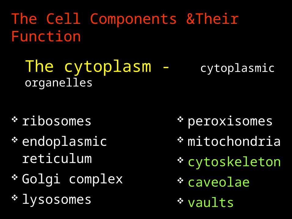
The Cell Components &Their Function
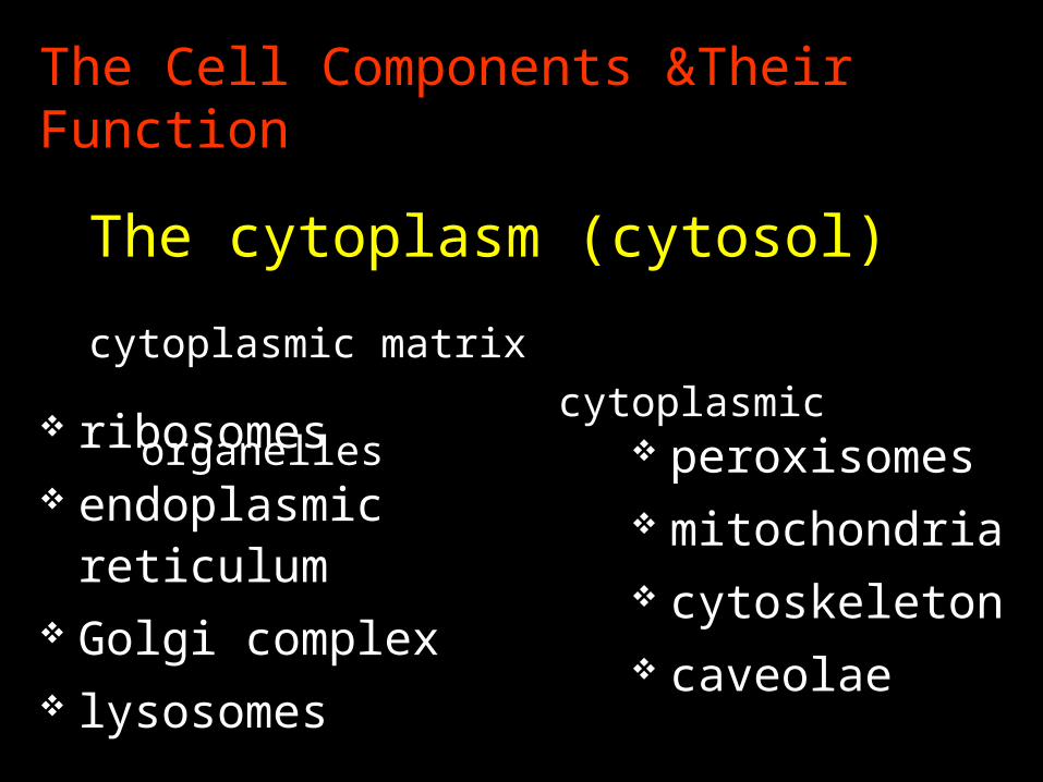
ribosomes endoplasmic
reticulum Golgi complex lysosomes
peroxisomes mitochondria cytoskeleton caveolae
The cytoplasm (cytosol) cytoplasmic matrix
cytoplasmic organelles
The Cell Components &Their Function
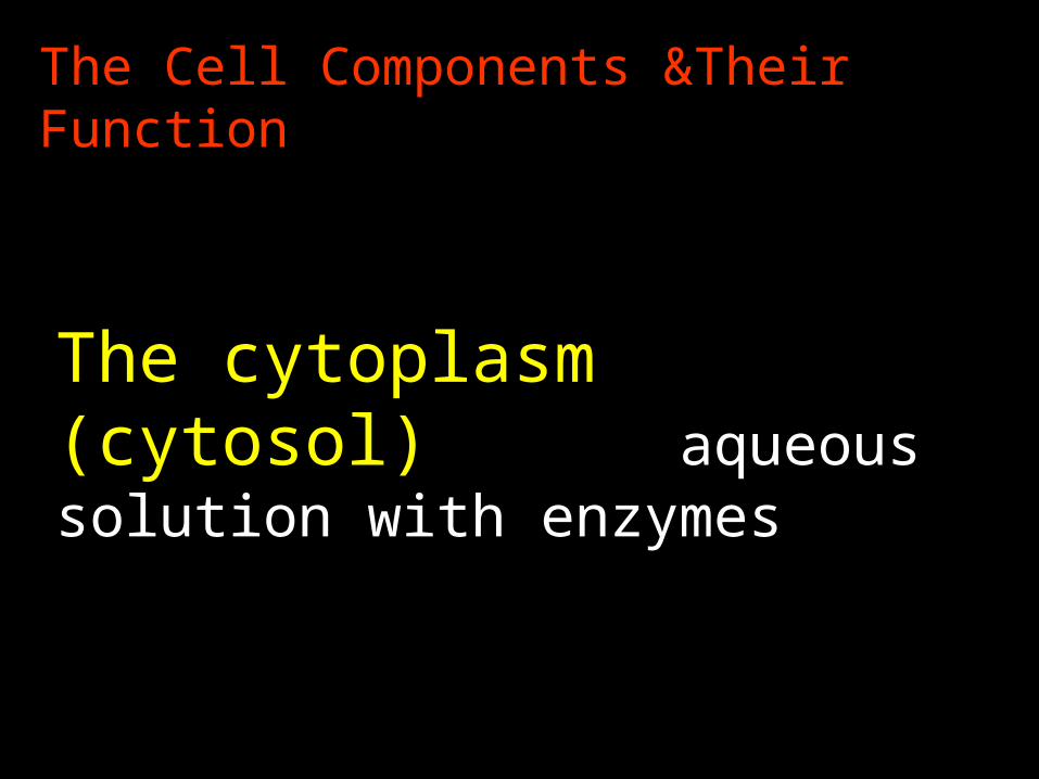
The cytoplasm (cytosol) aqueous solution with enzymes
The Cell Components &Their Function
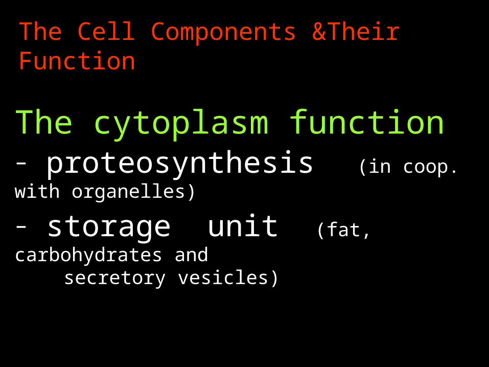
The cytoplasm function– proteosynthesis (in coop. with organelles)
– storage unit (fat, carbohydrates and secretory vesicles)
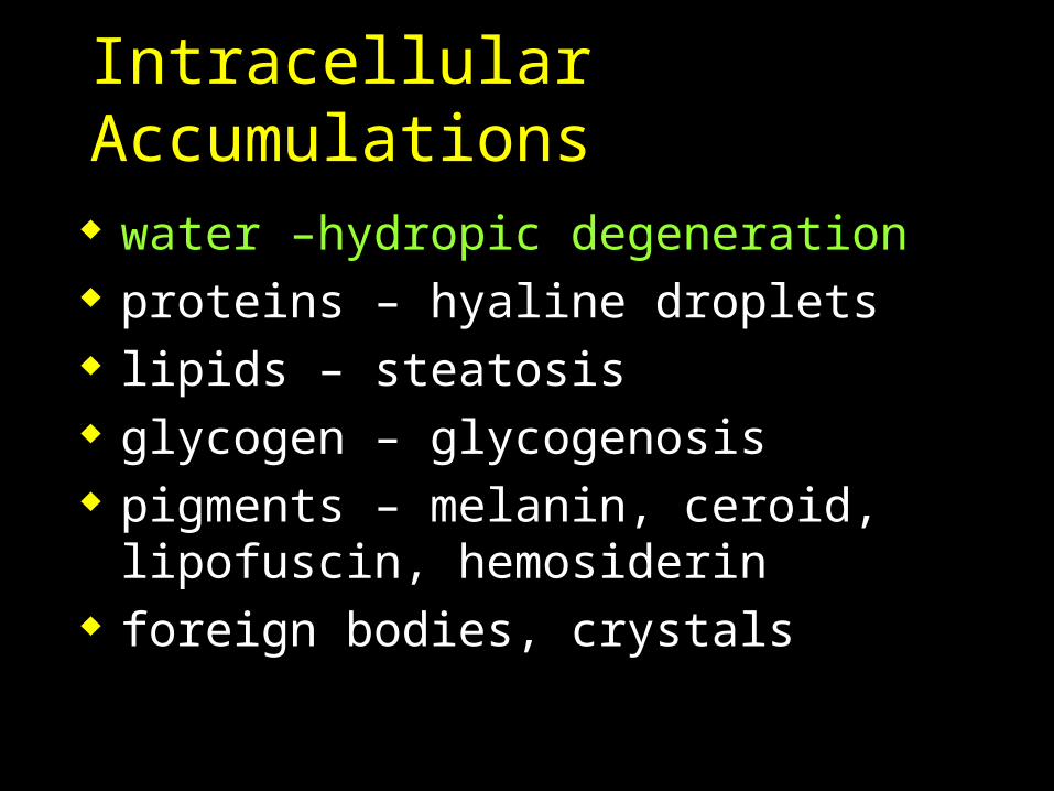
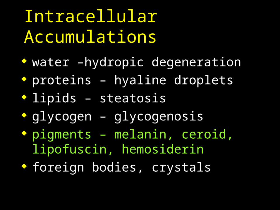
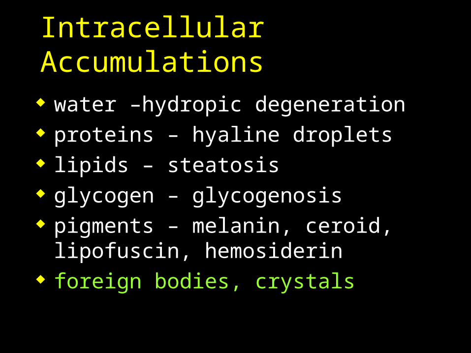
Intracellular Accumulations
water –hydropic degeneration proteins – hyaline droplets lipids – steatosis glycogen – glycogenosis pigments – melanin, ceroid, lipofuscin,
hemosiderin foreign bodies, crystals
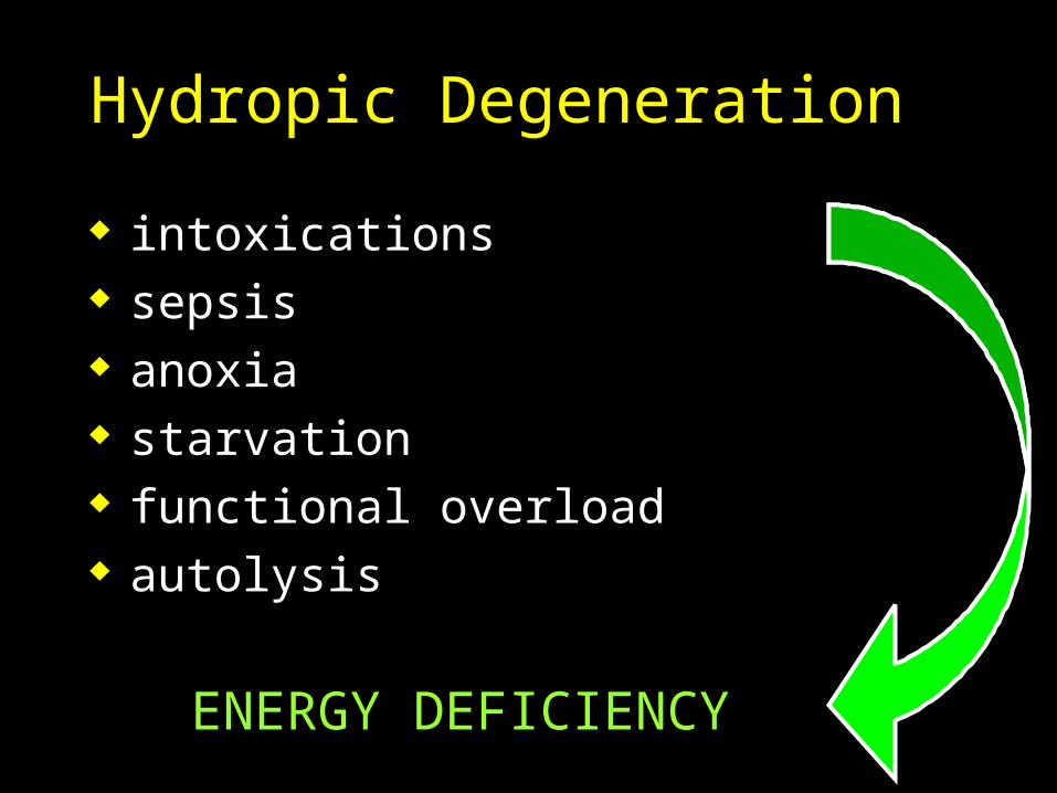
Hydropic Degeneration
intoxications sepsis anoxia starvation functional overload autolysis
ENERGY DEFICIENCY

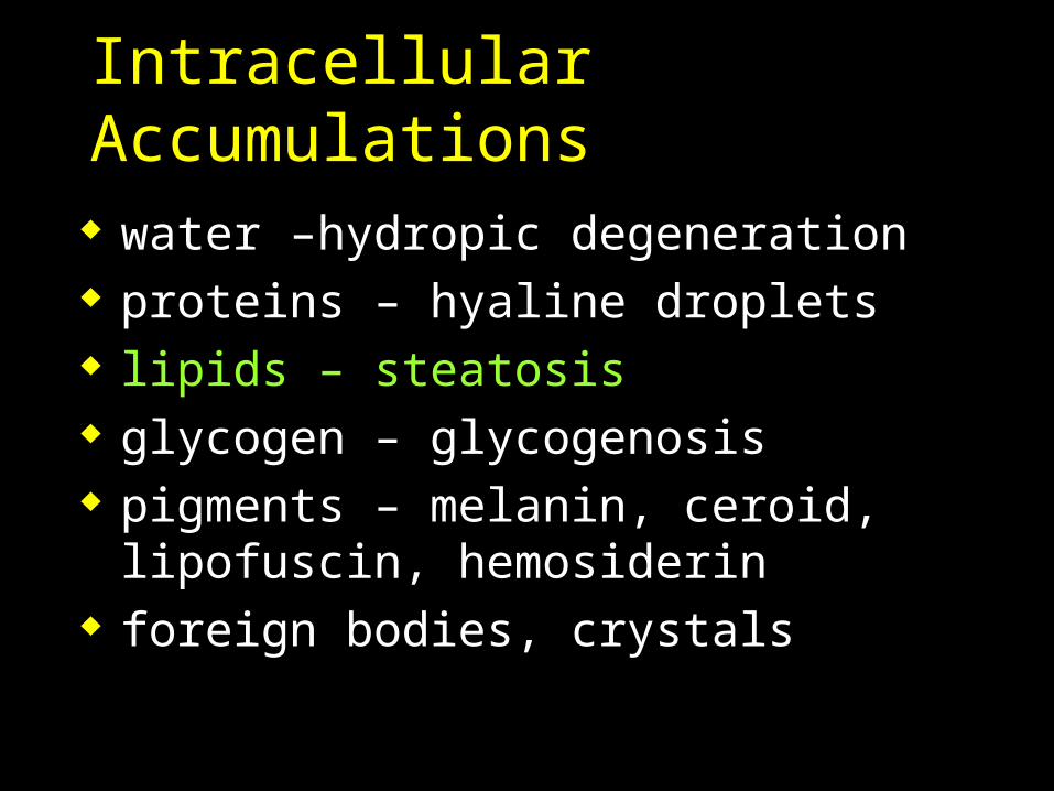
Intracellular Accumulations
water –hydropic degeneration proteins – hyaline droplets lipids – steatosis glycogen – glycogenosis pigments – melanin, ceroid, lipofuscin,
hemosiderin foreign bodies, crystals
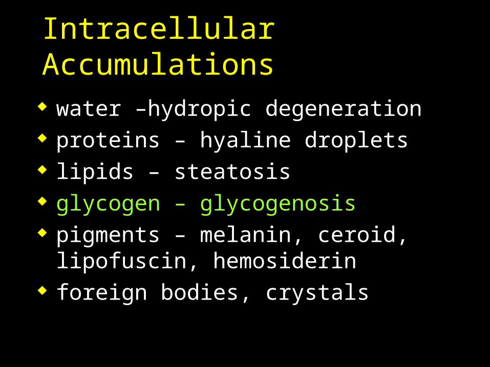
Intracellular Accumulations
water –hydropic degeneration proteins – hyaline droplets lipids – steatosis glycogen – glycogenosis pigments – melanin, ceroid, lipofuscin,
hemosiderin foreign bodies, crystals
Intracellular Accumulations
water –hydropic degeneration proteins – hyaline droplets lipids – steatosis glycogen – glycogenosis pigments – melanin, ceroid, lipofuscin,
hemosiderin foreign bodies, crystals
Intracellular Accumulations
water –hydropic degeneration proteins – hyaline droplets lipids – steatosis glycogen – glycogenosis pigments – melanin, ceroid, lipofuscin,
hemosiderin foreign bodies, crystals
Intracellular Accumulations
water –hydropic degeneration proteins – hyaline droplets lipids – steatosis glycogen – glycogenosis pigments – melanin, ceroid, lipofuscin,
hemosiderin foreign bodies, crystals
The Cell Components &Their Function
ribosomes endoplasmic
reticulum Golgi complex lysosomes
peroxisomes mitochondria cytoskeleton caveolae vaults
The cytoplasm -cytoplasmic organelles
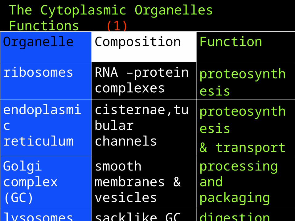
The Cytoplasmic Organelles Functions (1)
Organelle Composition Function
ribosomes RNA –protein complexes
proteosynthesis
endoplasmic reticulum
cisternae,tubular channels
proteosynthesis
& transport
Golgi complex (GC)
smooth membranes & vesicles
processing and packaging
lysosomes sacklike GC derived
digestion
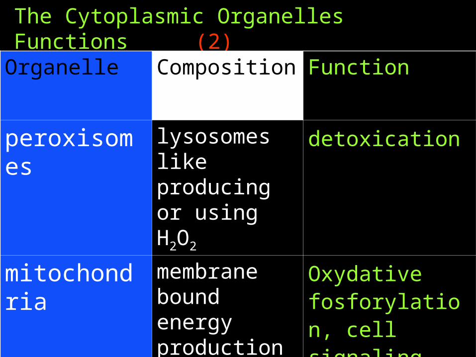
The Cytoplasmic Organelles Functions (2)
Organelle Composition Function
peroxisomes lysosomes like producing or using H2O2
detoxication
mitochondria membrane bound energy production
Oxydative fosforylation, cell signaling, pH control, Ca homeostasis
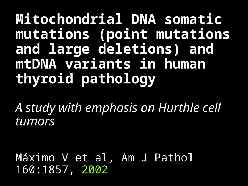
Mitochondrial DNA somatic mutations (point mutations and large deletions) and mtDNA variants in human thyroid pathology
A study with emphasis on Hurthle cell tumors
Máximo V et al, Am J Pathol 160:1857, 2002
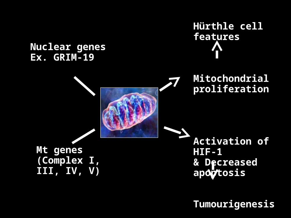
Nuclear genesEx. GRIM-19
Mt genes(Complex I, III, IV, V)
Hürthle cell features
Mitochondrial proliferation
Activation of HIF-1 & Decreased apoptosis
Tumourigenesis
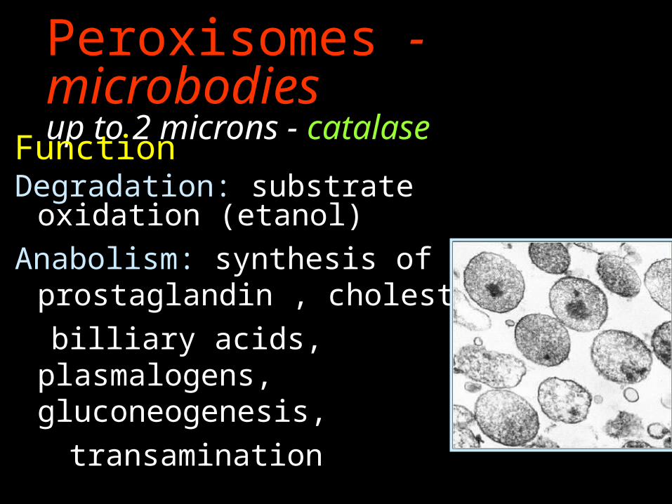
Peroxisomes - microbodiesup to 2 microns - catalase
FunctionDegradation: substrate oxidation
(etanol)
Anabolism: synthesis of prostaglandin , cholesterol,
billiary acids, plasmalogens, gluconeogenesis,
transamination
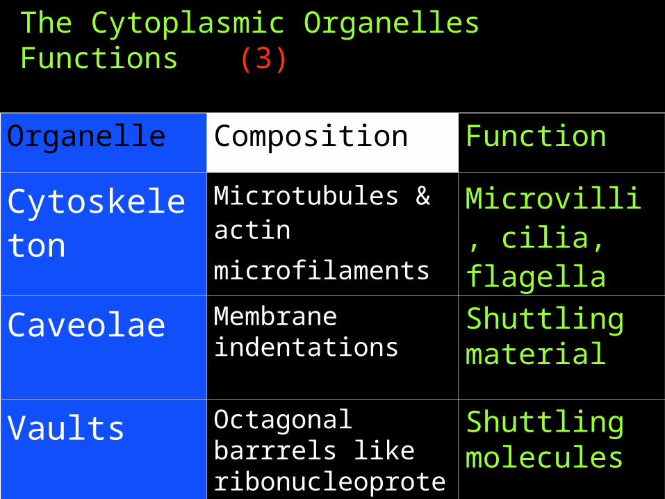
The Cytoplasmic Organelles Functions (3)
Organelle Composition Function
Cytoskeleton Microtubules & actin
microfilaments
Microvilli, cilia, flagella
Caveolae Membrane indentations
Shuttling material
Vaults Octagonal barrrels like ribonucleoproteins
Shuttling molecules
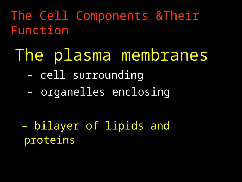
The Cell Components &Their Function
The plasma membranes
– cell surrounding
– organelles enclosing
– bilayer of lipids and proteins
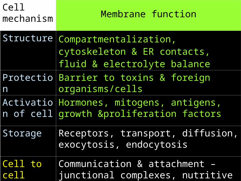
Cell mechanism Membrane function
Structure Compartmentalization, cytoskeleton & ER contacts, fluid & electrolyte balance
Protection Barrier to toxins & foreign organisms/cells
Activation of cell
Hormones, mitogens, antigens, growth &proliferation factors
Storage Receptors, transport, diffusion, exocytosis, endocytosis
Cell to cell interaction
Communication & attachment –junctional complexes, nutritive relationship, enzymes and antibody release
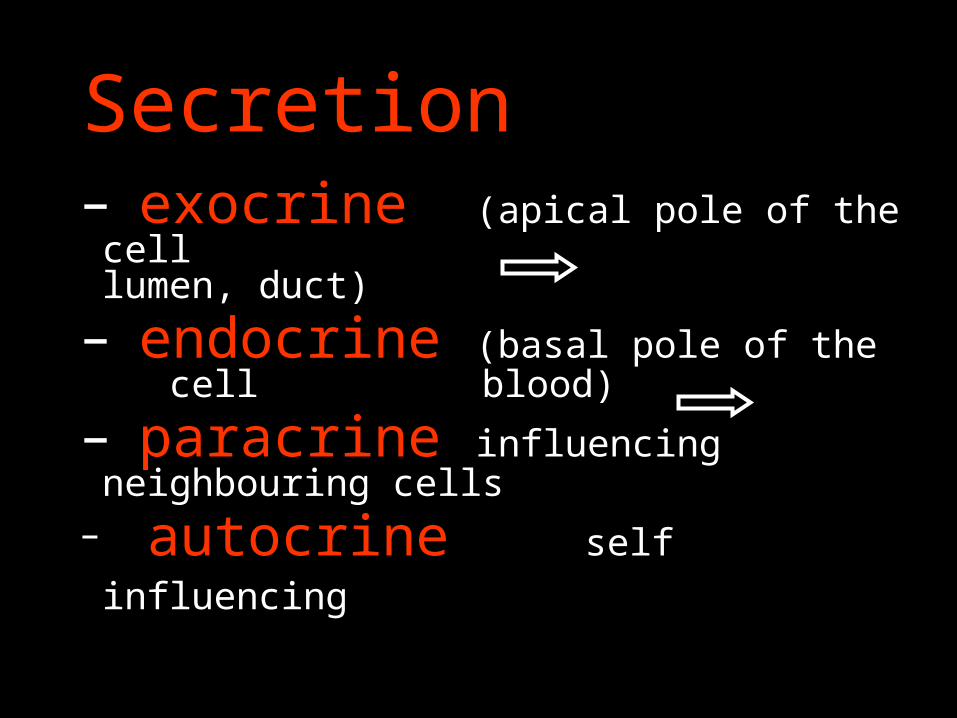
Secretion– exocrine (apical pole of the cell
lumen, duct)
– endocrine (basal pole of the cell blood)
– paracrine influencing neighbouring cells
– autocrine self influencing
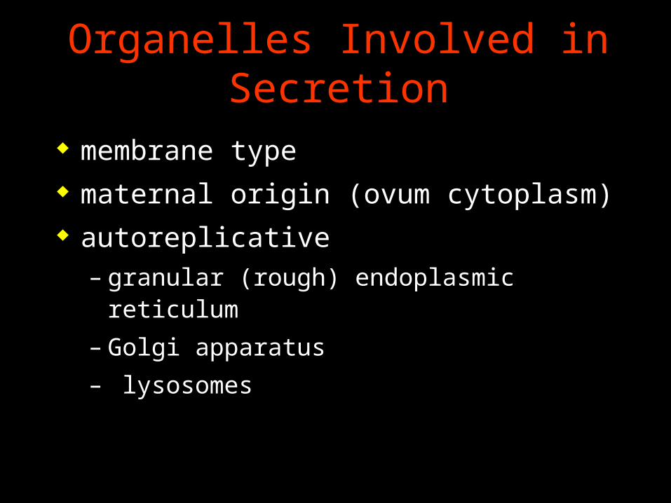
Organelles Involved in Secretion
membrane type maternal origin (ovum cytoplasm) autoreplicative
– granular (rough) endoplasmic reticulum
– Golgi apparatus
– lysosomes
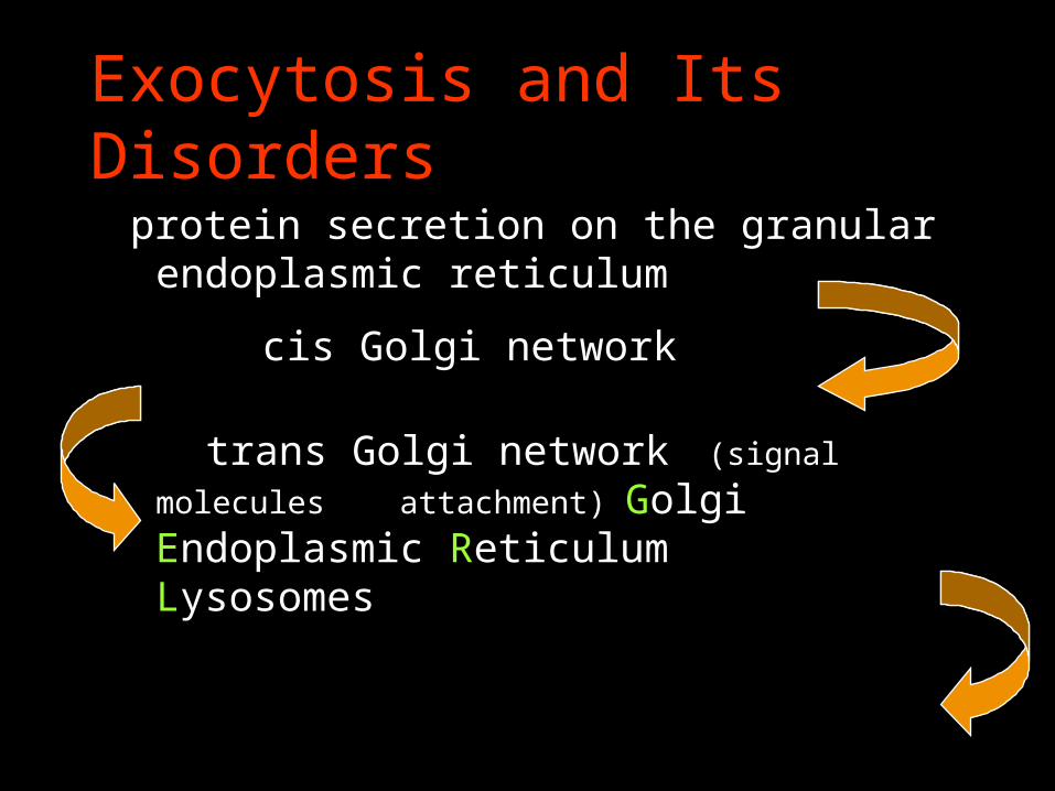
Exocytosis and Its Disorders
protein secretion on the granular endoplasmic reticulum
cis Golgi network
trans Golgi network (signal molecules
attachment) Golgi Endoplasmic Reticulum Lysosomes
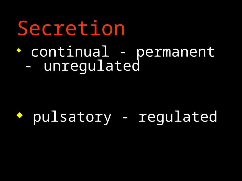
Secretion continual - permanent -
unregulated
pulsatory - regulated
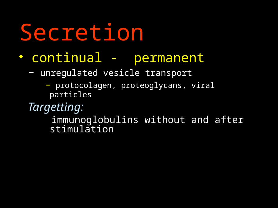
Secretion continual - permanent
– unregulated vesicle transport– protocolagen, proteoglycans, viral particles
Targetting: immunoglobulins without and after stimulation
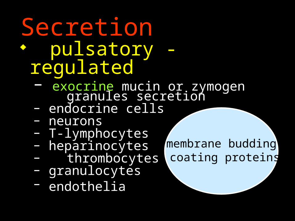
Secretion pulsatory - regulated
– exocrine mucin or zymogen granules
secretion– endocrine cells– neurons– T-lymphocytes– heparinocytes– thrombocytes– granulocytes– endothelia
membrane budding coating proteins
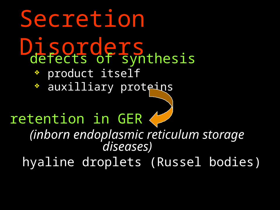
Secretion Disorders defects of synthesis
product itself auxilliary proteins
retention in GER (inborn endoplasmic reticulum storage
diseases)hyaline droplets (Russel bodies)
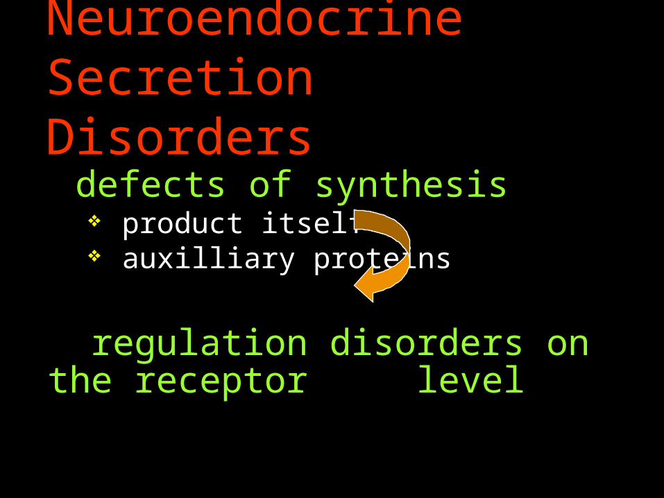
Neuroendocrine Secretion Disorders
defects of synthesis product itself auxilliary proteins
regulation disorders on the receptor
level
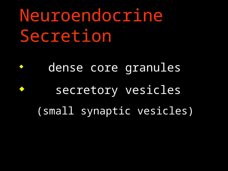
Neuroendocrine Secretion
dense core granules
secretory vesicles
(small synaptic vesicles)
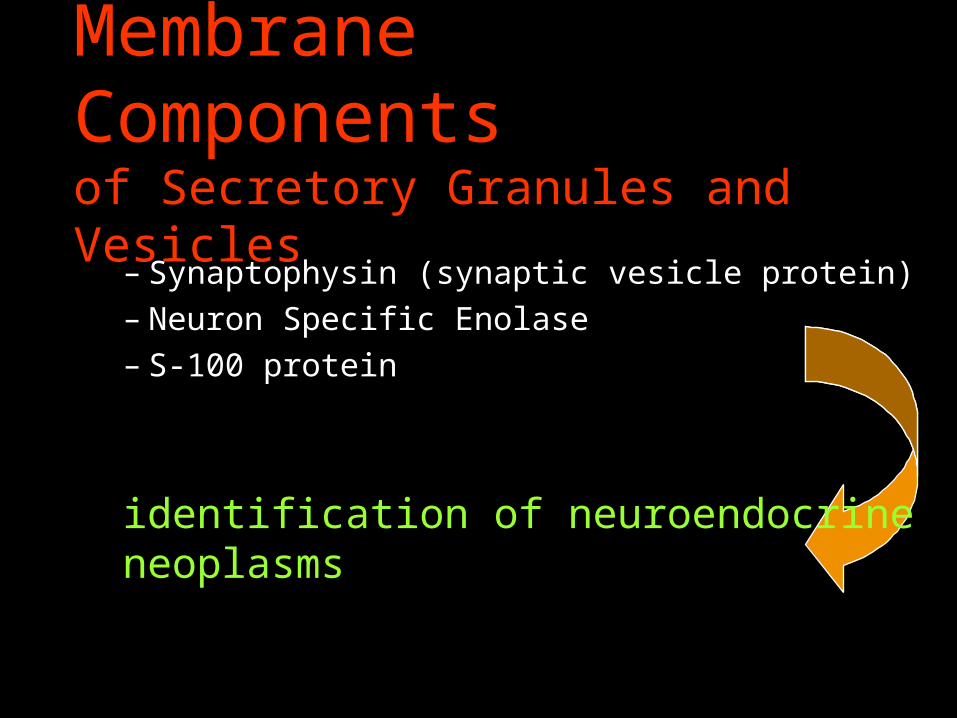
Membrane Components of Secretory Granules and Vesicles
– Synaptophysin (synaptic vesicle protein)– Neuron Specific Enolase– S-100 protein
identification of neuroendocrine neoplasms
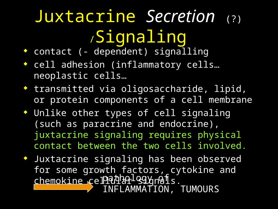
Juxtacrine Secretion (?) /Signaling contact (- dependent) signalling cell adhesion (inflammatory cells… neoplastic cells… transmitted via oligosaccharide, lipid, or protein
components of a cell membrane Unlike other types of cell signaling (such as paracrine
and endocrine), juxtacrine signaling requires physical contact between the two cells involved.
Juxtacrine signaling has been observed for some growth factors, cytokine and chemokine cellular signals.
pathology of INFLAMMATION, TUMOURS
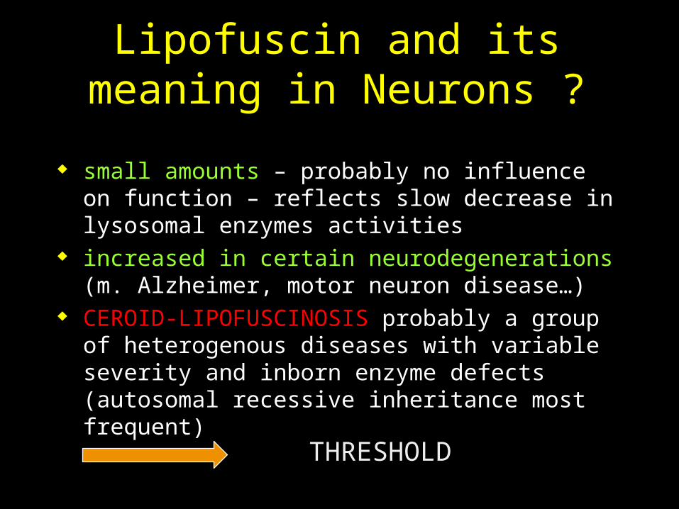
Lipofuscin and its meaning in Neurons ?
small amounts – probably no influence on function – reflects slow decrease in lysosomal enzymes activities
increased in certain neurodegenerations (m. Alzheimer, motor neuron disease…)
CEROID-LIPOFUSCINOSIS probably a group of heterogenous diseases with variable severity and inborn enzyme defects (autosomal recessive inheritance most frequent)
THRESHOLD