Embed Size (px)
Citation preview

1
GENERAL FEATURES OF FATTY ACIDS BIOSYNTHESIS
1. Fatty acids may be synthesized from dietary glucose via pyruvate. 2. Fatty acids are the preferred fuel source for the heart and the primary form
in which excess fuel is stored in adipose tissue. 3. The major site of fatty acid synthesis is the liver. 4. The enzymes that synthesize fatty acids are localized in the cytosol, and
they are completely different from the mitochondrial enzymes that catalyze fatty acid degradation.
5. The major synthetic pathway involves polymerization of two-carbon units derived from acetyl coenzyme A (acetyl CoA) to form a sixteen-carbon saturated fatty acid, palmitic acid.
6. Reduced nicotinamide adenine dinucleotide phosphate (NADPH) and adenosine triphosphate (ATP) are required for fatty acid synthesis.
PDF created with FinePrint pdfFactory Pro trial version http://www.fineprint.com

2
Sources of acetyl CoA and NADPH
1. Cytosol a. Acetyl CoA is derived from the oxidation of glucose via glycolysis and pyruvate
dehydrogenase. b. NADPH is generated by the pentose phosphate pathway.
2. Mitochondria: The acetyl CoA shuttle system. Acetyl CoA cannot cross the inner mitochondrial membrane and is transported to the cytosol via this shuttle, which also produces NADPH in the cytosol.
a. Citrate synthetase catalyzes the reaction of acetyl CoA with oxaloacetate to form citrate in the mitochondria.
b. Citrate is transported into the cytosol by a tricarboxylic acid transport system. c. Citrate reacts with CoA in the cytosol to form acetyl CoA and oxaloacetate
(OAA). d. OAA is converted to malate by an oxidized nicotinamide adenine dinucleotide
(NAD+)-dependent cytosolic malate dehydrogenase. e. Malate is decarboxylated to form pyruvate by the malic enzyme, which also
forms NADPH from oxidized nicotinamide adenine dinucleotide phosphate (NADP+).
f. Pyruvate is transported into the mitochondria by an active transport system. g. OAA is regenerated from pyruvate by pyruvate carboxylase.
PDF created with FinePrint pdfFactory Pro trial version http://www.fineprint.com

3
Transfer of Acetyl CoA to the Cytosol. Acetyl CoA is transferred from mitochondria to the cytosol, and the reducing potential NADH is concomitantly converted into that of NADPH by this series of reactions.
PDF created with FinePrint pdfFactory Pro trial version http://www.fineprint.com
4
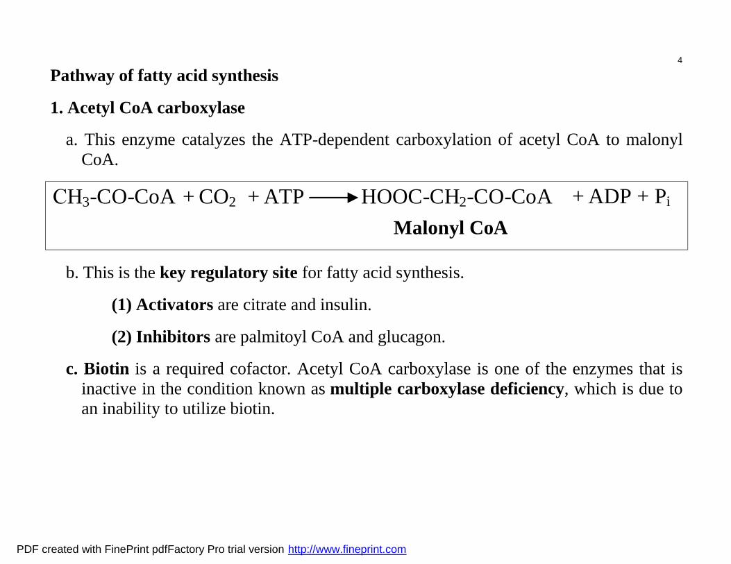
Pathway of fatty acid synthesis
1. Acetyl CoA carboxylase
a. This enzyme catalyzes the ATP-dependent carboxylation of acetyl CoA to malonyl CoA.
СH3-CO-CoA + CO2 ATP HOOC-CH2-CO-CoA Malonyl CoA
+ + ADP + Pi
b. This is the key regulatory site for fatty acid synthesis.
(1) Activators are citrate and insulin.
(2) Inhibitors are palmitoyl CoA and glucagon.
c. Biotin is a required cofactor. Acetyl CoA carboxylase is one of the enzymes that is inactive in the condition known as multiple carboxylase deficiency, which is due to an inability to utilize biotin.
PDF created with FinePrint pdfFactory Pro trial version http://www.fineprint.com

5
The fatty acid synthase enzyme complex catalyzes several reactions that convert acetyl CoA and malonyl CoA to butyryl CoA.
a. The acetyl transacylase component catalyzes the conversion of
acetyl CoA + acyl carrier protein (ACP) → acetyl-ACP + CoA.
(1) ACP possesses a phosphopantetheine prosthetic group, which is derived from the vitamin pantothenic acid and is identical to that portion of the CoA molecule.
(2) The acyl groups of the substrate molecules are covalently bound to ACP via a thioester linkage with the terminal sulfhydryl group.
b. The malonyl transacylase component catalyzes the conversion of
malonyl CoA + ACP → malonyl-ACP + CoA.
Fig. Phosphopantetheine.
Both acyl carrier protein and CoA include phosphopantetheine as their reactive units. ACP, a single polypeptide chain of 77 residues, can be regarded as a giant prosthetic group, a “macro CoA.”
PDF created with FinePrint pdfFactory Pro trial version http://www.fineprint.com

6
c. The 3-ketoacyl synthase component catalyzes the conversion of
malonyl-ACP + acetyl-ACP → acetoacetyl-ACP + ACP + CO2
d. The 3-ketoacyl reductase component catalyzes the NADPH-dependent conversion of
acetoacetyl-ACP → 3-hydroxybutyryl-ACP
e. The 3-hydroxybutyryl-ACP dehydratase component catalyzes the conversion of
3-hydroxybutyryl-ACP → crotonyl-ACP.
f. The enoyl-ACP reductase component catalyzes the NADPH-dependent conversion of
crotonyl-ACP → butyryl-ACP
PDF created with FinePrint pdfFactory Pro trial version http://www.fineprint.com

7
3. Formation of palmitic acid
a. Reaction. Butyryl-ACP (a four-carbon unit) reacts with another malonyl group via the reactions described above to form a six-carbon unit, and the reactions of the fatty acid synthase complex continue.
b. Reaction repeats. After seven turns of this cycle, palmitic-ACP (a 16-carbon unit) is synthesized.
c. Release. Palmitic acid is released from ACP and the fatty acid synthase complex by palmitoyl deacylase (thioesterase).
4. Stoichiometry of the synthesis of palmitate
8 Acetyl CoA + 7 ATP + 14 NADPH + 14 H+ + H2O → Palmitate + 7 ADP + 7Pi + 8 CoASH + 14 NADP+
PDF created with FinePrint pdfFactory Pro trial version http://www.fineprint.com

8
Animal Fatty Acid Synthase
1 2- attachment of a substrate to ACP 5- отщепление воды 3- lengthening of a chain 6- reduction 4- reduction 7- product release
Palmitate
3
1 2 6
7
4 5
ACP
SH
4 5 6
2 1
3 7
SH-Cys ACP
SH
P-пантетеин
SH-Cys
Acetyl-CоА
Malonyl-CоА
Domain 1
Domain 3
Domain 2
1 [ACP]-S-acetyl-transferase 2.3.1.38 2 [ACP]-S-malonyl-transferase 2.3.1.39 3 3-oxoacyl-[ACP]-syntase 2.3.1.41 4 3- oxoacyl -[ACP]-reductase 1.1.1.100 5 3-hydroxypalmitoyl-[ACP]-dehydrotase 4.2.1.61 6 enoyl-[ACP]-reductase (NADPH) 1.3.1.10 7 acyl-[ACP]-hydrolase 3.1.2.14
PDF created with FinePrint pdfFactory Pro trial version http://www.fineprint.com

9
Schematic Representation of Animal Fatty Acid Synthase. Each of the identical chains in the dimer contains three domains. Domain 1 (blue) contains acetyl transferase (AT), malonyl transferase (MT), and condensing enzyme (CE). Domain 2 (yellow) contains acyl carrier protein (ACP), β-ketoacyl reductase (KR), dehydratase (DH), and enoyl reductase (ER). Domain 3 (red) contains thioesterase (TE). The flexible phosphopantetheinyl group (green) carries the fatty acyl chain from one catalytic site on a chain to another, as well as between chains in the dimer.
PDF created with FinePrint pdfFactory Pro trial version http://www.fineprint.com

10
Elongation of fatty acids. Fatty acids longer than 16 carbons can be formed via one of two elongation systems adding 2-carbon units. 1. The endoplasmic reticulum system is the most active. It adds malonyl CoA onto palmitate in a manner similar to the action of fatty acid synthase, except that CoASH is used rather than the ACP. Stearic acid (an 18-carbon unit) is the product. 2. A mitochondrial elongation system uses acetyl CoA units, rather than malonyl CoA units, to elongate fatty acids for the synthesis of structural lipids in this organelle. Desaturation of fatty acids 1. The two most common monounsaturated fatty acids in mammals are palmitoleic acid (16:1:∆9) and oleic acid (18:1:∆9). 2. In the endoplasmic reticulum, double bonds are introduced between carbons 9 and 10 by fatty acid oxygenase, which requires molecular oxygen (O2) and NADPH.
PDF created with FinePrint pdfFactory Pro trial version http://www.fineprint.com

11
FATTY ACID OXIDATION. Fatty acids are an important energy source. Oxidation of fatty acids generates the high-energy compounds reduced NAD (NADH) and reduced flavin adenine dinucleotide (FADH2) and yields acetyl CoA, which is the substrate for the citric acid cycle.
β-Oxidation of fatty acids is the principal pathway. It occurs in the mitochondrial matrix and involves oxidation of the β-carbon to form a β-keto acid.
1. Activation of free fatty acids. Free fatty acids are taken up from the circulation by cells and are activated by formation of acyl CoA derivatives.
a. An endoplasmic reticulum acyl CoA synthetase (thiokinase) activates long-chain fatty acids of 12 or more carbons.
Fatty acid + ATP + CoASH → acyl CoA + PPi + AMP
b. Inner mitochondrial acyl CoA synthetases activate medium-chain (4-10 carbons) and short-chain (acetate and propionate) fatty acids. These fatty acids enter the mitochondria freely from the cytoplasm, whereas the long-chain fatty acids cannot.
PDF created with FinePrint pdfFactory Pro trial version http://www.fineprint.com

12
2. Role of carnitine. Long-chain fatty acyl CoAs cannot freely diffuse across the inner mitochondrial membrane. Carnitine in the inner mitochondrial membrane mediates transfer of fatty acyl groups from the cytosol to the mitochondrial matrix where they are oxidized.
a. Transfer reaction. On the outer surface of the inner mitochondrial membrane, the
enzyme carnitine palmitoyl transferase I (CPT I) catalyzes the transfer of the acyl group from CoA to carnitine. The fatty acyl group is translocated across the membrane to the inner surface, where the enzyme carnitine palmitoyl transferase II (CPT II) catalyzes the transfer of the acyl group to CoA drawn from the matrix CoA pool.
b. Sources of carnitine (1) Dietary sources include red meat and dairy products. (2) Synthesis. Carnitine (β-hydroxy-γ-trimethylammonium butyrate) may be
synthesized in the body from the amino acid lysine. c. Carnitine deficiency in humans may be classified as:
(1) Systemic, in which levels are reduced in all tissues (2) Myopathic, in which levels are reduced only in muscle, including the heart
PDF created with FinePrint pdfFactory Pro trial version http://www.fineprint.com

13
Fig. Acyl Carnitine Translocase. The entry of acyl carnitine into the mitochondrial matrix is mediated by a translocase. Carnitine returns to the cytosolic side of the inner mitochondrial membrane in exchange for acyl carnitine
PDF created with FinePrint pdfFactory Pro trial version http://www.fineprint.com

14
3. Pathway of β-oxidation. In the mitochondrial matrix, long-chain fatty acyl CoAs are subjected to a repeated four-step process that successively removes two-carbon units from the chain until the last two-carbon fragment remains.
a. Acyl CoA → enoyl CoA (1) This dehydrogenation reaction is catalyzed by a family of acyl CoA dehy-
drogenases with varying specificity for different length acyl CoA chains. (2) The prosthetic group of this
enzyme, flavin adenine dinucleotide (FAD), is reduced to FADH2 during this reaction.
(3) Genetic defects in acyl CoA dehydrogenases (e.g., medium-chain acyl CoA dehydrogenase deficiency) impair β-oxidation.
b. Enoyl CoA → 3-hydroxyacyl CoA. This hydration reaction is catalyzed by enoyl CoA hydratase.
c. 3-Hydroxyacyl CoA → 3-ketoacyl CoA (1) This dehydrogenation reaction is catalyzed by 3-hydroxyacyl CoA
dehydrogenase. (2) NAD+ is reduced to NADH during this reaction.
d. 3-Ketoacyl CoA + CoASH → acetyl CoA + tetradecanoyl CoA. This thiolytic cleavage reaction is catalyzed by thiolase (β-ketothiolase).
PDF created with FinePrint pdfFactory Pro trial version http://www.fineprint.com

15
Fig. Reaction Sequence for the Degradation of Fatty Acids. Fatty acids are degraded by the repetition of a four-reaction sequence consisting of oxidation, hydration, oxidation, and thiolysis.
PDF created with FinePrint pdfFactory Pro trial version http://www.fineprint.com

16
Fig. First Three Rounds in the Degradation of Palmitate. Two-carbon units are sequentially removed from the carboxyl end of the fatty acid.
PDF created with FinePrint pdfFactory Pro trial version http://www.fineprint.com

17
4. Stoichiometry of β-oxidation
a. β-Oxidation of palmitate (16 carbons) Palmitoyl CoA + 7 CoASH + 7 FAD + 7 NAD+ + 7 H2O → 8 acetyl CoA + 7 FADH2 + 7 NADH + 7 H+ 1
A total of 7 moles of FADH2 yields 14 moles of ATP by electron transport and oxidative phosphorylation. A total of 7 moles of NADH yields 21 moles of ATP by electron transport and oxidative phosphorylation. Therefore,
Palmitoyl CoA + 7 CoASH + 7 O2 + 35 ADP + 35 Pi → 8 acetyl CoA + 35 ATP + 42 H2O 2 Because it costs 2 moles of ATP to activate the free palmitate, 33 moles of ATP are formed per mole of palmitate via β-oxidation.
b. Total oxidation. The acetyl CoA derived from β-oxidation of fatty acyl CoAs may be oxidized to carbon dioxide (CO2) and water (H2O) by the citric acid cycle.
8 Acetyl CoA + 16 O2 + 96 ADP + 96 Pi → 8 CoASH + 96 ATP + 16 CO2 + 104 H2O 3 The combined yield of ATP is obtained by addition of equations (2) and (3)
Palmitoyl CoA + 23 O2 + 131 ADP + 131 Pi → 8 CoASH + 131 ATP + 16 CO2 + 146 H2O 4 After subtraction of 2 moles of ATP for activation of the fatty acyl CoA, 129 moles of ATP are formed by total oxidation of palmitate.
5. Respiratory quotient (RQ) is defined as moles of CO2 produced, divided by moles of oxygen consumed during complete oxidation of a metabolic fuel to CO2 and H2O. a. For palmitate, the RQ is given by:
(16 moles CO2 produced)/(23 moles O2 consumed) = 0.7 b. In contrast, the complete oxidation of glucose yields an RQ of 1.0.
PDF created with FinePrint pdfFactory Pro trial version http://www.fineprint.com

18
Oxidation of fatty acids with an odd number of carbon atoms. Fatty acids that have an odd number of carbon atoms are a minor species in human tissues.
1. They undergo β-oxidation as acyl CoA derivatives until a three-carbon fragment, propionyl CoA, is formed.
2. Propionyl CoA is carboxylated to methylmalonyl CoA by biotin-dependent propionyl CoA carboxylase.
3. Methylmalonyl CoA is converted to succinyl CoA by methylmalonyl CoA mutase.
4. Succinyl CoA can be metabolized via the citric acid cycle, of which it is an intermediate.
Fig. Conversion of Propionyl CoA Into Succinyl CoA. Propionyl CoA, generated from fatty acids with an odd number of carbons as well as some amino acids, is converted into the citric acid cycle intermediate succinyl CoA.
PDF created with FinePrint pdfFactory Pro trial version http://www.fineprint.com

19
Oxidation of unsaturated fatty acids. In the human body, 50% of the fatty acids are unsaturated. Many unsaturated fatty acids are degraded by the β-oxidation pathway with the help of two additional enzymes.
1. Double bonds in naturally occurring fatty acids are cis. The β-oxidation pathway can only process trans double bonds using enoyl CoA hydratase.
2. The cis double bond is removed by the sequential reactions catalyzed by 2,4-dienoyl CoA reductase and enoyl CoA isomerase to yield a ∆2-trans enoyl CoA.
Fig. Oxidation of Linoleoyl CoA. The complete oxidation of the diunsaturated fatty acid linoleate is facilitated by the activity of enoyl CoA isomerase and 2,4-dienoyl CoA reductase.
PDF created with FinePrint pdfFactory Pro trial version http://www.fineprint.com

20
α-Oxidation of fatty acids
1. This pathway involves the oxidation of long-chain fatty acids to 2-hydroxy fatty acids, which are constituents of brain lipids, followed by oxidation to a fatty acid with one less carbon. Removal of one carbon from the carboxyl end of a fatty acid has been demonstrated in microsomal fractions from brain tissue.
2. Refsum's disease (phytanic acid storage disease) is caused by a defect of α-oxidation.
a. Phytanic acid (derived from animal fat and cow's milk and probably originally from chlorophyll) accumulates in affected individuals. Phytanic acid cannot alternatively be oxidized by β-oxidation because the beta position is blocked by a methyl group.
b. Symptoms include retinitis pigmentosa, failing night vision, peripheral neuropathy, and cerebellar ataxia.
ω-Oxidation involves oxidation of the terminal methyl group to form an ω-hydroxy fatty acid. This is a minor pathway observed with liver microsomal preparations.
PDF created with FinePrint pdfFactory Pro trial version http://www.fineprint.com

21
BIOSYNTHESIS OF TRIACYLGLYCEROLS (TRIGLYCERIDES).
Fatty acids are converted to triacylglycerols for transport to tissues and storage.
Formation involves the acylation of the three hydroxyl groups of glycerol
1. Activation of the fatty acid. Formation of the CoA ester of the fatty acid is catalyzed in the cytosol by an ATP-dependent acyl CoA synthetase.
Fatty acid + ATP + CoASH → Acyl CoA + AMP + PPi
2. Acylation of glycerol a. First acylation. There are two routes for the acylation of the
first hydroxyl of glycerol. (1) One route uses dihydroxyacetone phosphate
(DHAP), derived from glycoly-sis, as the acceptor of an acyl moiety from a fatty acyl CoA. (a) This is followed by reduction, with NADPH as the
electron acceptor, to form lysophosphatidate. (b) The fatty acid preferentially introduced to form
lysophosphatidate is saturated. (2) The second route gives the same product and shows
the same preference for a saturated fatty acid, but the order is reversed, and reduction of DHAP to glycerol-3-phosphate occurs before acylation of the C-1 hydroxyl.
b. Second acylation. An unsaturated fatty acyl CoA thioester is introduced to the 2-hydroxyl of lysophosphatidate. An exception occurs in the human mammary gland, where a saturated fatty acyl CoA is used.
c. Third acylation. The phosphate group on C-3 is removed by a phosphatase and either a saturated or unsaturated fatty acid is incorporated at that position.
PDF created with FinePrint pdfFactory Pro trial version http://www.fineprint.com

22
Storage of triacylglycerols in adipose tissue cells (adipocytes)
1. The esterification of fatty acids in adipocytes to form triacylglycerols depends on carbohydrate metabolism for the formation of DHAP.
2. Adipocytes lack a glycerol kinase and cannot phosphorylate glycerol to glycerol-3-phosphate. The only source of glycerol-3-phosphate for triacylglycerol synthesis is from DHAP formed during glycolysis.
3. The entry of glucose into adipocytes is insulin dependent. Thus, insulin is an essential requirement for triacylglycerol synthesis in adipocytes.
PDF created with FinePrint pdfFactory Pro trial version http://www.fineprint.com

23
Lipolysis of triacylglycerols 1. Hormonal control of lipolysis in adipocytes
a. Hormone-sensitive lipase is activated by covalent phosphorylation of the lipase by a cyclic adenosine monophosphate (cAMP)-dependent protein kinase
b. Epinephrine circulating in the blood in response to stress, or norepinephrine released by neural connections to adipose tissue, activates the cell membrane ade-nylate cyclase to produce cAMP. Thus, under conditions of stress or when neural signals indicate low levels of metabolic fuel, the hormone-sensitive lipase is activated.
c. Insulin inhibits lipolysis by two mechanisms. (1) It reduces cAMP levels, probably by inhibiting adenylate cyclase activity. (2) It increases glucose entry into the adipocytes, so that the formation of DHAP and glycerol-3-
phosphate is increased. The availability of these products of glycolysis increases the rate of re-esterification of free fatty acids to triacylglycerols, thus reducing the rate of release of the fatty acids from adipocytes.
d. Prostaglandins inhibit lipolysis by reducing cAMP levels. 2. Enzymatic steps of lipolysis
a. Three different lipases are required to release the three fatty acids from the triacylglycerols. (1) Hormone-sensitive triacylglycerol lipase converts triacylglycerol to diacyl-glycerol plus a fatty acid. (2) Diacylglycerol lipase converts diacylglycerol to monoacylglycerol plus a fatty acid. (3) Monoacylglycerol lipase converts monoacylglycerol to glycerol plus a fatty acid.
b. The rate-limiting step of lipolysis in adipocytes is the reaction catalyzed by the hormone-sensitive triacylglycerol lipase. Diacyl- and monoacylglycerol lipases are present in excess, so that once triacylglycerol lipase is activated, lipolysis of triacylglycerols goes to completion.
3. Lipolysis in other tissues. Triacylglycerols are stored mainly in adipocytes. However, other tissues, including muscle and liver, store small amounts of triacylglycerols as intracellular lipid droplets for their own use. These fat stores appear to be mobilized by hormonal controls similar to those found in adipocytes.
PDF created with FinePrint pdfFactory Pro trial version http://www.fineprint.com

24
KETONE BODY METABOLISM. Ketone bodies (i.e., acetoacetate, β-hydroxybutyrate, acetone) are the preferred energy substrates of the heart, skeletal muscle, and kidney during the fasting state. If blood levels of β-hydroxybutyrate and acetoacetate increase sufficiently, as they do during starvation, they also become the primary energy substrate for the brain.
Biosynthesis of ketone bodies.
1. Thiolase catalyzes the formation of two acetyl CoA molecules from acetoacetyl CoA.
2. 3-Hydroxy-3-methylglutaryl CoA (HMG CoA) synthase catalyzes the addition of another acetyl CoA to acetoacetyl CoA to form HMG CoA.
3. HMG CoA lyase catalyzes the cleavage of HMG CoA to acetoacetate and acetyl CoA.
4. β-Hydroxybutyrate dehydrogenase catalyzes the formation of P-hydroxybutyrate from acetoacetate when the NADH/NAD+ ratio is high, as it is in the liver during fasting.
5. Acetone is formed spontaneously from a small fraction of the circulating acetoacetate and is exhaled by the lungs. In patients with untreated diabetes, the odor of acetone is apparent on the patient's breath.
PDF created with FinePrint pdfFactory Pro trial version http://www.fineprint.com

25
Fig. Formation of Ketone Bodies. The Ketone bodies-acetoacetate, d-3-hydroxybutyrate, and acetone from acetyl CoA—are formed primarily in the liver. Enzymes catalyzing these reactions are (1) 3-ketothiolase, (2) hydroxymethylglutaryl CoA synthase, (3) hydroxymethylglutaryl CoA cleavage enzyme, and (4) d-3-hydroxybutyrate dehydrogenase. Acetoacetate spontaneously decarboxylates to form acetone.
PDF created with FinePrint pdfFactory Pro trial version http://www.fineprint.com

26
Ketone body oxidation
1. A 3-keto acid CoA transferase catalyzes the formation of the CoA thioester of acetoacetate. This is a nonhepatic enzyme that prevents ketone bodies from being used as an energy source by the liver.
2. Thiolase catalyzes the cleavage of acetoacetyl CoA to two acetyl CoA molecules with the use of CoA, providing two-carbon fragments for oxidation by the citric acid cycle.
Energy yield from oxidation of ketone bodies
1. Each mole of acetyl CoA that is formed yields 12 moles of ATP via the citric acid cycle, electron transport, and oxidative phosphorylation.
2. Conversion of P-hydroxybutyrate to acetoacetate yields an NADH molecule, which in turn yields three more ATP molecules by electron transport and oxidative phosphorylation.
3. The activation reaction requires the equivalent of 1 mole of ATP.
4. Therefore, oxidation of acetoacetate yields 23 moles of ATP; oxidation of p-hy-droxybutyrate yields 26 moles of ATP.
PDF created with FinePrint pdfFactory Pro trial version http://www.fineprint.com

27
CH
OH
CH3
CH2
COOH
3-hydroxybutirate
NAD+
NADH+H+hydroxybutirate-dehydrohenase
CH3CO
CH2CO
OH
Acetoacetate
Succenyl-CоА
Succinate
SuccinylCоА-acetoacetate-CоА-transferase
CH3
C
O
CH2
C
O
SCoA
Acetoacetyl-CоА
HSCoAThiolase
CH3 C
O
SCoA
CH3 C
O
SCoA+
2 Acetyl-CоА
Citric cycle
Keton body oxidation
PDF created with FinePrint pdfFactory Pro trial version http://www.fineprint.com