Embed Size (px)
Citation preview
GENE EXPRESSION IN ENDOMETRIAL TISSUES OF NORMAL MARES AND MARES WITH DELAYED UTERINE CLEARANCE
By
G. Anthony Gray, DVM, Diplomate ABVP
Virginia Polytechnic Institute and State University
And
Virginia-Maryland Regional College of Veterinary Medicine
A Thesis Submitted To the Faculty of the Virginia Polytechnic Institute and State University In Partial Fulfillment of the Requirements for the Degree of
Master of Science
in Biomedical and Veterinary Sciences
Approved By: John J. Dascanio, VMD, DACT, DABVP; Committee Chair
Beverly J. Purswell, DVM, PhD, DACT
Mark V. Crisman, MS, DVM, DACVIM
Will H. Eyestone, MS, PhD
April 4th, 2006
Blacksburg, Virginia
Keywords: Endometrium, Delayed Uterine Clearance, Mare, Gene Expression,
Real-Time PCR, Infertility, Reproduction
GENE EXPRESSION IN ENDOMETRIAL TISSUES OF NORMAL MARES AND
MARES WITH DELAYED UTERINE CLEARANCE
by
G. Anthony Gray, DVM, Diplomate ABVP John J. Dascanio, VMD, DACT, DABVP; Chairman
Department of Large Animal Clinical Sciences
(ABSTRACT)
Delayed uterine clearance (DUC) is a significant problem contributing to subfertility and
infertility in the mare, characterized by an accumulation of fluid and inflammatory debris
in the uterine lumen following breeding events, venereal disease or an estral cycle. This
syndrome is typically seen in older, multiparous mares and mares with poor reproductive
tract conformation. The etiopathogenesis of DUC has not been fully elucidated but
suggested causes include poor genital conformation, a cranioventrally tilted uterus,
defective myometrial contractions, decreased intrauterine immune activity, inappropriate
lymphatic drainage or mucus overproduction. The objective of this research was to
evaluate gene expression of selected genes in endometrial tissue samples taken from three
categories of mares (young fertile [YF], older clinically normal [ON] and older
susceptible [OS]). The genes assayed in this research were oxytocin receptor, PGF2α
receptor and progesterone receptor. The expression of each of these genes was
normalized using the expression of two housekeeping genes, beta actin and ribosomal
18S RNA. Quantitative real-time polymerase chain reaction (QPCR) was used to
evaluate gene expression of the selected genes. Results indicated that there was no
statistically significant difference in the expression of any of the three experimental genes
among any of the three categories of mares. From this research, the direction of further
research regarding the pathogenesis of DUC can be made: myometrial tissues can be
assayed for similar genes, the expression of other genes regulating myometrial
contraction can be assayed or the expression of uterorelaxants can be studied.

Dedication
To my wife, Kerri, for her support, inspiration and toleration of me throughout my
program and my job, during both highs and lows.
To my loving parents for their prayers and encouragement in all of my academic,
personal and equine undertakings.
iii

Acknowledgements
I would like to take a moment to thank the people who have been instrumental in
the completion of this research and thesis. First and foremost, I want to thank God for
the opportunities, abilities and successes He has granted me thus far in my life. Without
His love and guidance, I’d be nothing. I want to thank my wife, Kerri, for her support of
me through thick and thin. She is a special person and I’m fortunate to get to share all
my days with her. I love you, dear. I want to thank my parents for their enduring
support, encouragement and prayers for me from the very beginning. Thanks, Mom and
Dad.
I want to thank Dr. Dascanio for his help and support for the past few years. He
has been an inspiration and a great mentor, both in the truck, the hospital and in the lab.
Thanks, Doc. I would like to thank Dr. Rigby and her group at Texas A&M for the
endometrial tissue samples. I owe a huge debt of gratitude to Kim Bridges for her long
hours and attention to detail in the processing of all of my samples. I want to thank
Jolynne Tschetter for always giving me an experienced opinion on molecular techniques
when needed (which was quite often). I would like to thank Dr. Francois Elvinger and
Jolynne Tschetter for their assistance with the statistical portion of this research. Also I
appreciate the input and support from the rest of my committee members: Drs. Purswell,
Crisman and Eyestone – thanks very much.
iv
Table of Contents Abstract ii Dedication iii Acknowledgements iv Table of Contents v List of Figures vi List of Tables vii List of Abbreviations viii Introduction and Literature Review Pages 1-16
Chapter 1: Introduction 1.1 - Molecular Biology in Equine Practice
Chapter 2: Literature Review 2.1 - Delayed Uterine Clearance Defined; Historical Work
Involving Delayed Uterine Clearance 2.2 - Physiology of Smooth Muscle Contraction 2.3 - Physiologic Compounds Involved in the Pathogenesis
and Treatment of Delayed Uterine Clearance Materials and Methods Pages 17-26 Subjects Endometrial Tissue Collection Extraction of RNA From Endometrial Biopsy Samples Quantification of RNA
Complementary DNA (cDNA) Synthesis Using Reverse-Transcriptase (RT)
Primer Design and DNA Sequencing Plasmid Standard Preparation Quantitative Real Time Polymerase Chain Reaction (QPCR) Statistical Analysis Results Pages 27-32 Discussion and Conclusions Pages 33-37 Literature Cited Pages 38-41 Endnotes Page 42 Vita Page 43
v

List of Figures
Figure 1 : The events involved in smooth muscle contraction Page 12
Figure 2 : Example of Quantitative Real-Time PCR Display Page 22
Figure 3 : Example of QPCR Melt Curve Page 23
Figure 4 : Regression Analysis Plot
Constructed from Plasmid Standards Page 25
Figure 5 : Average Ct Values Generated by QPCR Page 30
Figure 6 : Calculated Average Initial cDNA Concentrations Page 31
vi

List of Tables
Table 1 : Average Ct Values + Standard Deviations Page 28
Table 2 : Calculated Average Initial cDNA
Concentrations + Standard Deviations Page 29
vii

List of Abbreviations bps – Base Pairs Ca2+ - Calcium cAMP – Cyclic Adenosine Monophosphate cDNA – Complementary Deoxyribonucleic Acid CEM – Contagious Equine Metritis Ct- Threshold Cycle DNA – Deoxyribonucleic Acid DUC – Delayed Uterine Clearance EMG – Electromyogram EPM – Equine Protozoal Myeloencephalitis G3PDH – Glyceraldehyde-3-Phosphate Dehydrogenase IgA – Immunoglobulin Type A IgG – Immunoglobulin Type G iNOS – Inducible Nitric Oxide Synthase IP3 – Inositol 1,4,5-Triphosphate IU – International Unit IV – Intravenous Mg2+ - Magnesium mL - Milliliter MLCK – Myosin Light Chain Kinase MLCP – Myosin Light Chain Phosphatase mRNA – Messenger Ribonucleic Acid ng - Nanogram NO – Nitric Oxide OT-NP – Oxytocin-Neurophysin PCR – Polymerase Chain Reaction pg - Picogram PGF2α – Prostaglandin F2αPHF – Potomac Horse Fever PMIE – Post-Mating Induced Endometritis QPCR – Quantitative Real-Time Polymerase Chain Reaction RNA – Ribonucleic Acid ROK – Rho A-Associated Kinase RT– Reverse Transcriptase SR – Sarcoplasmic Reticulum μL - Microliter
viii

Chapter 1: Introduction
Delayed uterine clearance (DUC) and subsequent endometritis are major
problems contributing to subfertility and infertility in the mare. The clinical syndrome of
DUC is characterized by an accumulation and retention of fluid within the uterine lumen.
This often occurs following breeding events, during the estral cycle, or with venereal
infection. DUC is most commonly recognized in older, multiparous mares and those
mares with poor reproductive tract conformation. Post-mating induced endometritis
(PMIE) is a naturally occurring event following insemination. The inflammatory by-
products and fluid that are generated in response to spermatozoa and contaminant
microbes in semen are cleared in a timely fashion by normal mares, but are often retained
for prolonged periods of several hours to several days in mares with DUC1. The resultant
inflammatory fluid creates an inhospitable environment for an embryo to enter five to six
days following ovulation and fertilization. Fluid within the uterus prior to breeding,
likewise, creates an unfavorable condition for equine sperm and can be deleterious to
sperm motility and survival, lending to infertility. Intrauterine fluid can be identified
during routine pre-breeding transrectal palpation and ultrasound examination or during a
breeding soundness examination.
There are three physical barriers that prevent entry of foreign substances into the
equine uterus: (1) a tight seal of the vulvar lips, (2) the vagino-vestibular fold and (3) the
cervix. Several factors have been implicated in the mechanics of intrauterine fluid
accumulation. Poor anatomical conformation of the mare is a large contributor to uterine
fluid pooling. A vulva that is tilted forward with the dorsal commisure recessed into the
pelvic canal along with the anus allows for fecal contamination of the reproductive tract,
providing a source for pathogens (most commonly Streptococcus zooepidemicus or E.
coli) to colonize the structures of the reproductive tract internal to the vulva. An
incomplete seal of the vulvar lips and/or an incompetent vaginovestibular junction allows
contaminants and air to be aspirated into the vagina. This condition, also known as
”windsucking”, frequently accompanies this abnormal perineal conformation. Poor
1

perineal conformation as well as a uterus that has sunk into the abdomen ventral to the
pelvis occurs with increasing age and parity, contributing to poor uterine clearance.
Other factors in the origin or retention of intrauterine fluid include defective myometrial
contractility, lowered immune defenses within the uterus, overproduction of mucus,
inadequate lymphatic drainage, or a combination of these factors2.
Mares with normal uterine clearance evacuate accumulated fluid by sequential
contractions of the myometrium, moving the fluid caudally and eventually pushing it out
through the cervix3. DUC mares may have deficient myometrial contractions or may
have cervical pathology (ex. fibrosis, scarring, adhesions) that prevents fluid from being
expelled normally4.
Therapy for DUC is aimed at eliminating the intrauterine fluid by both
mechanical and pharmacologic means. Uterine lavage using sterile isotonic fluids can be
performed to eliminate fluid. Intrauterine antibiotics may be administered following
uterine lavage, depending on the existence of a bacterial infection. Exogenous oxytocin
therapy has historically been the cornerstone medical therapy for DUC. This therapy is
provided to cause contraction the myometrium, thereby augmenting mechanical clearance
of intrauterine contents2,5. Prostaglandins are also known to have ecbolic properties and
can be used to help expel intraluminal uterine contents as well as cause lysis of the corpus
luteum and a return to estrus 4,6. The use of ecbolic drugs such as fenprostalene or the
synthetic prostaglandin analogue cloprostenol has been suggested because of the
prolonged duration of action as compared with oxytocin or other prostaglandins.
However, the potential ill effects of prostaglandins on the development and function of
luteal tissue is concerning and has lead to hesitation in using these drugs for uterine
contractile properties in late estrus or within the first 36 hours of diestrus7.
The research presented in this thesis is aimed at analyzing gene expression in the
endometrial tissues of normal mares without delayed uterine clearance versus those
mares with the clinical condition. The expression of three important hormonal receptors,
oxytocin, progesterone and prostaglandin F2α, was evaluated to determine if DUC could
be explained by over- or under-expression of particular genes in equine endometrial
2
tissue. Two additional genes, beta actin and ribosomal 18S RNA, were selected as
“housekeeping” genes, genes that should be expressed uniformly in all tissue types of
normal and diseased animals. The experimental gene expressions were evaluated with
respect to beta actin and ribosomal 18S RNA internal control expression using
quantitative real time polymerase chain reaction (QPCR).
3

1.1: Molecular Biology in Equine Practice
Recent advances in molecular biology in the last few decades have contributed
new diagnostic and therapeutic tools to the arenas of equine medicine, surgery and
reproduction. The polymerase chain reaction (PCR) is now widely used in the diagnosis
of many equine bacterial, protozoal and viral diseases such as salmonellosis, equine
protozoal myeloencephalitis, contagious equine metritis, herpesviral diseases and
Potomac horse fever. PCR allows for the detection of the causative microorganisms of
these diseases in equine blood, spinal fluid, feces and tissue8. Many breed registries are
using PCR, restriction enzyme digestions, agarose gel electrophoreses and Southern
Blotting to determine parentage of horses to verify their breed before allowing them to be
registered8. Cloning of equines has recently been accomplished successfully in Idaho
with the aid of molecular biological technology; injecting enucleated horse ova with
DNA from mule fetuses has resulted in healthy cloned mules. Molecular biology has
also led to the development of new recombinant vaccines in the equine industry.
Gene expression is another useful molecular biological tool that has been recently
developed and utilized. Equine tissues can be homogenized, the RNA extracted, and then
be subjected to reverse transcriptase (RT) to synthesize cDNA. That cDNA is then
amplified and quantified using real time PCR (QPCR, also known as kinetic PCR) and
specific, selected primer sequences. QPCR is a faster and more sensitive form of PCR
based on the fundamentals of traditional PCR and provides more accurate and
reproducible results than standard, end-point amplicon analysis. QPCR can use
specialized markers that fluoresce when bound to double-stranded DNA or use
quenchers, specialized fluorogenic probes that bind and cause fluorescence. This
binding-induced fluorescence is captured and recorded by a camera; the greater the
amount of target DNA in the sample, the greater the fluorescence. Results are transferred
to a computer and the DNA quantified. QPCR quantification is based on a threshold
value of fluorescence rather than an endpoint value of copy numbers of PCR product.
The threshold value, also known as the Ct value, is defined as the PCR cycle at which the
reaction shifts from baseline to exponential DNA production. Early in the PCR
4

amplification process, the exponential phase, conditions are optimal and the amount of
double-stranded DNA theoretically doubles at every cycle. Therefore, during this
exponential phase of the reaction, the fluorescent signal increase is directly proportional
to the amount of PCR product that is produced. The Ct value is related directly to the
initial number of cDNA copies present at the onset of the reaction; the greater the initial
number of cDNA present in the sample, the earlier the exponential phase is achieved.
This method of determining quantity of cDNA is more accurate than traditional PCR and
allows for comparison of amounts of cDNA between experimental samples as the
reaction proceeds rather than at the end of the reaction. Real-time PCR also eliminates
the need for post-reaction processing of DNA such as gel electrophoresis or blotting. It is
with this technology that the experiment outlined in this thesis was performed. The
ultimate goal of this experiment was to lay the foundation for future experiments in
reproductive molecular biology that may benefit the equine field practitioner in making
diagnostic and therapeutic decisions during breeding soundness examinations.
5
Chapter 2: Literature Review
2.1: Delayed Uterine Clearance Defined; Historical Work Involving Delayed
Uterine Clearance
The accumulation of fluid within the lumen of the uterus is an established, well-
defined clinical problem occurring following breeding in the affected mare. This fluid
can interfere with pregnancy if it persists until the conceptus enters the uterus at day five
or six post-ovulation. The cellular and inflammatory by-products within the uterus are
lethal to the early embryo2. Zent et al proposed that the prevalence of delayed uterine
clearance (DUC) in a Thoroughbred mare population could reach as high as 15%9;
LeBlanc et al stated that, “…recurrent bouts of endometritis after mating represent the
majority of infertility and subfertility problems in equine practice”5. A survey of over
1000 equine veterinarians in the Unites States revealed that endometritis was the third
most frequently occurring problem in adult horses10. Mares naturally respond to
intrauterine deposition of foreign, antigenic material, such as bacteria or spermatozoa,
with an inflammatory reaction. Mares with normal uterine clearance remove the by-
products of this reaction via coordinated contractions of the myometrial smooth muscle
and by immune system phagocytosis. Mares with DUC retain this fluid for greater than
24-36 hours resulting in persistent mating-induced endometritis (PMIE). Several groups
of researchers have performed experiments to examine the phenomenon of uterine
inflammation following uterine challenge. Substances such as Streptococcus bacteria,
charcoal, chromium-labeled microspheres and technetium-albumin radiocolloid have
been infused into mares’ uteruses to evaluate the response and rate of clearance5,11-13.
Evans et al performed one of the earliest studies in 1987 in which estrus and diestrus
intrauterine environments were created using exogenous, intramuscular administration of
estradiol and progesterone. These mares, of varying ages and parity, were then
challenged by intrauterine inoculation of Streptococcus zooepidemicus, charcoal and 51Cr-labeled microspheres. The mares then had their uteruses flushed after five hours.
The results of this work showed that there was a significant positive correlation between
6
age and stage of estrous and numbers of bacteria and microspheres retained in the
uterus14. This study suggested that the estrogen-influenced uterus has decreased physical
clearance but that younger mares have increased ability to evacuate uterine contents. In a
similar experiment, Troedsson and Liu used non-antigenic chromium microspheres
followed by Streptococcus zooepidemicus intrauterine infusions to demonstrate the
uterus’ capacity for physical clearance of foreign material and inflammatory products.
This group found that mares potentially susceptible to chronic uterine infections had a
significant delay in physical clearance of microspheres from the uterus compared to
resistant mares11.
Nikolakopoulos and Watson showed that uterine contractility is important in the
clearance of uterine fluid, but not necessarily for the elimination of bacteria15. This group
infused bacteria into the uteruses of five normal estral mares during a control heat and
during a heat cycle in which the mares were treated with a uterine-relaxing drug (a beta2-
agonist, clenbuterol). During the period of pharmacologic-induced uterine relaxation, all
mares accumulated fluid whereas none of the mares accumulated fluid during the control
estrus. The cellularity, color, percentage of neutrophils and bacterial content of the fluid
did not differ between the two cycles15. This work supports the theory that impaired
contractility and subsequent decreased mechanical clearance leads to persistent mating-
induced endometritis. LeBlanc et al designed an experiment to examine uterine clearance
mechanisms in the early post-ovulatory period in nine mares, four that were considered
resistant to endometritis and five considered susceptible to endometritis. This study also
used an inoculum of Streptococcus organisms, charcoal and 51Cr-microspheres instilled
into the uterus. At ovulation, the uterus was cultured and then lavaged with saline. The
effluent was then analyzed to determine the amount of retained charcoal and
microspheres. The group found that mares susceptible to endometritis had more positive
bacterial cultures and accumulated more fluid within the uterine lumen after ovulation
than did resistant mares, leading the group to conclude that the uterine cellular defense
mechanisms were dysfunctional in the early post-ovulatory period, but that no decrease in
mechanical clearance was apparent12. A 1994 study by LeBlanc et al, building on the
7
information and data collected from previous work, used radiolabeled technetium infused
into the uterus and measured by scintigraphy. This experiment demonstrated that normal
mares in estrus, those resistant to post-mating induced endometritis, clear greater than
50% of the radiocolloid within two hours of infusion whereas mares with poor cervical
dilation or those susceptible to endometritis and DUC did not adequately clear a
significant amount of radiocolloid13. In a follow-up experiment, this same group looked
at the effect of exogenous oxytocin administration on clearance of technetium
radiocolloid and found that 20 IU of oxytocin given intravenously immediately following
scintigraphy caused evacuation of >90% of the radiocolloid within 30 minutes of
administration5. This supports the current use of exogenous oxytocin as a treatment for
DUC.
In 2001, Rigby et al hypothesized that DUC was a result of an intrinsic contractile
defect in myometrial smooth muscle4. This group performed an in vitro experiment using
strips of myometrium harvested following hysterectomy of young nulliparous mares,
older reproductively normal mares, and older mares with delayed uterine clearance. The
tension generated in the circular and longitudinal strips was measured as the muscle strips
were subjected to potassium chloride, oxytocin or PGF2α. The intracellular calcium
concentration of the muscle strips was also determined by laser cytometry. It was found
that mares with DUC consistently generated less tension in the myometrium when
exposed to any of the contractile agents but that the intracellular calcium levels were the
same for all groups. This led to the supposition that the failure to contract in DUC was
not related to calcium transport but may be related to intracellular second messengers
further downstream4. In a related experiment, von Reitzenstein et al studied the patterns
and strength of uterine contractions in normal and DUC mares treated with oxytocin and
detomidine, an alpha-2 agonist used a sedative. It was shown that normal mares had an
increased number of contractions, the contractions lasted longer and were of greater
intensity than in DUC mares when given either of three treatments: (1) 0.001 mg/kg
intravenous (IV) detomidine alone, (2) 0.001 mg/kg IV detomidine followed in ten
minutes by 10 IU oxytocin IV and (3) 0.5mL IV saline followed by 10 IU oxytocin IV.
8
When detomidine was given before oxytocin, normal mares demonstrated an increase in
the number and strength of myometrial contractions but no similar, significant effect in
contractions was measured in DUC mares. Detomidine also led to an increased number
and percentage of total propagating contractions as opposed to non-propagating
contractions in normal mares but caused no change in propagating contraction patterns in
DUC mares3. This data suggested that mares with DUC have a defect in myoelectrical
signaling and a decrease in the contractile strength of the uterine muscle3.
Another significant factor in the clearance of accumulated intrauterine fluid is the
position of the uterus within the mare’s abdomen. With increasing age and parity, the
uterus of the mare becomes more cranioventrally located within the abdomen. LeBlanc
et al used scintigraphy during estrus in 44 mares, 24 reproductively normal and 20 with
DUC. This group infused radiocolloid into the uteruses of these primiparous and
nulliparous mares and found that the angle formed between the cervix and uterus of
normal mares was more horizontal in position than the more vertically oriented angle
formed in DUC mares. This suggests that failure to evacuate intrauterine contents may
be due to the cranioventral location of the uterus17. Impaired lymphatic drainage of
intrauterine contents has also been suggested to play a role in the pathogenesis of delayed
uterine clearance. LeBlanc et al used intrauterine India ink to evaluate clearance by
lymphatics in normal mares and mares susceptible to endometritis. Her group found that
lymphatic clearance of India ink was impaired in the endometritis mares compared to the
normal mares based on the post-mortem identification of reduced amounts of ink
recovered in the regional lymph nodes that drain the uterus18.
A 2005 study by Alghamdi et al investigated the role of nitric oxide (NO), a
known smooth muscle relaxant, and an inducible form of nitric oxide synthase (iNOS) in
the etiopathogenesis of PMIE and DUC. This group took endometrial biopsy samples
and uterine secretion samples from susceptible and resistant mares at 13 hours post-
insemination and used PCR technology to assay these two components (iNOS and NO),
respectively. Their data suggested a possible role for these two compounds in the cause
of DUC by showing that susceptible mares had higher levels of both NO and iNOS than
9

did resistant mares19. Fang et al used ovariectomized rats and inoculated their uteruses
with E.coli. The rats were then euthanized and their uteruses assayed for multiple forms
of NOS. This group found that there was a localized increase in type II nitric oxide
synthase expression and nitric oxide production in response to intrauterine infection and
that the nitric oxide system may play a role in host response to restrict the infection20.
10

2.2: Physiology of Smooth Muscle Contraction
Myometrial smooth muscle plays a very important role in the evacuation of
uterine contents. This muscle is capable of stretching to accommodate a growing fetus, is
strong enough to cause the expulsion of the fetus and fetal membranes and then is able
retract to normal, non-gravid size within seven to ten days of parturition. A failure of
contraction of this muscle would lead to the retention of any accumulated fluid or debris
in the nongravid uterus. Calcium plays a critical, essential role in the contraction of all
muscles within the body, including the smooth muscle of the myometrium. The
contraction of smooth muscle differs from that of skeletal muscle. Skeletal muscles store
large quantities of calcium ions in their sarcoplasmic reticulum and are stimulated to
release it by action potentials generated by the somatic nervous system. Released
calcium binds to a protein molecule called troponin, resulting in a shift in the
configuration of this protein and starting a cascade of sliding of actin and myosin
filaments. Smooth muscle cells behave differently. When stimulated by the autonomic
nervous system or by extracellular agonists (ex. oxytocin, PGF2α), smooth muscle cells
release calcium ions that bind to a molecule called calmodulin instead of troponin. This
calcium-calmodulin complex activates myosin directly and initiates the swiveling or
sliding of the myocytes, which then contract as a unit16. A schematic drawing of the
events leading to smooth muscle contraction is shown in Figure 1.
11

The Events Involved in Smooth Muscle Contraction
Figure 1: Schematic illustrating the events leading to uterine myometrial contraction or
relaxation. The increase in intracellular [Ca2+] can result from influx from extracellular
sources through voltage- or receptor-gated channels or via release from intracellular
sarcoplasmic reticulum stores. AGO=agonist; IP3=inositol 1,4,5-triphosphate;
SR=sarcoplasmic reticulum; MLCK=myosin light chain kinase; MLCP=myosin light
chain phosphatase; LCP=phosphorylated myosin light chain; Mg=magnesium24.
12
2.3: Physiologic Compounds Involved in the Pathogenesis and Treatment of
Delayed Uterine Clearance
There are numerous hormones and circulating substances that cause a change in
the tone and environment within the mare’s tubular reproductive tract. Substances that
increase uterine tone are known as ecbolic substances and include oxytocin,
prostaglandins (especially PGF2α) and, anecdotally, progesterone. Oxytocin is a hormone
that is synthesized and packaged into secretory vesicles by the hypothalamus and
eventually secreted from the posterior pituitary into the general circulation to effect
smooth muscle contraction. In circulation, oxytocin is transported bound to a protein
carrier called a neurophysin21,22. Oxytocin is associated with myometrial contractions
and mammary gland milk “let-down” in the female and has been shown to be involved in
semen transit in the testes of the male. It has been recently shown that oxytocin can be
produced in sites other than the hypothalamo-neurohypophyseal axis. Bae and Wilson
found that oxytocin was localized in the endometrium of the mare at the light
microscopic and ultrastructural level21. This group used immunostaining and
immunogold labeling of endometrial biopsy specimens collected from mares during
estrus21. The local production of oxytocin has great implications in the autocrine and
paracrine control of myometrial contraction in the mare. To investigate the abundance of
oxytocin-neurophysin (OT-NP) in mare’s endometrium, Behrendt-Adam et al performed
a molecular biological research study in which endometrial biopsy samples were taken
from nonpregnant mares during estrus and at five, ten, and 15 days post-ovulation and
from pregnant mares at days ten, 15 and 20 post-ovulation22. Biopsy samples were
subjected to RT-PCR and Southern blotting to determine the amounts of OT-NP and
glyceraldehyde-3-phosphate dehydrogenase (G3PDH, a constitutively-produced enzyme
found in most tissues and frequently used in molecular biology as a reference for
expression, commonly referred to as a “housekeeping” gene) mRNA. The data collected
from this research demonstrated that the amount of OT-NP was positively correlated with
serum estradiol levels in open mares and negatively correlated with serum progesterone
levels in all mares. This suggests that uterine oxytocin may be involved in regulation of
13

reproductive tract function during the estrous cycle and/or establishment of pregnancy in
horses22. In 1999, Cadario et al concluded that oxytocin caused a dose-dependent
increase in intrauterine pressures in both normal and DUC mares23. This group used
intrauterine catheters with pressure sensors connected to a physiograph to determine the
intrauterine pressure and direction of myometrial contractions following injections of
oxytocin23.
ProstaglandinF2α is another important compund in uterine contraction.
ProstaglandinF2α is one of a host of eicosanoids produced by the metabolism of
arachadonic acid by the enzyme cyclooxygenase. Prostaglandins are liberated by most
tissues within the body and serve important roles in the inflammatory cascade as well as
effecting vasoactive and smooth muscle changes such as contraction or relaxation of
vascular endothelial or gastrointestinal smooth muscles. Prostaglandin F2α has been
shown to cause a dose-dependent elevation of calcium in vascular smooth muscle cells,
calcium being intimately involved in the generation of smooth muscle contraction24.
ProstaglandinF2α released from the uterine endometrium is known to cause regression of
a mature corpus luteum in most domestic species, thereby allowing the animal to return
to estrus. ProstaglandinF2α also has ecbolic or uterotonic effects, causing myometrial
contraction, and plays a role in ovulation at the follicular level. Exogenous PGF2α
administered intramuscularly to mares will cause physical and behavioral changes such
as sweating, apparent abdominal pain from myometrial smooth muscle contractions,
diarrhea and/or anorexia. These effects are transient and short-lived when natural PGF2α
is used. Synthetic PGF2α-analogues such as cloprostenol (Estrumate®) appear to have
fewer adverse side effects and exert their ecbolic activity over a longer period of time.
Obstetrical manipulations such as manual cervical dilation or intrauterine infusion of
materials will also cause endogenous PGF2α release. Cadario et al measured serum
concentrations of a metabolite of PGF2α in normal and DUC mares following intrauterine
infusion of radiocolloid with subsequent administration of exogenous oxytocin. The data
from this experiment showed that DUC mares had an increase in PGF2α metabolite levels
after oxytocin administration whereas normal mares had no such increase25. Jones et al
14
used electromyography (EMG) and intraluminal catheters to measure electrical activity of
the myometrium and intrauterine pressure when mares were treated with a variety of
ecbolic and uterorelaxant substances. It was found that oxytocin and the synthetic
prostaglandin cloprostenol caused prolonged EMG activity followed initially by a short
burst pattern that was most pronounced in estrus and least in diestrus, suggesting that
uterine motility is stimulated to a greater extent during estrus26. A related study was done
by Troedsson et al using EMG and increased durations of myometrial activity during
estrus similar to the results found by Jones et al were obtained27.
Progesterone and estrogen are the steroid reproductive hormones and are
intimately involved in the physical, behavioral and immunologic components of the
estrous cycle. Estradiol, the principal circulating estrogenic compound in the mare, is
synthesized in the ovary by granulosa and thecal cells of the ovarian follicles. Estradiol
is responsible for the behavioral signs of sexual receptivity as well as physical changes in
the tubular reproductive tract, including relaxation of the cervix and uterus, endometrial
edema and increased secretions from the tract. Estrogens have been shown to stimulate
local immunity within the endometrium so that the intrauterine environment is less
susceptible to infection14. Estrogens are at their highest level during estrus and decline
during diestrus. Progesterone is produced by the corpus luteum and by the placenta
during pregnancy. In the mare, early luteal cell formation occurs prior to ovulation so
that the progesterone levels begin to rise before the end of estrus7. Progesterone levels
are highest during diestrus and are barely detectable during estrus (<1 ng/mL). This
steroid hormone has activities antagonistic to those of estrogen. Progesterone suppresses
behavioral signs of estrus, inhibits endometrial edema, suppresses reproductive
secretions, causes an increase in tone of the uterus and cervix, and causes reduction of the
cervical canal diameter28. It has been shown in murine models that progesterone causes a
decrease in local production of the immunoglobulins IgG and IgA by the endometrial
immune cells, therefore giving the hormone an immunosuppressive effect within the
uterus29. Progesterone , although having a known effect on increasing tone of the
myometrium, has been shown to reduce myometrial contractility in humans, causing
15
uterine quiescence30. This suggests that progesterone’s effects are not influential on
propulsive contractions but more on tonic contraction.
The three genes assayed in this research (oxytocin receptor, PGF2α receptor and
progesterone receptor) were selected based on their respective clinical relevances.
Oxytocin and PGF2α possess ecbolic properties that make them essential for evaluation in
DUC mares. Progesterone, although not an ecbolic hormone, has a clinical effect on
uterine tone and levels begin to rise in the early pre-ovulatory period. Therefore,
progesterone receptor was selected because the expression of the gene encoding this
protein may influence the development of DUC.
16

Materials and Methods
Subjects
Endometrial biopsy samples from 25 mares were used in this study. These mares
were utilized in a previous research project conducted at the Texas A&M College of
Veterinary Medicine4. The mares were categorized into three groups: (1) six were young
fertile, nulliparous mares (non-DUC), (2) seven were older, reproductively normal mares
(non-DUC) and (3) twelve were age-matched, older, susceptible mares (DUC). Mares
were determined to be susceptible or resistant to DUC based on results of breeding
soundness examinations, reproductive history and measurements of uterine function. The
measurements of uterine function included: (1) maximal height and location of free fluid
within the uterus during estrus, (2) free fluid within the uterus during diestrus, (3) uterine
fluid 72 hours following insemination, (4) uterine clearance of a radiocolloid using
nuclear scintigraphy, (5) uterine fluid, and (6) positive endometrial culture 96 hours
following bacterial challenge with Streptococcus zooepidemicus.
Endometrial Tissue Collection
One month following categorization and uterine challenge studies, each mare was
inseminated with equivalent volumes of fresh semen. Eighteen hours post-breeding, the
mares were anesthetized and hysterectomized via a ventral midline celiotomy.
Endometrial and myometrial tissues were harvested from the surgically removed uteri.
Tissue samples were flash frozen in liquid nitrogen and stored at –70° C until the time of
RNA extraction.
Extraction of RNA from Endometrial Biopsy Samples
Frozen endometrial biopsy samples from each mare were mechanically disrupted
and homogenized in liquid nitrogen with a mortar and pestle. Qiagen’s RNeasy Maxi
Kita was used to extract RNA from each tissue sample and to digest genomic DNA. This
kit utilizes a silica-gel-based membrane and high-salt buffer to adsorb RNA. Samples
were lysed and homogenized in the presence of a denaturing guanidine isothiocyanate
17
buffer; the sample was then applied to a membrane-containing column that bound RNA
and washed away contaminants. All RNA molecules greater than 200 base pairs (bps)
are recovered with this technique. This procedure allows for enrichment of mRNA
because most RNA molecules less than 200 bps are ribosomal, transfer or mitochondrial
RNA.
Quantification of RNA
RNA was quantified using Tecan’s GENiosb machine with the Magellanb
fluorointensity program and Invitrogen’s RiboGreenc dye. A dilution series of
experimental RNA samples was made and the fluorointensity of these serial dilutions was
compared to a known standard RNA fluorointensity (standard curve) to determine
experimental RNA concentration. The concentration of RNA was obtained so that the
proper volume of sample could be added to the following reactions to get optimal results
based on manufacturer’s suggestions of quantity of sample to be added per reaction.
Complementary DNA (cDNA) Synthesis Using Reverse Transcriptase (RT)
Bio-Rad’s iScript cDNA Synthesisd kits were used to synthesize cDNA from a
standard amount of each endometrial RNA sample. These kits use a two-step RT
reaction with oligo dT nucelotides and random hexamer primers to generate cDNA. An
Eppendorf Master Cycler thermal cyclere was used for RT reactions. The reaction was
run using the following three-step synthesis protocol: five minutes at 25° C for primer
annealing, 30 minutes at 42° C for RT-mediated chain elongation and 5 minutes at 85° C
for inactivation of reverse transcriptase enzyme. To verify reaction results (ie cDNA
synthesis), agarose gel electrophoresis reactions were performed on multiple
experimental samples alongside no-reverse transcriptase (no-RT) and no template
controls. Gels were stained with ethidium bromide and viewed with ultraviolet light to
check for appropriate sized fragment bands in the experimental sample lanes.
18

Primer Design and DNA Sequencing
Custom oligonucleotide primers were designed for each gene sequence of interest
(beta actin, ribosomal 18S RNA, oxytocin receptor, progesterone receptor and
prostaglandin F2α receptor) using the commercial primer design program Primer 3f.
Gene sequences for equine beta actin (AF035774), ribosomal 18S RNA (AJ311673) and
progesterone receptor (AF007798) had been previously determined and published and
were located using NCBI’s PubMed Nucleotide Search Engineg.
Equine gene sequences for oxytocin and PGF2α had not been published, so a
consensus sequence was constructed using known human, bovine and murine sequences.
SeqWeb’s online consensus design softwareh was used for construction of the equine
consensus sequence. Once a consensus sequence was produced, primers were designed
using “intron-spanning” criteria such that left and right primers were located on different
exons, as extrapolated from intron and exon breaks found on the human genome. By
having forward and reverse primers located on different exons, genomic DNA
amplification from potential, inadvertent genomic DNA contamination of RNA samples
was lessened during QPCR. These custom primers were then constructed by Operoni
and rehydrated according to manufacturer’s recommendations. The first set of primers
was designed such that a 400-500 base pair region of the gene was produced during the
PCR reaction. This amplified portion was then sequenced at the Virginia Bioinformatics
Institutej (VBI) at Virginia Tech. The sequence produced by VBI was then compared to
known, published gene sequences using NCBI Blast Enginek to confirm that the proper
sequence had been amplified. Then, a second set of internal primers was designed to
produce a 100-150 base pair segment of DNA that spanned the previously identified
intron. Once all primers had been designed and constructed, optimization PCR reactions
were performed using temperature and cDNA concentration gradients to determine
optimal annealing temperatures and sample concentrations for each primer. Reactions
were determined to be optimal when their efficiencies exceeded 90% and correlation
coefficients met or exceeded 0.99.
19

Plasmid Standard Preparation
cDNA for each specific gene of interest (beta actin, ribosomal 18S RNA, oxytocin
receptor, progesterone receptor and PGF2α receptor) was isolated from agarose gel
electrophoresis and was ligated into pCR2.1 plasmid vectors using TOPO TA cloning
kitsc. These plasmids were then purified using Qiagen’s QiaPrep MiniPrep Systema.
Top10 E.coli c bacteria from Invitrogen were transformed using the prepared plasmids.
Quantitative Real Time Polymerase Chain Reaction (QPCR)
Once cDNA and plasmid standards were synthesized and primers were designed
for each tissue sample, Bio-Rad’s iQ SYBR Green Supermix kitsd were used to prepare
samples for QPCR. A Bio-Rad iCycler QPCR machined was used to run each reaction.
Samples were run in 96 well plates using 25 μL reaction volumes. Based on the results
of optimization reactions using temperature and concentration gradients, the optimal
temperature for amplification of each gene and its respective primer set was determined.
The following annealing temperatures were selected for each respective gene: Beta actin
and oxytocin receptor at 65°C, ribosomal 18S RNA at 63.3°C, progesterone receptor at
51°C, and PGF2α receptor at 60°C.
The following three-step protocol was used for each QPCR reaction:
Step 1: 95°C for 3 minutes to activate polymerase and denature double-stranded
cDNA
Step 2: 45 cycles at 95°C for 30 seconds each to denature DNA and primers
1 cycle at selected temperature for each gene as determined during
optimization reactions (51, 60, 63.3 or 65°C) for 30 seconds to allow
primer annealing
1 cycle at 72°C for 20 seconds for chain extension
Step 3: 1 cycle at 95°C for 1 minute for final denaturation of double-stranded
20

structures
Step 4: 1 cycle at 55°C for 1 minute to prepare for melt curve synthesis; allows
double-stranded products to re-anneal
Step 5: 80 cycles at 10 seconds each, starting at 55°C
and increasing by 0.5°C each cycle for melt curve construction
SYBR Green is a marker dye that does not bind to single-stranded nucleic acids
(ie. mRNA) but that binds with great affinity to double-stranded nucleic acids. Once
bound to the double-stranded molecules, the dye fluoresces. A camera records the
fluorescence and displays the results as threshold cycle or Ct (Figure 2). All experimental
samples were run in triplicate along with their respective no-reverse transcriptase (no RT)
controls and a no-template (no cDNA) negative control. Each QPCR plate also contained
triplicate plasmid standards. Melt curve analysis was performed on each plate to verify
product purity. During melt curve construction, the temperature of the thermal cycler is
increased incrementally until double stranded products denature and the SYBR Green
dye is released, causing a sharp decrease in fluorescence. An example of a melt curve
graph is depicted in Figure 3.
21
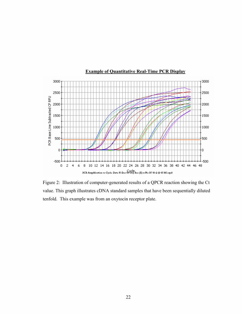
Example of Quantitative Real-Time PCR Display
Figure 2: Illustration of computer-generated results of a QPCR reaction showing the Ct
value. This graph illustrates cDNA standard samples that have been sequentially diluted
tenfold. This example was from an oxytocin receptor plate.
22

Example of QPCR Melt Curve
Figure 3: Graph depicting melt curve analysis obtained following QPCR reaction of
oxytocin receptor cDNA. The melt curve is obtained by plotting the negative first
derivative values of the fluorescence intensity as a function of the temperature, which
results in a peak. The single peak verifies product purity.
23

Once the Ct values were produced by QPCR in triplicate for each mare in the
experiment, a spreadsheet was constructed. The Ct values for each mare were entered
and the arithmetic mean of each triplicate was calculated. The plasmid standard Ct
values were used to construct a linear regression plot with the average Ct value plotted on
the y-axis and the plasmid concentration on the x-axis. From this logistic regression plot,
an R2 value was determined based on the slope of the generated line. A slope-intercept
equation (y=mx+b) was then made and solved for “x” for each mare. This “x” value
represents the cDNA concentration in the initial sample of each mare. An example of
one of the regression plots is shown in Figure 4.
24
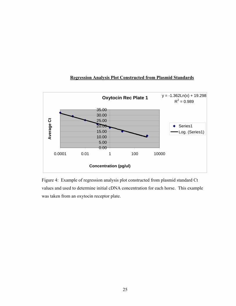
Regression Analysis Plot Constructed from Plasmid Standards
Oxytocin Rec Plate 1 y = -1.362Ln(x) + 19.298R2 = 0.989
0.005.00
10.0015.0020.0025.0030.0035.00
0.0001 0.01 1 100 10000
Concentration (pg/ul)
Ave
rage
Ct
Series1Log. (Series1)
Figure 4: Example of regression analysis plot constructed from plasmid standard Ct
values and used to determine initial cDNA concentration for each horse. This example
was taken from an oxytocin receptor plate.
25
Statistical Analysis
SAS statistical softwarel was used for generation of statistical results. Within this
program, a Kruskal-Wallis test was used for analysis of the data. This method of analysis
is a non-parametric test used to compare three or more independent groups of sampled
data. Unlike parametric tests, the Kruskal-Wallis makes no assumptions about the
distribution of the data (e.g., normality); the test uses the ranks of the data rather than
their raw values to calculate the statistic. Statistical significance was defined as p< 0.05.
26

Results
The expression of the experimental genes oxytocin receptor, PGF2α receptor and
progesterone receptor was normalized using each of two “housekeeping” genes,
ribosomal 18S RNA and beta actin. Regardless of which housekeeping gene was used
for normalization, there was no statistically significant difference discovered among
expression levels of each of the three experimental genes in any of the three categories of
mares examined in this research. The mean Ct values and initial cDNA concentrations
for YF, ON and OS mares were calculated and are reported in Tables 1 and 2. Figure 5
shows graphically the average Ct values + standard deviation for each of the five genes
assayed. Figure 6 shows graphically the calculated initial cDNA concentrations for each
gene.
27

Average Ct Values
Mare
Category
Beta Actin Ribosomal
18S RNA
Oxytocin
Receptor
PGF2α
Receptor
Progesterone
Receptor
Young Fertile
(YF)
26.32 + 2.86 15.64 + 2.45 30.91 + 2.69 34.06 + 2.01 28.87 + 2.64
Older Normal
(ON)
26.62 + 3.05 16.65 + 0.56 31.41 + 2.69 29.47 + 13.06 29.88 + 3.19
Older
Susceptible
(OS)
26.81 + 2.51 15.65 + 2.36 30.71 + 2.76 34.08 + 2.74 29.05 + 2.49
Table 1: Average Ct + standard deviation values for each of the three categories of mares
for each of the five genes assayed.
28
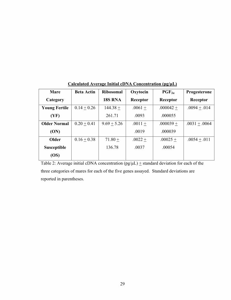
Calculated Average Initial cDNA Concentration (pg/μL)
Mare
Category
Beta Actin Ribosomal
18S RNA
Oxytocin
Receptor
PGF2α
Receptor
Progesterone
Receptor
Young Fertile
(YF)
0.14 + 0.26 144.38 +
261.71
.0061 +
.0093
.000042 +
.000055
.0094 + .014
Older Normal
(ON)
0.20 + 0.41 9.69 + 5.26 .0011 +
.0019
.000039 +
.000039
.0031 + .0064
Older
Susceptible
(OS)
0.16 + 0.38 71.80 +
136.78
.0022 +
.0037
.00025 +
.00054
.0054 + .011
Table 2: Average initial cDNA concentration (pg/μL) + standard deviation for each of the
three categories of mares for each of the five genes assayed. Standard deviations are
reported in parentheses.
29

Figure 5: Graphical representation of average Ct values for each category of mare for
each of the two housekeeping genes and three experimental genes assayed.
30
Calculated Initial cDNA Concentrations
0.00001
0.0001
0.001
0.01
0.1
1
10
100
1000
Beta
Act
in
18S
RN
A
Oxy
toci
n R
ec
PGF2
alph
a R
ec
Prog
este
rone
Rec
Gene
Log
Initi
al c
DN
A C
once
ntra
tion
(pg/
uL)
YFONOS
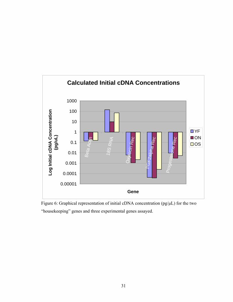
Figure 6: Graphical representation of initial cDNA concentration (pg/μL) for the two
“housekeeping” genes and three experimental genes assayed.
31
The data for each of the three experimental genes was normalized using the
housekeeping gene data. This was accomplished by dividing each mare’s experimental
value by the corresponding mare’s value for either beta actin or ribosomal 18S RNA (ie.
calculated initial cDNA concentration of experimental gene divided by concentration of
beta actin or 18S RNA). When the expression of the three experimental genes was
normalized using beta actin, the difference was not statistically significant at the p<0.05
level; for oxytocin receptor, p=0.0841, for PGF2α receptor, p=0.3881 and for
progesterone receptor, p=0.1507. When the expression was normalized using ribosomal
18S RNA, statistical significance was also not found; for oxytocin receptor, p=0.6674,
for PGF2α receptor, p=0.5866 and for progesterone receptor, p=0.3746.
There was an apparently large degree of variability in the expression of ribosomal
18S RNA among the three categories of mares. This variability reduces the validity of
this gene as a true housekeeping gene that, by definition, should be expressed at similar
levels regardless of age or clinical condition. The large standard deviations reported
indicate the highly variant, non-normally distributed data. For example, the standard
deviation of the average Ct for PGF2α receptor for the older normal mare group was
aberrantly high (13.06) because two of the seven mares’ samples did not recover any
cDNA product and therefore had no average Ct value associated with it. This spuriously
low value caused a large standard deviation to be calculated. The data was re-analyzed
after exclusion of these two mare’s data; statistical significance was still not found at the
p<0.05 level.
32
Discussion and Conclusions
The pathogenesis of delayed uterine clearance was investigated at the molecular
level using real-time PCR to assess the expression of the receptors of the uterotonic
substances oxytocin, progesterone and PGF2α in the endometrium of normal and
clinically affected mares. No significant differences were discovered among any of the
three categories of mares included in this experiment (YF, ON, OS). However, there
remains a great deal of investigation that can be pursued using this same technology
using different intermediates and different tissue types.
The uterus is a myogenic organ, meaning that its smooth musculature can contract
in the absence of hormonal or neural input. This, however, does not imply that agonists
such as oxytocin and PGF2α do not play critical roles in modulating the activity of the
organ. The contraction of myometrial smooth muscle is dependent on calcium, the release
of which is stimulated by action potentials, oxytocin, prostaglandins and other stimuli31.
Molnar and Hertelendy used rat myometrium exposed to oxytocin, PGF2α, endothelin and
inositol 1,4,5-triphosphate (IP3, an intracellular second messenger) to show that the
primary action of PGF2α in myometrial cells is to enhance extracellular Ca2+ influx
through gated channels, whereas oxytocin and endothelin receptors are coupled to
phospholipase C, generating IP3 and raising the intracellular concentration of free Ca2+
from intracellular (sarcoplasmic reticulum) as well as extracellular sources32. Wray also
corroborates this finding in her review on the physiologic mechanisms of uterine
contractility33. Endothelin is a potent vasoconstrictor and has been shown to cause
uterine contractions in multiple species34. The rise in intracellular calcium initiates the
next phases of contraction: the formation of the calcium-calmodulin molecule complex,
activation of myosin light-chain kinase (MLCK), myosin phosphorylation and, finally,
the myosin head’s interaction with actin 35. This work identifies several other
intermediates in the contractile pathway that could be assayed using QPCR (ie MLCK,
MLCP, calmodulin, IP3, endothelin).
33

Although there were no statistically significant differences among oxytocin,
prostaglandin and progesterone receptors in the mares assayed in this study, the
expression of mRNA for oxytocin and progesterone receptors was approaching
significance at the p< 0.05 level (p=0.0841 and p=0.1507, respectively). In future work
using similar study design and molecular techniques, the inclusion of greater numbers of
mares may lead to the finding of statistically significant results with less influence of the
non-normally distributed data. In addition to increasing sample size, the inclusion of
other genes of interest encoding such proteins as MLCK, iNOS, MLCP, relaxin,
endothelin and others as well as the analysis of myometrial tissues may uncover
important factors in the etiology or development of delayed uterine clearance.
It has been suggested, based on the work of Rigby et al, that DUC is ultimately a
result of defective myometrial contraction4. This group determined that intracellular
calcium concentrations of myometrial cells were not altered either in health, with
increasing age or with DUC. This leads to the speculation that downstream calcium
intermediates may play a significant role in the pathogenesis of DUC. After calcium
concentrations have risen, the calmodulin, MLCK and myofibrillar protein molecules
become the integral intermediates leading to myometrial contraction. Future applications
of molecular biological technology should focus on the expression of these molecules in
normal and DUC mares to evaluate their contribution to the disease process.
Rigby’s group looked at myometrial samples whereas our research was focused
on cell surface receptors for uterotonic agonists found in the endometrium. This may
have lead to the absence of significant results in this research. While the endometrium
plays an important role in uterine activity and health, the myometrium is ultimately
responsible for the contractions that lead to the expulsion of inflammatory debris.
Although no significant differences were found in the expression of mRNA encoding the
oxytocin, progesterone and PGF2α receptors, there may indeed be differences in the
expression of these molecules in the myometrium or in the cell-to-cell signaling between
endometrial and myometrial cells. Cell-to-cell signaling molecules and
endometrium/myometrium interactions are sources of future investigation into the
34
pathogenesis of DUC. Work by Carsten and Miller has shown that prostaglandin
receptors are found on the membrane of the sarcoplasmic reticulum in addition to the cell
membrane and that prostaglandin-mediated rises in intracellular calcium may be a result
of a direct intracellular action in combination with a cell surface membrane receptor-
coupled action24. However, the current study is assaying mRNA production for
receptors, regardless of their eventual site of placement and should still be an adequate
gauge of up- or down-regulation of these receptors in normal and DUC mares.
The converse of Rigby’s group’s hypothesis that inappropriate myometrial
contraction causes DUC may also need to be investigated; that is, does excessive
relaxation of the myometrium become a significant contributor to the pathogenesis of
DUC? Uterorelaxants such as nitric oxide19, which is formed by the inducible enzyme
nitric oxide synthase 19,20, the hormone relaxin36, or substances that potentiate myosin
light-chain phosphatase (MLCP) may be over-expressed in DUC mares. MLCP is
responsible for the dephosphorylation of MLCK, turning off the stimulus for
contraction35. MLCP activity is influenced by another phosphorylating molecule, rho A-
associated kinase (ROK); ROK phosphorylates MLCP and renders it inactive37.
Therefore, down-regulation of ROK or a defect in its production may lead to increased
MLCP activity, reducing MLCK-mediated myosin phosphorylation and, hence, inhibition
of myometrial contraction. Cyclic AMP (cAMP) is generated from the enzyme adenylate
cyclase and this intracellular second-messenger has been shown to cause a relaxation of
myometrial and other smooth muscles via the activation of cAMP-dependent protein
kinase38. Up-regulation of adenylate cyclase may lead to excessive uterine relaxation and
DUC. The uterus is also endowed with adrenergic receptors, both α and β; α receptors,
particularly α1, are primarily associated with uterine contraction whereas β receptors are
associated with relaxation34. The expression of these β receptors may be aberrantly over-
expressed in DUC mares, leading to myometrial relaxation. Myometrial relaxation can
be assessed in vivo using pressure transducers or in vitro via myometrial muscle biopsy
tension generation as described by Rigby4. Herein lie other arms of the myometrial
35

regulatory pathway that must be investigated at the molecular or ultrastructural level to
help elucidate the pathogenesis of DUC in mares.
The role of magnesium in DUC mares may be an important avenue to investigate.
Magnesium is known to be a co-factor necessary for proper oxytocin/oxytocin receptor
interaction39 Magnesium is a potent tocolytic used for uterine relaxation in pre-term
labor in humans35. Hurd et al demonstrated the antagonistic effect that magnesium has
on oxytocin-induced myometrial contraction. This group showed that pretreatment of
cultured myometrial cells with magnesium caused a reduction in intracellular calcium in
response to exposure to oxytocin40. Laser flow cytometric analysis of fluorescent
intensity using magnesium-sensitive dyes to assay magnesium levels from mares’
myometrium could be performed to see if the amount of magnesium in DUC mares
differs from that found in normal mares. This flow cytometry technology has been used
to assay magnesium in other tissues and could be applied to uterine tissues, as well41.
The hypothesis that DUC mares would under-express receptors for oxytocin was
made prior to this investigation; the results of this research did not support this
hypothesis. This could mean that the receptors are constitutively expressed in heath or
diseased uteruses and the oxytocin secretion or binding is impaired with increasing age or
with disease. The work of Sharp et al suggests that the affinity of oxytocin receptors for
oxytocin is reduced in pregnant mares, which are under the influence of progesterone42.
Murata et al has shown that oxytocin receptor mRNA is up-regulated in the presence of
increased circulating estrogen concentrations in the rat43. However, in this research
project, the oxytocin receptor mRNA is ubiquitously expressed in these mares which
were in late estrus or early diestrus, and should have been under the influence of high
estrogen concentrations.
Myometrial cell prostaglandin production stimulated by oxytocin has not been
reported for the mare as it has been in other species4. Nikolakopoulos et al showed that
DUC mares had reduced circulating levels of PGF2α following both endogenous oxytocin
release and after exogenous oxytocin administration44. In the face of normal, ubiquitous
PGF2α receptor production as demonstrated in the current project, this may explain why
36

DUC mares do not generate appropriate myometrial contractions—the receptors exist but
the circulating levels of PGF2α are inadequate to cause signal transduction and initiation
of the influx of intracellular calcium.
37
Literature Cited
1. Fumuso E, Giguere S, Wade J, et al. Endometrial IL-1beta, IL-6 and TNF-alpha, mRNA expression in mares resistant or susceptible to post-breeding endometritis. Effects of estrous cycle, artificial insemination and immunomodulation. Vet Immunol Immunopathol 2003;96:31-41.
2. Watson ED. Post-breeding endometritis in the mare. Anim Reprod Sci 2000;60-61:221-232.
3. von Reitzenstein M, Callahan MA, Hansen PJ, et al. Aberrations in uterine contractile patterns in mares with delayed uterine clearance after administration of detomidine and oxytocin. Theriogenology 2002;58:887-898.
4. Rigby SL, Barhoumi R, Burghardt RC, et al. Mares with delayed uterine clearance have an intrinsic defect in myometrial function. Biol Reprod 2001;65:740-747.
5. LeBlanc M, Neuwirth L, Mauragis D, et al. Oxytocin enhances clearance of radiocolloid from the uterine lumen of reproductively normal mares and mares susceptible to endometritis. Equine Vet J 1994;26:279-282.
6. Ohmichi M, Koike K, Kimura A, et al. Role of mitogen-activated protein kinase pathway in prostaglandin F2alpha-induced rat puerperal uterine contraction. Endocrinology 1997;138:3103-3111.
7. Troedsson MH, Ababneh MM, Ohlgren AF, et al. Effect of periovulatory prostaglandin F2alpha on pregnancy rates and luteal function in the mare. Theriogenology 2001;55:1891-1899.
8. Belknap J. Molecular Biology in Equine Medicine: Current and Future Applications. Compend on Cont Ed 1997;19:224-237.
9. Zent WW, Troedsson MH, Xue JL. Postbreeding uterine fluid accumulation in a normal population of thoroughbred mares: A field study. Proc Am Assoc Equine Pract 1998;44:64-65.
10. Traub-Dargatz JL, Salman MD, Voss JL. Medical problems of adult horses, as ranked by equine practitioners. J Am Vet Med Assoc 1991;198:1745-1747.
11. Troedsson MH, Liu IK. Uterine clearance of non-antigenic markers (51Cr) in response to a bacterial challenge in mares potentially susceptible and resistant to chronic uterine infections. J Reprod Fertil Suppl 1991;44:283-288.
38
12. LeBlanc MM, Asbury AC, Lyle SK. Uterine clearance mechanisms during the early postovulatory period in mares. Am J Vet Res 1989;50:864-867.
13. LeBlanc MM, Neuwirth L, Asbury AC, et al. Scintigraphic measurement of uterine clearance in normal mares and mares with recurrent endometritis. Equine Vet J 1994;26:109-113.
14. Evans MJ, Hamer JM, Gason LM, et al. Factors affecting uterine clearance of inoculated materials in mares. J Reprod Fertil Suppl 1987;35:327-334.
15. Nikolakopoulos E, Watson ED. Uterine contractility is necessary for the clearance of intrauterine fluid but not bacteria after bacterial infusion in the mare. Theriogenology 1999;52:413-423.
16. Ritchison G. Human Physiology-Muscle, http://people.eku.edu/ritchisong/301notes3.htm; 2006.
17. LeBlanc MM, Neuwirth L, Jones L, et al. Differences in uterine position of reproductively normal mares and those with delayed uterine clearance detected by scintigraphy. Theriogenology 1998;50:49-54.
18. Le Blanc MM, Johnson, R.D., Calderwood Mays, M.B. and Valderrama, C. Lymphatic clearance of India ink in reproductively normal mares and mares susceptible to endometritis. Biol Reprod Mono Ser 1995;1:501-506.
19. Alghamdi AS, Foster DN, Carlson CS, et al. Nitric oxide levels and nitric oxide synthase expression in uterine samples from mares susceptible and resistant to persistent breeding-induced endometritis. Am J Reprod Immunol 2005;53:230-237.
20. Fang L, Nowicki BJ, Dong YL, et al. Localized increase in nitric oxide production and the expression of nitric oxide synthase isoforms in rat uterus with experimental intrauterine infection. Am J Obstet Gynecol 1999;181:601-609.
21. Bae SE, Watson ED. A light microscopic and ultrastructural study on the presence and location of oxytocin in the equine endometrium. Theriogenology 2003;60:909-921.
22. Behrendt-Adam CY, Adams MH, Simpson KS, et al. Oxytocin-neurophysin I mRNA abundance in equine uterine endometrium. Domest Anim Endocrinol 1999;16:183-192.
23. Cadario ME, Merritt AM, Archbald LF, et al. Changes in intrauterine pressure after oxytocin administration in reproductively normal mares and in those with a delay in uterine clearance. Theriogenology 1999;51:1017-1025.
39
24. Carsten ME, Miller J.D. Calcium Control Mechanisms in the Myometrial
Cell and the Role of the Phosphoinositide Cycle In: Carsten ME MJ, ed. Uterine Function: Molecular and Cellular Aspects. New York: Plenum Press, 1990;121-167.
25. Cadario ME, Thatcher WW, Klapstein E, et al. Dynamics of prostaglandin secretion, intrauterine fluid and uterine clearance in reproductively normal mares and mares with delayed uterine clearance. Theriogenology 1999;52:1181-1192.
26. Jones DM, Fielden ED, Carr DH. Some physiological and pharmacological factors affecting uterine motility as measured by electromyography in the mare. J Reprod Fertil Suppl 1991;44:357-368.
27. Troedsson MH, Wistrom AO, Liu IK, et al. Registration of myometrial activity using multiple site electromyography in cyclic mares. J Reprod Fertil 1993;99:299-306.
28. Squires EL. Progesterone In: McKinnon AOaV, J.L., ed. Equine Reproduction. Phildadelphia: Lea and Febiger, 1993;57-64.
29. Canning MB, Billington WD. Hormonal regulation of immunoglobulins and plasma cells in the mouse uterus. J Endocrinol 1983;97:419-424.
30. Fomin VP, Cox BE, Word RA. Effect of progesterone on intracellular Ca2+ homeostasis in human myometrial smooth muscle cells. Am J Physiol 1999;276:C379-385.
31. Luckas MJ, Taggart MJ, Wray S. Intracellular calcium stores and agonist-induced contractions in isolated human myometrium. Am J Obstet Gynecol 1999;181:468-476.
32. Molnar M, Hertelendy F. Signal transduction in rat myometrial cells: comparison of the actions of endothelin-1, oxytocin and prostaglandin F2 alpha. Eur J Endocrinol 1995;133:467-474.
33. Wray S, Kupittayanant S, Shmygol A, et al. The physiological basis of uterine contractility: a short review. Exp Physiol 2001;86:239-246.
34. Wray S. Uterine contraction and physiological mechanisms of modulation. Am J Physiol 1993;264:C1-18.
35. Wray S, Jones K, Kupittayanant S, et al. Calcium signaling and uterine contractility. J Soc Gynecol Investig 2003;10:252-264.
40
36. Nishikori K, Weisbrodt NW, Sherwood OD, et al. Effects of relaxin on rat uterine myosin light chain kinase activity and myosin light chain phosphorylation. J Biol Chem 1983;258:2468-2474. 37. Woodcock NA, Taylor CW, Thornton S. Effect of an oxytocin receptor antagonist and rho kinase inhibitor on the [Ca++]i sensitivity of human myometrium. Am J Obstet Gynecol 2004;190:222-228.
38. Price SA, Bernal AL. Uterine quiescence: the role of cyclic AMP. Exp Physiol 2001;86:265-272.
39. Gimpl G, Fahrenholz F. The oxytocin receptor system: structure, function, and regulation. Physiol Rev 2001;81:629-683.
40. Hurd WW, Natarajan V, Fischer JR, et al. Magnesium sulfate inhibits the oxytocin-induced production of inositol 1,4,5-trisphosphate in cultured human myometrial cells. Am J Obstet Gynecol 2002;187:419-424.
41. Mooren FC, Golf SW, Lechtermann A, et al. Alterations of ionized Mg2+ in human blood after exercise. Life Sci 2005;77:1211-1225.
42. Sharp DC, Thatcher MJ, Salute ME, et al. Relationship between endometrial oxytocin receptors and oxytocin-induced prostaglandin F2 alpha release during the oestrous cycle and early pregnancy in pony mares. J Reprod Fertil 1997;109:137-144.
43. Murata T, Narita K, Honda K, et al. Changes of receptor mRNAs for oxytocin and estrogen during the estrous cycle in rat uterus. J Vet Med Sci 2003;65:707-712.
44. Nikolakopoulos E KH, Watson ED. Oxytocin and PGF-2 release in mares resistant and susceptible to persistent mating-induced endometritis. Proceedings of the 7th International Symposium on Equine Reproduction 1998;Abstract 95:Abstract 95.
41

Endnotes a Qiagen Inc., Valencia, CA 91355 b Tecan Group Ltd, Männedorf, Switzerland c Invitrogen Corporation, Carlsbad, California 92008 d Bio-Rad Laboratories, Hercules, CA 94547 e Eppendorf North America, Westbury, NY 11590 f http://frodo.wi.mit.edu/cgi-bin/primer3/primer3_www.cgi g http://www.ncbi.nlm.nih.gov/entrez/query.fcgi?db=Nucleotide&itool=toolbarh SeqWeb v.3; Accelrys Inc., San Diego, CA 92121 i Operon Biotechnologies, Inc., Huntsville, AL 35805 j https://www.vbi.vt.edu/k http://www.ncbi.nlm.nih.gov/BLAST/lSAS Institute Inc., Cary, NC 27513
42

Vita
Giles Anthony Gray is the son of Mr. and Mrs. Giles Gray, Jr. of Castlewood,
VA. Anthony was born in Bristol, TN and raised outside of Lebanon, VA. He graduated
valedictorian of Lebanon High School in 1992 and then attended the Virginia Military
Institute on a full academic scholarship. He graduated from VMI in 1996 with a B.S. in
Biology with minors in Chemistry and Psychology. He attended graduate school at the
University of Virginia before coming to veterinary school at the Virginia-Maryland
Regional College of Veterinary Medicine in 1997. He graduated from VMRCVM in
2001 and completed a rotating internship in Equine Field Services and Large Animal
Medicine at VMRCVM in 2002. He worked as an associate veterinarian at Davie County
Large Animal Hospital in Mocksville, NC for a year before accepting a position as a
resident in Equine Field Services at VMRCVM. Anthony has passed the certification
exam to become a Diplomate in the American Board of Veterinary Practitioners as an
Equine Specialist. Anthony is married to Kerri Carico Gray, a senior veterinary student
at VMRCVM, class of 2006. The couple will be moving to Bristol, TN where Anthony
will be a partner at Abingdon Equine Veterinary Services and Kerri will be a small
animal emergency vet at the Airport Pet Emergency Clinic in Blountville, TN.
43


![Control of the MARES Autonomous Underwater Vehicledee09011/Bruno_Ferreiras_personal_webpage/... · A. MARES AUV MARES, or Modular Autonomous Robot for Environment Sampling [1-2],](https://img.pdfslide.us/doc/110x75/5ecae8c4906b58548b38bd0a/control-of-the-mares-autonomous-underwater-vehicle-dee09011brunoferreiraspersonalwebpage.jpg)