Embed Size (px)
Citation preview
article are available in the G&G Data Depository(www.gia.edu/gandg).
MATERIALS AND METHODSFrom the parcel of rough, we randomly selectedabout 150 specimens for examination that rangedfrom ~0.4 to 3 g. We studied 15 samples in detail fortheir crystallographic features, surface markings,cleavage directions, and pleochroism with respect tocrystallographic orientation. These samples exhibit-ed a variety of colors: green, yellowish green tobrownish green, brownish pink, and distinctly bicol-ored green/pink. In addition, we selected six samplesof the pinkish brown to brownish pink variety chias-tolite, which displayed distinct (sometimes partial)cross-shaped arrangements of dark inclusions. About20% of the 150 specimens were chiastolite, and theyranged in diaphaneity from transparent to opaque.
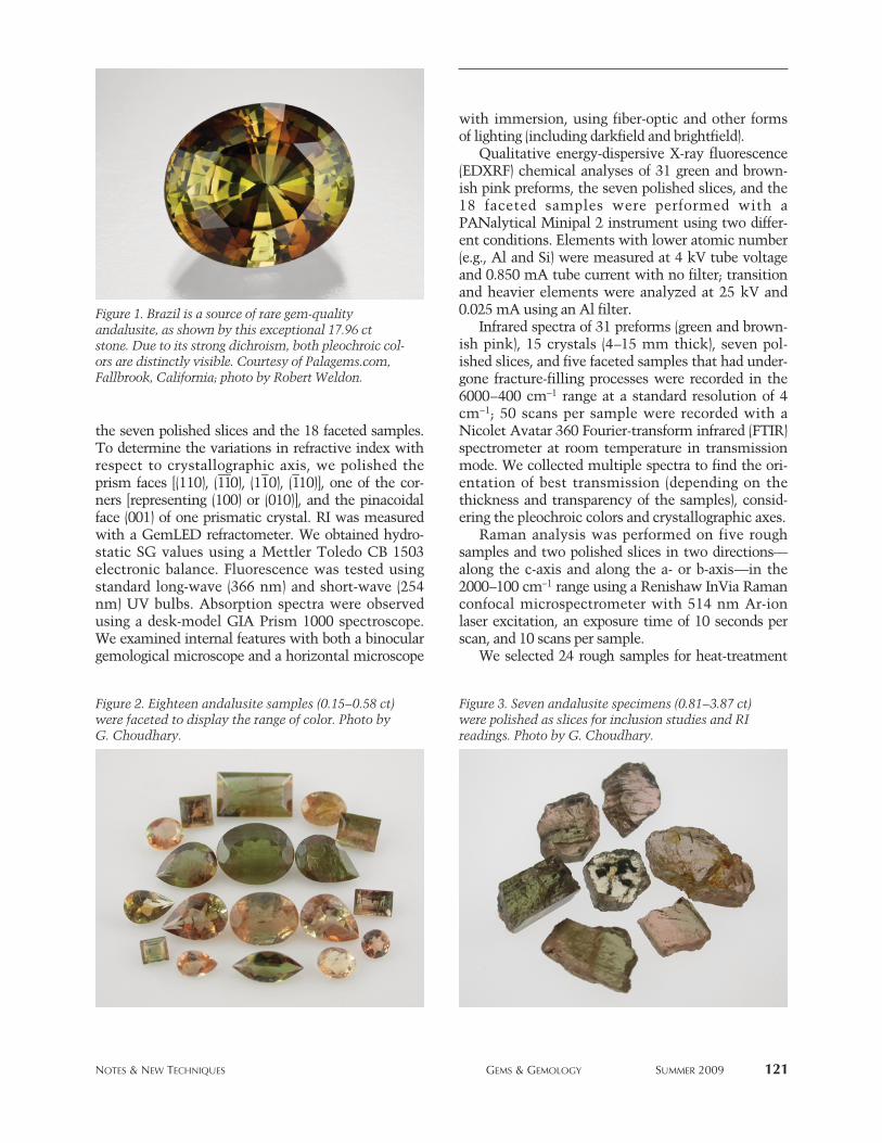
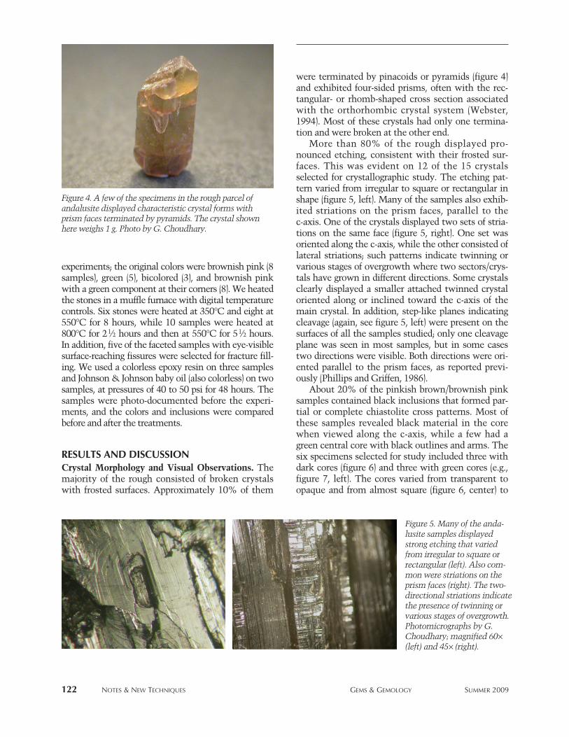
We selected 72 of the 150 specimens, of moderatecommercial quality, for fashioning on the basis oftheir coloration, clarity, and projected yield. Of these,18 were faceted (figure 2) and the other 54 were leftpreformed. The preformed stones were categorizedinto three color groups: green (17 specimens), brown-ish pink (14), and strongly pleochroic pinkishbrown/yellowish green (23). On an additional sevenspecimens (figure 3), we polished the largest parallelsurfaces so we could obtain RI readings and examinetheir internal features.
Standard gemological tests—including color obser-vations, SG measurements, fluorescence reactions,and visible-range spectroscopy—were performed onall samples. RI and optic sign were measured only on
GEM-QUALITY ANDALUSITEFROM BRAZIL
Shyamala Fernandes and Gagan Choudhary
See end of article for About the Authors and Acknowledgments.GEMS & GEMOLOGY, Vol. 45, No. 2, pp. 120–129.© 2009 Gemological Institute of America
120 NOTES & NEW TECHNIQUES GEMS & GEMOLOGY SUMMER 2009
he name andalusite is derived from the Andalusiaregion of southern Spain, where the mineral was
first discovered (Dana and Ford, 1992). Gem-qualitymaterial is known from Brazil (Espírito Santo andMinas Gerais States), Sri Lanka, the United States,Madagascar, Russia (Siberia), Myanmar (O’Donoghue,2006), and China (Liu, 2006). Andalusite varietiesinclude highly pleochroic brownish pink/green (e.g.,figure 1), Mn-rich bright green (“viridine”), and chias-tolite. The last is typically opaque and features a darkcross-shaped pattern formed by carbonaceous inclu-sions (Webster, 1994; O’Donoghue, 2006).
This article reports the properties recorded froman ~1 kg parcel of medium-quality andalusite thatwas purchased from a gem merchant in Jaipur, India,who deals in rough from Brazil. The exact source ofthe andalusite in Brazil was, unfortunately, notavailable. Additional photos to accompany this
NOTES & NEW TECHNIQUES
T
Widely known as a rock-forming mineral,andalusite is not frequently encountered as agem material. This article documents the gemo-logical and spectroscopic properties of numer-ous rough and cut samples of gem-qualityandalusite from Brazil. The color varied fromyellowish green and green to brownish pink(including bicolored green and brownish pink),and some were faceted with the pleochroic col-ors attractively oriented. Transparent samples ofthe chiastolite variety of andalusite were partic-ularly interesting. All the samples displayed acomplex growth pattern, with the various inclu-sions oriented along the growth directions. Heattreatment produced subtle changes in color, ifany, but fracture filling with resin resulted inobvious improvements in apparent clarity.
the seven polished slices and the 18 faceted samples.To determine the variations in refractive index withrespect to crystallographic axis, we polished theprism faces [(110), (11
—0), (11
–0), (1
–10)], one of the cor-
ners [representing (100) or (010)], and the pinacoidalface (001) of one prismatic crystal. RI was measuredwith a GemLED refractometer. We obtained hydro-static SG values using a Mettler Toledo CB 1503electronic balance. Fluorescence was tested usingstandard long-wave (366 nm) and short-wave (254nm) UV bulbs. Absorption spectra were observedusing a desk-model GIA Prism 1000 spectroscope.We examined internal features with both a binoculargemological microscope and a horizontal microscope
with immersion, using fiber-optic and other forms of lighting (including darkfield and brightfield).
Qualitative energy-dispersive X-ray fluorescence(EDXRF) chemical analyses of 31 green and brown-ish pink preforms, the seven polished slices, and the18 faceted samples were performed with aPANalytical Minipal 2 instrument using two differ-ent conditions. Elements with lower atomic number(e.g., Al and Si) were measured at 4 kV tube voltageand 0.850 mA tube current with no filter; transitionand heavier elements were analyzed at 25 kV and0.025 mA using an Al filter.
Infrared spectra of 31 preforms (green and brown-ish pink), 15 crystals (4–15 mm thick), seven pol-ished slices, and five faceted samples that had under-gone fracture-filling processes were recorded in the6000–400 cm−1 range at a standard resolution of 4cm−1; 50 scans per sample were recorded with aNicolet Avatar 360 Fourier-transform infrared (FTIR)spectrometer at room temperature in transmissionmode. We collected multiple spectra to find the ori-entation of best transmission (depending on thethickness and transparency of the samples), consid-ering the pleochroic colors and crystallographic axes.
Raman analysis was performed on five roughsamples and two polished slices in two directions—along the c-axis and along the a- or b-axis—in the2000–100 cm−1 range using a Renishaw InVia Ramanconfocal microspectrometer with 514 nm Ar-ionlaser excitation, an exposure time of 10 seconds perscan, and 10 scans per sample.
We selected 24 rough samples for heat-treatment
NOTES & NEW TECHNIQUES GEMS & GEMOLOGY SUMMER 2009 121
Figure 2. Eighteen andalusite samples (0.15–0.58 ct)were faceted to display the range of color. Photo byG. Choudhary.
Figure 3. Seven andalusite specimens (0.81–3.87 ct)were polished as slices for inclusion studies and RIreadings. Photo by G. Choudhary.
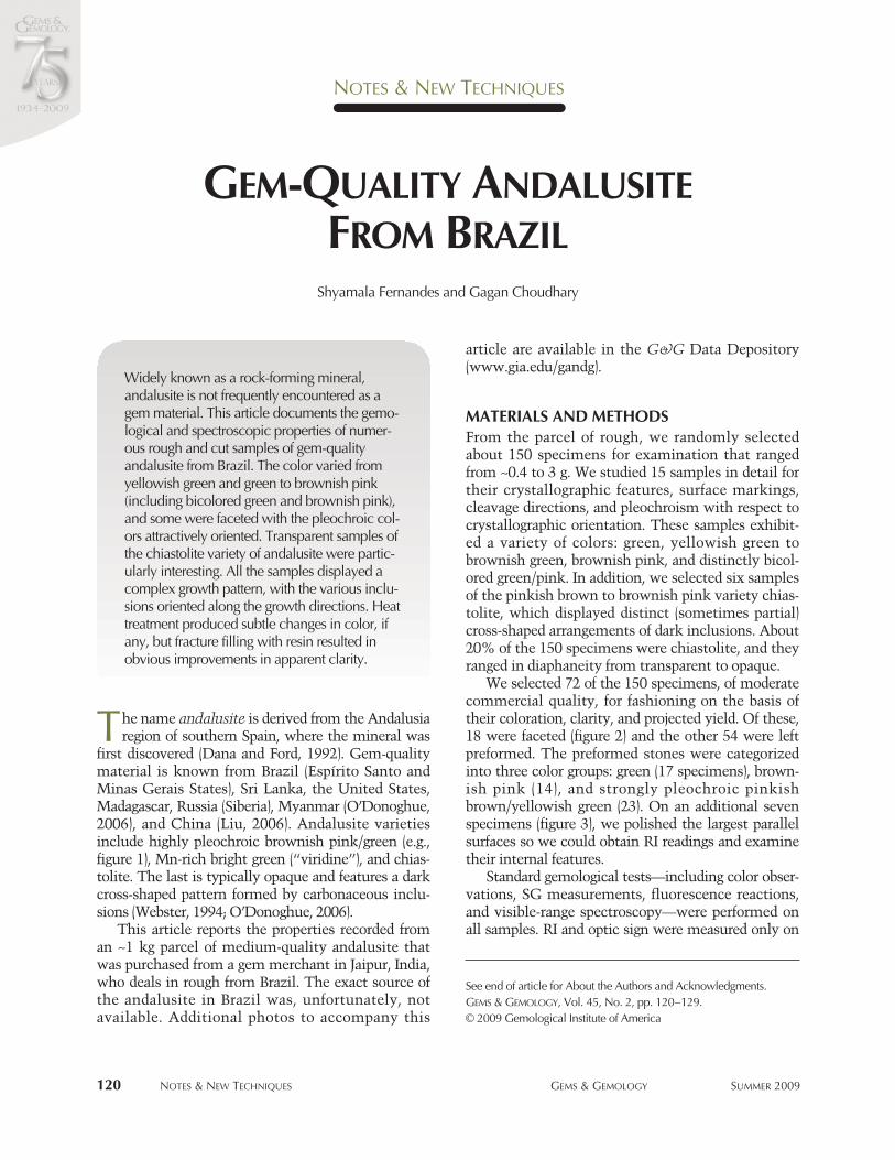
Figure 1. Brazil is a source of rare gem-qualityandalusite, as shown by this exceptional 17.96 ctstone. Due to its strong dichroism, both pleochroic col-ors are distinctly visible. Courtesy of Palagems.com,Fallbrook, California; photo by Robert Weldon.
122 NOTES & NEW TECHNIQUES GEMS & GEMOLOGY SUMMER 2009
experiments; the original colors were brownish pink (8samples), green (5), bicolored (3), and brownish pinkwith a green component at their corners (8). We heatedthe stones in a muffle furnace with digital temperaturecontrols. Six stones were heated at 350°C and eight at550°C for 8 hours, while 10 samples were heated at800°C for 21⁄2 hours and then at 550°C for 51⁄2 hours.In addition, five of the faceted samples with eye-visiblesurface-reaching fissures were selected for fracture fill-ing. We used a colorless epoxy resin on three samplesand Johnson & Johnson baby oil (also colorless) on twosamples, at pressures of 40 to 50 psi for 48 hours. Thesamples were photo-documented before the experi-ments, and the colors and inclusions were comparedbefore and after the treatments.
RESULTS AND DISCUSSIONCrystal Morphology and Visual Observations. Themajority of the rough consisted of broken crystalswith frosted surfaces. Approximately 10% of them
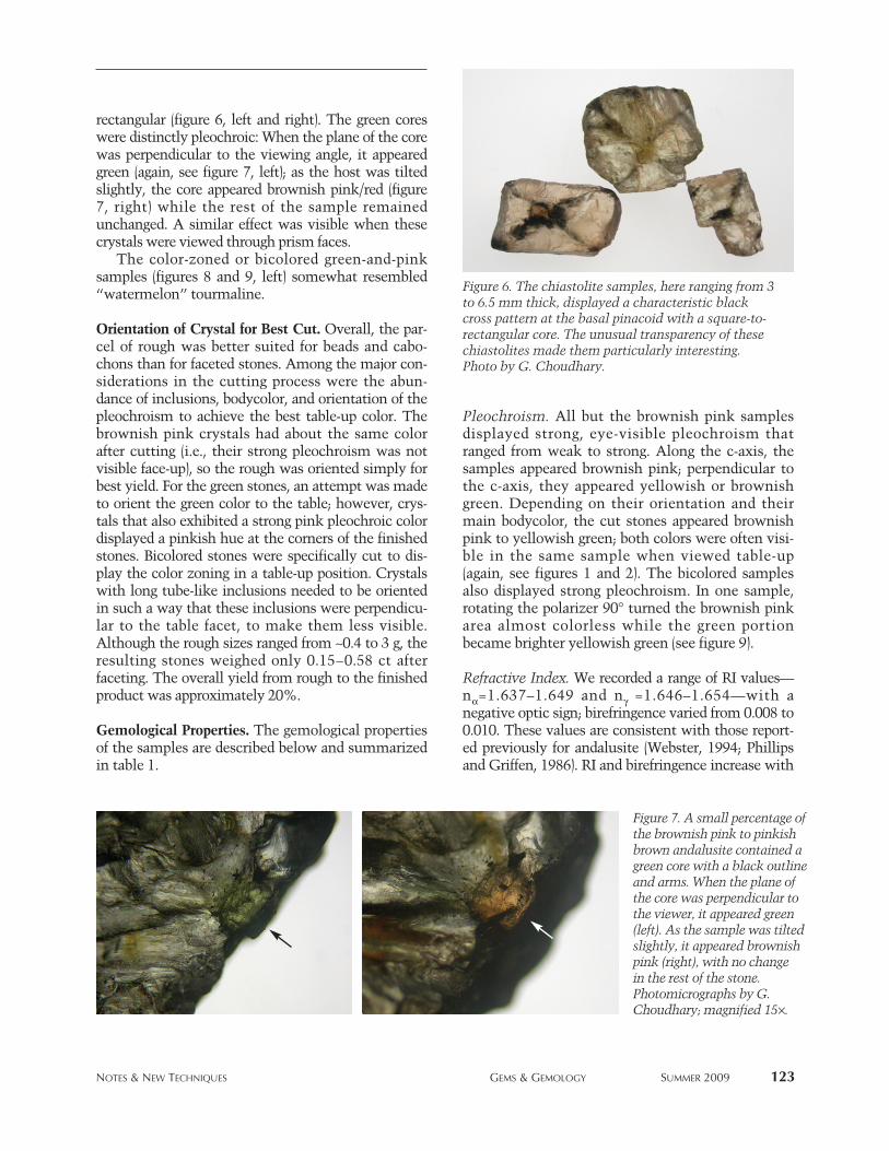
were terminated by pinacoids or pyramids (figure 4)and exhibited four-sided prisms, often with the rec-tangular- or rhomb-shaped cross section associatedwith the orthorhombic crystal system (Webster,1994). Most of these crystals had only one termina-tion and were broken at the other end.
More than 80% of the rough displayed pro-nounced etching, consistent with their frosted sur-faces. This was evident on 12 of the 15 crystalsselected for crystallographic study. The etching pat-tern varied from irregular to square or rectangular inshape (figure 5, left). Many of the samples also exhib-ited striations on the prism faces, parallel to the c-axis. One of the crystals displayed two sets of stria-tions on the same face (figure 5, right). One set wasoriented along the c-axis, while the other consisted oflateral striations; such patterns indicate twinning orvarious stages of overgrowth where two sectors/crys-tals have grown in different directions. Some crystalsclearly displayed a smaller attached twinned crystaloriented along or inclined toward the c-axis of themain crystal. In addition, step-like planes indicatingcleavage (again, see figure 5, left) were present on thesurfaces of all the samples studied; only one cleavageplane was seen in most samples, but in some casestwo directions were visible. Both directions were ori-ented parallel to the prism faces, as reported previ-ously (Phillips and Griffen, 1986).
About 20% of the pinkish brown/brownish pinksamples contained black inclusions that formed par-tial or complete chiastolite cross patterns. Most ofthese samples revealed black material in the corewhen viewed along the c-axis, while a few had agreen central core with black outlines and arms. Thesix specimens selected for study included three withdark cores (figure 6) and three with green cores (e.g.,figure 7, left). The cores varied from transparent toopaque and from almost square (figure 6, center) to
Figure 4. A few of the specimens in the rough parcel ofandalusite displayed characteristic crystal forms withprism faces terminated by pyramids. The crystal shownhere weighs 1 g. Photo by G. Choudhary.
Figure 5. Many of the anda-lusite samples displayedstrong etching that variedfrom irregular to square orrectangular (left). Also com-mon were striations on theprism faces (right). The two-directional striations indicatethe presence of twinning orvarious stages of overgrowth.Photomicrographs by G.Choudhary; magnified 60×(left) and 45× (right).
NOTES & NEW TECHNIQUES GEMS & GEMOLOGY SUMMER 2009 123
rectangular (figure 6, left and right). The green coreswere distinctly pleochroic: When the plane of the corewas perpendicular to the viewing angle, it appearedgreen (again, see figure 7, left); as the host was tiltedslightly, the core appeared brownish pink/red (figure7, right) while the rest of the sample remainedunchanged. A similar effect was visible when thesecrystals were viewed through prism faces.
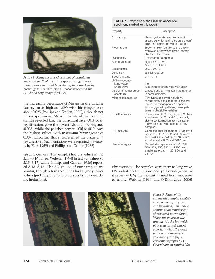
The color-zoned or bicolored green-and-pinksamples (figures 8 and 9, left) somewhat resembled“watermelon” tourmaline.
Orientation of Crystal for Best Cut. Overall, the par-cel of rough was better suited for beads and cabo-chons than for faceted stones. Among the major con-siderations in the cutting process were the abun-dance of inclusions, bodycolor, and orientation of thepleochroism to achieve the best table-up color. Thebrownish pink crystals had about the same colorafter cutting (i.e., their strong pleochroism was notvisible face-up), so the rough was oriented simply forbest yield. For the green stones, an attempt was madeto orient the green color to the table; however, crys-tals that also exhibited a strong pink pleochroic colordisplayed a pinkish hue at the corners of the finishedstones. Bicolored stones were specifically cut to dis-play the color zoning in a table-up position. Crystalswith long tube-like inclusions needed to be orientedin such a way that these inclusions were perpendicu-lar to the table facet, to make them less visible.Although the rough sizes ranged from ~0.4 to 3 g, theresulting stones weighed only 0.15–0.58 ct afterfaceting. The overall yield from rough to the finishedproduct was approximately 20%.
Gemological Properties. The gemological propertiesof the samples are described below and summarizedin table 1.
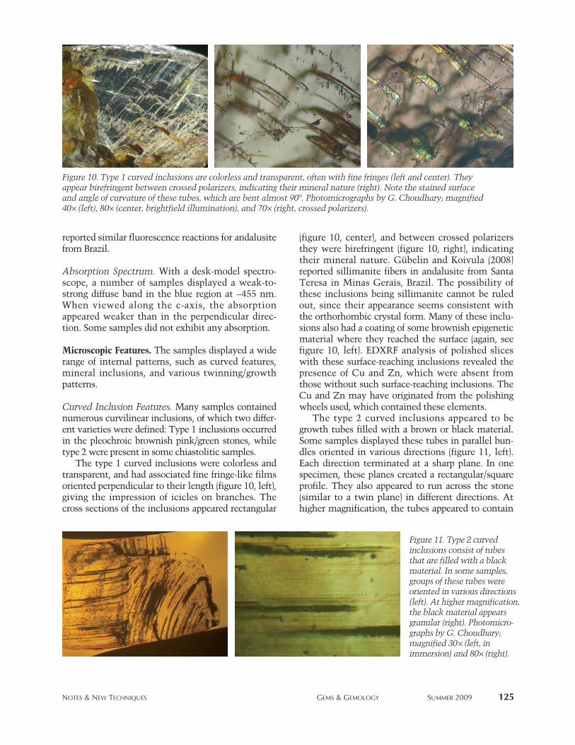
Pleochroism. All but the brownish pink samplesdisplayed strong, eye-visible pleochroism thatranged from weak to strong. Along the c-axis, thesamples appeared brownish pink; perpendicular tothe c-axis, they appeared yellowish or brownishgreen. Depending on their orientation and theirmain bodycolor, the cut stones appeared brownishpink to yellowish green; both colors were often visi-ble in the same sample when viewed table-up(again, see figures 1 and 2). The bicolored samplesalso displayed strong pleochroism. In one sample,rotating the polarizer 90° turned the brownish pinkarea almost colorless while the green portionbecame brighter yellowish green (see figure 9).
Refractive Index. We recorded a range of RI values—nα=1.637–1.649 and nγ =1.646–1.654—with a negative optic sign; birefringence varied from 0.008 to0.010. These values are consistent with those report-ed previously for andalusite (Webster, 1994; Phillipsand Griffen, 1986). RI and birefringence increase with
Figure 6. The chiastolite samples, here ranging from 3to 6.5 mm thick, displayed a characteristic blackcross pattern at the basal pinacoid with a square-to-rectangular core. The unusual transparency of thesechiastolites made them particularly interesting.Photo by G. Choudhary.
Figure 7. A small percentage ofthe brownish pink to pinkishbrown andalusite contained agreen core with a black outlineand arms. When the plane ofthe core was perpendicular tothe viewer, it appeared green(left). As the sample was tiltedslightly, it appeared brownishpink (right), with no change in the rest of the stone.Photomicrographs by G.Choudhary; magnified 15×.
the increasing percentage of Mn (as in the viridinevariety) to as high as 1.690 with birefringence ofabout 0.025 (Phillips and Griffen, 1986), although notin our specimens. Measurements of the orientedsample revealed that the pinacoidal face (001), or α-ray direction, gave the lowest RIs and birefringence(0.008), while the polished corner (100) or (010) gavethe highest values (with maximum birefringence of0.009), indicating that it represented the b-axis or γ-ray direction. Such variations were reported previous-ly by Kerr (1959) and Phillips and Griffen (1986).
Specific Gravity. The samples had SG values in the3.11–3.16 range. Webster (1994) listed SG values of3.15–3.17, while Phillips and Griffen (1986) report-ed 3.13–3.16. The SG values of our samples aresimilar, though a few specimens had slightly lowervalues (probably due to fractures and surface-reach-ing inclusions).
Fluorescence. The samples were inert to long-waveUV radiation but fluoresced yellowish green toshort-wave UV; the intensity varied from moderateto strong. Webster (1994) and O’Donoghue (2006)
124 NOTES & NEW TECHNIQUES GEMS & GEMOLOGY SUMMER 2009
TABLE 1. Properties of the Brazilian andalusite specimens studied for this report.
Property Description
Color range Green, yellowish green to brownish green, brownish pink, bicolored green/pink, and pinkish brown (chiastolite)
Pleochroism Brownish pink (parallel to the c-axis)Yellowish or brownish green (perpen-dicular to the c-axis)
Diaphaneity Transparent to opaqueRefractive index nα = 1.637–1.649
nγ = 1.646–1.654Birefringence 0.008-0.010Optic sign Biaxial negative Specific gravity 3.11–3.16UV fluorescence
Long-wave InertShort-wave Moderate to strong yellowish green
Visible-range absorption Diffuse band at ~455 (weak to strong)spectrum in some samples
Microscopic features Two types of curved inclusions, minute films/discs, numerous mineral inclusions, “fingerprints,” pinpoints, twinning/growth patterns, cross pat-terns in chiastolite varieties
EDXRF analysis Presence of Al, Si, Fe, Ca, and Cr (twospecimens had Zn and Cu, probably due to contamination from the polish-ing wheels); no Mn detected in any samples
FTIR analysis Complete absorption up to 2100 cm−1;peaks at ~3687, 3652, and 3624 cm−1;twin peaks at ~3520 and 3460 cm−1; shoulders at ~3280 and 3095 cm−1
Raman analysis Several sharp peaks at ~1063, 917, 550, 463, 356, 325, and 290 cm−1; smaller peaks at ~1120, 850, and 717 cm−1
Figure 9. Many of theandalusite samples exhibit-ed color zoning in greenand brownish pink (left), acombination reminiscentof bicolored tourmalines.When the polarizer wasrotated 90°, the brownishpink area turned almostcolorless, while the greenportion became brighteryellowish green (right).Photomicrographs by G.Choudhary; magnified 25×.
Figure 8. Many bicolored samples of andalusiteappeared to display various growth stages, withtheir colors separated by a sharp plane marked bybrown granular inclusions. Photomicrograph byG. Choudhary; magnified 25×.
reported similar fluorescence reactions for andalusitefrom Brazil.
Absorption Spectrum. With a desk-model spectro-scope, a number of samples displayed a weak-to-strong diffuse band in the blue region at ~455 nm.When viewed along the c-axis, the absorptionappeared weaker than in the perpendicular direc-tion. Some samples did not exhibit any absorption.
Microscopic Features. The samples displayed a widerange of internal patterns, such as curved features,mineral inclusions, and various twinning/growthpatterns.
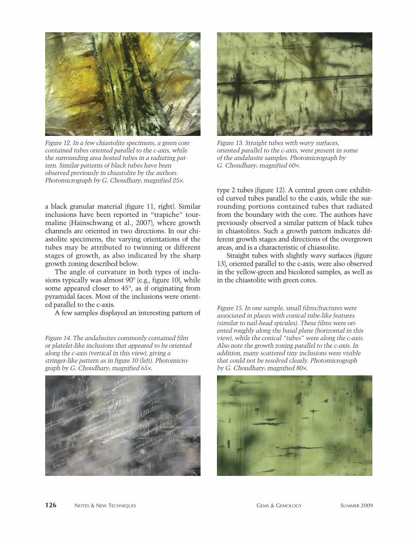
Curved Inclusion Features. Many samples containednumerous curvilinear inclusions, of which two differ-ent varieties were defined: Type 1 inclusions occurredin the pleochroic brownish pink/green stones, whiletype 2 were present in some chiastolitic samples.
The type 1 curved inclusions were colorless andtransparent, and had associated fine fringe-like filmsoriented perpendicular to their length (figure 10, left),giving the impression of icicles on branches. Thecross sections of the inclusions appeared rectangular
(figure 10, center), and between crossed polarizersthey were birefringent (figure 10, right), indicatingtheir mineral nature. Gübelin and Koivula (2008)reported sillimanite fibers in andalusite from SantaTeresa in Minas Gerais, Brazil. The possibility ofthese inclusions being sillimanite cannot be ruledout, since their appearance seems consistent withthe orthorhombic crystal form. Many of these inclu-sions also had a coating of some brownish epigeneticmaterial where they reached the surface (again, seefigure 10, left). EDXRF analysis of polished sliceswith these surface-reaching inclusions revealed thepresence of Cu and Zn, which were absent fromthose without such surface-reaching inclusions. TheCu and Zn may have originated from the polishingwheels used, which contained these elements.
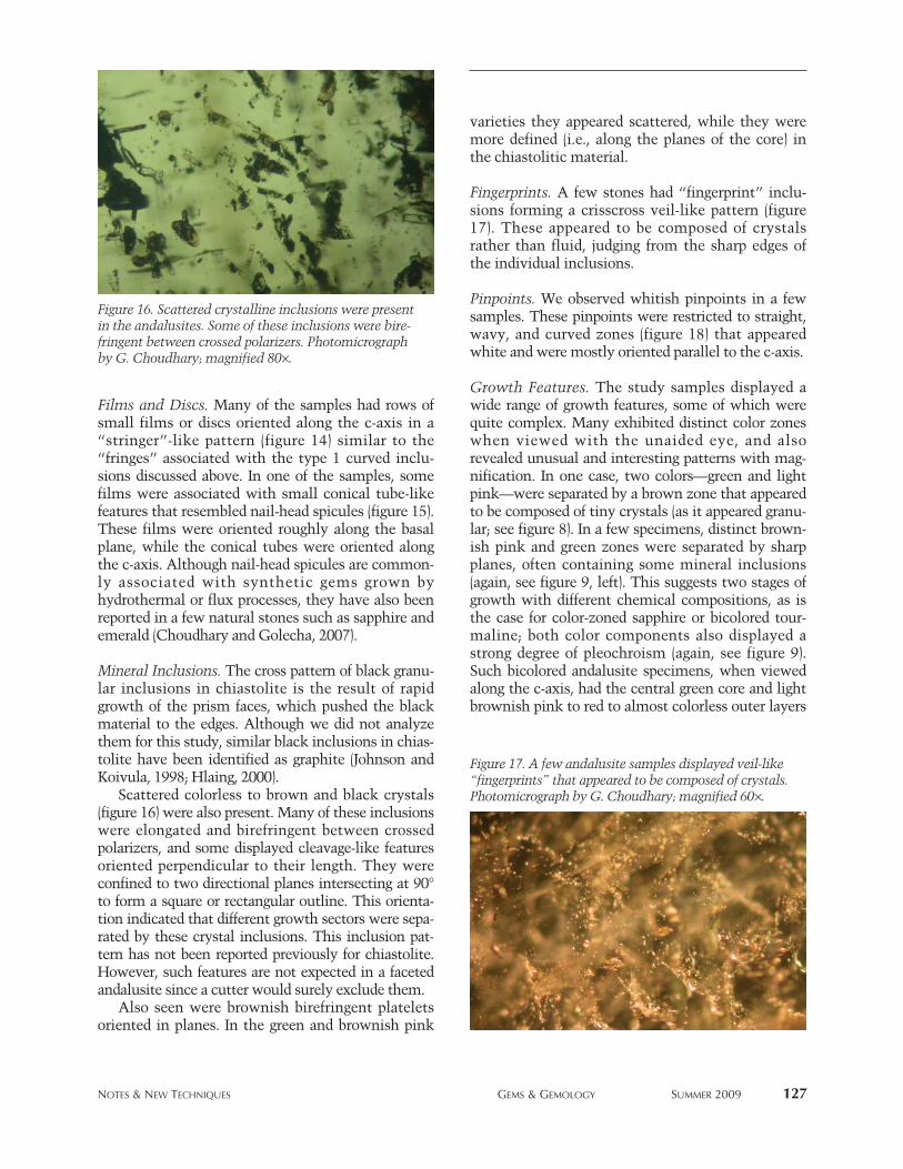
The type 2 curved inclusions appeared to begrowth tubes filled with a brown or black material.Some samples displayed these tubes in parallel bun-dles oriented in various directions (figure 11, left).Each direction terminated at a sharp plane. In onespecimen, these planes created a rectangular/squareprofile. They also appeared to run across the stone(similar to a twin plane) in different directions. Athigher magnification, the tubes appeared to contain
NOTES & NEW TECHNIQUES GEMS & GEMOLOGY SUMMER 2009 125
Figure 11. Type 2 curvedinclusions consist of tubesthat are filled with a blackmaterial. In some samples,groups of these tubes were oriented in various directions(left). At higher magnification,the black material appearsgranular (right). Photomicro-graphs by G. Choudhary;magnified 30× (left, in immersion) and 80× (right).
Figure 10. Type 1 curved inclusions are colorless and transparent, often with fine fringes (left and center). Theyappear birefringent between crossed polarizers, indicating their mineral nature (right). Note the stained surfaceand angle of curvature of these tubes, which are bent almost 90°. Photomicrographs by G. Choudhary; magnified40× (left), 80× (center, brightfield illumination), and 70× (right, crossed polarizers).
126 NOTES & NEW TECHNIQUES GEMS & GEMOLOGY SUMMER 2009
a black granular material (figure 11, right). Similarinclusions have been reported in “trapiche” tour-maline (Hainschwang et al., 2007), where growthchannels are oriented in two directions. In our chi-astolite specimens, the varying orientations of thetubes may be attributed to twinning or differentstages of growth, as also indicated by the sharpgrowth zoning described below.
The angle of curvature in both types of inclu-sions typically was almost 90° (e.g., figure 10), whilesome appeared closer to 45°, as if originating frompyramidal faces. Most of the inclusions were orient-ed parallel to the c-axis.
A few samples displayed an interesting pattern of
type 2 tubes (figure 12). A central green core exhibit-ed curved tubes parallel to the c-axis, while the sur-rounding portions contained tubes that radiatedfrom the boundary with the core. The authors havepreviously observed a similar pattern of black tubesin chiastolites. Such a growth pattern indicates dif-ferent growth stages and directions of the overgrownareas, and is a characteristic of chiastolite.
Straight tubes with slightly wavy surfaces (figure13), oriented parallel to the c-axis, were also observedin the yellow-green and bicolored samples, as well asin the chiastolite with green cores.
Figure 12. In a few chiastolite specimens, a green corecontained tubes oriented parallel to the c-axis, whilethe surrounding area hosted tubes in a radiating pat-tern. Similar patterns of black tubes have beenobserved previously in chiastolite by the authors.Photomicrograph by G. Choudhary; magnified 25×.
Figure 13. Straight tubes with wavy surfaces, oriented parallel to the c-axis, were present in someof the andalusite samples. Photomicrograph by G. Choudhary; magnified 60×.
Figure 14. The andalusites commonly contained filmor platelet-like inclusions that appeared to be orientedalong the c-axis (vertical in this view), giving astringer-like pattern as in figure 10 (left). Photomicro-graph by G. Choudhary; magnified 65×.
Figure 15. In one sample, small films/fractures wereassociated in places with conical tube-like features(similar to nail-head spicules). These films were ori-ented roughly along the basal plane (horizontal in thisview), while the conical “tubes” were along the c-axis.Also note the growth zoning parallel to the c-axis. Inaddition, many scattered tiny inclusions were visiblethat could not be resolved clearly. Photomicrographby G. Choudhary; magnified 80×.
NOTES & NEW TECHNIQUES GEMS & GEMOLOGY SUMMER 2009 127
Films and Discs. Many of the samples had rows ofsmall films or discs oriented along the c-axis in a“stringer”-like pattern (figure 14) similar to the“fringes” associated with the type 1 curved inclu-sions discussed above. In one of the samples, somefilms were associated with small conical tube-likefeatures that resembled nail-head spicules (figure 15).These films were oriented roughly along the basalplane, while the conical tubes were oriented alongthe c-axis. Although nail-head spicules are common-ly associated with synthetic gems grown byhydrothermal or flux processes, they have also beenreported in a few natural stones such as sapphire andemerald (Choudhary and Golecha, 2007).
Mineral Inclusions. The cross pattern of black granu-lar inclusions in chiastolite is the result of rapidgrowth of the prism faces, which pushed the blackmaterial to the edges. Although we did not analyzethem for this study, similar black inclusions in chias-tolite have been identified as graphite (Johnson andKoivula, 1998; Hlaing, 2000).
Scattered colorless to brown and black crystals(figure 16) were also present. Many of these inclusionswere elongated and birefringent between crossedpolarizers, and some displayed cleavage-like featuresoriented perpendicular to their length. They wereconfined to two directional planes intersecting at 90°to form a square or rectangular outline. This orienta-tion indicated that different growth sectors were sepa-rated by these crystal inclusions. This inclusion pat-tern has not been reported previously for chiastolite.However, such features are not expected in a facetedandalusite since a cutter would surely exclude them.
Also seen were brownish birefringent plateletsoriented in planes. In the green and brownish pink
varieties they appeared scattered, while they weremore defined (i.e., along the planes of the core) inthe chiastolitic material.
Fingerprints. A few stones had “fingerprint” inclu-sions forming a crisscross veil-like pattern (figure17). These appeared to be composed of crystalsrather than fluid, judging from the sharp edges ofthe individual inclusions.
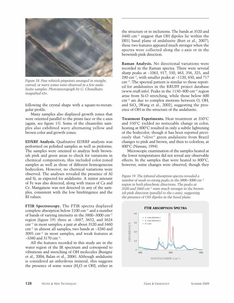
Pinpoints. We observed whitish pinpoints in a fewsamples. These pinpoints were restricted to straight,wavy, and curved zones (figure 18) that appearedwhite and were mostly oriented parallel to the c-axis.
Growth Features. The study samples displayed awide range of growth features, some of which werequite complex. Many exhibited distinct color zoneswhen viewed with the unaided eye, and alsorevealed unusual and interesting patterns with mag-nification. In one case, two colors—green and lightpink—were separated by a brown zone that appearedto be composed of tiny crystals (as it appeared granu-lar; see figure 8). In a few specimens, distinct brown-ish pink and green zones were separated by sharpplanes, often containing some mineral inclusions(again, see figure 9, left). This suggests two stages ofgrowth with different chemical compositions, as isthe case for color-zoned sapphire or bicolored tour-maline; both color components also displayed astrong degree of pleochroism (again, see figure 9).Such bicolored andalusite specimens, when viewedalong the c-axis, had the central green core and lightbrownish pink to red to almost colorless outer layers
Figure 16. Scattered crystalline inclusions were presentin the andalusites. Some of these inclusions were bire-fringent between crossed polarizers. Photomicrographby G. Choudhary; magnified 80×.
Figure 17. A few andalusite samples displayed veil-like“fingerprints” that appeared to be composed of crystals.Photomicrograph by G. Choudhary; magnified 60×.
128 NOTES & NEW TECHNIQUES GEMS & GEMOLOGY SUMMER 2009
following the crystal shape with a square-to-rectan-gular profile.
Many samples also displayed growth zones thatwere oriented parallel to the prism face or the c-axis(again, see figure 15). Some of the chiastolite sam-ples also exhibited wavy alternating yellow andbrown color and growth zones.
EDXRF Analysis. Qualitative EDXRF analysis wasperformed on polished samples as well as preforms.The samples were oriented to analyze both brown-ish pink and green areas to check for variations inchemical composition; this included color-zonedsamples as well as those of different homogeneousbodycolors. However, no chemical variations wereobserved. The analyses revealed the presence of Aland Si, as expected for andalusite. A minor amountof Fe was also detected, along with traces of Ca andCr. Manganese was not detected in any of the sam-ples, consistent with the low birefringence and theRI values.
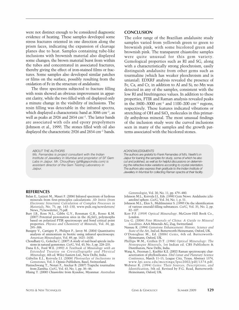
FTIR Spectroscopy. The FTIR spectra displayedcomplete absorption below 2100 cm−1 and a numberof bands of varying intensity in the 3800–3000 cm−1
region (figure 19): three at ~3687, 3652, and 3624cm−1 in most samples; a pair at about 3520 and 3460cm−1 in almost all samples; two bands at ~3280 and3095 cm−1 in most samples; and weak features at~3380 and 3170 cm−1.
All the features recorded in this study are in thewater region of the IR spectrum and correspond tovibrations and stretching of OH molecules (Busignyet al., 2004; Balan et al., 2006). Although andalusiteis considered an anhydrous mineral, this suggeststhe presence of some water (H2O or OH), either in
the structure or in inclusions. The bands at 3520 and3460 cm−1 suggest that OH dipoles lie within the{001} basal plane of andalusite (Burt et al., 2007);these two features appeared much stronger when thespectra were collected along the c-axis or in thebrownish pink direction.
Raman Analysis. No directional variations wererecorded in the Raman spectra. There were severalsharp peaks at ~1063, 917, 550, 463, 356, 325, and290 cm−1, with smaller peaks at ~1120, 850, and 717cm−1. The spectral pattern is similar to those report-ed for andalusites in the RRUFF project database(www.rruff.info). Peaks in the 1150–800 cm−1 regionarise from Si-O stretching, while those below 600cm−1 are due to complex motions between O, OH,and SiO4 (Wang et al., 2002), suggesting the pres-ence of OH in the structure of the andalusite.
Treatment Experiments. Heat treatment at 350°Cand 550°C yielded no noticeable change in color;heating at 800°C resulted in only a subtle lighteningof the bodycolor, though it has been reported previ-ously that “olive” green andalusite from Brazilchanges to pink and brown, and then to colorless, at800°C (Nassau, 1994).
Microscopic examination of the samples heated atthe lower temperatures did not reveal any observableeffects. In the samples that were heated to 800°C,however, some changes were observed, though they
Figure 19. The infrared absorption spectra revealed anumber of weak-to-strong peaks in the 3800–3000 cm−1
region in both pleochroic directions. The peaks at3520 and 3460 cm−1 were much stronger in the brown-ish pink direction (parallel to the c-axis), suggestingthe presence of OH dipoles in the basal plane.
Figure 18. Fine whitish pinpoints arranged in straight,curved, or wavy zones were observed in a few anda-lusite samples. Photomicrograph by G. Choudhary;magnified 65×.
NOTES & NEW TECHNIQUES GEMS & GEMOLOGY SUMMER 2009 129
were not distinct enough to be considered diagnosticevidence of heating. These samples developed somestress fractures oriented in one direction along theprism faces, indicating the expansion of cleavageplanes due to heat. Samples containing tube-likeinclusions with brownish material also displayedsome changes; the brown material burst from withinthe tubes and concentrated in associated fractures,thereby giving the effect of iron-stained films or frac-tures. Some samples also developed similar patchesor films on the surface, possibly resulting from theoxidation of Fe in the structure of andalusite.
The three specimens subjected to fracture fillingwith resin showed an obvious improvement in appar-ent clarity, while the two filled with oil displayed onlya minute change in the visibility of inclusions. Theresin filling was detectable in the infrared spectra,which displayed a characteristic band at 3046 cm−1, aswell as peaks at 2926 and 2854 cm−1. The latter bandsare associated with oils and epoxy prepolymers(Johnson et al., 1999). The stones filled with oil alsodisplayed the characteristic 2926 and 2854 cm−1 bands.
CONCLUSIONThe color range of the Brazilian andalusite studysamples varied from yellowish green to green tobrownish pink, with some bicolored green andbrownish pink. The transparent chiastolite sampleswere quite unusual for this gem variety.Gemological properties such as RI and SG, alongwith a characteristically strong pleochroism, easilydistinguish andalusite from other gems such astourmaline (which has weaker pleochroism and isuniaxial). EDXRF analysis revealed the presence ofFe, Ca, and Cr, in addition to Al and Si; no Mn wasdetected in any of the samples, consistent with thelow RI and birefringence values. In addition to theseproperties, FTIR and Raman analysis revealed peaksin the 3800–3000 cm−1 and 1100–200 cm−1 regions,respectively. These features indicated vibrations orstretching of OH and SiO4 molecules in this primar-ily anhydrous mineral. The most unusual findingsof the inclusion study were the curved inclusionsseen in many of the samples and the growth pat-terns associated with the bicolored stones.
REFERENCESBalan E., Lazzeri M., Mauri F. (2006) Infrared spectrum of hydrous
minerals from first-principles calculations. Ab Initio (fromElectronic Structure) Calculation of Complex Processes inMaterials, No. 75, pp. 143–150, www.psik.org/newsletters/News_75/newsletter_75.pdf.
Burt J.B., Ross N.L., Gibbs G.V., Rossman G.R., Rosso K.M.(2007) Potential protonation sites in the Al2SiO5 polymorphsbased on polarized FTIR spectroscopy and bond critical pointproperties. Physics and Chemistry of Minerals, Vol. 34, pp.295–306.
Busigny V., Cartigny P., Philipot P., Javoy M. (2004) Quantitativeanalysis of ammonium in biotite using infrared spectroscopy.American Mineralogist, Vol. 89, pp. 1625–1630.
Choudhary G., Golecha C. (2007) A study of nail-head spicule inclu-sions in natural gemstones. G&G, Vol. 43, No. 3, pp. 228–235.
Dana E.S., Ford W.E. (1992) A Textbook of Mineralogy with anExtended Treatise on Crystallography and PhysicalMineralogy, 4th ed. Wiley Eastern Ltd., New Delhi, India.
Gübelin E.J., Koivula J.I. (2008) Photoatlas of Inclusions inGemstones, Vol. 3. Opinio Publishers, Basel, Switzerland.
Hainschwang T., Notari F., Anckar B. (2007) Trapiche tourmalinefrom Zambia. G&G, Vol. 43, No. 1, pp. 36–46.
Hlaing T. (2000) Chiastolite from Kyaukse, Myanmar. Australian
Gemmologist, Vol. 20, No. 11, pp. 479–480.Johnson M.L., Koivula J.I., Eds. (1998) Gem News: Andalusite (chi-
astolite) sphere. G&G, Vol. 34, No. 1, p. 51.Johnson M.L., Elen S., Muhlmeister S. (1999) On the identification
of various emerald filling substances. G&G, Vol. 35, No. 2, pp.82–107.
Kerr P.F. (1959) Optical Mineralogy. McGraw-Hill Book Co.,London.
Liu G. (2006) Fine Minerals of China: A Guide to MineralLocalities. AAA Minerals AG, Zug, Switzerland.
Nassau K. (1994) Gemstone Enhancement: History, Science andState of the Art, 2nd ed. Butterworth-Heinemann, Oxford, UK.
O’Donoghue M., Ed. (2006) Gems, 6th ed. Butterworth-Heinemann, Oxford, UK.
Phillips W.M., Griffen D.T. (1986) Optical Mineralogy: TheNonopaque Minerals, 1st Indian ed. CBS Publishers &Distributors, New Delhi, India.
Wang A., Freeman J., Kuebler K.E. (2002) Raman spectroscopic char-acterization of phyllosilicates. 33rd Lunar and Planetary ScienceConference, March 11–15, League City, Texas, Abstract 1374,www.lpi.usra.edu/meetings/lpsc2002/pdf/1374.pdf.
Webster R. (1994) Gems: Their Sources, Descriptions, andIdentification, 5th ed. Revised by P.G. Read, Butterworth-Heinemann, Oxford, UK.
ABOUT THE AUTHORSMs. Fernandes is project consultant with the IndianInstitute of Jewellery in Mumbai and proprietor of SF GemLabs in Jaipur. Mr. Choudhary ([email protected]) isassistant director of the Gem Testing Laboratory inJaipur.
ACKNOWLEDGMENTSThe authors are grateful to Frank Fernandes of M/s. Neethi’s inJaipur for loaning the samples for study, some of which he alsocut and polished, as well as for helpful discussions on determin-ing the refractive index variations according to crystal orientation.The authors also express their gratitude to the Indian Institute ofJewellery in Mumbai for collecting Raman spectra at their facility.