Embed Size (px)
Citation preview
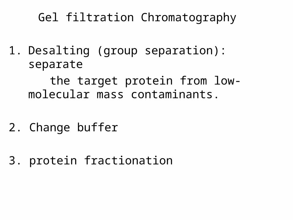
Gel filtration Chromatography
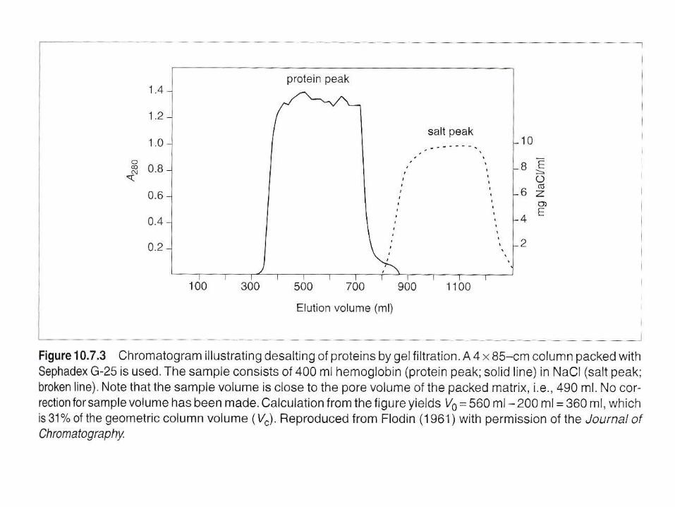
1. Desalting (group separation): separate
the target protein from low-molecular mass contaminants.
2. Change buffer
3. protein fractionation
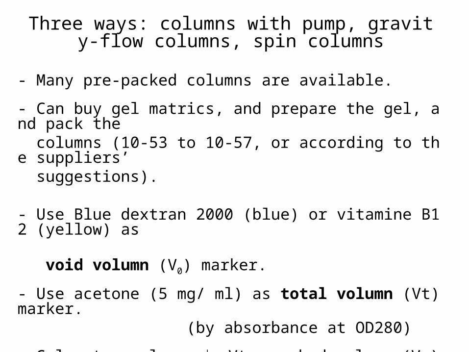
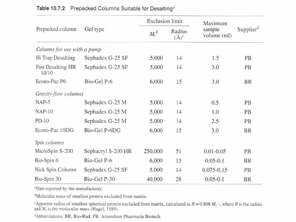
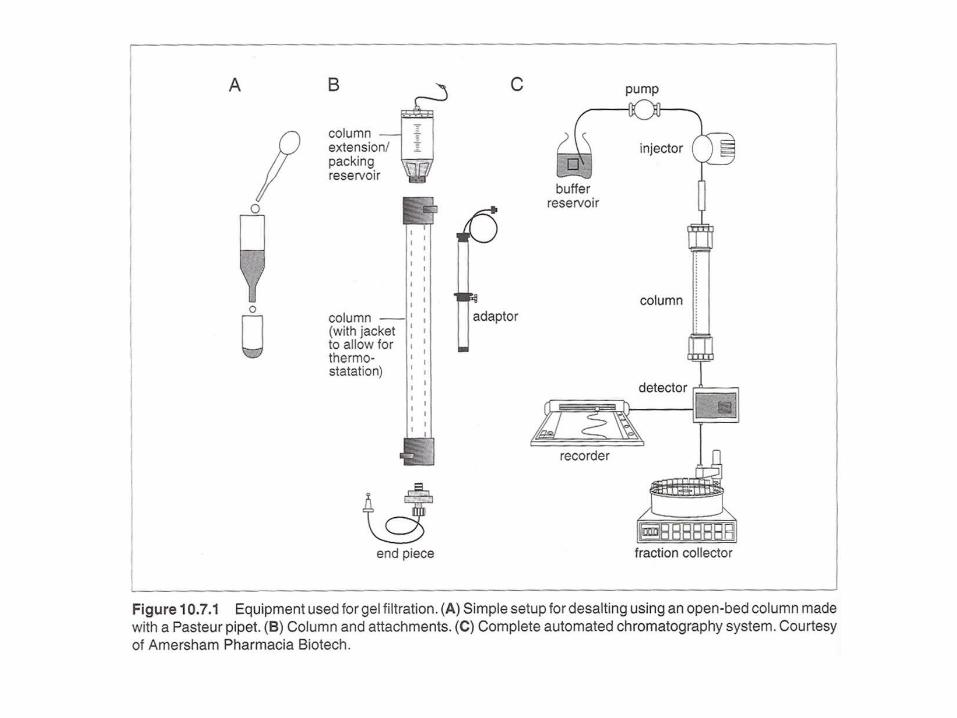
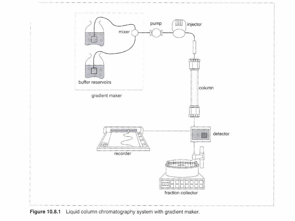
Three ways: columns with pump, gravity-flow columns, spin columns
- Many pre-packed columns are available.
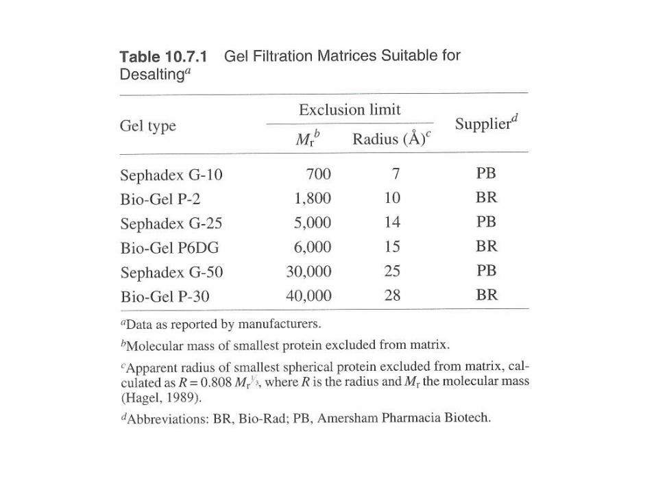
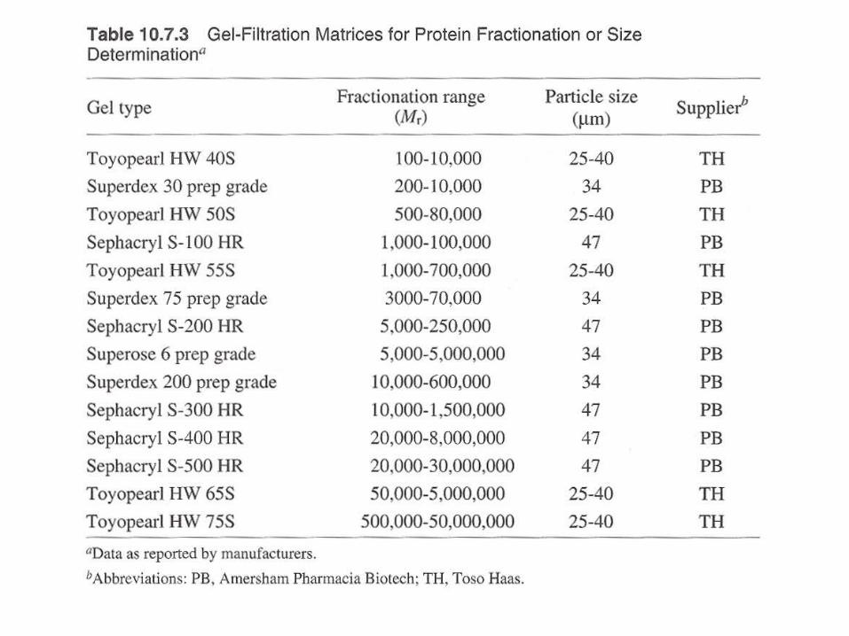
- Can buy gel matrics, and prepare the gel, and pack the columns (10-53 to 10-57, or according to the suppliers’ suggestions).
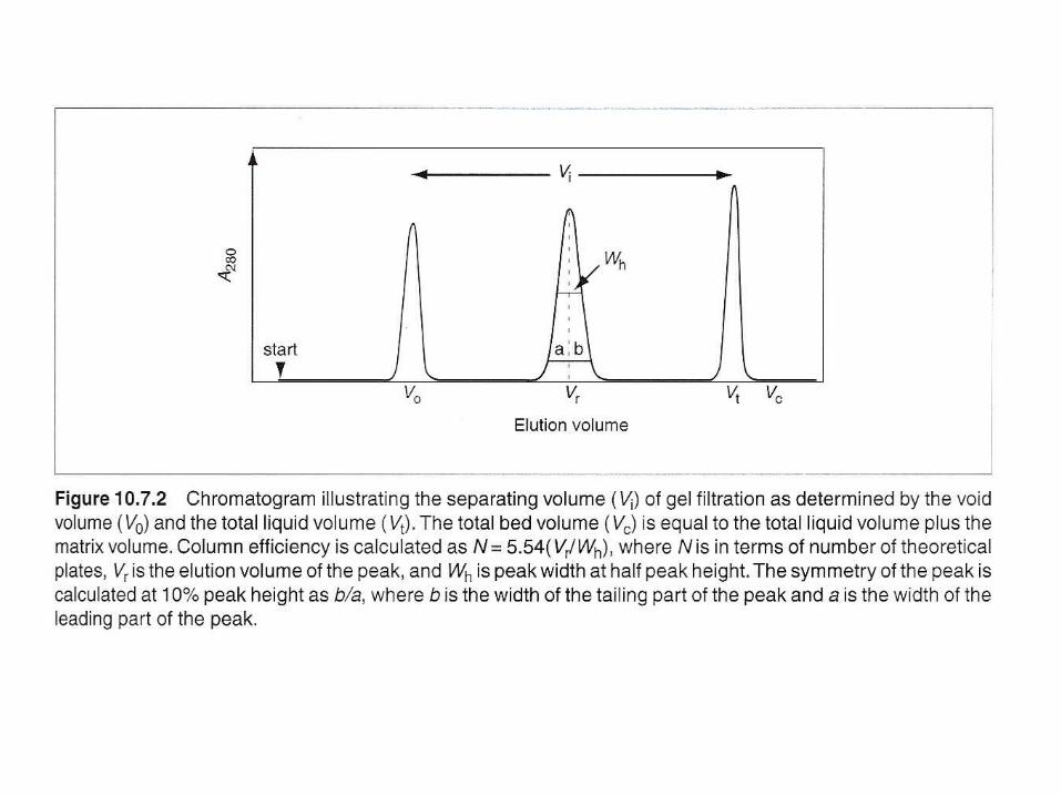
- Use Blue dextran 2000 (blue) or vitamine B12 (yellow) as void volumn (V0) marker.
- Use acetone (5 mg/ ml) as total volumn (Vt) marker. (by absorbance at OD280)
- Gel matrx volumn + Vt = bed-volumn (Vc)
- Vi (separating volumn) = Vt - V0
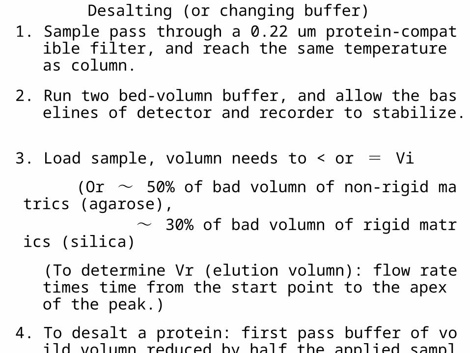
Desalting (or changing buffer)1. Sample pass through a 0.22 um protein-compatible filter, and
reach the same temperature as column.
2. Run two bed-volumn buffer, and allow the baselines of detector and recorder to stabilize.
3. Load sample, volumn needs to < or = Vi
(Or ~ 50% of bad volumn of non-rigid matrics (agarose), ~ 30% of bad volumn of rigid matrics (silica)
(To determine Vr (elution volumn): flow rate times time from the start point to the apex of the peak.)
4. To desalt a protein: first pass buffer of voild volumn reduced by half the applied sample volumn – waste
5. then apply buffer of applied sample volumn – collect.
6. Wash column with > 1 column volumn of buffer with an antibacterial agent and store.
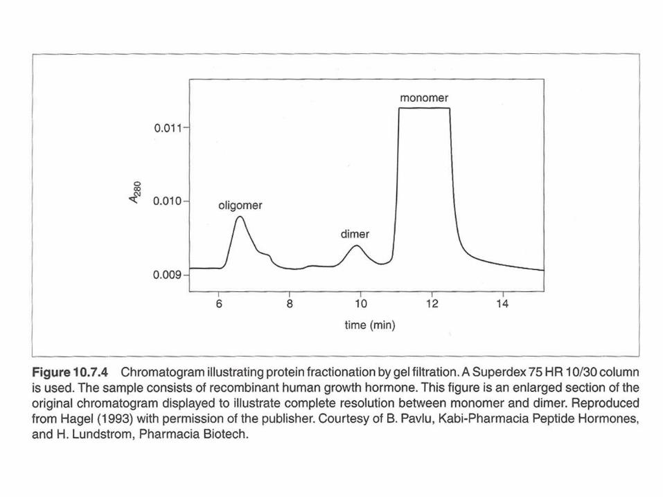
Gel filtration for protein fractionation (usually used in the polishing step in protein purification)
- Columns are different from desalting
- Sample volumn about 2% of the bed volumn.
- Purity of the peak can be checked by HPLC or electrophotresis.
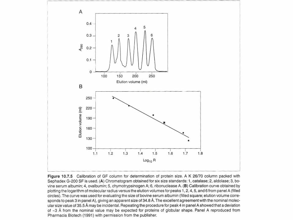
- Proteins molecular weight could be determined by Vr (elution volumn).
But mass spectrophotometry (MS) can do better job in determination of molecular mass.

Ion-Exchange Chromatography
A. Selecting a buffer system If the pI of the target protein is known, use anion-exchange medium with operating pH > pI, or cation-exchange medium with operating pH < pI. If the pI is unknown, determine the pI by isoelectric focusing (1-D of 2-D electrophoresis). The optimal pH for binding, elution, and the binding capability at the optimal condition can be determined.
Most proteins in cells has pI below pH 7, can start an anion- exchange medium and operating pH of 8.5 to start, then evaluate the results and optimize conditions. .

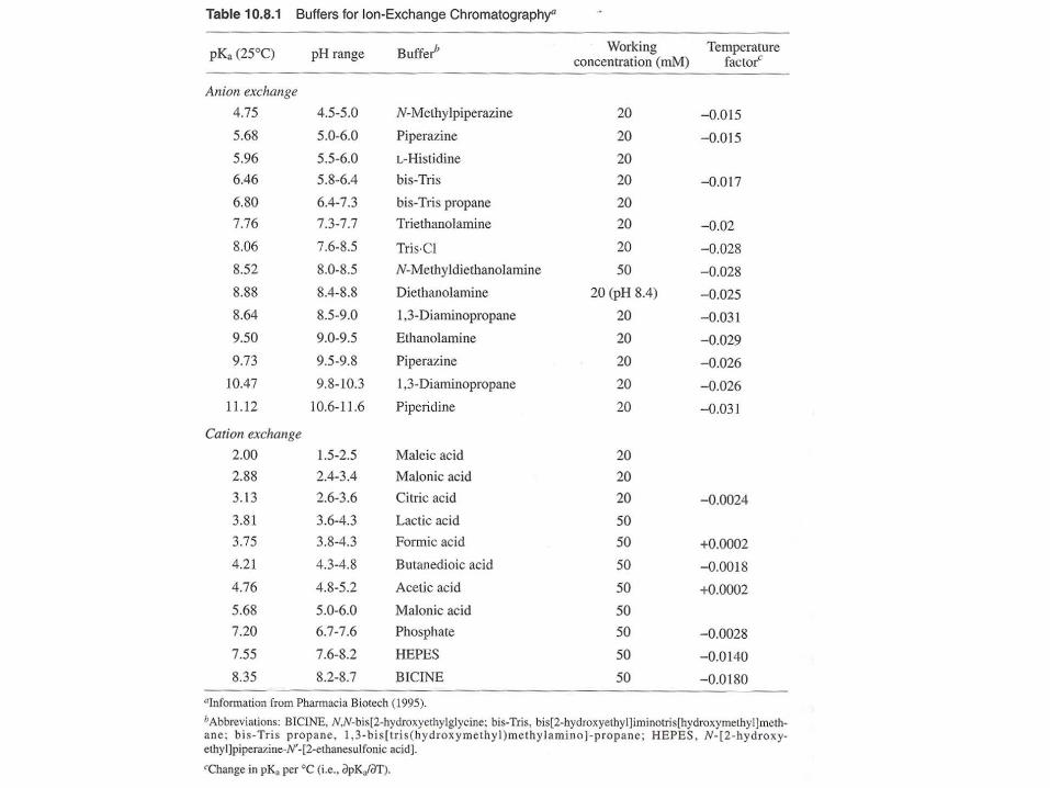
B. Selecting a buffer system
- Consider the pH stability of the sample.
- Anionic buffers for cation exchange,
- Cationic buffers for anion exchange.
(Thus, buffering ions will not bind gel matrix)


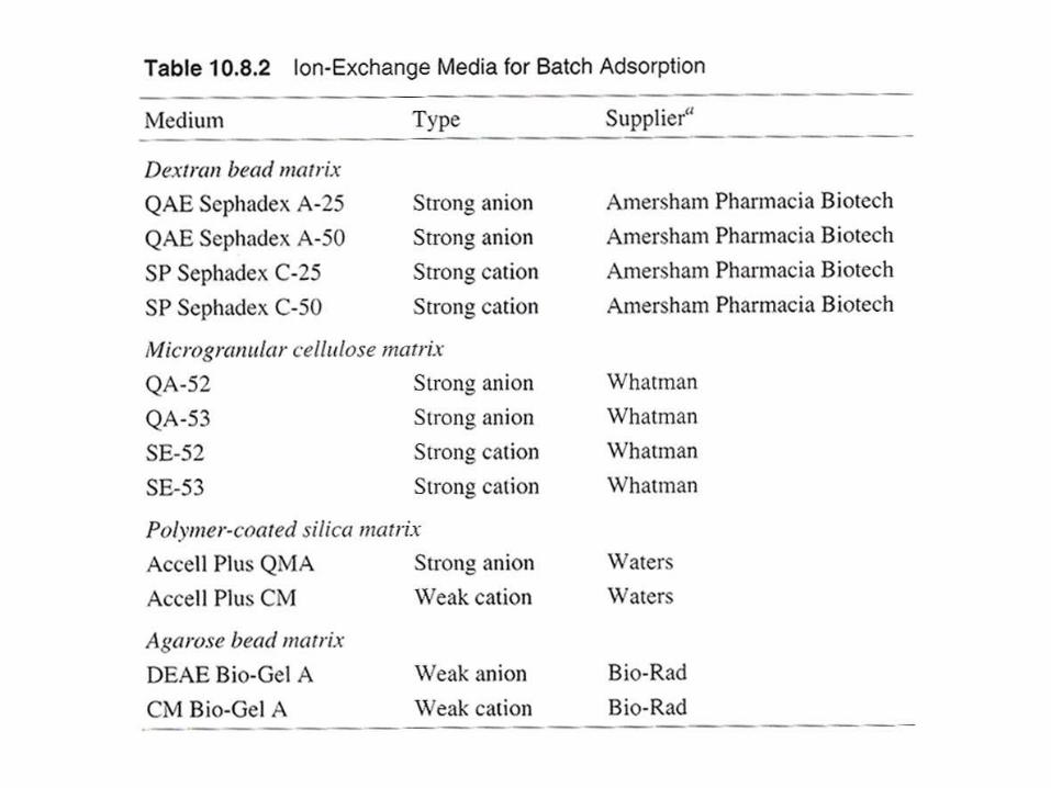
Batch adsorption and step-gradient elution (with increasing salt concentration)
eg. anion exchange gel,
binding buffer: 20 mM Tris.Cl, pH 7.5
washing buffer: 20 mM Tris.Cl, pH 7.5/100 mM NaCl
elution buffer: 20 mM Tris.Cl, pH 7.5/350 mM NaCl
regeneration buffer: 20 mM Tris.Cl, pH 7.5/ 2 M NaCl
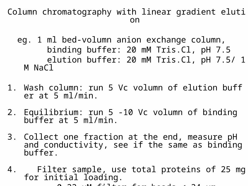
Column chromatography with linear gradient elution
eg. 1 ml bed-volumn anion exchange column, binding buffer: 20 mM Tris.Cl, pH 7.5 elution buffer: 20 mM Tris.Cl, pH 7.5/ 1 M NaCl
1. Wash column: run 5 Vc volumn of elution buffer at 5 ml/min.
2. Equilibrium: run 5 -10 Vc volumn of binding buffer at 5 ml/min.
3. Collect one fraction at the end, measure pH and conductivity, see if the same as binding buffer.
4. Filter sample, use total proteins of 25 mg for initial loading. 0.22 uM filter for beads < 34 um, 0.45 um filter for beads between 34 um and 90 um, 1 um filter for beads > 90 um

5. Open injection valve for sample injection, and begin to collect fractions at 1 ml.
Can reduce flow rate for very concentrated sample, or increase flow rate for very diluted sample.
6. After sample injected, wash with 3 to 5 Vc volumn of binding buffer at flow rate 5 ml/min. Monitor signal to baseline.
7. Close sample injection valves to reduce system dead volumn.
8. Elute with a linear gradient from 0% to 100% elution buffer in 20Vc (20 ml).
9. Regenerate colunmn by washing with 5 Vc of elution buffer.
10. Reequilibrate column with 5 to 10 Vc of binding buffer.


Preparation of antibody-sepharose
Covalently linking an antibody to Sepharose (CL-4B, or CL-2b for high MW antigen), using CNBr activation method.
1. dialyze 1-30 m g/ml antibody against 0.1 M NaHCO3 /0.5 M NaCl at 40C with 3 buffer changes over 24 hrs, use dialysis solutions 500 times of the antibody volumn.
2. Centrifuge
3. Measure A 280 (mg/ml IgG = A 280 / 1.44), dilute to 5 mg/ml with 0.1 M NaHCO3 /0.5 M NaCl
4. Wash the sepharose with 10 vol water, use Waterman no. 1 filter and Buchner funnel.
5. Add equal volumn of 0.2 M Na2CO3 to Sepharose.
6. Add CNBr/ acetonitrile dropwise (in hood).7. Filter, dry, and add 0.1 M HCl, and add antibody solution, fo
r 2 hr at rt. 8. Add glycine to saturate the active group on Sepharose, and
measure A 280 to determine percentage coupling