Embed Size (px)
Citation preview
Take the Pink Link!
www. .com
Gel Electrophoresis Size Marker
Size Marker • © 2008 AppliChem
Lyophilised markers
Starting materialThe DNA in our lyophilised length markers is amplified in low-nuclease host bacteria, restricted with quality enzymes, and is deproteinised. This ensures that they contain high-quality, low-protein or nuclease-free products with very good migration properties on agarose, agar and polyacrylamide gels.
Quality controlNumerous gel separations of undigested plasmids and digested fragments form part of the manufacturing process. The fragment mass is determined using two calibrated photometers via conversion (1 OD260 nm = 50 µg dsDNA) and is aliquoted at 100 % (+2 %). Each batch of DNA and gel loading buffer is subject to a final quality check using gel electrophoresis.
StabilityLyophilisation of the DNA markers results in very stable products, without any significant impairment of quality at room temperature after transport (even at the height of summer, > 35°C for several days) or after long storage times (> 4 years at -20°C).
ResuspensionBefore use, lyophilised DNA markers can be dissolved in gel loading buffer or optional for end-labeling in sterile, bidistilled water. The lyophilised form allows you the highest degree of flexibility (labeling, concentration, intensity of staining bands).
Legal noteThe DNA ladders were manufactured by digestion of plasmids with EcoR I. The plasmids are legally protected. To produce copies apply for a permission from the manufacturer.
Tips on the use of DNA length markers
Correct storageDeproteinised and lyophilised DNA samples are extremely stable (> 5 years). Problems do not usually occur unless DNA markers in solution are stored (> 6 weeks) at room temperature, they become contaminated with bacteria, or are frequent-ly thawed and refrozen (> 20 times). After dissolution of the DNA, we therefore recommend aliquoting of the DNA marker in ready-to-use amounts for storage at -20°C (for 2 – 4 years). At +4°C, markers in solution are stable for several weeks or even months. Here, however, there is the risk of bacterial or nuclease contamination. This can be prevented by storage at -20°C.
End-labelingThanks to their deproteinised and lyophilised form, these DNA markers are suitable for universal use for end-labeling with modified nucleotides. Labeling in four positions via the terminal EcoR I generated recessed ends is possible, especially with the DNA ladder 100 bp (A3470) and the DNA Ladder Mix 100 – 5000 (A3660). For the labeling of the DNA, the product is simply dissolved in TE buffer or bidistilled water.
DNA staining with methylene blueOccasionally, ethidium bromide is not suitable for staining DNA. An alternative is methylene blue. In such cases, however, it must be kept in mind that the mass of DNA used must be increased by about 30 % and that about 1.5 h more must be planned for destaining steps. With a methylene blue concentrate supplied by AppliChem, you achieve very good results, even for small fragments of about 200 bp.
i n t r o d u c t i o n
© 2008 AppliChem • Size Marker 1
Mass calculations for single fragmentsUsing DNA markers of a defined origin, the calculation of the mass of single fragments is relatively easy. The amount loaded per lane, e.g. 1 µg/10 µl, is divided by the number of base pairs of the DNA used and is multiplied by the fragment size. For example: the 267 bp fragment of the pBR322 Hae III marker with a loading amount of 1 µg: 1 µg : 4361 bp (pBR322) = 0.229 ng/base pair x 267 bp of the fragment = 61.2 ng/267 fragment.Assuming comparable staining (saturation with dye), the mass of unknown fragments can be determined in this way. If required, concentration gradients can be loaded.
Less is often moreThe different gel formats for agarose and polyacrylamide gel electrophoresis and the varying sensitivity of staining or detec-tion mean that it is only possible to give an approximation of the recommended DNA amount to be loaded. Most DNA mar-kers show the best separation with loading amounts of 0.5 – 1 µg on agarose gels. The general rule is: the lower the number of marker bands, the smaller the total amount needed. A low loading amount is also of advantage with short migration distances or very sensitive detection. When using polyacrylamide gels, generally only about 1/5 – 1/2 the amount required for agarose gels is necessary. For DNA markers with large fragments and a high mass (e.g. equimolar mixtures or lambda DNA markers), depending on the agarose concentration, a minimum separation distance is required to separate or comple-tely distinguish between the dominant upper bands in terms of width. This separation distance can often be reduced by 10 – 15 % by reducing the marker amount. By using equalized markers, in addition to a marked reduction of the marker amount, the separation distance can be reduced by a further 10 – 15 %, from e.g. 70 to 50 mm, on a standard format gel, to obtain the best results.
How to achieve results quicklyEspecially deproteinised DNA length markers can be separated very well using high voltages, e.g. within 20 minutes with a migration distance of 70 mm on a 1.2 % agarose gel at 120 Volt. This can only be realized, however, with a high quality gel migration buffer (the water quality must be taken into account!) and an adequate amount of buffer (400 – 600 ml). Poor buffer quality results in overheating of the agarose gel at higher voltages and this therefore causes problems. A high buffer volume (the buffer can usually be reused 4 – 6 times in one week without problems) leads the heat off, provided the gel chamber for submersion is of adequate size.
Staining front too intense?In some cases, users who regularly perform assays with our products sometimes comment that the dye concentration in the gel loading buffer we supply is too high (only lyophilised markers). If this is the case, simply dissolve the DNA length marker in double concentration in the gel loading buffer and further dilute it with 1/2 volume TE buffer. Even when using a 50 % solution, there are no problems with the resulting 7 % glycerol concentration to increase the solution density.
Plausibility of the results and ion concentrations:Non-plausible results (e.g. unexpected fragment runs at 500 bp instead of 350 bp) may occur under relatively extreme conditions (separation of restriction or PCR samples with a very high salt content > 150 mM) or at very high voltages with relatively short separation distances. To identify errors, 1/10 volume restriction buffer can be added to the aliquot of the DNA marker, for example, or the restriction sample can be appropriately diluted with gel loading buffer (usually 1/2 volume). In many cases, lowering the voltage during separation is also helpful. The accurate determination of the size of fragments or PCR products may also be impaired, when salt fronts combined with local problems of leading off heat with conducting away heat and very high electrophoresis voltages, result in partial denaturation of the DNA fragments. These are conditions that theoretically may also occur when processing samples from Maxam-Gilbert sequencing in combination with sample buffers containing formamide.
i n t r o d u c t i o n

The quality of this DNA sizing standard differs considerably from products offered by most other manufacturers
thanks to the elaborate manufacturing method, which also fulfils the highest quality requirements! The digested
DNA is not simply precipitated and resuspended, but also extracted in phenol. This guarantees the extremely
long shelf-life without any loss of quality. Not only the prices should therefore be compared.
Prod. No. DNA Marker Bands Fragment Sizes [bp]
A3470 DNA Ladder 100 bp (lyophilised) 11 1000 900 800 700 600 500 400 300 200 150 100
A5191 DNA Ladder 100 bp 10 1000 900 800 700 600 500 400 300 200 100
A3302 DNA Ladder 100 bp equalized (lyophilised) 11 1000 900 800 700 600 500 400 300 200 150 100
A5216 DNA Ladder 100 bp plus 11 1500 1000 900 800 700 600 500 400 300 200 100
A3660 DNA Ladder Mix 100 – 5000 (lyophilised) 17 5000 4000 3000 2500 2000 1500 1000 900 800 700 600 500 400 300 200 150 100
A2667 DNA Ladder 1 kb (lyophilised) 11 10000 8000 6000 5000 4000 3000 2500 2000 1500 1000 500
A3982 DNA Ladder 250 bp (lyophilised) 16 8000 6000 5000 4000 3000 2750 2500 2250 2000 1750 1500 1250 1000 750 500 250
A5207 DNA Ladder 1 kb 13 10000 8000 6000 5000 4000 3000 2500 2000 1500 1000 750 500 250
A6430 DNA Ladder 1 kb concatamer (lyophlised) >25 1000 bp steps (1000 – approx. 25000)
A7215 DNA Marker quick-run (lyophylised) 5 2500 2000 1500 1000 500
A7222 DNA Marker quick-run extended (lyophylised) 9 6000 4000 25000 2000 1500 1000 500 200 100
A4406 DNA Marker pBR322 – Hae III (lyophilised) 22 587 540 502 458 434 267 234 213 192 184 124 123 104 89 80 64 57 51 21 18 11 8
A5229 DNA Marker pBR322 – Hae III 22 587 540 502 458 434 267 234 213 192 184 124 123 104 89 80 64 57 51 21 18 11 8
A6927 DNA Marker pBR328 Mix (lyophilised) 12 2176 1766 1230 1033 653 517 453 394 298 234 220 154
A5194 DNA Marker Phage Lambda Sty I 11 19329 7743 6223 4254 3472 2690 1882 1489 925 421 74
A4412 DNA Marker Phage Lambda BstE II (lyophilised) 14 8454 7242 6369 5686 4822 4324 3675 2323 1929 1371 1264 702 224 117
A5220 DNA Marker Phage Lambda BstE II 14 8454 7242 6369 5686 4822 4324 3675 2323 1929 1371 1264 702 224 117
A5589 DNA Marker Phage Lambda Hind III (lyophilised) 8 23130 9416 6557 4361 2322 2027 564 125
A5223 DNA Marker Phage Lambda Hind III 8 23130 9400 6557 4361 2322 2027 564 125
A5235 DNA Marker pUC19 – Msp I 12 501 489 404 331 242 190 147 111 110 67 34 26
A3996 DNA Marker pUC19 – Msp I (lyophilised) 12 501 489 404 331 242 190 147 111 110 67 34 26
Protein Marker
A5238 Protein Marker I (14 – 116) 7 116.0 97.4 66.2 37.6 28.5 18.4 14.0
A5418 Protein Marker II (6.5 – 200) prestained 8 200.0 116.0 68.0 43.0 30.0 20.0 14.4 6.5
A4402 Protein Marker III (6.5 – 200) 8 200.0 116.0 68.0 43.0 30.0 20.0 14.4 6.5
A3993 Protein Marker IV (10 – 150) 8 150 100 80 60 40 30 20 10
DNA marker
2 Size Marker • © 2008 AppliChem
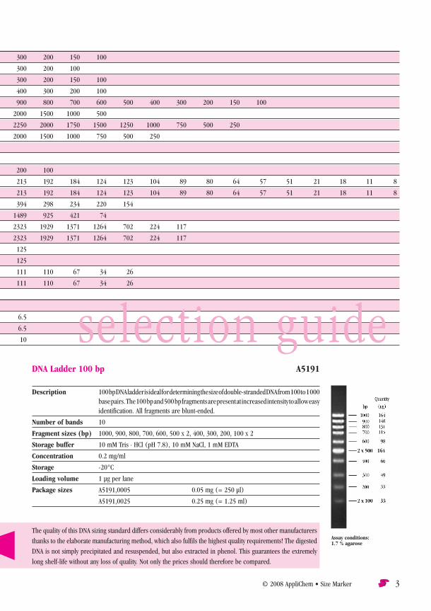
DNA Ladder 100 bp (lyophilised) A3470DNA size standard for medium-sized fragments and PCR products
Description The fragments of this size marker occur in equimolar proportions, i.e. also all possible marking positions on the terminal EcoR I generated recessed ends. The band mass increases with fragment length. The DNA is deproteinized and lyophilized. This marker is particularly suitable for com-parisons with medium-sized DNA fragments and PCR products. The 500 base pair band mass has been doubled for easier orientation on 1.5 – 2.0 agarose gels. The size and mass of plasmid in- sertions or PCR products can be determined precisely in the range of up to 1000 base pairs at intervals of 100 base pairs. An additional band with 150 base pairs has been included for the determination of smaller PCR products. The best results are achieved after a migration distance of approx. 70 – 80 mm on agarose gels. Each lane of a normal sized agarose gel (approx. 80 ml)
should be loaded with approx. 0.4 – 0.8 µg of marker. Loading buffer is supplied separately.
Number of bands 11
Fragment sizes (bp) 1000, 900, 800, 700, 600, 500 (x2), 400, 300, 200, 150, 100
Supplied with 1 ml 10 mM Tris · HCl (pH 7.5); 5 mM sodium acetate; 2 mM EDTA; 10 % glycerol; loading buffer (1X) 0.02 % bromophenol blue; 0.015 % xylene cyanol
Storage -20°C
Loading volume 0.4 – 0.8 µg per lane
Package size A3470,0050 50 µg
The quality of this DNA sizing standard differs considerably from products offered by most other manufacturers
thanks to the elaborate manufacturing method, which also fulfils the highest quality requirements! The digested
DNA is not simply precipitated and resuspended, but also extracted in phenol. This guarantees the extremely
long shelf-life without any loss of quality. Not only the prices should therefore be compared.
Prod. No. DNA Marker Bands Fragment Sizes [bp]
A3470 DNA Ladder 100 bp (lyophilised) 11 1000 900 800 700 600 500 400 300 200 150 100
A5191 DNA Ladder 100 bp 10 1000 900 800 700 600 500 400 300 200 100
A3302 DNA Ladder 100 bp equalized (lyophilised) 11 1000 900 800 700 600 500 400 300 200 150 100
A5216 DNA Ladder 100 bp plus 11 1500 1000 900 800 700 600 500 400 300 200 100
A3660 DNA Ladder Mix 100 – 5000 (lyophilised) 17 5000 4000 3000 2500 2000 1500 1000 900 800 700 600 500 400 300 200 150 100
A2667 DNA Ladder 1 kb (lyophilised) 11 10000 8000 6000 5000 4000 3000 2500 2000 1500 1000 500
A3982 DNA Ladder 250 bp (lyophilised) 16 8000 6000 5000 4000 3000 2750 2500 2250 2000 1750 1500 1250 1000 750 500 250
A5207 DNA Ladder 1 kb 13 10000 8000 6000 5000 4000 3000 2500 2000 1500 1000 750 500 250
A6430 DNA Ladder 1 kb concatamer (lyophlised) >25 1000 bp steps (1000 – approx. 25000)
A7215 DNA Marker quick-run (lyophylised) 5 2500 2000 1500 1000 500
A7222 DNA Marker quick-run extended (lyophylised) 9 6000 4000 25000 2000 1500 1000 500 200 100
A4406 DNA Marker pBR322 – Hae III (lyophilised) 22 587 540 502 458 434 267 234 213 192 184 124 123 104 89 80 64 57 51 21 18 11 8
A5229 DNA Marker pBR322 – Hae III 22 587 540 502 458 434 267 234 213 192 184 124 123 104 89 80 64 57 51 21 18 11 8
A6927 DNA Marker pBR328 Mix (lyophilised) 12 2176 1766 1230 1033 653 517 453 394 298 234 220 154
A5194 DNA Marker Phage Lambda Sty I 11 19329 7743 6223 4254 3472 2690 1882 1489 925 421 74
A4412 DNA Marker Phage Lambda BstE II (lyophilised) 14 8454 7242 6369 5686 4822 4324 3675 2323 1929 1371 1264 702 224 117
A5220 DNA Marker Phage Lambda BstE II 14 8454 7242 6369 5686 4822 4324 3675 2323 1929 1371 1264 702 224 117
A5589 DNA Marker Phage Lambda Hind III (lyophilised) 8 23130 9416 6557 4361 2322 2027 564 125
A5223 DNA Marker Phage Lambda Hind III 8 23130 9400 6557 4361 2322 2027 564 125
A5235 DNA Marker pUC19 – Msp I 12 501 489 404 331 242 190 147 111 110 67 34 26
A3996 DNA Marker pUC19 – Msp I (lyophilised) 12 501 489 404 331 242 190 147 111 110 67 34 26
Protein Marker
A5238 Protein Marker I (14 – 116) 7 116.0 97.4 66.2 37.6 28.5 18.4 14.0
A5418 Protein Marker II (6.5 – 200) prestained 8 200.0 116.0 68.0 43.0 30.0 20.0 14.4 6.5
A4402 Protein Marker III (6.5 – 200) 8 200.0 116.0 68.0 43.0 30.0 20.0 14.4 6.5
A3993 Protein Marker IV (10 – 150) 8 150 100 80 60 40 30 20 10
© 2008 AppliChem • Size Marker 3
selection guideDNA Ladder 100 bp A5191
Description 100 bp DNA ladder is ideal for determining the size of double-stranded DNA from 100 to 1 000 base pairs. The 100 bp and 500 bp fragments are present at increased intensity to allow easy
identification. All fragments are blunt-ended.
Number of bands 10
Fragment sizes (bp) 1000, 900, 800, 700, 600, 500 x 2, 400, 300, 200, 100 x 2
Storage buffer 10 mM Tris · HCl (pH 7.8), 10 mM NaCl, 1 mM EDTA
Concentration 0.2 mg/ml
Storage -20°C
Loading volume 1 µg per lane
Package sizes A5191,0005 0.05 mg (= 250 µl)
A5191,0025 0.25 mg (= 1.25 ml)
Assay conditions: 1.7 % agarose
4 Size Marker • © 2008 AppliChem
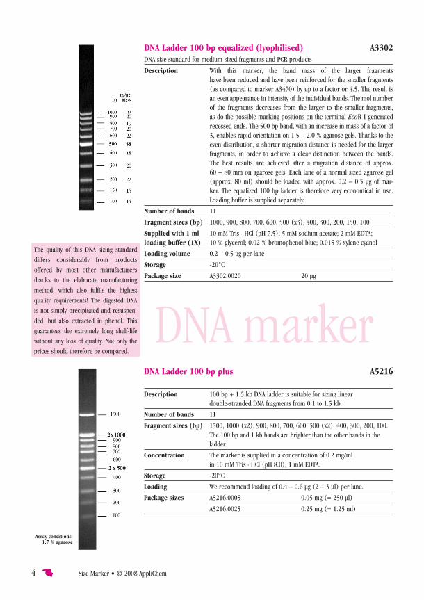
DNA Ladder 100 bp plus A5216
Description 100 bp + 1.5 kb DNA ladder is suitable for sizing linear double-stranded DNA fragments from 0.1 to 1.5 kb.
Number of bands 11
Fragment sizes (bp) 1500, 1000 (x2), 900, 800, 700, 600, 500 (x2), 400, 300, 200, 100. The 100 bp and 1 kb bands are brighter than the other bands in the
ladder.
Concentration The marker is supplied in a concentration of 0.2 mg/ml in 10 mM Tris · HCl (pH 8.0), 1 mM EDTA.
Storage -20°C
Loading We recommend loading of 0.4 – 0.6 µg (2 – 3 µl) per lane.
Package sizes A5216,0005 0.05 mg (= 250 µl)
A5216,0025 0.25 mg (= 1.25 ml)
Assay conditions: 1.7 % agarose
DNA Ladder 100 bp equalized (lyophilised) A3302DNA size standard for medium-sized fragments and PCR products
Description With this marker, the band mass of the larger fragments have been reduced and have been reinforced for the smaller fragments
(as compared to marker A3470) by up to a factor or 4.5. The result is an even appearance in intensity of the individual bands. The mol number of the fragments decreases from the larger to the smaller fragments, as do the possible marking positions on the terminal EcoR I generated recessed ends. The 500 bp band, with an increase in mass of a factor of 3, enables rapid orientation on 1.5 – 2.0 % agarose gels. Thanks to the even distribution, a shorter migration distance is needed for the larger fragments, in order to achieve a clear distinction between the bands. The best results are achieved after a migration distance of approx. 60 – 80 mm on agarose gels. Each lane of a normal sized agarose gel (approx. 80 ml) should be loaded with approx. 0.2 – 0.5 µg of mar-ker. The equalized 100 bp ladder is therefore very economical in use.
Loading buffer is supplied separately.
Number of bands 11
Fragment sizes (bp) 1000, 900, 800, 700, 600, 500 (x3), 400, 300, 200, 150, 100
Supplied with 1 ml 10 mM Tris · HCl (pH 7.5); 5 mM sodium acetate; 2 mM EDTA; loading buffer (1X) 10 % glycerol; 0.02 % bromophenol blue; 0.015 % xylene cyanol
Loading volume 0.2 – 0.5 µg per lane
Storage -20°C
Package size A3302,0020 20 µg
The quality of this DNA sizing standard
differs considerably from products
offered by most other manufacturers
thanks to the elaborate manufacturing
method, which also fulfils the highest
quality requirements! The digested DNA
is not simply precipitated and resuspen-
ded, but also extracted in phenol. This
guarantees the extremely long shelf-life
without any loss of quality. Not only the
prices should therefore be compared.
DNA marker

© 2008 AppliChem • Size Marker 5
DNA Ladder Mix 100 – 5000 (lyophilised) A3660DNA size standard for the determination of the size of medium-sized fragments and plasmids
Description The 100 bp ladder (A3470) was extended with fragments in the size range of 1500 – 5000. The extension by 40 % mass in the larger fragments means easy
orientation from 1500 bp upwards on 1.2 – 1.5 % agarose gels. In addition to small and large plasmid insertions and PCR products, the size and quantity of plasmid vectors can also be determined. The best results are achieved after a migration distance of 80 – 90 mm on agarose gels with loading volumes of 0.5 – 0.8 µg per lane on
standard sized gels.
Number of bands 17
Fragment sizes (bp) 5000, 4000, 3000, 2500, 2000, 1500, 1000, 900, 800, 700, 600, 500 (x2), 400, 300, 200, 150, 100.
Supplied with 1 ml 10 mM Tris · HCl (pH 7.5); 5 mM sodium acetate; 2 mM EDTA; loading buffer (1X) 10 % glycerol; 0.02 % bromophenol blue; 0.015 % xylene cyanol
Loading volume 0.5 – 0.8 µg per lane (for good staining properties with ethidium bromide)
Migration distance Best results are achieved after a migration distance of 70 – 90 mm.
Storage -20°C. Avoid repeated thawing and refreezing. Portioning in small aliquots is recommended. The lyophilised product is stable for many weeks even at room temperature!
Package size A3660,0050 50 µg
This sizing standard was manufactured using digestion of 7 plasmids with EcoR I. The plasmids are legally
protected. Permission to produce copies must be sought from the manufacturer. The DNA was deproteinised,
precipitated and lyophilised after digestion with phenol/chloroform. If the fragments are to be labeled, the marker
should be dissolved in distilled water (10 minutes at room temperature, shaking occasionally).
The quality of this DNA sizing standard differs considerably from products offered by most other manufacturers
thanks to the elaborate manufacturing method, which also fulfils the highest quality requirements! The digested
DNA is not simply precipitated and resuspended, but also extracted in phenol. This guarantees the extremely long
shelf-life without any loss of quality. Not only the prices should therefore be compared.
DNA Ladder 1 kb (lyophilised) A2667
Description The size of medium-sized plasmids and fragments can be determined with 11 DNA bands between 10,000 and 500 base pairs. Unlike the equimolar distribution of the bands in other
DNA markers and the long migration distance necessary with these for the separation of large fragments, the mass of the larger bands of the 1 kbp DNA ladder has been reduced. This permits relatively short separation distances for a 1 kbp DNA ladder (approx. 70 mm on 1.2 % agarose gels) and means that it is very economical in use (0.4 – 0.6 µg per lane, or 400 separations) on normal sized gels. Loading buffer is supplied separately. The fragments have terminal EcoR I generated recessed ends.
The DNA is deproteinized and lyophilized.
Number of bands 11
Fragment sizes (bp): 10000, 8000, 6000, 5000, 4000, 3000, 2500, 2000, 1500, 1000, 500
Supplied with 1 ml 10 mM Tris · HCl (pH 7.5); 5 mM sodium acetate; 2 mM EDTA; 10 % glycerol; loading buffer (1X) 0.02 % bromophenol blue; 0.015 % xylene cyanol
Loading volume 0.4 – 0.6 µg per lane
Storage -20°C
Package size A2667,0050 50 µg
A2667,0200 4 x 50 µg
DNA marker6 Size Marker • © 2008 AppliChem
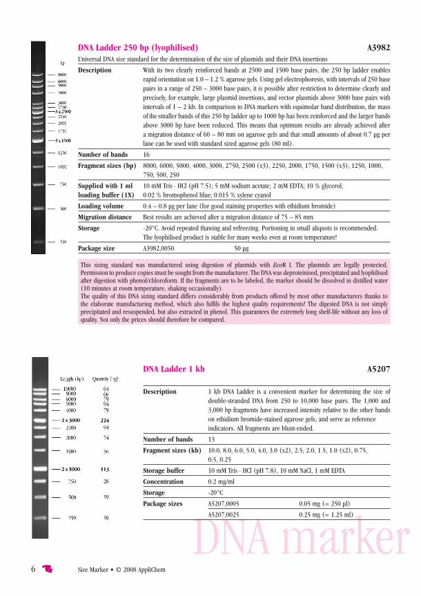
DNA Ladder 250 bp (lyophilised) A3982Universal DNA size standard for the determination of the size of plasmids and their DNA insertions
Description With its two clearly reinforced bands at 2500 and 1500 base pairs, the 250 bp ladder enables rapid orientation on 1.0 – 1.2 % agarose gels. Using gel electrophoresis, with intervals of 250 base
pairs in a range of 250 – 3000 base pairs, it is possible after restriction to determine clearly and precisely, for example, large plasmid insertions, and vector plasmids above 3000 base pairs with intervals of 1 – 2 kb. In comparison to DNA markers with equimolar band distribution, the mass of the smaller bands of this 250 bp ladder up to 1000 bp has been reinforced and the larger bands above 3000 bp have been reduced. This means that optimum results are already achieved after a migration distance of 60 – 80 mm on agarose gels and that small amounts of about 0.7 µg per
lane can be used with standard sized agarose gels (80 ml).
Number of bands 16
Fragment sizes (bp) 8000, 6000, 5000, 4000, 3000, 2750, 2500 (x3), 2250, 2000, 1750, 1500 (x3), 1250, 1000, 750, 500, 250
Supplied with 1 ml 10 mM Tris · HCl (pH 7.5); 5 mM sodium acetate; 2 mM EDTA; 10 % glycerol; loading buffer (1X) 0.02 % bromophenol blue; 0.015 % xylene cyanol
Loading volume 0.4 – 0.8 µg per lane (for good staining properties with ethidium bromide)
Migration distance Best results are achieved after a migration distance of 75 – 85 mm.
Storage -20°C. Avoid repeated thawing and refreezing. Portioning in small aliquots is recommended. The lyophilised product is stable for many weeks even at room temperature!
Package size A3982,0050 50 µg
This sizing standard was manufactured using digestion of plasmids with EcoR I. The plasmids are legally protected. Permission to produce copies must be sought from the manufacturer. The DNA was deproteinised, precipitated and lyophilised after digestion with phenol/chloroform. If the fragments are to be labeled, the marker should be dissolved in distilled water (10 minutes at room temperature, shaking occasionally).The quality of this DNA sizing standard differs considerably from products offered by most other manufacturers thanks to the elaborate manufacturing method, which also fulfils the highest quality requirements! The digested DNA is not simply precipitated and resuspended, but also extracted in phenol. This guarantees the extremely long shelf-life without any loss of quality. Not only the prices should therefore be compared.
DNA Ladder 1 kb A5207
Description 1 kb DNA Ladder is a convenient marker for determining the size of double-stranded DNA from 250 to 10,000 base pairs. The 1,000 and
3,000 bp fragments have increased intensity relative to the other bands on ethidium bromide-stained agarose gels, and serve as reference
indicators. All fragments are blunt-ended.
Number of bands 13
Fragment sizes (kb) 10.0, 8.0, 6.0, 5.0, 4.0, 3.0 (x2), 2.5, 2.0, 1.5, 1.0 (x2), 0.75, 0.5, 0.25
Storage buffer 10 mM Tris · HCl (pH 7.8), 10 mM NaCl, 1 mM EDTA
Concentration 0.2 mg/ml
Storage -20°C
Package sizes A5207,0005 0.05 mg (= 250 µl)
A5207,0025 0.25 mg (= 1.25 ml)
DNA marker © 2008 AppliChem • Size Marker 7
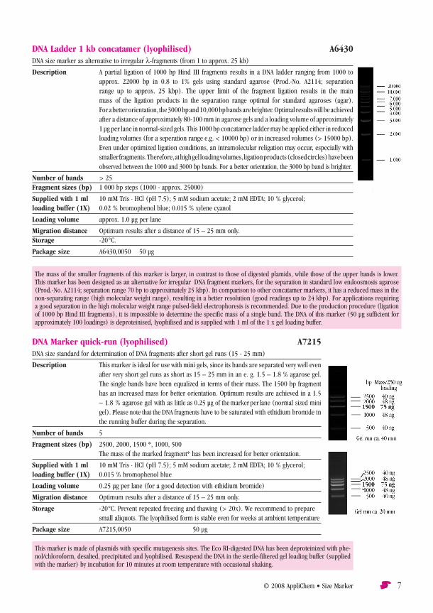
DNA Marker quick-run (lyophilised) A7215DNA size standard for determination of DNA fragments after short gel runs (15 - 25 mm)
Description This marker is ideal for use with mini gels, since its bands are separated very well even after very short gel runs as short as 15 – 25 mm in an e. g. 1.5 – 1.8 % agarose gel. The single bands have been equalized in terms of their mass. The 1500 bp fragment has an increased mass for better orientation. Optimum results are achieved in a 1.5 – 1.8 % agarose gel with as little as 0.25 µg of the marker per lane (normal sized mini gel). Please note that the DNA fragments have to be saturated with ethidium bromide in
the running buffer during the separation.
Number of bands 5
Fragment sizes (bp) 2500, 2000, 1500 *, 1000, 500 The mass of the marked fragment* has been increased for better orientation.
Supplied with 1 ml 10 mM Tris · HCl (pH 7.5); 5 mM sodium acetate; 2 mM EDTA; 10 % glycerol; loading buffer (1X) 0.015 % bromophenol blue
Loading volume 0.25 µg per lane (for a good detection with ethidium bromide)
Migration distance Optimum results after a distance of 15 – 25 mm only.
Storage -20°C. Prevent repeated freezing and thawing (> 20x). We recommend to prepare small aliquots. The lyophilised form is stable even for weeks at ambient temperature
Package size A7215,0050 50 µg
DNA Ladder 1 kb concatamer (lyophilised) A6430DNA size marker as alternative to irregular l-fragments (from 1 to approx. 25 kb)
Description A partial ligation of 1000 bp Hind III fragments results in a DNA ladder ranging from 1000 to approx. 22000 bp in 0.8 to 1% gels using standard agarose (Prod.-No. A2114; separation
range up to approx. 25 kbp). The upper limit of the fragment ligation results in the main mass of the ligation products in the separation range optimal for standard agaroses (agar). For a better orientation, the 3000 bp and 10,000 bp bands are brighter. Optimal results will be achieved after a distance of approximately 80-100 mm in agarose gels and a loading volume of approximately 1 µg per lane in normal-sized gels. This 1000 bp concatamer ladder may be applied either in reduced loading volumes (for a seperation range e.g. < 10000 bp) or in increased volumes (> 15000 bp). Even under optimized ligation conditions, an intramolecular religation may occur, especially with smaller fragments. Therefore, at high gel loading volumes, ligation products (closed circles) have been
observed between the 1000 and 3000 bp bands. For a better orientation, the 3000 bp band is brighter.
Number of bands > 25Fragment sizes (bp) 1 000 bp steps (1000 - approx. 25000)
Supplied with 1 ml 10 mM Tris · HCl (pH 7.5); 5 mM sodium acetate; 2 mM EDTA; 10 % glycerol; loading buffer (1X) 0.02 % bromophenol blue; 0.015 % xylene cyanol
Loading volume approx. 1.0 µg per lane
Migration distance Optimum results after a distance of 15 – 25 mm only.Storage -20°C.
Package size A6430,0050 50 µg
The mass of the smaller fragments of this marker is larger, in contrast to those of digested plamids, while those of the upper bands is lower. This marker has been designed as an alternative for irregular λDNA fragment markers, for the separation in standard low endoosmosis agarose (Prod.-No. A2114; separation range 70 bp to approximately 25 kbp). In comparison to other concatamer markers, it has a reduced mass in the non-separating range (high molecular weight range), resulting in a better resolution (good readings up to 24 kbp). For applications requiring a good separation in the high molecular weight range pulsed-field electrophoresis is recommended. Due to the production procedure (ligation of 1000 bp Hind III fragments), it is impossible to determine the specific mass of a single band. The DNA of this marker (50 µg sufficient for approximately 100 loadings) is deproteinised, Iyophilised and is supplied with 1 ml of the 1 x gel loading buffer.
This marker is made of plasmids with specific mutagenesis sites. The Eco RI-digested DNA has been deproteinized with phe-nol/chloroform, desalted, precipitated and lyophilised. Resuspend the DNA in the sterile-filtered gel loading buffer (supplied with the marker) by incubation for 10 minutes at room temperature with occasional shaking.

8 Size Marker • © 2008 AppliChem
DNA Marker pBR322 – Hae III (lyophilised) A4406DNA size standard for high-percentage agarose gels and polyacrylamide gels
Description Plasmid pBR322 was digested with Hae III, deproteinized and lyophilized. This marker is particularly suited to comparisons with small PCR fragments and DNA
from restriction samples on polyacrylamide gels or high-percentage agarose gels (> 2.2 %). The best results are achieved after a migration distance of approx. 70 – 80 mm on agarose gels or 100 mm on 6 – 8 % polyacrylamide gels. Each lane of a normal sized gel (approx. 80 ml) should be loaded with approx. 1 µg of marker
for agarose gels or 0.5 µg for PAA gels. Loading buffer is supplied separately.
Number of bands 22
Fragment sizes (bp) 587, 540, 502, 458, 434, 267, 234, 213, 192, 184, 124, 123, 104, 89, 80, 64, 57, 51, 21, 18, 11, 8
Supplied with 1 ml 10 mM Tris · HCl (pH 7.5); 5 mM sodium acetate; 2 mM EDTA; 10 % glycerol; loading buffer (1X) 0.02 % bromophenol blue; 0.015 % xylene cyanol
Loading volume 0.5 – 1.0 µg per lane
Storage -20°C
Package size A4406,0050 50 µgPlease note! Extremely small fragments can only be visualized on high-percentage PAA gels.
DNA Marker quick-run extended (lyophilised) A7222DNA size standard for determination of DNA fragments after short gel runs (35 mm)
Description This marker is ideal for use with mini or midi gels, since its bands are separated very well even after very short gel runs as short as 35 mm in an e. g. 1.2 % agarose gel. The single bands have been equalized in terms of their mass. The 1500 bp frag-ment has an increased mass for better orientation. Optimum results are achieved
with as little as 0.3 – 0.5 µg of the marker per lane (normal-sized mini/midi gel).
Number of bands 9
Fragment sizes (bp) 6000, 4000, 2500, 2000, 1500*, 1000, 500, 200, 100 The mass of the marked fragment* has been increased for better orientation.
Supplied with 1 ml 10 mM Tris · HCl (pH 7.5); 5 mM sodium acetate; 2 mM EDTA; 10 % glycerol; loading buffer (1X) 0.015 % bromophenol blue
Loading volume 0.3 - 0.5 µg per lane (for a good detection with ethidium bromide in a mini or midi gel)
Storage -20°C. Prevent repeated freezing and thawing (> 20x). We recommend to prepare
small aliquots. The lyophilised form is stable even for weeks at ambient temperature!
Package size A7222,0050 50 µg
This marker is made of plasmids with specific mutagenesis sites. The Eco RI-digested DNA has been deproteinized
with phenol/chloroform, desalted, precipitated and lyophilised. Resuspend the DNA in the sterile-filtered gel
loading buffer (supplied with the marker) by incubation for 10 minutes at room temperature with occasional
shaking.

© 2008 AppliChem • Size Marker 9
DNA Marker pBR328 Mix (lyophilised) A6927DNA size standard for middle-sized fragments and PCR productsDescription Plasmid pBR328 was digested with Hinf I and Bgl I, respectively, deproteinised and
lyophilised. The fragments have been mixed in an equimolar ratio. This marker is particularly suitable for comparison with PCR fragments (e.g. food diagnostics) and DNA from restriction samples on agarose gels with sizes between 150 and 2000 base pairs. The concise distribution of fragments of the marker makes this product ideal for long and short gel runs with running distances of 70 – 80 mm (1.5 % agarose gel). Load 1 µg of the marker per lane of a normal sized agarose gel (approx. 80 ml
Loading buffer is supplied separately.
Number of bands 12
Fragment sizes (bp) 2176; 1766; 1230; 1033; 653; 517; 453; 394; 298; 234; 220; 154
Supplied with 1 ml 10 mM Tris · HCl (pH 7.5); 5 mM sodium acetate; 2 mM EDTA; 10 % glycerol; loading buffer (1X) 0.02 % bromophenol blue; 0.015 % xylene cyanol
Loading volume 1.0 µg per lane
Storage -20°C. Prevent repeated freezing and thawing (> 20x). We recommend to prepare small aliquots. The lyophilised form is stable even for weeks at ambient temperature!
Package sizes A6927,0050 50 µg
Resuspend the DNA in the sterile-filtered gel loading buffer (supplied with the marker) for 10 minutes at room
temperature by occasional shaking.
DNA Marker pBR322 – Hae III A5229
Description This marker is generated by digestion of the plasmid pBR322 with Hae III.
Number of bands 22
Fragment sizes (bp) 587, 540, 502, 458, 434, 267, 234, 213, 192, 184, 124, 123, 104, 89, 80, 64, 57, 51, 21, 18, 11, 8
Storage buffer 10 mM Tris · HCl (pH 7.8), 10 mM NaCl, 1 mM EDTA
Concentration 0.2 mg/ml
Storage -20°C
Package sizes A5229,0005 0.05 mg (= 250 µL)
A5229,0025 0.25 mg (= 1.25 ml)
Assay conditions: 1.7 % agarose
DNA markerThe quality of this DNA sizing standard differs con-
siderably from products offered by most other
manufacturers thanks to the elaborate manufacturing
method, which also fulfils the highest quality require-
ments! The digested DNA is not simply precipitated
and resuspended, but also extracted in phenol. This
guarantees the extremely long shelf-life without any
loss of quality. Not only the prices should therefore
be compared.
10 Size Marker • © 2008 AppliChem
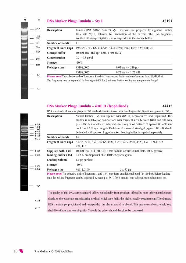
DNA Marker Phage Lambda – Sty I A5194
Description Lambda DNA (cI857 Sam 7) Sty I markers are prepared by digesting Lambda DNA with Sty I, followed by inactivation of the enzyme. The DNA fragments are then ethanol-precipitated and resuspended in the storage buffer.
Number of bands 11
Fragment sizes (bp) 19329*; 7743; 6223; 4254*; 3472; 2690; 1882; 1489; 925; 421; 74
Storage buffer 10 mM Tris · HCl (pH 8.0), 1 mM EDTA
Concentration 0.2 – 0.5 µg/µl
Storage -20°C
Package sizes A5194,0005 0.05 mg (= 250 µl)
A5194,0025 0.25 mg (= 1.25 ml)Please note! The cohesive ends of fragments 1 and 4 (*) may cause the formation of an extra band (23583 bp). The fragments may be separated by heating to 65°C for 3 minutes before loading the sample onto the gel.
The quality of this DNA sizing standard differs considerably from products offered by most other manufacturers
thanks to the elaborate manufacturing method, which also fulfils the highest quality requirements! The digested
DNA is not simply precipitated and resuspended, but also extracted in phenol. This guarantees the extremely long
shelf-life without any loss of quality. Not only the prices should therefore be compared.
DNA Marker Phage Lambda – BstE II (lyophilised) A4412DNA size standard made of phage l DNA for the determination of large DNA fragments (digestion of genomic DNA)
Description Natural lambda DNA was digested with BstE II, deproteinized and lyophilized. This marker is suitable for comparisons with fragment sizes between 8400 and 700 base
pairs. The best results are achieved after a migration distance of approx. 80 – 90 mm on 1.0 – 1.2 % agarose gels. Each lane of a normal sized gel (approx. 80 ml) should
be loaded with approx. 1 µg of marker. Loading buffer is supplied separately.
Number of bands 14
Fragment sizes (bp) 8454*, 7242, 6369, 5686*, 4822, 4324, 3675, 2323, 1929, 1371, 1264, 702, 224, 117
Supplied with 1 ml 10 mM Tris · HCl (pH 7.5); 5 mM sodium acetate; 2 mM EDTA; 10 % glycerol;loading buffer (1X) 0.02 % bromophenol blue; 0.015 % xylene cyanol
Loading volume 1.0 µg per lane
Storage -20°C
Package size A4412,0100 2 x 50 µgPlease note! The cohesive ends of fragments 1 and 4 (*) may form an additional band (14140 bp). Before loading onto the gel, the fragments can be separated by heating to 65°C for 5 minutes with subsequent incubation on ice.
© 2008 AppliChem • Size Marker 11
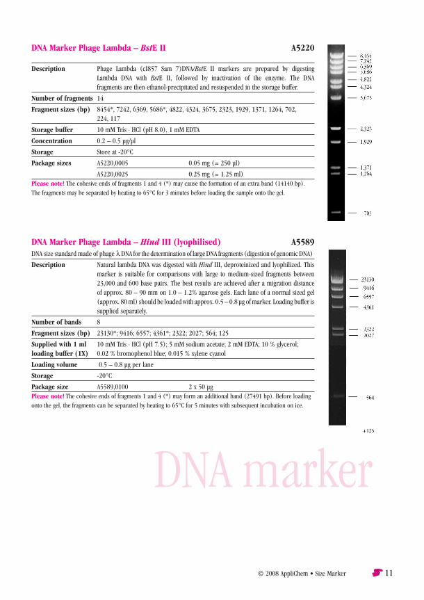
DNA Marker Phage Lambda – BstE II A5220
Description Phage Lambda (cI857 Sam 7)DNA/BstE II markers are prepared by digesting Lambda DNA with BstE II, followed by inactivation of the enzyme. The DNA fragments are then ethanol-precipitated and resuspended in the storage buffer.
Number of fragments 14
Fragment sizes (bp) 8454*, 7242, 6369, 5686*, 4822, 4324, 3675, 2323, 1929, 1371, 1264, 702, 224, 117
Storage buffer 10 mM Tris · HCl (pH 8.0), 1 mM EDTA
Concentration 0.2 – 0.5 µg/µl
Storage Store at -20°C
Package sizes A5220,0005 0.05 mg (= 250 µl)
A5220,0025 0.25 mg (= 1.25 ml)Please note! The cohesive ends of fragments 1 and 4 (*) may cause the formation of an extra band (14140 bp). The fragments may be separated by heating to 65°C for 3 minutes before loading the sample onto the gel.
DNA Marker Phage Lambda – Hind III (lyophilised) A5589DNA size standard made of phage l DNA for the determination of large DNA fragments (digestion of genomic DNA)
Description Natural lambda DNA was digested with Hind III, deproteinized and lyophilized. This marker is suitable for comparisons with large to medium-sized fragments between
23,000 and 600 base pairs. The best results are achieved after a migration distance of approx. 80 – 90 mm on 1.0 – 1.2% agarose gels. Each lane of a normal sized gel (approx. 80 ml) should be loaded with approx. 0.5 – 0.8 µg of marker. Loading buffer is
supplied separately.
Number of bands 8
Fragment sizes (bp) 23130*; 9416; 6557; 4361*; 2322; 2027; 564; 125
Supplied with 1 ml 10 mM Tris · HCl (pH 7.5); 5 mM sodium acetate; 2 mM EDTA; 10 % glycerol; loading buffer (1X) 0.02 % bromophenol blue; 0.015 % xylene cyanol
Loading volume 0.5 – 0.8 µg per lane
Storage -20°C
Package size A5589,0100 2 x 50 µgPlease note! The cohesive ends of fragments 1 and 4 (*) may form an additional band (27491 bp). Before loading onto the gel, the fragments can be separated by heating to 65°C for 5 minutes with subsequent incubation on ice.
DNA marker
DNA marker
12 Size Marker • © 2008 AppliChem
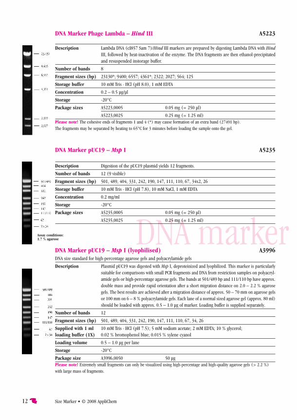
DNA Marker Phage Lambda – Hind III A5223
Description Lambda DNA (cI857 Sam 7)/Hind III markers are prepared by digesting Lambda DNA with Hind III, followed by heat-inactivation of the enzyme. The DNA fragments are then ethanol-precipitated
and resuspended instorage buffer.
Number of bands 8
Fragment sizes (bp) 23130*; 9400; 6557; 4361*; 2322; 2027; 564; 125
Storage buffer 10 mM Tris · HCl (pH 8.0), 1 mM EDTA
Concentration 0.2 – 0.5 µg/µl
Storage -20°C
Package sizes A5223,0005 0.05 mg (= 250 µl)
A5223,0025 0.25 mg (= 1.25 ml)Please note! The cohesive ends of fragments 1 and 4 (*) may cause formation of an extra band (27491 bp). The fragments may be separated by heating to 65°C for 3 minutes before loading the sample onto the gel.
DNA Marker pUC19 – Msp I A5235
Description Digestion of the pUC19 plasmid yields 12 fragments.
Number of bands 12 (9 visible)
Fragment sizes (bp) 501, 489, 404, 331, 242, 190, 147, 111, 110, 67, 34x2, 26
Storage buffer 10 mM Tris · HCl (pH 7.8), 10 mM NaCl, 1 mM EDTA
Concentration 0.2 mg/ml
Storage -20°C
Package sizes A5235,0005 0.05 mg (= 250 µl)
A5235,0025 0.25 mg (= 1.25 ml)
Assay conditions: 1.7 % agarose
DNA Marker pUC19 – Msp I (lyophilised) A3996DNA size standard for high-percentage agarose gels and polyacrylamide gels
Description Plasmid pUC19 was digested with Msp I, deproteinized and lyophilized. This marker is particularly suitable for comparisons with small PCR fragments and DNA from restriction samples on polyacryl-amide gels or high-percentage agarose gels. The bands at 501/489 bp and 111/110 bp have approx. double mass and provide rapid orientation after a short migration distance on 2.0 – 2.2 % agarose gels. The best results are achieved after a migration distance of approx. 50 – 70 mm on agarose gels or 100 mm on 6 – 8 % polyacrylamide gels. Each lane of a normal sized agarose gel (approx. 80 ml)
should be loaded with approx. 0.5 – 1.0 µg of marker. Loading buffer is supplied separately.
Number of bands 12
Fragment sizes (bp) 501, 489, 404, 331, 242, 190, 147, 111, 110, 67, 34, 26
Supplied with 1 ml 10 mM Tris · HCl (pH 7.5); 5 mM sodium acetate; 2 mM EDTA; 10 % glycerol; loading buffer (1X) 0.02 % bromophenol blue; 0.015 % xylene cyanol
Loading volume 0.5 – 1.0 µg per lane
Storage -20°C
Package size A3996,0050 50 µgPlease note! Extremely small fragments can only be visualized using high-percentage and high-quality agarose gels (> 2.2 %) with large mass of fragments.

© 2008 AppliChem • Size Marker 13
Protein Marker I (14 – 116) A5238
Description The bands of 3 µl marker are easily visualized with Coomassie staining in a
polyacrylamide gel. Protein Marker I is a mixture of 7 purified proteins supplied in gel loading buffer for direct application to
an SDS polyacrylamide gel.
Number of bands 7
Fragment sizes (kd) 116.0; 97.4; 66.2; 37.6; 28.5; 18.4; 14.0
Loading buffer 50 mM Tris · HCl (pH 6.8), 100 mM dithiothreitol, 2 % SDS, 0.1% bromophenol blue, 10 % glycerol
Assay conditions 3 µl/12 % PAGE
Package size A5235,0500 500 µl
Protein Marker II (6.5 – 200) prestained A5418Prestained protein size marker for gel electrophoresis
Description This is a ready-to-use marker containing covalent prestained proteins. The product contains formamide.
Number of bands 8
Protein sizes (kd) 200.0; 116.0; 68.0; 43.0; 30.0; 20.0; 14.4; 6.5
Assay conditions 10 µl/4 – 20 % PAGE gradient gel Tris-Glycine; left lane prestained but not
Coomassie-stained; right lane prestained and additional Coomassie staining.
Storage -20°C, if stored longer than 1 month. Please note that repeated freezing and
thawing will reduce product quality. Pre- paration of small aliquots is recommended.
Package size A5418,0250 250 µl
Application ● The marker has to be “preheated” to room tem-perature to guarantee that all components are dissolved. ● Apply 5 µl of the marker to a mini-gel (10 x 10 cm, 1 – 0.75 mm thick, 7 mm slot). ● To minimize myosin aggregation, the aliquot to be loaded on the gel may be heated to 95°C for 1 – 2 minutes. ● Prestained markers are not recommended for the determination of the molecular weight since their behaviour during electrophoresis strongly depends on the electrophoresis conditions. ● The transfer of proteins during blotting depends on their size. Lysozyme is fully transferred after 30 minutes, while myosin requires 2.5 hours (1 V/cm2).
protein marker
14 Size Mark er • © 2008 AppliChem
prot
ein
mar
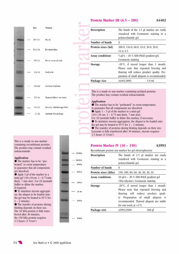
ker Protein Marker III (6.5 – 200) A4402
Description The bands of the 2.5 µl marker are easily visualized with Coomassie staining in a polyacrylamide gel.
Number of bands 8
Protein sizes (kd) 200.0; 116.0; 68.0; 43.0; 30.0; 20.0; 14.4; 6.5
Assay conditions 5 µl/4 – 20 % SDS-PAGE gradient gel; Coomassie staining
Storage -20°C, if stored longer than 1 month. Please note that repeated freezing and
thawing will reduce product quality. Pre- paration of small aliquots is recommended.
Package size A4402,0001 1.0 ml
Protein Marker IV (10 – 150) A3993Recombinant protein size marker for gel electrophoresis
Description The bands of 2.5 µl marker are easily visualized with Coomassie staining in a polyacrylamide gel.
Number of bands 8
Protein sizes (kDa) 150; 100; 80; 60; 40; 30; 20; 10
Assay conditions 10 µl/4 – 20 % SDS-PAGE gradient gel (Tris-Glycine); Coomassie staining
Storage -20°C, if stored longer than 1 month. Please note that repeated freezing and thawing will reduce product quali-
ty. Preparation of small aliquots is recommended. Thawed aliquots are stable
for one week at +4°C.
Package size A3993,0500 500 µl
This is a ready-to-use marker containing acylated proteins. The product may contain residual iodoacetamide.
Application ● The marker has to be “preheated” to room temperature, to guarantee that all components are dissolved. ● Apply 1 – 5 µl of the marker to a mini-gel (10 x 10 cm, 1 – 0.75 mm thick, 7 mm slot). Use 1X Laemmli buffer to dilute this marker, if necessary. ● To minimize myosin aggregation, the aliquot to be loaded onto the gel may be heated to 95°C for 1 – 2 minutes. ● The transfer of proteins during blotting depends on their size. Lysozyme is fully transferred after 30 minutes, myosin requires 2.5 hours (1 V/cm2).
This is a ready-to-use marker containing recombinant proteins. The product may contain residual iodoacetamide.
Application● The marker has to be “pre-heated” to room temperature to guarantee that all components are dissolved.● Apply 5 µl of the marker to a mini-gel (10 x 10 cm, 1 – 0.75 mm thick, 7 mm slot). Use 1X Laemmli buffer to dilute the marker, if required.● To minimize myosin aggregati-on, the aliquot to be loaded onto the gel may be heated to 95°C for 1 – 2 minutes.● The transfer of proteins during blotting depends on their size. The 10 kDa protein is fully trans-ferred after 30 minutes, the 150 kDa protein requires 2.5 hours (1 V/cm2).
© 2008 AppliChem • Size Marker 15
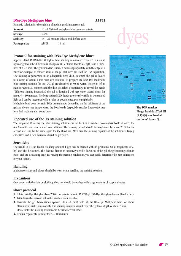
Protocol for staining with DNA-Dye Methylene blue:Approx. 50 ml 1X DNA-Dye Methylene blue staining solution are required to stain an agarose gel with the dimensions of approx. 80 x 60 mm (width x length) and a thick-ness of 3 – 4 mm. The gel should be trimmed down appropriately, with the edge of a ruler for example, to remove areas of the gel that were not used for DNA separation. The staining is performed in an adequately sized dish, in which the gel is floated to a depth of about 5 mm with dye solution. To prepare the DNA-Dye Methylene blue staining solution for use, 250 µl are dissolved in 50 ml water. The gel is left to stain for about 20 minutes and the dish is shaken occasionally. To reveal the bands (different staining intensities) the gel is destained with tap water several times for about 5 – 10 minutes. The blue-stained DNA bands are clearly visible in transmitted light and can be measured with a ruler or documented photographically.Methylene blue does not stain DNA permanently: depending on the thickness of the gel and the storage temperature, the DNA bands (especially smaller fragments) may lose their staining after some time.
Repeated use of the 1X staining solutionThe prepared 1X methylene blue staining solution can be kept in a suitable brown-glass bottle at +4°C for 4 – 6 months and can be used several times. The staining period should be lengthened by about 20 % for the second use, and by the same again for the third use. After this, the staining capacity of the solution is largely exhausted and a new solution should be prepared.
SensitivityThe bands in a 1 kb ladder (loading amount 1 µg) can be stained with no problems. Small fragments (150 bp) can also be stained. The decisive factors in sensitivity are the thickness of the gel, the gel:staining solution ratio, and the destaining time. By varying the staining conditions, you can easily determine the best conditions for your system.
HandlingA laboratory coat and gloves should be worn when handling the staining solution.
PrecautionOn contact with the skin or clothing, the area should be washed with large amounts of soap and water.
Short protocol1. Dilute DNA-Dye Methylene blue 200X concentrate down to 1X (250 µl DNA-Dye Methylene blue + 50 ml water)2. Trim down the agarose gel to the smallest area possible.3. Incubate the gel (dimensions approx. 80 x 60 mm) with 50 ml DNA-Dye Methylene blue for about
20 minutes; shake occasionally. The staining solution should cover the gel to a depth of about 5 mm. Please note: the staining solution can be used several times!4. Destain repeatedly in water for 5 – 10 minutes.
DNA-Dye Methylene blue A5595Nontoxic solution for the staining of nucleic acids in agarose gels
Amount 10 ml 200-fold methylene blue dye concentrate
Storage +4°C
Stability 18 – 24 months (shake well before use)
Package size A5595 10 ml
The DNA marker Phage Lambda-Hind III (A5589) was loaded on the 4th lane (*).
dyes

16 Size Mark er • © 2008 AppliChem
Protocol for staining of gels with Proteo-Dye Blue-Vis: SDS-PAGE minigels (1 mm thickness, 10 % acrylamide, Tris-glycine buffer system) are treated for 1 hour with 30 % v/v methanol after electrophoresis to remove the excess of SDS and to fix the proteins in the gel matrix. The gels are stained for 2 hours in a volume of 100 ml Proteo-Dye Blue-Vis. After this, the gels are washed with acetate buffer (0.2 M; pH 4.5), containing 20 % v/v methanol for 90 - 120 minutes until the dark red background is reduced to a weak pink background in contrast to the blue protein bands. The gels are documented with the help of a video system (transilluminator 312 nm, white light plate) or on a scanner in the transmission mode.
Proteo-Dye Blue-Vis A6810
Synomym Protein dye in the visible range
Storage 2 – 8°C protected from light
HS-No. 38220000
Package size A6810,1000 1L
1 L is sufficient for approx. 30 minigels, since 3x reusable detection limit 3 – 5 ng/mm² reversible staining of proteins
Protocol for staining of gels with Proteo-Dye Red-Fluo: SDS-PAGE minigels (5x8 cm, 1 mm thickness, 10 % acrylamide, Tris-glycine buffer system) are fixed for 30 minutes in a solution of 7.5 % v/v acetic acid, 20 % v/v ethanol. After this, the gels are stained with Proteo-Dye Red-Fluo (100 ml) for 2 - 3 hours. The background fluorescence can be removed by repeated washing with the fixation solution (7.5 % v/v acetic acid, 20 % v/v ethanol; 3 - 4 washing steps, 30 minutes each). The documentation of the gels is carried out with a video system (transilluminator 312 nm, adapting filter 590 nm).
Proteo-Dye Red-Fluo A6803
Synomym Protein dye with red fluorescence
Storage 2 – 8°C protected from light
HS-No. 38220000
Specification Emission maximum: 630 nm
Exicitation wave lenght: 312 nm
Package size A6803,1000 1L
1 L is sufficient for approx. 30 minigels, since 3x reusable detection limit 1-3 ng/mm² reversible staining of proteins more sensitive than most other products available in the market
dyes

© 2008 AppliChem • Size Marker
Proteo-Dye Green-Fluo is a fluorescence protein dye, based on a metal-chelate complex in conjunction with a detergent in submicellar concentration in an aqueous solution. This dye solution enables to detect very small amounts of proteins and the staining is fully reversible. Use the dye solution up to three times!
Protocol for staining of gels with Proteo-Dye Green-Fluo: SDS-PAGE minigels (5x8 cm; 1 mm thickness, 10% acrylamide, Tris-glycine buffer system) are postelectrophoretically treated for 30 minutes with a methanol solution (30 %, v/v) while gently shaking (100 rpm). After this, the gels are stained for 2 hours in a volume of 100 ml Proteo-Dye Green-Fluo. Reduction of the background fluorescence is achieved by repeated washing steps (3 – 4 times, 30 min. each) with 30 % v/v methanol. The gels are documented with a video system (transilluminator 365 nm, adapting filter 520 nm).
Protocol for staining of blots with Proteo-Dye Green-Fluo: By staining with Coomassie or silver the proteins in the gel become irreversibly denaturated and are difficult to transfer to blotting membranes. Metal chelate staining methods are reversible, resulting in good preconditions for the following blot transfer. Blots were performed by a typical semi-dry method. After the blotting process, the blots were washed in distilled water for 1 hour and dried at room temperature. Blots were then stained with Proteo-Dye Green-Fluo for 2 hours, briefly washed in 30 % v/v ethanol and dried again. Only in the dried state, intensely gree n fluorescent protein bands become visible in UV toplight (365 nm UV light) and are documented with a video system (adapting filter 420 nm). The stain may be easily and fastly removed just by rinsing in 30% v/v ethanol.
Proteo-Dye Green-Fluo A6794
Synomym Protein dye with green fluorescence
Storage 2 – 8°C protected from light
HS-No. 38220000
Specification Emission maximum: 520 nm
Excitation wave lenght: 365 nm
Package size A6794,1000 1L
1 L is sufficient for approx. 30 minigels, since 3x reusable detection limit 3 – 5 ng/mm² reversible staining of proteins suitable for gels and for blots
dyes

There is another top address in Darmstadt:AppliChem GmbH Ottoweg 4 D - 64291 Darmstadt Phone +49 (0)6151 / 9357-0 Fax +49 (0)6151 / 9357-11
eMail [email protected] internet www.applichem.com
4t M
atth
es +
Tra
ut ·
Darm
stadt