Embed Size (px)
Citation preview
8/7/2019 Gaussian higher Order Derivative based Structural Enhancement of Digital Bone X-Ray Images
http://slidepdf.com/reader/full/gaussian-higher-order-derivative-based-structural-enhancement-of-digital-bone 1/5
Gaussian Higher Order Derivative Based Structural
Enhancement of Digital Bone X-ray Images
a,1Raka Kundu,
b,2Ratnesh Kumar,
a,3Biswajit Biswas,
a,4Amlan Chakrabarti
aA. K. Choudhury School of Information Technology,
University of Calcutta, Kolkata - 700 009, India.1
[email protected], [email protected]
bNational Institute for The Orthopaedically Handicapped,
B. T. Road, Bon-Hoogly, Kolkata- 700 090, [email protected]
Abstract
A novel method for enhancement of digital X-ray
images of bones is presented in this paper. It has come
to observation that the proposed method based on the
Gaussian higher order derivative shows an appreciable
enhancement of edges in digital X-ray images of
bones that can be used for detection of various bone
deformities as well as for the better understanding of
the bone structure. We have achieved a level of
improvement in distinguishing the bone information
from the other parts of the digital X-ray images.
Keywords: Gaussian function, higher order derivative
operator, digital X-ray image, image enhancement.
1. Introduction
Digital X-ray images are the common form of
electromagnetic radiation image. X-ray images [1]
play a significant role in medical images for the
diagnosis of diseases and deformities of bone in
human body. Edges are the basic features of a given
image and they are the regions of rapid intensitychange i.e. high frequency regions in the spatial
domain. Edges characterize the boundaries, thus
preserving the important structural properties of an
image. Therefore enhancing high-frequency
components of the image can sharpen edges
effectively.
Due to lack of sharpness, digital X-ray images
sometimes do not hold enough information for
medical diagnosis. Here, we have undergone a process
of image enhancement of bone structures in digital X-
ray images. The basic common method of sharpening
[1], [2] is addition or subtraction of Laplacian image
[1] to the original input image. Therefore a study was
carried out on the third order Gaussian operator for
increasing the sharpness of the digital bone X-ray
image. The higher order Gaussian operators are easyto realize but, are very sensitive to noise. So, prior to
application of the higher order Gaussian operator there
is need of smoothing the image by noise removal
filter. Here, in this paper we mainly focus our research
on the derivation of Gaussian higher order derivative
operator and its use in highlighting the regions of
bones of digital X-ray image by detection of
meaningful edges of the image.
The organization of this paper is as follows. Section II
contains the proposed method of our paper work. Here
we have discussed the algorithms. Section IIIcompares and illustrates the results. Concluding
remarks are in Section IV.
2. Methodology
2.1. Proposed Gaussian Operator Algorithm
The formulation of the proposed higher order
derivative is from Gaussian function [3]. The one-
ISSN : 2229-60
Raka Kundu,Ratnesh Kumar,biswajit Biswas,Amlan Chakrabarti, Int. J. Comp. Tech. Appl., Vol 2 (1), 142-146
8/7/2019 Gaussian higher Order Derivative based Structural Enhancement of Digital Bone X-Ray Images
http://slidepdf.com/reader/full/gaussian-higher-order-derivative-based-structural-enhancement-of-digital-bone 2/5
dimensional Gaussian function is given by:
2
2
2
)(
2exp*
2
1)(
x
x D (1)
μ = mean, with μ = 0 , σ 2= variance.
2
2
2
2exp*
2
1)(
x
x D
(2)
The two dimensional Gaussian function is the product
of two such Gaussians function one in each dimension.
The two dimensional Gaussian function is:
2
22
2
)(
2exp*
2
1),(
y x
y x D
(3)
For simplicity we drop2
2
1
.
2
22
2
)(
exp),(
y x
y x D
(4)
Now, x
y x D y x D x
)),((),('
2
22
2
)(
2
' exp*),(
y x
x
x y x D
(5)
x
y x D y x D x
x
),((),(
'"
2
22
2
)(
4
22" exp*),(
y x
x
x y x D
(6)
x
y x D y x D x
x
)),((),(
"
'"
2
22
2
)(
6
32'" exp*
3),(
y x
x
x x y x D
(7)
Similarly,
y
y x D y x D
y
y
)),((),(
"
'"
2
22
2
)(
6
32'" exp*
3),(
y x
y
y y y x D
(8)
So,
),(),(),('" '"'" y x D y x D y x D y x
2
22
2
)(
6
3232
exp*33
y x y y x x
(9)
),('"
y x D , the Gaussian third order operator is
convolved with input image to obtain the third order
Gaussian image. Figure 1 and Figure 2 show the input
image and Gaussian third order image respectively.
Figure 1: Original Image
Figure 2: Results of Gaussian operator applied on
Figure 1
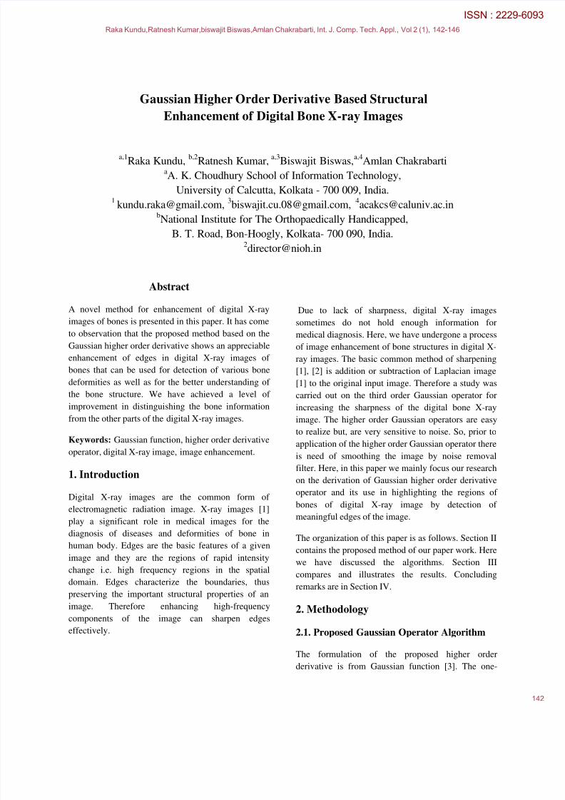
Figure 3 is the plot of 1D Gaussian derivative function
for order of 3, it shows the variation of the function
w.r.t. x. It is a zero crossing operator whose
polynomial is (3x σ 2
- x3) ∕ σ
6. This polynomial is
also known as Hermit polynomial [4].
Figure 3 : 1D plot of the 3rd order Gaussian operator
The 3x3 convolution mask of equation (9) with the
adjustment of variance (σ 2) from 0.25 to 0.3 gives the
Raka Kundu,Ratnesh Kumar,biswajit Biswas,Amlan Chakrabarti, Int. J. Comp. Tech. Appl., Vol 2 (1), 142-146
8/7/2019 Gaussian higher Order Derivative based Structural Enhancement of Digital Bone X-Ray Images
http://slidepdf.com/reader/full/gaussian-higher-order-derivative-based-structural-enhancement-of-digital-bone 3/5
desirable result for the enhancement of bone structure
of digital X-ray image.
2.2. Image enhancement
The 3x3 window obtained from third order Gaussian
derivative is next used for digital X-ray image
sharpening. Considering the centre pixel of the
obtained window as positive, we add the third order
Gaussian image with the original digital X-ray image.
Resultant image obtained after addition is the edge
sharpened image.
Let,
)],([),(),( 3 y x I y x I y xS (10)
Where, I(x,y) and S(x,y) are the input image and
structural enhanced image respectively. ),(3 y x I is
the third order Gaussian image. Figure4 describes the
proposed method.
),( y x I ),(3
y x I ),( y xS
Figure4: Procedure for the generation of the enhanced
image
The resultant image is significantly sharper than the
original input image and is able to confer the structure
of the bone of the digital image.
3. Results and Discussions
Let us demonstrate the structural enhancement of the
digital X-ray image from our experimental results.
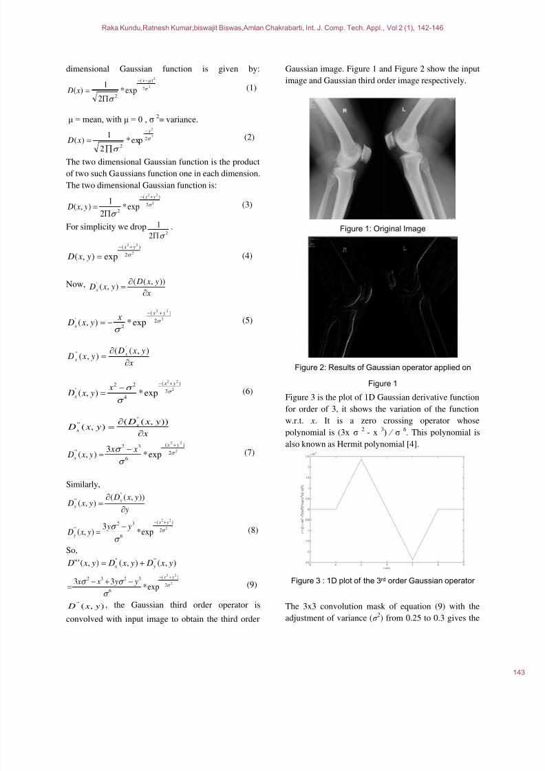
Figure 5 and Figure 6 illustrate the mesh plot of the
input digital X-ray image (Figure 9) and the enhanced
digital X-ray image (Figure 10). If we compare thevalues of Figure 6 with Figure 5 we see that the Figure
6 have much higher peaks for the edges whereas the
non-edges remains almost same as compared to the
random distribution in Figure 5. Thus it can be
visualized from the results that the edge regions of the
bone of the processed image is much sharper than the
original image. This gives a clear view of the structure
of the bone.
Figure 5: Mesh plot of the test image (Figure 9)
Figure 6: Mesh plot of the enhanced image (Figure 10)
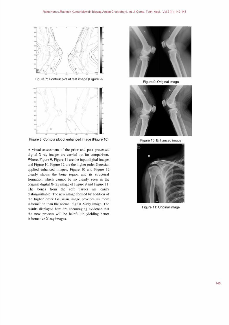
The contour plot of the original image and enhanced
image gives more clear idea of the edge enhancement.
The result in Figure 8 shows that the contour of the
bones has been well detected in the enhanced image
compared to the initial input as in Figure 7. This
proves that our algorithm is efficient and serves the
purpose of edge enhancement.
Raka Kundu,Ratnesh Kumar,biswajit Biswas,Amlan Chakrabarti, Int. J. Comp. Tech. Appl., Vol 2 (1), 142-146
8/7/2019 Gaussian higher Order Derivative based Structural Enhancement of Digital Bone X-Ray Images
http://slidepdf.com/reader/full/gaussian-higher-order-derivative-based-structural-enhancement-of-digital-bone 4/5
Figure 7: Contour plot of test image (Figure 9)
Figure 8: Contour plot of enhanced image (Figure 10)
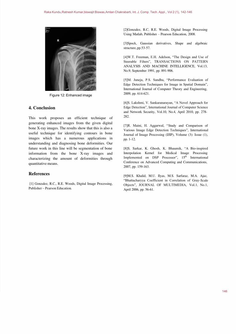
A visual assessment of the prior and post processed
digital X-ray images are carried out for comparison.
Where, Figure 9, Figure 11 are the input digital images
and Figure 10, Figure 12 are the higher order Gaussian
applied enhanced images. Figure 10 and Figure 12clearly shows the bone region and its structural
formation which cannot be so clearly seen in the
original digital X-ray image of Figure 9 and Figure 11.
The bones from the soft tissues are easily
distinguishable. The new image formed by addition of
the higher order Gaussian image provides us more
information than the normal digital X-ray image. The
results displayed here are encouraging evidence that
the new process will be helpful in yielding better
informative X-ray images.
Figure 9: Original image
Figure 10: Enhanced image
Figure 11: Original image
Raka Kundu,Ratnesh Kumar,biswajit Biswas,Amlan Chakrabarti, Int. J. Comp. Tech. Appl., Vol 2 (1), 142-146
8/7/2019 Gaussian higher Order Derivative based Structural Enhancement of Digital Bone X-Ray Images
http://slidepdf.com/reader/full/gaussian-higher-order-derivative-based-structural-enhancement-of-digital-bone 5/5
Figure 12: Enhanced image
4. Conclusion
This work proposes an efficient technique of
generating enhanced images from the given digital
bone X-ray images. The results show that this is also a
useful technique for identifying contours in bone
images which has a numerous applications in
understanding and diagnosing bone deformities. Our
future work in this line will be segmentation of bone
information from the bone X-ray images and
characterizing the amount of deformities through
quantitative means.
References
[1] Gonzalez, R.C., R.E. Woods, Digital Image Processing,
Publisher – Pearson Education.
[2]Gonzalez, R.C, R.E. Woods, Digital Image Processing
Using Matlab, Publisher – Pearson Education, 2008.
[3]Spock, Gaussian derivatives, Shape and algebraic
structure, pp.53-57.
[4]W.T. Freeman, E.H. Adelson, “The Design and Use of Steerable Filters”, TRANSACTIONS ON PATTERN
ANALYSIS AND MACHINE INTELLIGENCE, Vol.13,
No.9, September 1991, pp. 891-906.
[5]M. Juneja, P.S. Sandhu, “Performance Evaluation of
Edge Detection Techniques for Image in Spatial Domain”,
International Journal of Computer Theory and Engineering,
2009, pp. 614-621.
[6]S. Lakshmi, V. Sankaranarayan, “A Novel Approach for
Edge Detection”, International Journal of Computer Science
and Network Security, Vol.10, No.4, April 2010, pp. 278-
282.
[7]R. Maini, H. Aggarwal, “Study and Comparison of
Various Image Edge Detection Techniques”, International
Journal of Image Processing (IJIP), Volume (3): Issue (1),
pp. 1-12.
[8]S. Sarkar, K. Ghosh, K. Bhaumik, “A Bio-inspired
Interpolation Kernel for Medical Image Processing
Implemented on DSP Processor”, 15th International
Conference on Advanced Computing and Communications,
2007, pp. 159-163.
[9]M.S. Khalid, M.U. Ilyas, M.S. Sarfaraz, M.A. Ajaz,“Bhattacharyya Coefficient in Correlation of Gray-Scale
Objects”, JOURNAL OF MULTIMEDIA, Vol.1, No.1,
April 2006, pp. 56-61.
Raka Kundu,Ratnesh Kumar,biswajit Biswas,Amlan Chakrabarti, Int. J. Comp. Tech. Appl., Vol 2 (1), 142-146