Embed Size (px)
DESCRIPTION
GASTROINTESTINAL SYSTEM PROTOZOA -I-. Entamoeba histolytica Giardia lamblia Entamoeba coli. Doç.Dr.Hrisi BAHAR. Gastrointestinal system protozoon. Entamoeba histolytica. Pathogenic Entamoeba histolytica Balantidium coli Giardia lamblia Dientamoeba fragilis Cryptosporidium parvum - PowerPoint PPT Presentation
Citation preview
GASTROINTESTINAL SYSTEM PROTOZOA -I-
Entamoeba histolytica
Giardia lambliaEntamoeba coli
Doç.Dr.Hrisi BAHAR
Gastrointestinal system Gastrointestinal system protozoonprotozoon
Entamoeba histolytica
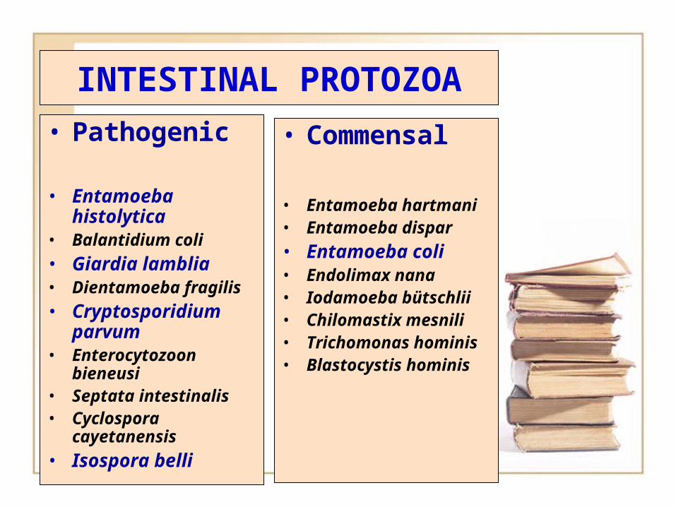
INTESTINAL PROTOZOA
• Pathogenic
• Entamoeba histolytica
• Balantidium coli• Giardia lamblia• Dientamoeba
fragilis• Cryptosporidium
parvum• Enterocytozoon
bieneusi• Septata intestinalis• Cyclospora
cayetanensis• Isospora belli
• Commensal
• Entamoeba hartmani
• Entamoeba dispar• Entamoeba coli• Endolimax nana• Iodamoeba
bütschlii• Chilomastix
mesnili• Trichomonas
hominis• Blastocystis
hominis
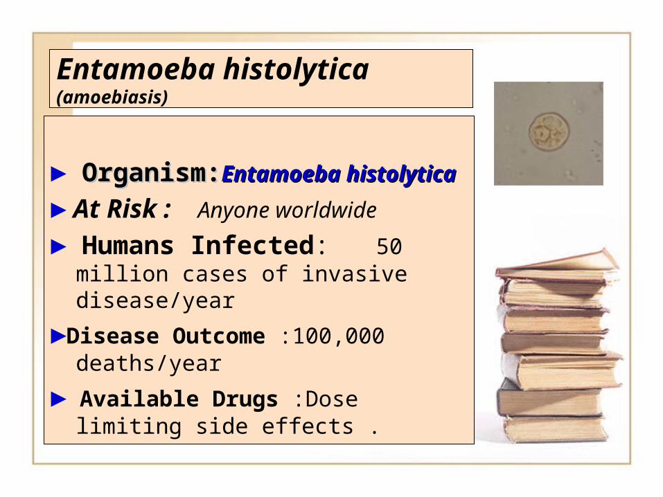
Entamoeba histolytica(amoebiasis)
► Organism:Organism:EntamoebaEntamoeba histolyticahistolytica
► At Risk : Anyone worldwide
► Humans Infected: 50 million cases of invasive disease/year
►Disease Outcome :100,000 deaths/year
► Available Drugs :Dose limiting side effects .
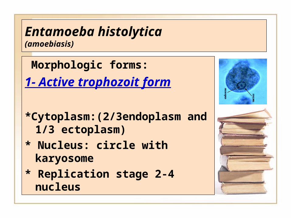
Entamoeba histolytica(amoebiasis)
Morphologic forms:
1- Active trophozoit form
*Cytoplasm:(2/3endoplasm and 1/3 ectoplasm)
* Nucleus: circle with karyosome
* Replication stage 2-4 nucleus
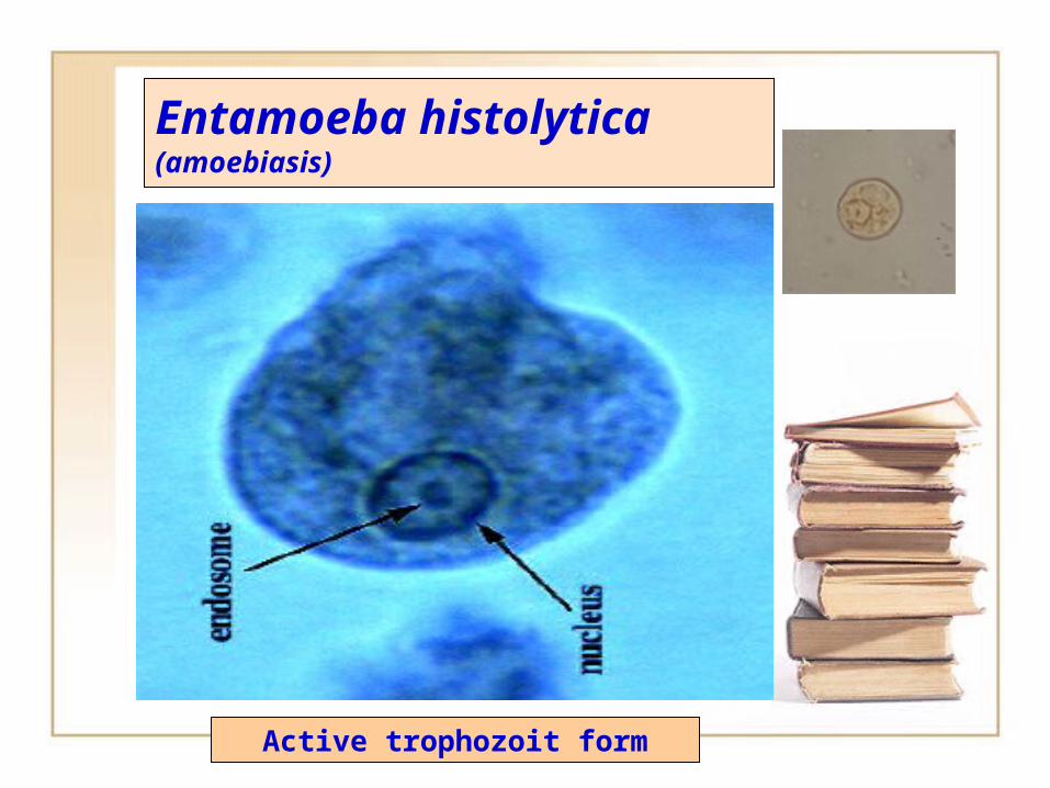
Entamoeba histolytica(amoebiasis)
Active trophozoit form
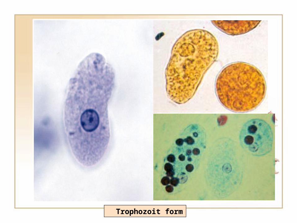
Trophozoit form
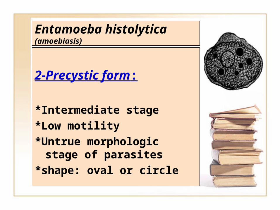
Entamoeba histolytica(amoebiasis)
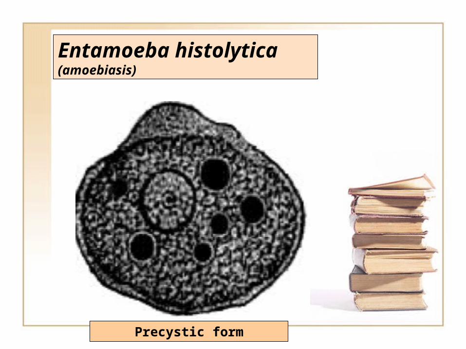
2-Precystic form:
*Intermediate stage
*Low motility
*Untrue morphologic stage of parasites
*shape: oval or circle
Entamoeba histolytica(amoebiasis)
Precystic form
Entamoeba histolytica(amoebiasis)
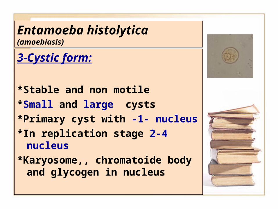
3-Cystic form:
*Stable and non motile*Small and large cysts*Primary cyst with -1- nucleus*In replication stage 2-4
nucleus*Karyosome,, chromatoide
body and glycogen in nucleus
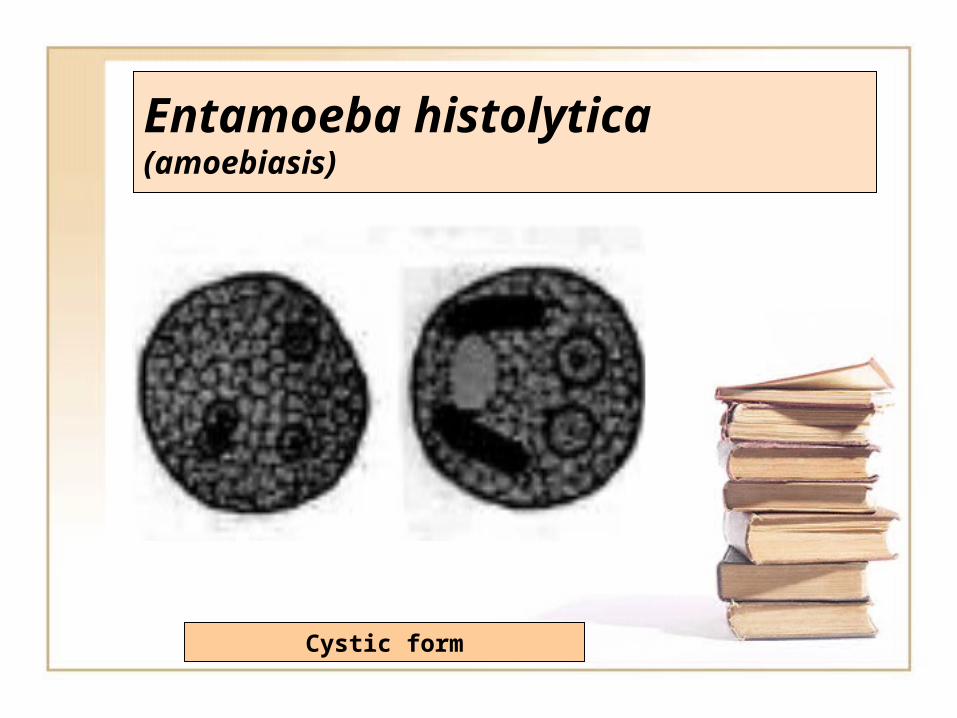
Entamoeba histolytica(amoebiasis)
Cystic form
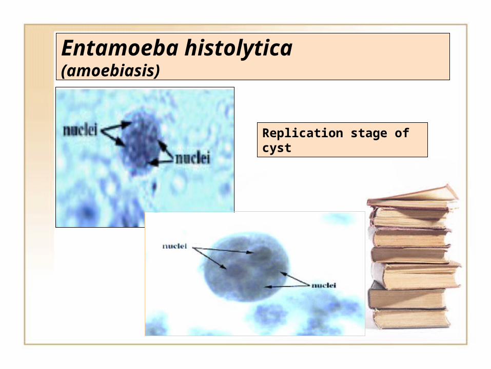
Entamoeba histolytica(amoebiasis)
Replication stage of cyst
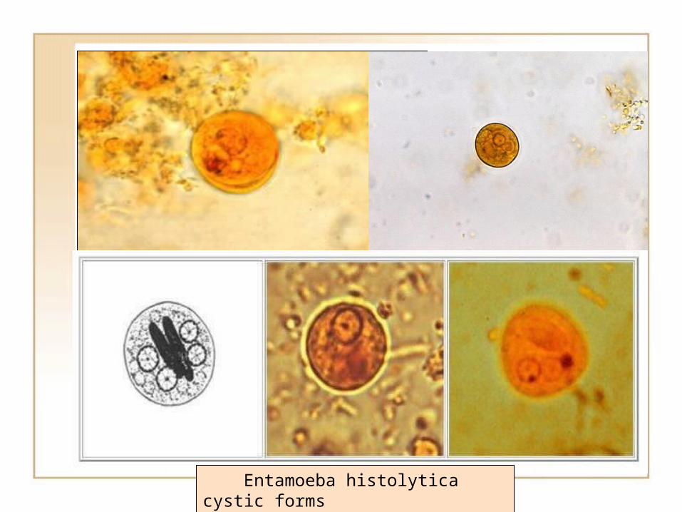
Entamoeba histolytica cystic forms
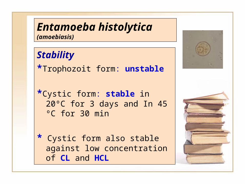
Entamoeba histolytica(amoebiasis)
Stability*Trophozoit form: unstable
*Cystic form: stable in 20ºC for 3 days and In 45 ºC for 30 min
* Cystic form also stable against low concentration of CL and HCL
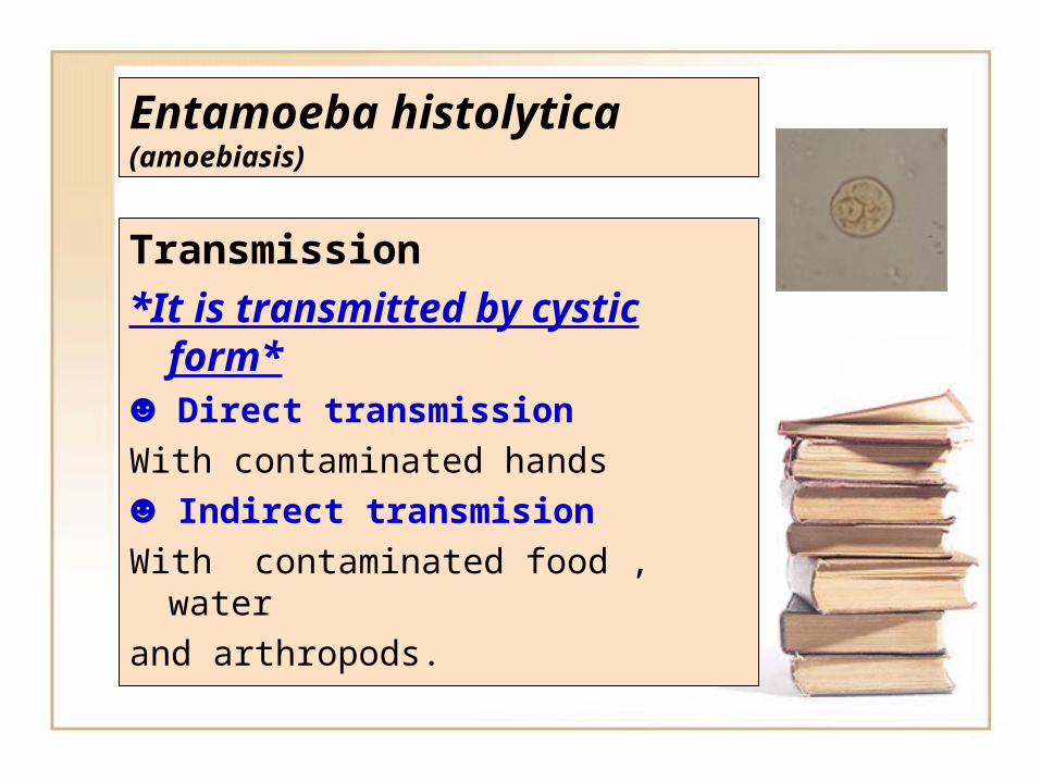
Entamoeba histolytica(amoebiasis)
Transmission*It is transmitted by cystic
form*☻ Direct transmissionWith contaminated hands☻ Indirect transmisionWith contaminated food , waterand arthropods.
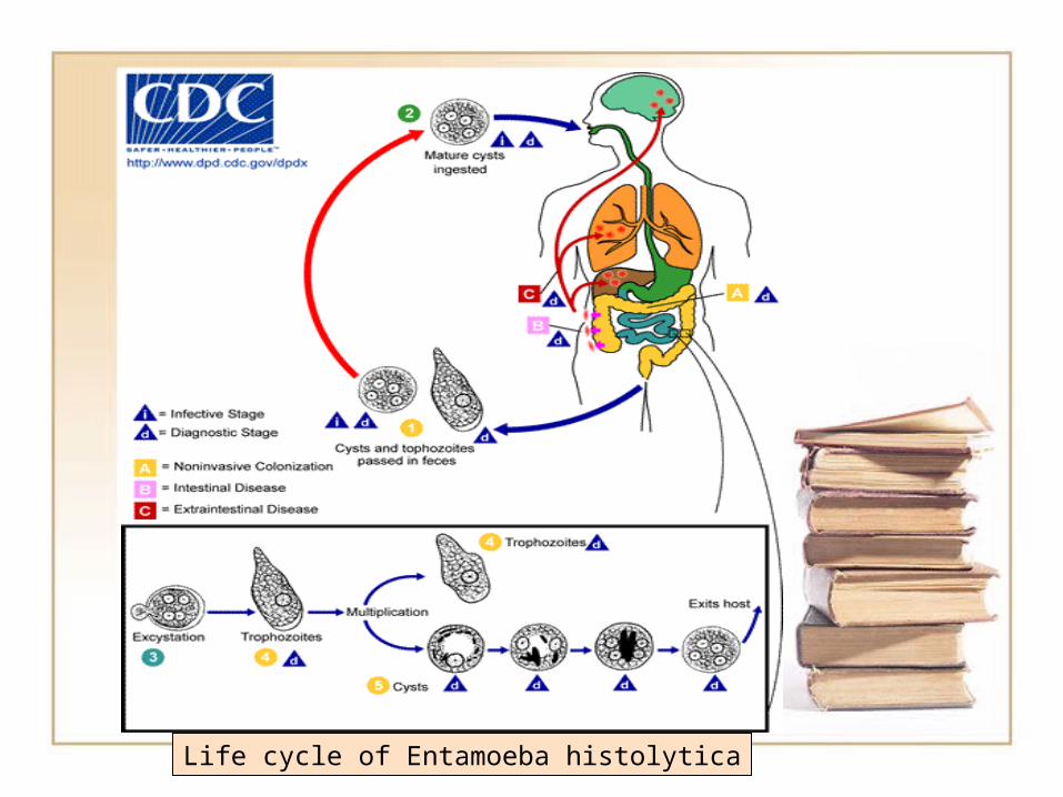
Life cycle of Entamoeba histolytica
Entamoeba histolytica(amoebiasis)
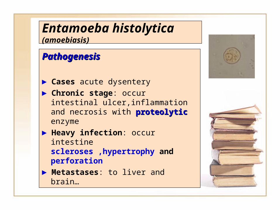
PathogenesisPathogenesis
► Cases acute dysentery► Chronic stage: occur intestinal
ulcer,inflammation and necrosis with proteolyticproteolytic enzyme
► Heavy infection: occur intestine scleroses ,hypertrophy and perforation
► Metastases: to liver and brain…
Entamoeba histolytica(amoebiasis)
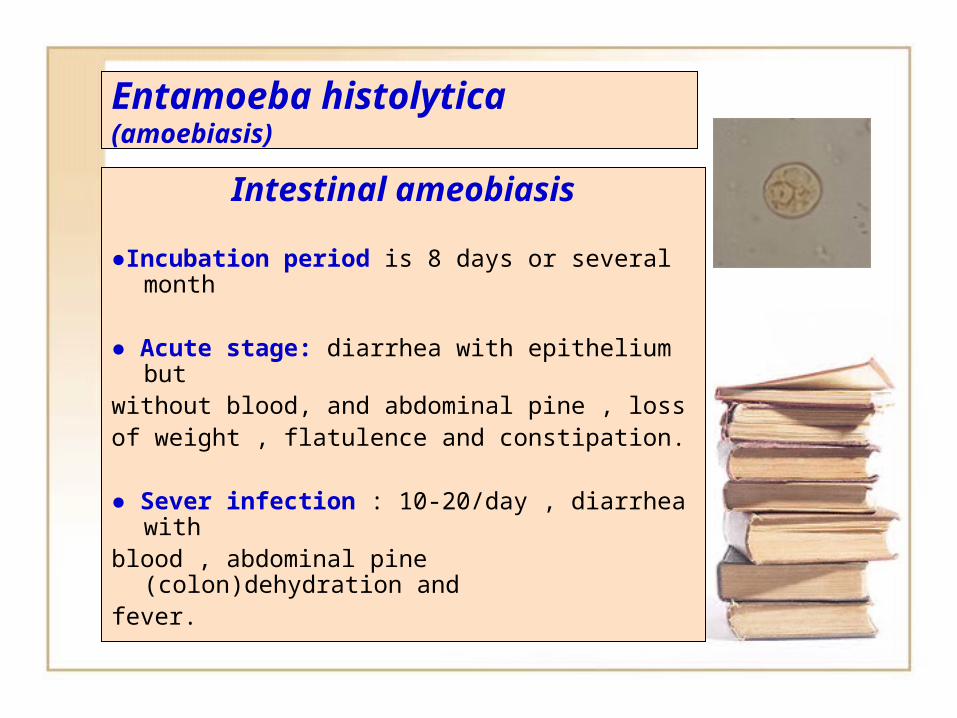
Intestinal ameobiasis
●Incubation period is 8 days or several month
● Acute stage: diarrhea with epithelium butwithout blood, and abdominal pine , lossof weight , flatulence and constipation.
● Sever infection : 10-20/day , diarrhea withblood , abdominal pine (colon)dehydration andfever.
Entamoeba histolytica(amoebiasis)
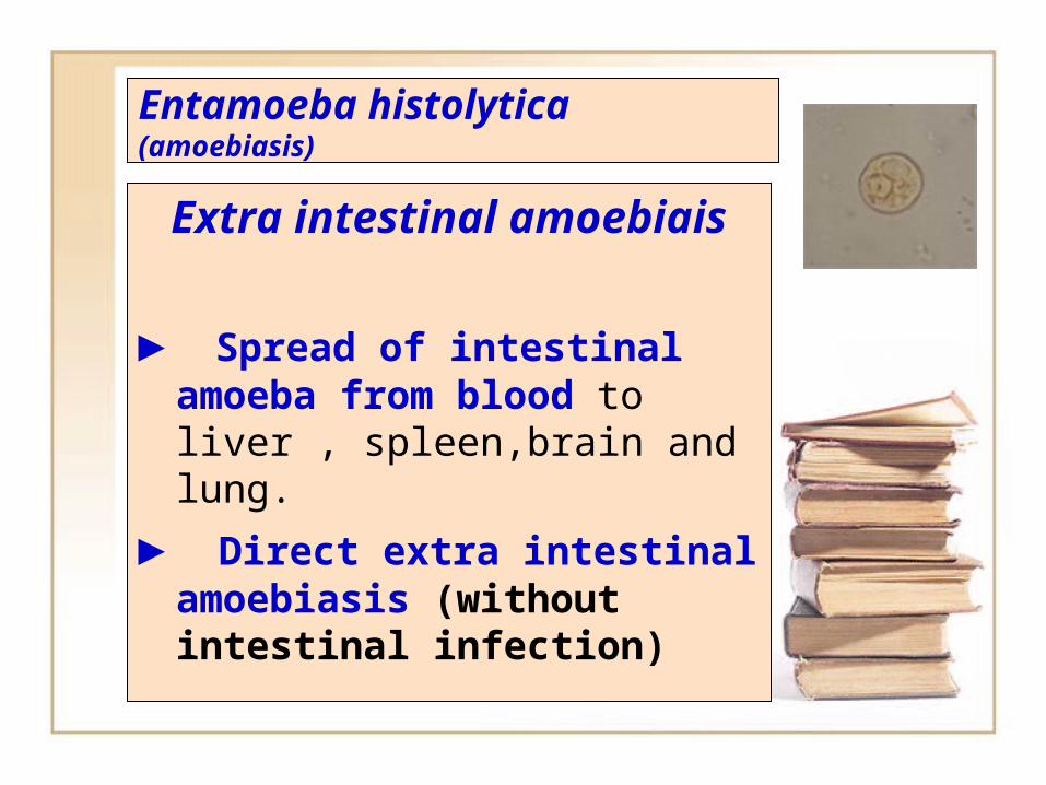
Extra intestinal amoebiais
► Spread of intestinal amoeba from blood to liver , spleen,brain and lung.
► Direct extra intestinal amoebiasis (without intestinal infection)
Entamoeba histolytica(amoebiasis)
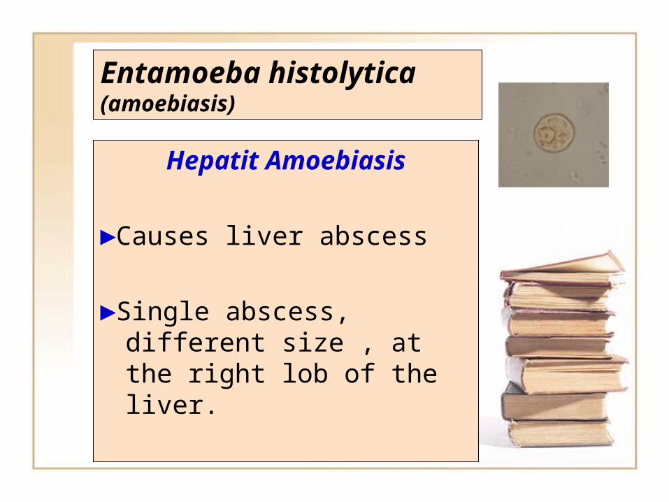
Hepatit Amoebiasis
►Causes liver abscess
►Single abscess, different size , at the right lob of the liver.
Entamoeba histolytica(amoebiasis)
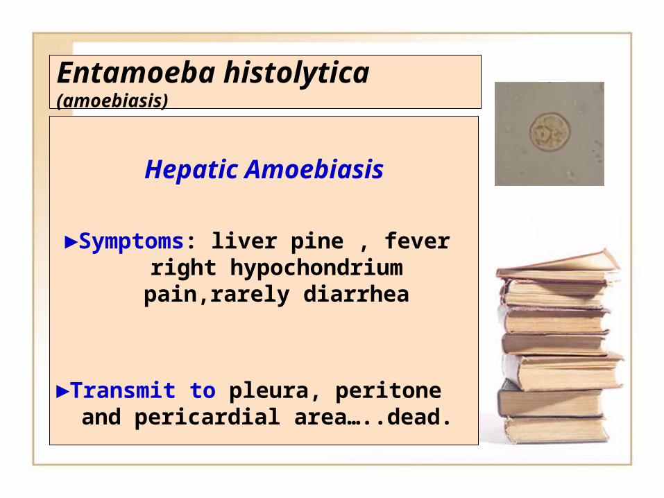
Hepatic Amoebiasis
►Symptoms: liver pine , fever right hypochondrium pain,rarely diarrhea
►Transmit to pleura, peritone and pericardial area…..dead.
Entamoeba histolytica(amoebiasis)
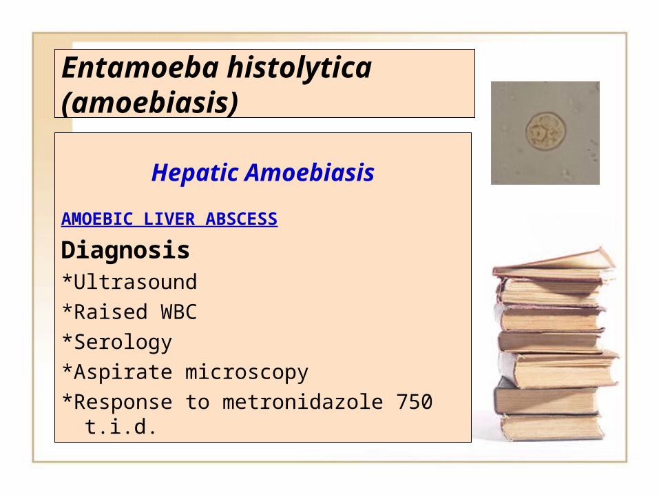
Hepatic Amoebiasis
AMOEBIC LIVER ABSCESS
Diagnosis*Ultrasound*Raised WBC*Serology*Aspirate microscopy*Response to metronidazole 750
t.i.d.
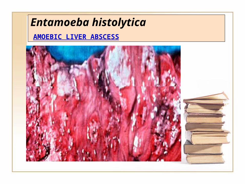
Entamoeba histolytica AMOEBIC LIVER ABSCESS
Entamoeba histolytica(amoebiasis)
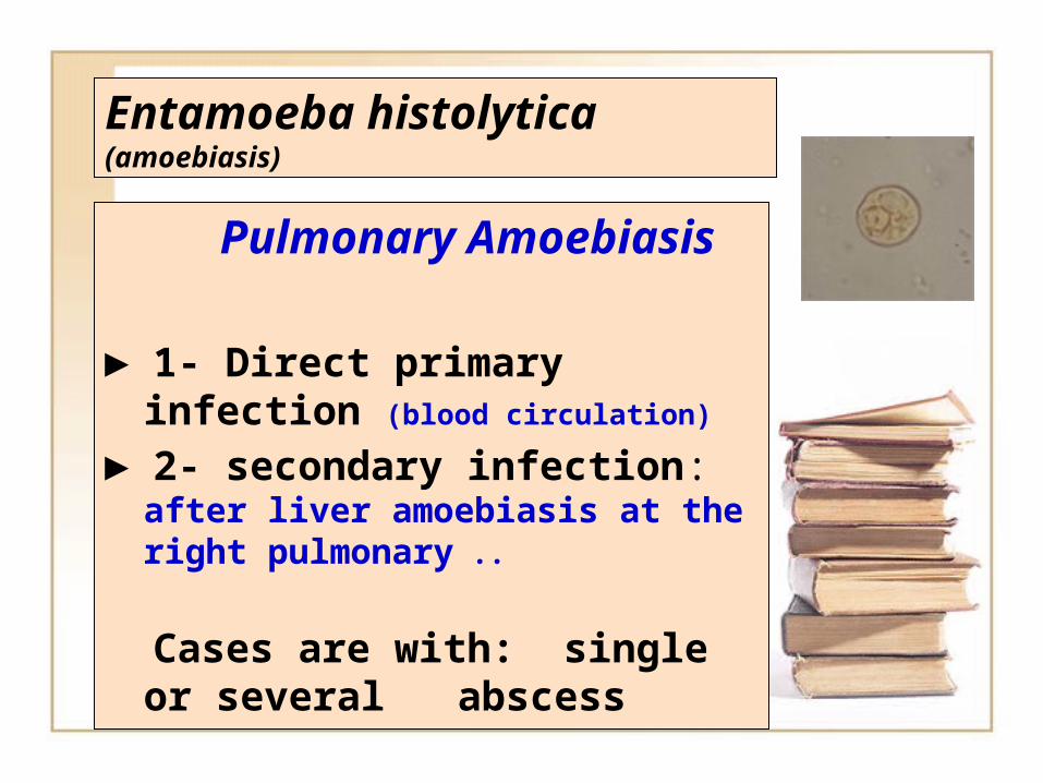
Pulmonary Amoebiasis
► 1- Direct primary infection (blood circulation)
► 2- secondary infection: after liver amoebiasis at the right pulmonary ..
Cases are with: single or several abscess
Entamoeba histolytica(amoebiasis)
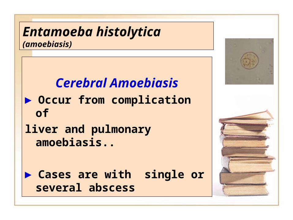
Cerebral Amoebiasis► Occur from complication of
liver and pulmonary amoebiasis..
► Cases are with single or several abscess
Entamoeba histolytica(amoebiasis
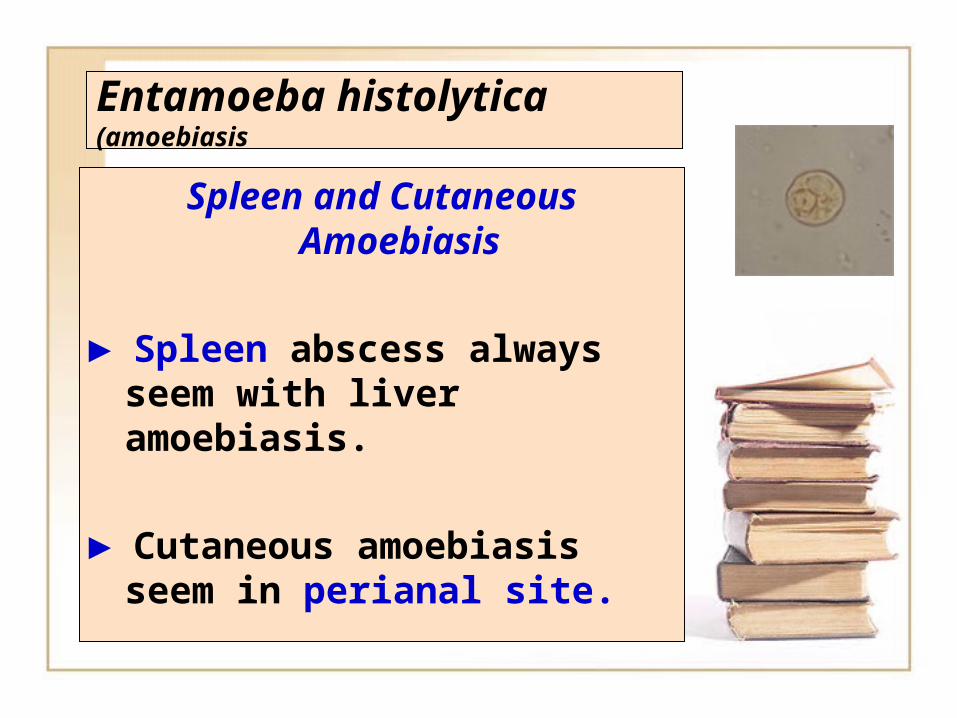
Spleen and Cutaneous Amoebiasis
► Spleen abscess always seem with liver amoebiasis.
► Cutaneous amoebiasis seem in perianal site.
Entamoeba histolytica(amoebiasis
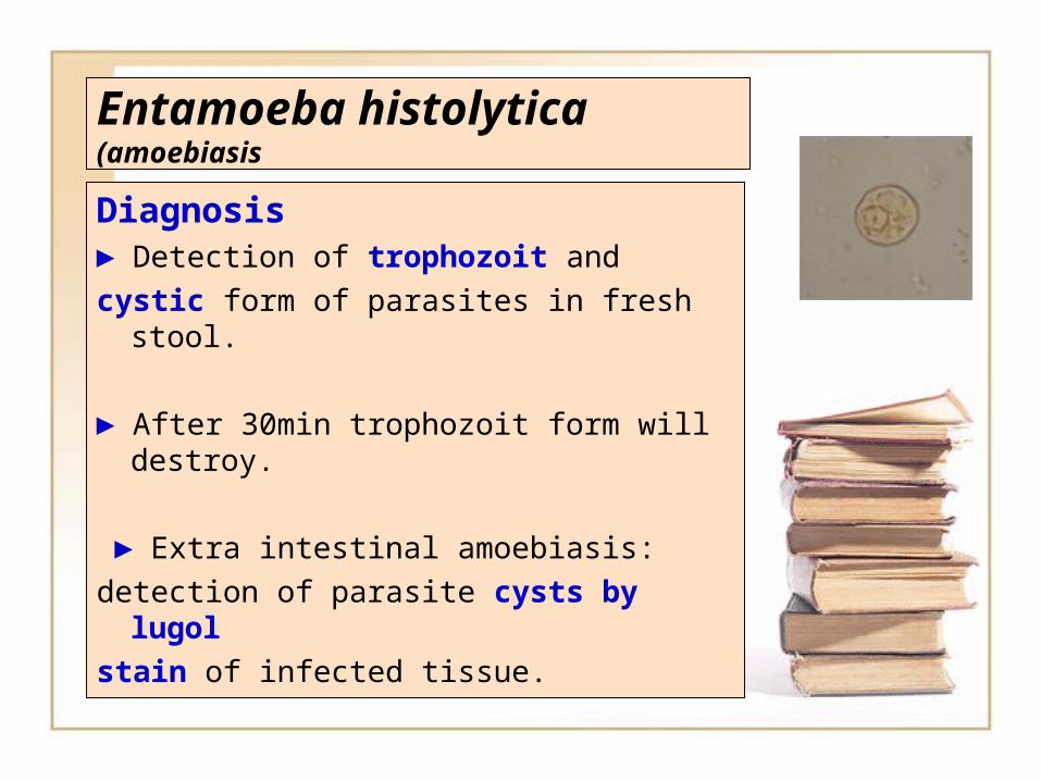
Diagnosis► Detection of trophozoit and
cystic form of parasites in fresh stool.
► After 30min trophozoit form will destroy.
► Extra intestinal amoebiasis:
detection of parasite cysts by lugol
stain of infected tissue.
Entamoeba histolytica(amoebiasis
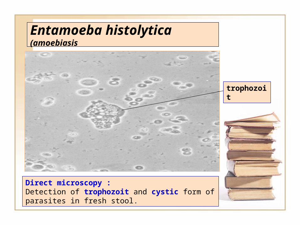
Direct microscopy :Detection of trophozoit and cystic form of parasites in fresh stool.
trophozoit
Entamoeba histolytica(amoebiasis
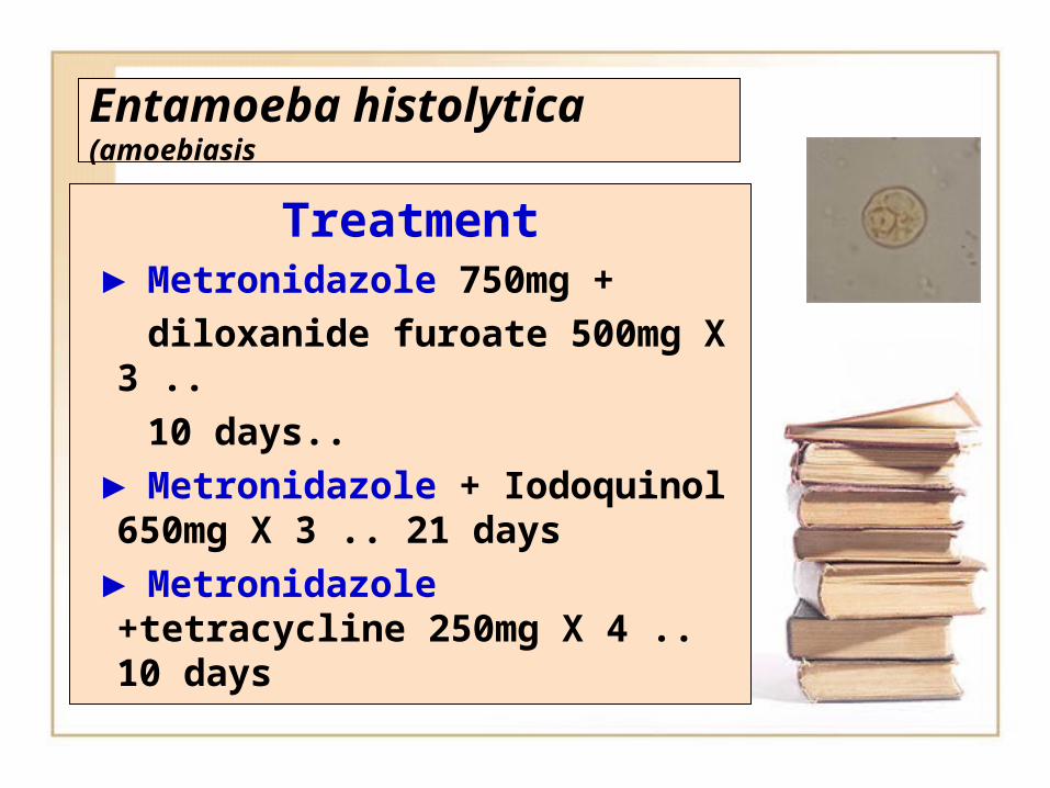
Treatment ► Metronidazole 750mg + diloxanide furoate 500mg X
3 .. 10 days.. ► Metronidazole + Iodoquinol
650mg X 3 .. 21 days ► Metronidazole
+tetracycline 250mg X 4 .. 10 days
Entamoeba histolytica(amoebiasis
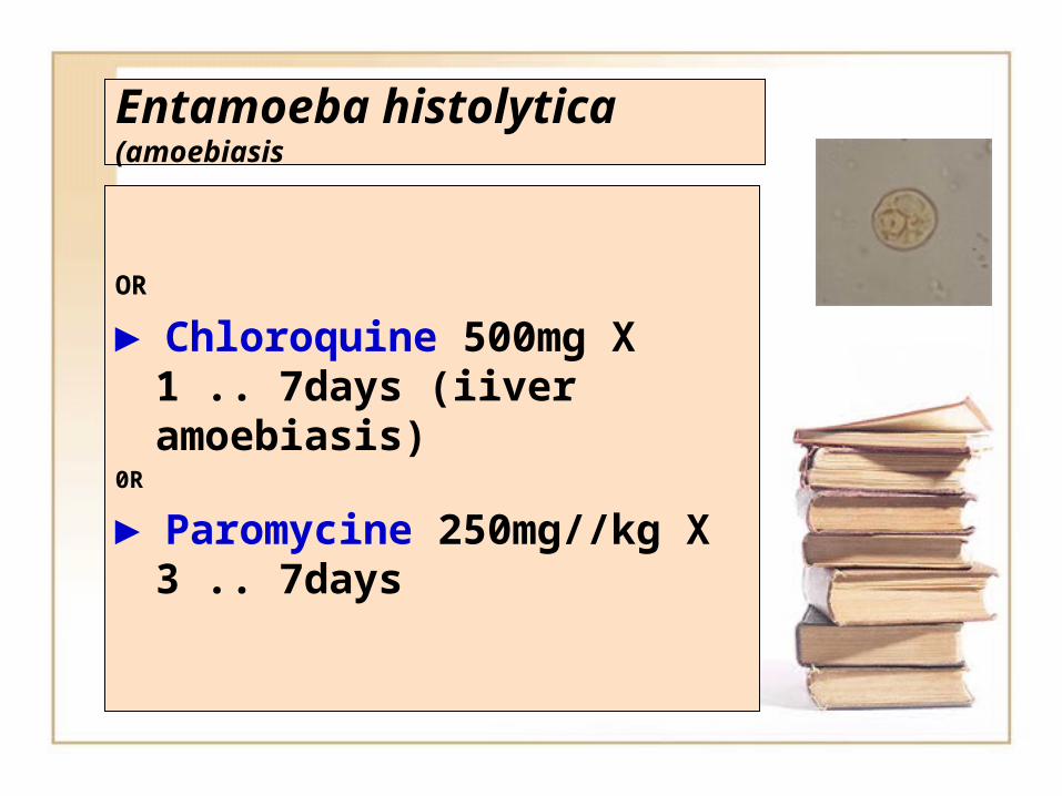
OR
► Chloroquine 500mg X 1 .. 7days (iiver amoebiasis)
0R
► Paromycine 250mg//kg X 3 .. 7days
Gastrointestinal system protozoonGastrointestinal system protozoon
Entamoeba coli
Entamoeba coli
► Entamoeba coli is a non-pathogenicnon-pathogenic species of Entamoeba that frequently exists as a commensal parasite in the human gastrointestinal tract especially in the colon.
Entamoeba coli
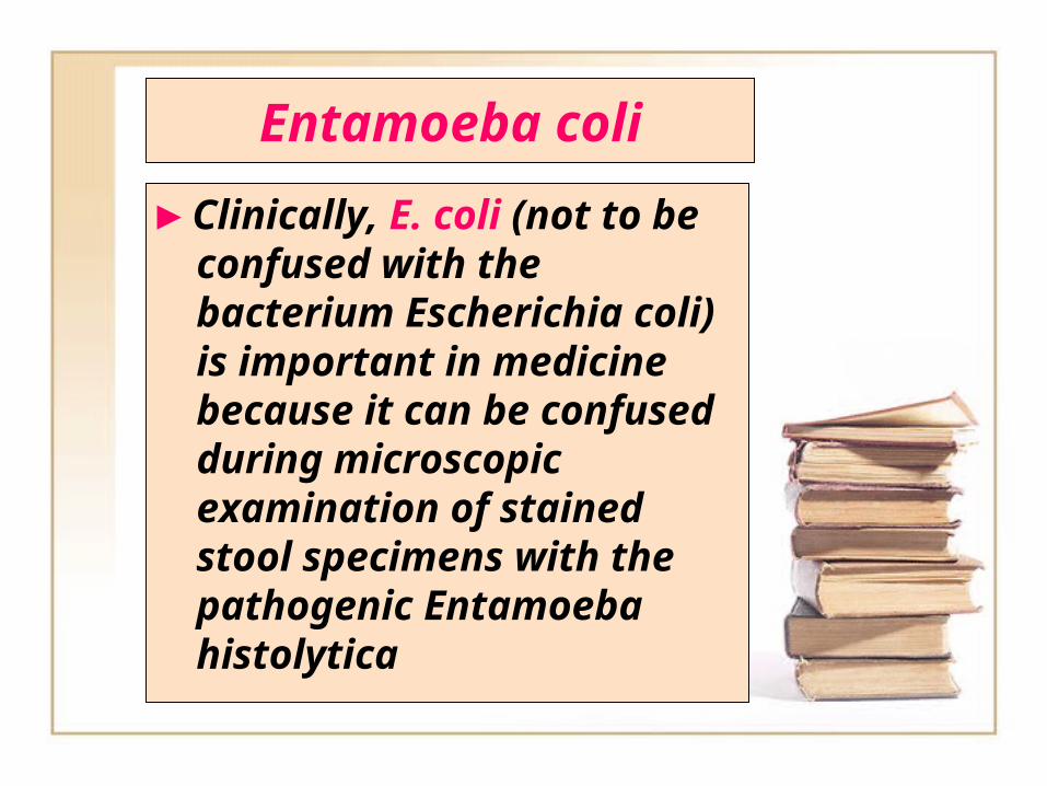
► Clinically, E. coli (not to be confused with the bacterium Escherichia coli) is important in medicine because it can be confused during microscopic examination of stained stool specimens with the pathogenic Entamoeba histolytica
Entamoeba coli
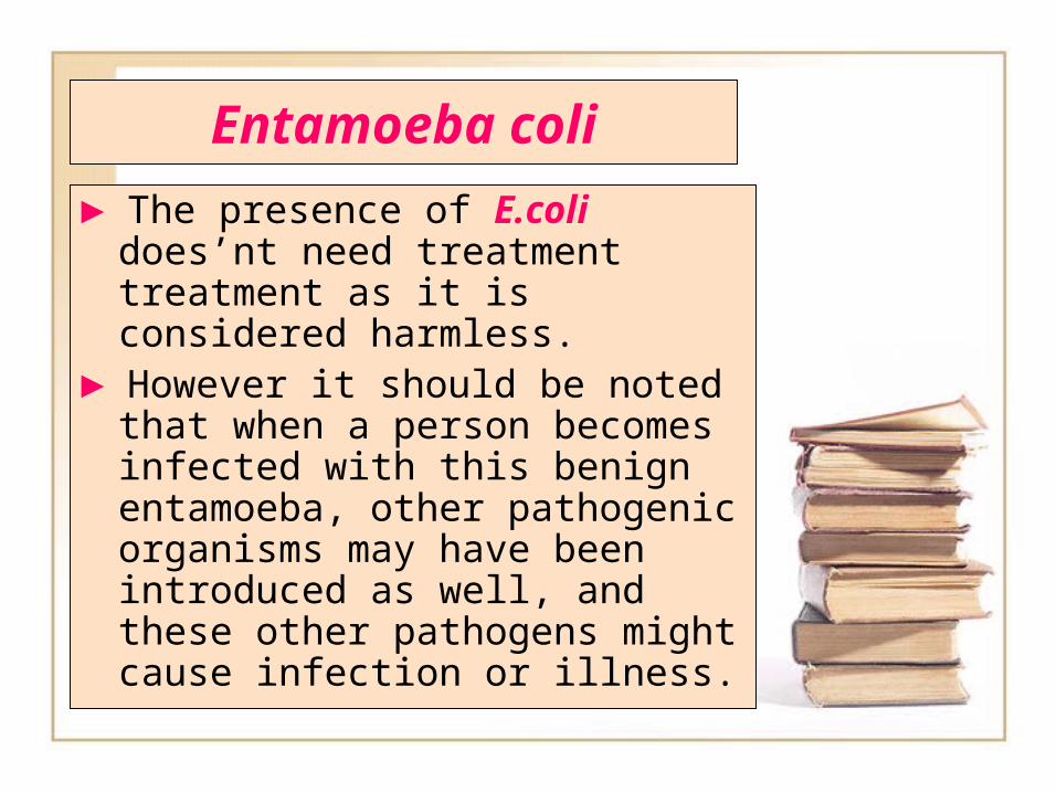
► The presence of E.coli does’nt need treatment treatment as it is considered harmless.
► However it should be noted that when a person becomes infected with this benign entamoeba, other pathogenic organisms may have been introduced as well, and these other pathogens might cause infection or illness.
Entamoeba coli
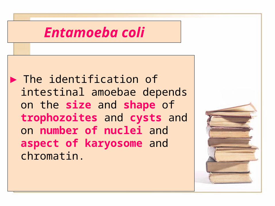
► The identification of intestinal amoebae depends on the size and shape of trophozoites and cysts and on number of nuclei and aspect of karyosome and chromatin.
Entamoeba coli
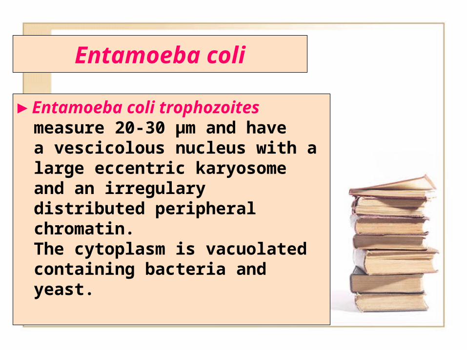
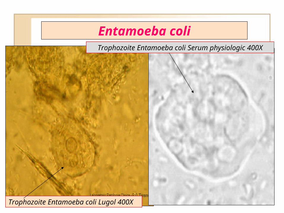
► Entamoeba coli trophozoites measure 20-30 µm and have a vescicolous nucleus with a large eccentric karyosome and an irregulary distributed peripheral chromatin. The cytoplasm is vacuolated containing bacteria and yeast.
Entamoeba coli
► Entamoeba coli trophozoites
► Entamoeba coli trophozoites
Trophozoite Entamoeba coli Lugol 400X
Trophozoite Entamoeba coli Serum physiologic 400X
Entamoeba coli
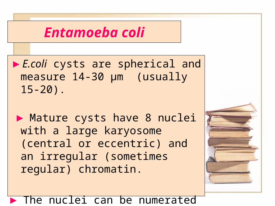
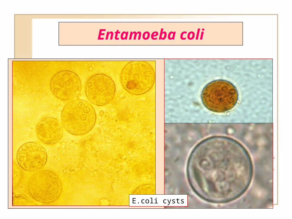
► ► E.coli cysts are spherical and measure 14-30 µm (usually 15-20).
► Mature cysts have 8 nuclei with a large karyosome (central or eccentric) and an irregular (sometimes regular) chromatin.
► The nuclei can be numerated with careful focusing.
Entamoeba coli
► While this differentiation is typically done by visual examination of the parasitic cysts via light microscopy, new methods using molecular biology techniques have been developed also.
Entamoeba coli
►
E.coli cysts
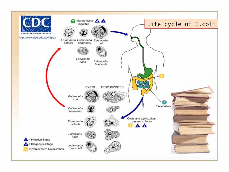
Life cycle of E.coli Life cycle of E.coli
Entamoeba coli
► Cysts and trophosoits of E.coli are larger then E.histolytica.
► E.coli is the only species in the genus encountered in humans with more then four nuclei in the cyst stage.
Entamoeba coli
► Cysts and trophosoits of E.coli are larger then E.histolytica.
► E.coli is found in the mouth between the gingival pockets.
Gastrointestinal system Gastrointestinal system protozoon protozoon
Giardia lamblia Giardia lamblia (Giardia intestinalis) (Giardia intestinalis)
Giardia lambliaGiardia lamblia
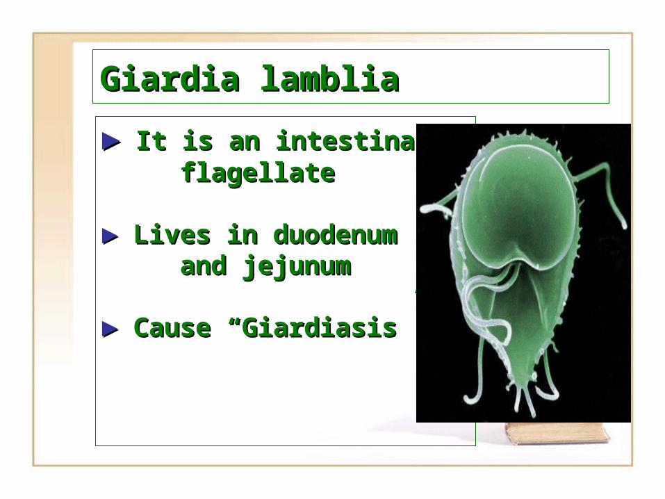
►► It is an iIt is an intestinal ntestinal flagellateflagellate
►► LLives in duodenum ives in duodenum and jejunumand jejunum
►► Cause “Cause “GiardiasisGiardiasis””
Giardia lambliaGiardia lamblia
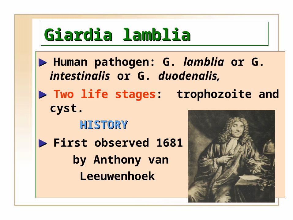
►► Human pathogen: G. lamblia or G. intestinalis or G. duodenalis,
►► Two life stages: trophozoite and cyst.
HISTORYHISTORY
►► First observed 1681
by Anthony van Leeuwenhoek
Giardia lambliaGiardia lamblia
►► First observed 1681 by Anthony van Leeuwenhoek
►► Described ~200 years later by Vilem Lambl
►► First cultured in 1960’s
►► Confirmed pathogen 1970’s
►► One of most common intestinal parasites
►► Causes Giardiasis (beaver fever)
►►Geographic region with poor water sanitation
Giardia lambliaGiardia lamblia
►► Species details– Single-celled protist– 5 species of Giardia
►► G. intestinalis/lamblia• G. muris in rodents, birds,
reptiles• G. agilis in amphibians• G. ardae in great blue
heron• G. psittaci in budgerigar
Giardia lambliaGiardia lamblia
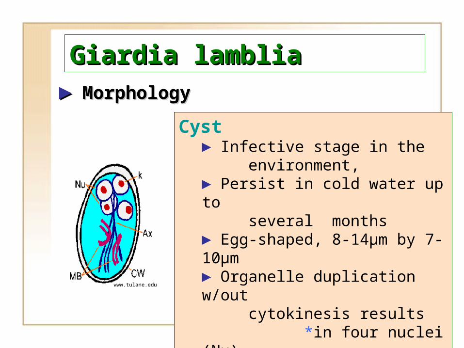
►► MorphologyMorphology
www.tulane.edu
Cyst► Infective stage in the environment, ► Persist in cold water up to several months► Egg-shaped, 8-14µm by 7-10µm► Organelle duplication w/out cytokinesis results *in four nuclei (Nu)
*four median bodies (MB) *four axonemes (Ax)
Giardia lambliaGiardia lamblia
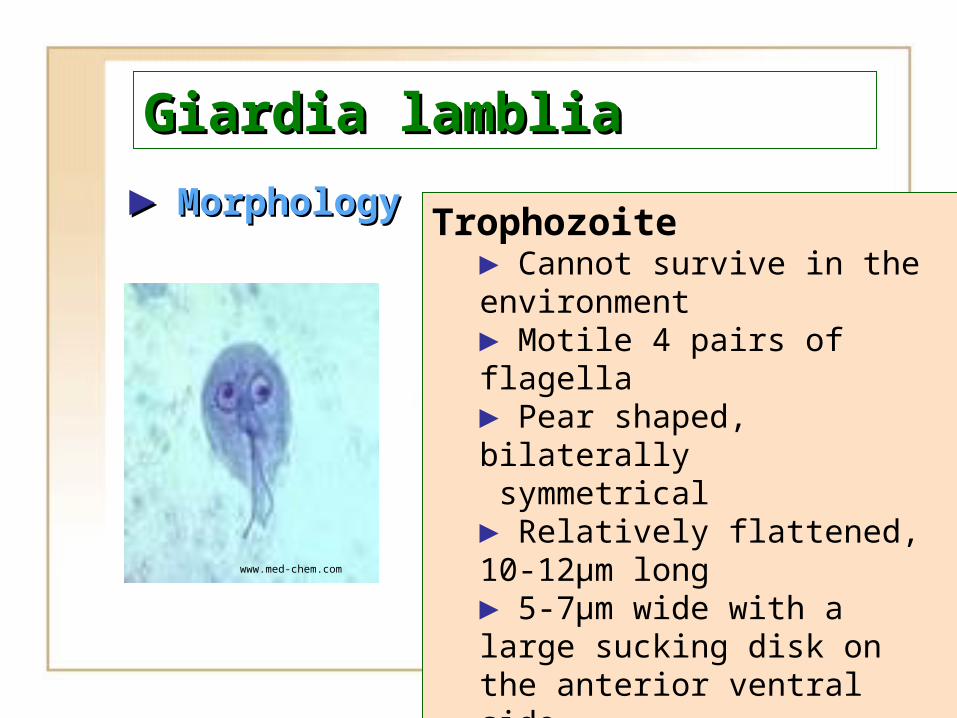
►► MorphologyMorphology
www.med-chem.com
Trophozoite► Cannot survive in the environment► Motile 4 pairs of flagella► Pear shaped, bilaterally symmetrical► Relatively flattened, 10-12µm long► 5-7µm wide with a large sucking disk on the anterior ventral side► Two nuclei
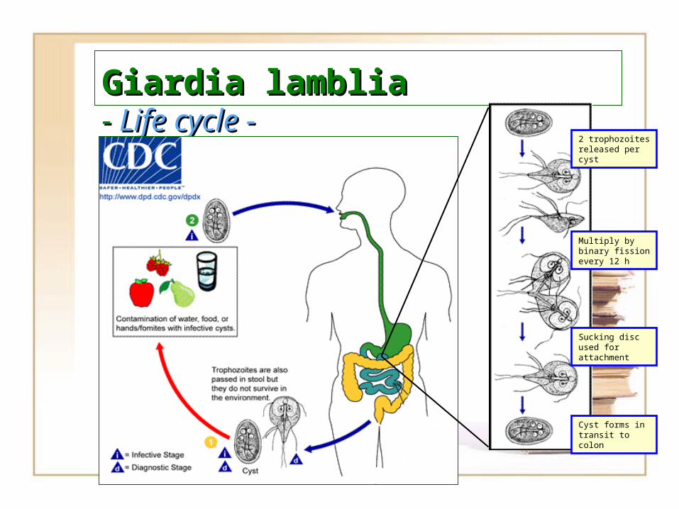
Giardia lambliaGiardia lamblia- - Life cycle -Life cycle -
2 trophozoites released per cyst
Multiply by binary fission every 12 h
Sucking disc used for attachment
Cyst forms in transit to colon
Giardia lambliaGiardia lamblia
Clinical Clinical SymptomsSymptoms and and PathogenesisPathogenesis
►► Ingested cysts excyst in response to stomach acidity.
►► Attach (via their sucking disk) to microvilli of epithelium in small intestine, causing epithelial damage and interfering with gut transport processes.
Giardia lambliaGiardia lamblia
Clinical Clinical SymptomsSymptoms and and PathogenesisPathogenesis
►►Epithelial mucus is thinned, lymphocytes and other inflammatory cells infiltrate, physical blocking of absorption may occur, and enterobacteria may proliferate, causing more epithelial damage.
►►Distention and flatulence can occur.
Giardia lambliaGiardia lamblia
Clinical Clinical SymptomsSymptoms and and PathogenesisPathogenesis
►► Symptoms include profuse and watery to semisolid, greasy, bulky and foul- smelling diarrhea; abdominal cramps; nausea; vomiting; anorexia; low-grade fever and flu-like headache. General malaise, weakness, weight loss.
Giardia lambliaGiardia lamblia
►► Symptoms more likely in children and immunosuppressed adults than in healthy adults.
►► Infectous dose is low (about 10 cysts; possibly less).
►► Incubation period from 7-11 days.
Giardia lambliaGiardia lamblia
►► Duration of illness varies: few days to months.
►►Infection may resolve spontaneously. often, subchronic stage develops with mild to moderate symptoms occurring periodically.
Giardia lambliaGiardia lamblia
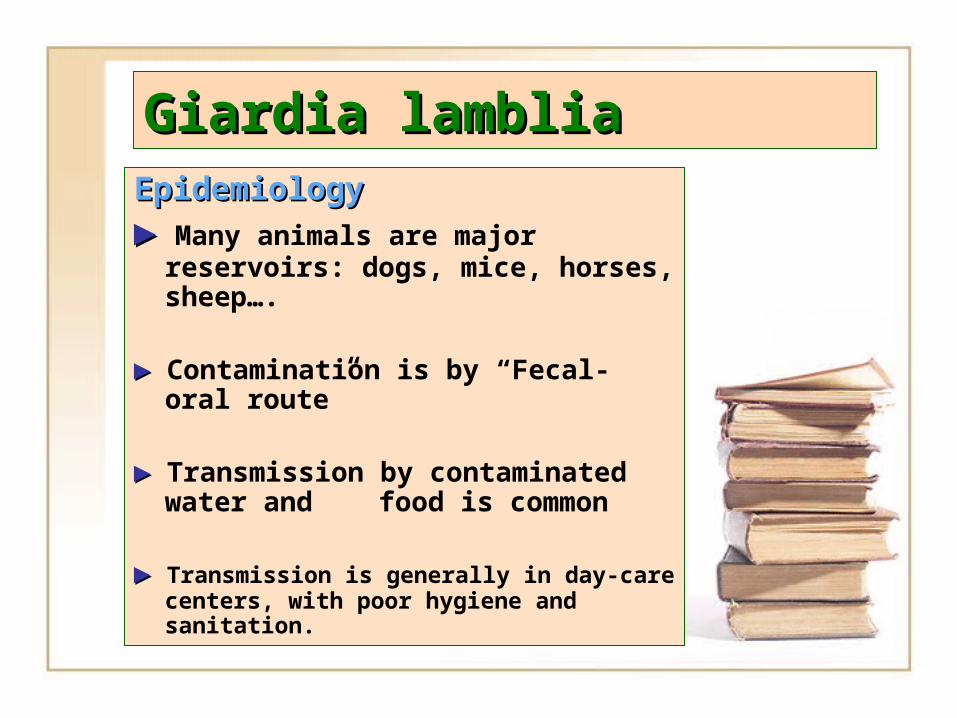
EpidemiologyEpidemiology
►► Many animals are major reservoirs: dogs, mice, horses, sheep….
►► Contamination is by “Fecal-oral route”
►► Transmission by contaminated water and food is common
►► Transmission is generally in day-care centers, with poor hygiene and sanitation.
Giardia lambliaGiardia lamblia
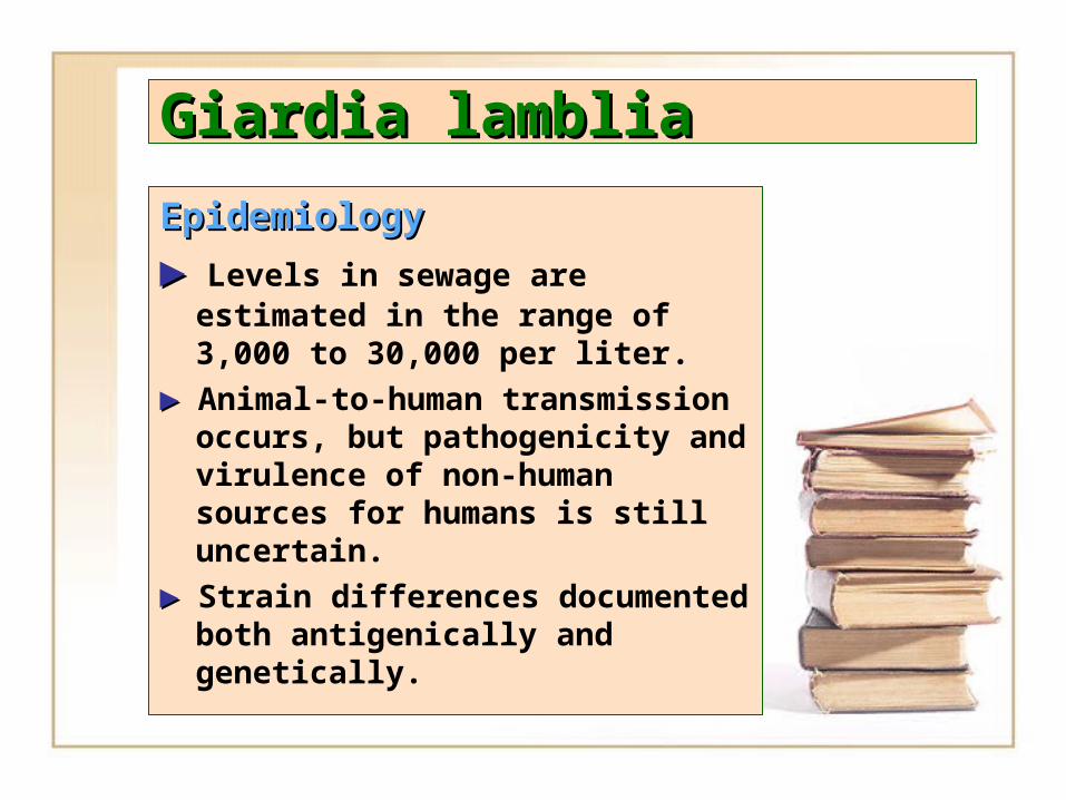
EpidemiologyEpidemiology
►► Levels in sewage are estimated in the range of 3,000 to 30,000 per liter.
►► Animal-to-human transmission occurs, but pathogenicity and virulence of non-human sources for humans is still uncertain.
►► Strain differences documented both antigenically and genetically.
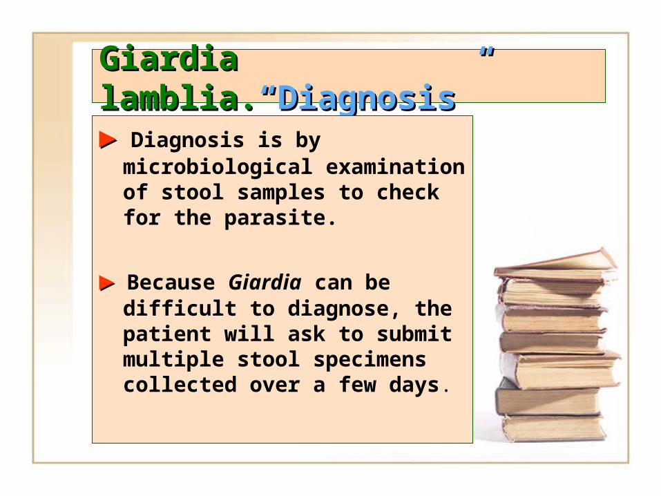
Giardia lamblia.Giardia lamblia.“Diagnosis”“Diagnosis”
►► Diagnosis is by microbiological examination of stool samples to check for the parasite.
►► Because Giardia can be difficult to diagnose, the patient will ask to submit multiple stool specimens collected over a few days.
• intestinalis trophozoite in a wet mount stained with iodine.
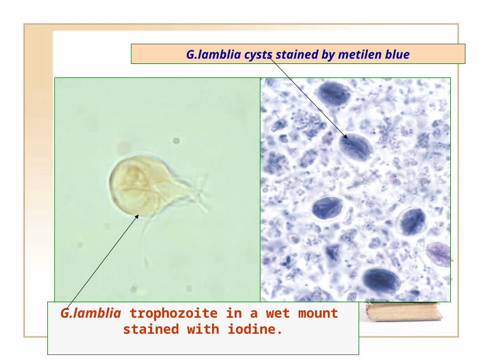
G.lamblia trophozoite in a wet mount stained with iodine.
G.lamblia cysts stained by metilen blue
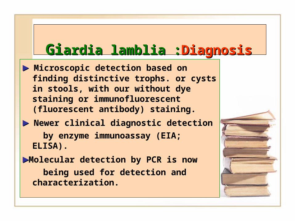
GiGiardia lamblia :ardia lamblia :DDiagnosisiagnosis ►► Microscopic detection based on finding
distinctive trophs. or cysts in stools, with our without dye staining or immunofluorescent (fluorescent antibody) staining.
►► Newer clinical diagnostic detection
by enzyme immunoassay (EIA; ELISA).
►►Molecular detection by PCR is now
being used for detection and characterization.
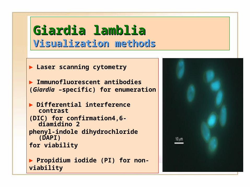
Giardia lambliaGiardia lambliaVVisualization isualization mmethodsethods
► Laser scanning cytometry
► Immunofluorescent antibodies (Giardia –specific) for enumeration
► Differential interference contrast (DIC) for confirmation4,6-diamidino
2phenyl-indole dihydrochloride
(DAPI)for viability
► Propidium iodide (PI) for non-viability
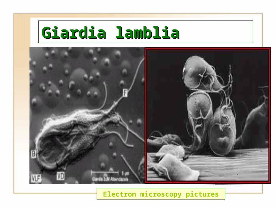
Giardia lambliaGiardia lamblia
Electron microscopy pictures
Environmental Control of GiardiaEnvironmental Control of Giardia
►► Water: physical and chemical treatment (coagulation-flocculation, sedimentation,
filtration and disinfection) will reduce Giardia
►►Relatively resistant to chemical desinfection
but relatively sensitive to physical disinfection by UV radiation or heat.
►► Pasteurization and thermal treatments is effective for foods
Giardia lambliaGiardia lambliaProtection from GiardiasisProtection from Giardiasis
► Washing hands thoroughly with soap and water.
► Not drinking untreated water or not use ice made from untreated water .
► Washing all food that is to be eaten raw by uncontaminated water..
Giardia lambliaGiardia lamblia
TreatmentTreatment
►► Quinacrine HCl Quinacrine HCl ,,Metronidazole Metronidazole (Flagyl) and Furazolidone (Flagyl) and Furazolidone (Furoxone).(Furoxone).
►► Oral QuinicrineOral Quinicrine HCl cures HCl cures aboutabout 90% of infections. 90% of infections.
►► Probiotics Probiotics could help could help the the
treatmenttreatment
Giardia lambliaGiardia lamblia
►► Giardia Giardia can infect all can infect all people ,young people ,young
children and pregnant women .children and pregnant women .
►► Rapid loss of fluids from Rapid loss of fluids from
diarrhea can be especially diarrhea can be especially
life threatening .life threatening .
►► Fluid replacementFluid replacement is an is an
important part of the important part of the treatmenttreatment


![BGD Lecture - Gastrointestinal System Development · [Expand] Historic drawing of the developing gastrointestinal tract (Kollman) BGD Lecture - Gastrointestinal System Development](https://img.pdfslide.us/doc/110x75/5d60c3df88c9937e068b5994/bgd-lecture-gastrointestinal-system-development-expand-historic-drawing.jpg)