Embed Size (px)
Citation preview
Gastrointestinal
0
AACN CCRN Review
Gastrointestinal
Presenter: Carol Rauen, RN, MS, PCCN, CCRN, CCNS, CEN
1
Gastrointestinal
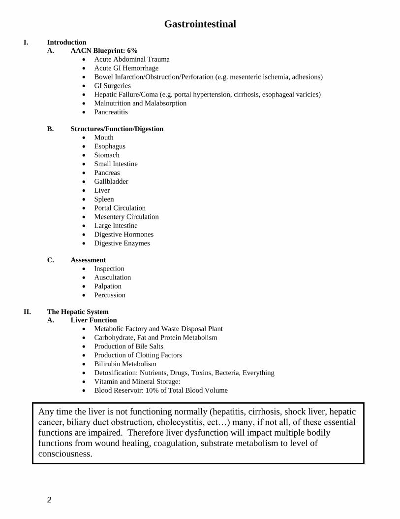
I. Introduction
A. AACN Blueprint: 6%
Acute Abdominal Trauma
Acute GI Hemorrhage
Bowel Infarction/Obstruction/Perforation (e.g. mesenteric ischemia, adhesions)
GI Surgeries
Hepatic Failure/Coma (e.g. portal hypertension, cirrhosis, esophageal varicies)
Malnutrition and Malabsorption
Pancreatitis
B. Structures/Function/Digestion
Mouth
Esophagus
Stomach
Small Intestine
Pancreas
Gallbladder
Liver
Spleen
Portal Circulation
Mesentery Circulation
Large Intestine
Digestive Hormones
Digestive Enzymes
C. Assessment
Inspection
Auscultation
Palpation
Percussion
II. The Hepatic System
A. Liver Function
Metabolic Factory and Waste Disposal Plant
Carbohydrate, Fat and Protein Metabolism
Production of Bile Salts
Production of Clotting Factors
Bilirubin Metabolism
Detoxification: Nutrients, Drugs, Toxins, Bacteria, Everything
Vitamin and Mineral Storage:
Blood Reservoir: 10% of Total Blood Volume
Any time the liver is not functioning normally (hepatitis, cirrhosis, shock liver, hepatic
cancer, biliary duct obstruction, cholecystitis, ect…) many, if not all, of these essential
functions are impaired. Therefore liver dysfunction will impact multiple bodily
functions from wound healing, coagulation, substrate metabolism to level of
consciousness.
2
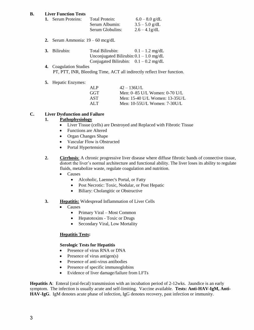
B. Liver Function Tests
1. Serum Proteins: Total Protein: 6.0 – 8.0 g/dL
Serum Albumin: 3.5 – 5.0 g/dL
Serum Globulins: 2.6 – 4.1g/dL
2. Serum Ammonia: 19 – 60 mcg/dL
3. Bilirubin: Total Bilirubin: 0.1 – 1.2 mg/dL
Unconjugated Bilirubin: 0.1 – 1.0 mg/dL
Conjugated Bilirubin: 0.1 – 0.2 mg/dL
4. Coagulation Studies
PT, PTT, INR, Bleeding Time, ACT all indirectly reflect liver function.
5. Hepatic Enzymes:
ALP 42 – 136U/L
GGT Men: 0–85 U/L Women: 0-70 U/L
AST Men: 15-40 U/L Women: 13-35U/L
ALT Men: 10-55U/L Women: 7-30U/L
C. Liver Dysfunction and Failure
1. Pathophysiology
Liver Tissue (cells) are Destroyed and Replaced with Fibrotic Tissue
Functions are Altered
Organ Changes Shape
Vascular Flow is Obstructed
Portal Hypertension
2. Cirrhosis: A chronic progressive liver disease where diffuse fibrotic bands of connective tissue,
distort the liver’s normal architecture and functional ability. The liver loses its ability to regulate
fluids, metabolize waste, regulate coagulation and nutrition.
Causes
Alcoholic, Laennec's Portal, or Fatty
Post Necrotic: Toxic, Nodular, or Post Hepatic
Biliary: Cholangitic or Obstructive
3. Hepatitis: Widespread Inflammation of Liver Cells
Causes
Primary Viral – Most Common
Hepatotoxins - Toxic or Drugs
Secondary Viral, Low Mortality
Hepatitis Tests:
Serologic Tests for Hepatitis
Presence of virus RNA or DNA
Presence of virus antigen(s)
Presence of anti-virus antibodies
Presence of specific immunoglobins
Evidence of liver damage/failure from LFTs
Hepatitis A: Enteral (oral-fecal) transmission with an incubation period of 2-12wks. Jaundice is an early
symptom. The infection is usually acute and self-limiting. Vaccine available. Tests: Anti-HAV-IgM, Anti-
HAV-IgG. IgM denotes acute phase of infection, IgG denotes recovery, past infection or immunity.
3
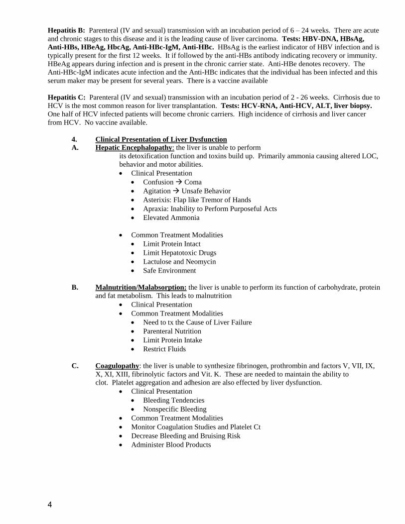
Hepatitis B: Parenteral (IV and sexual) transmission with an incubation period of 6 – 24 weeks. There are acute
and chronic stages to this disease and it is the leading cause of liver carcinoma. Tests: HBV-DNA, HBsAg,
Anti-HBs, HBeAg, HbcAg, Anti-HBc-IgM, Anti-HBc. HBsAg is the earliest indicator of HBV infection and is
typically present for the first 12 weeks. It if followed by the anti-HBs antibody indicating recovery or immunity.
HBeAg appears during infection and is present in the chronic carrier state. Anti-HBe denotes recovery. The
Anti-HBc-IgM indicates acute infection and the Anti-HBc indicates that the individual has been infected and this
serum maker may be present for several years. There is a vaccine available
Hepatitis C: Parenteral (IV and sexual) transmission with an incubation period of 2 - 26 weeks. Cirrhosis due to
HCV is the most common reason for liver transplantation. Tests: HCV-RNA, Anti-HCV, ALT, liver biopsy.
One half of HCV infected patients will become chronic carriers. High incidence of cirrhosis and liver cancer
from HCV. No vaccine available.
4. Clinical Presentation of Liver Dysfunction A. Hepatic Encephalopathy: the liver is unable to perform
its detoxification function and toxins build up. Primarily ammonia causing altered LOC,
behavior and motor abilities.
Clinical Presentation
Confusion Coma
Agitation Unsafe Behavior
Asterixis: Flap like Tremor of Hands
Apraxia: Inability to Perform Purposeful Acts
Elevated Ammonia
Common Treatment Modalities
Limit Protein Intact
Limit Hepatotoxic Drugs
Lactulose and Neomycin
Safe Environment
B. Malnutrition/Malabsorption: the liver is unable to perform its function of carbohydrate, protein
and fat metabolism. This leads to malnutrition
Clinical Presentation
Common Treatment Modalities
Need to tx the Cause of Liver Failure
Parenteral Nutrition
Limit Protein Intake
Restrict Fluids
C. Coagulopathy: the liver is unable to synthesize fibrinogen, prothrombin and factors V, VII, IX,
X, XI, XIII, fibrinolytic factors and Vit. K. These are needed to maintain the ability to
clot. Platelet aggregation and adhesion are also effected by liver dysfunction.
Clinical Presentation
Bleeding Tendencies
Nonspecific Bleeding
Common Treatment Modalities
Monitor Coagulation Studies and Platelet Ct
Decrease Bleeding and Bruising Risk
Administer Blood Products
4
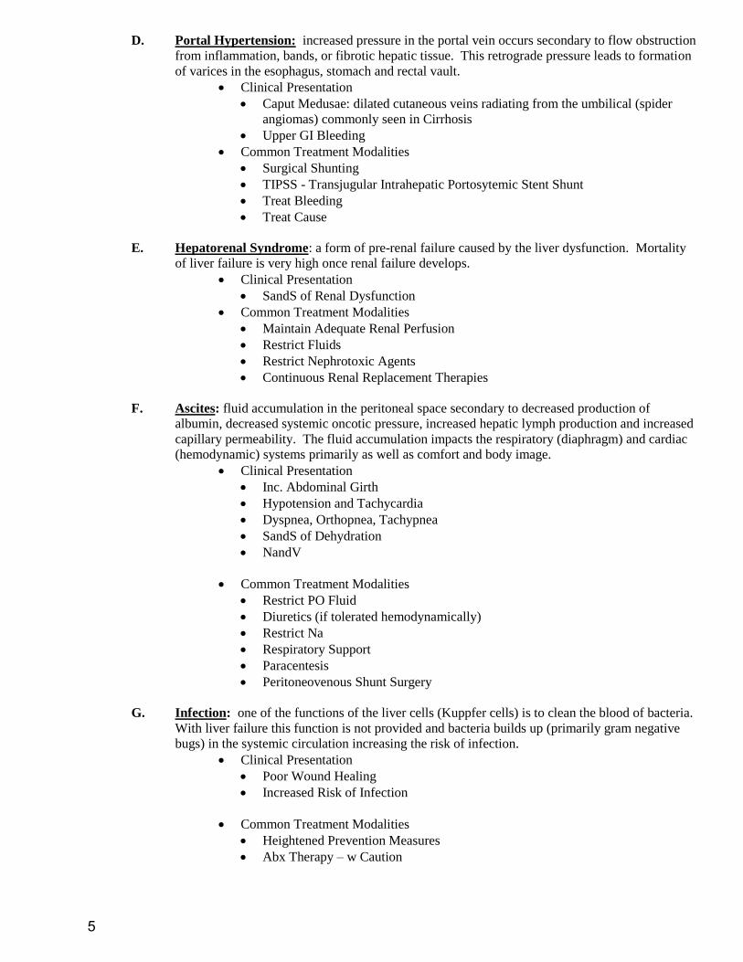
D. Portal Hypertension: increased pressure in the portal vein occurs secondary to flow obstruction
from inflammation, bands, or fibrotic hepatic tissue. This retrograde pressure leads to formation
of varices in the esophagus, stomach and rectal vault.
Clinical Presentation
Caput Medusae: dilated cutaneous veins radiating from the umbilical (spider
angiomas) commonly seen in Cirrhosis
Upper GI Bleeding
Common Treatment Modalities
Surgical Shunting
TIPSS - Transjugular Intrahepatic Portosytemic Stent Shunt
Treat Bleeding
Treat Cause
E. Hepatorenal Syndrome: a form of pre-renal failure caused by the liver dysfunction. Mortality
of liver failure is very high once renal failure develops.
Clinical Presentation
SandS of Renal Dysfunction
Common Treatment Modalities
Maintain Adequate Renal Perfusion
Restrict Fluids
Restrict Nephrotoxic Agents
Continuous Renal Replacement Therapies
F. Ascites: fluid accumulation in the peritoneal space secondary to decreased production of
albumin, decreased systemic oncotic pressure, increased hepatic lymph production and increased
capillary permeability. The fluid accumulation impacts the respiratory (diaphragm) and cardiac
(hemodynamic) systems primarily as well as comfort and body image.
Clinical Presentation
Inc. Abdominal Girth
Hypotension and Tachycardia
Dyspnea, Orthopnea, Tachypnea
SandS of Dehydration
NandV
Common Treatment Modalities
Restrict PO Fluid
Diuretics (if tolerated hemodynamically)
Restrict Na
Respiratory Support
Paracentesis
Peritoneovenous Shunt Surgery
G. Infection: one of the functions of the liver cells (Kuppfer cells) is to clean the blood of bacteria.
With liver failure this function is not provided and bacteria builds up (primarily gram negative
bugs) in the systemic circulation increasing the risk of infection.
Clinical Presentation
Poor Wound Healing
Increased Risk of Infection
Common Treatment Modalities
Heightened Prevention Measures
Abx Therapy – w Caution
5
III. The Pancreas
A. Function
Endocrine Functions
Synthesis and Release of Hormones: Glycogen, Insulin, Gastrin
Exocrine Functions
Pancreatic Enzymes Break Down Protein, Starch and Fat. > 2L/day
Bicarbonate Raise pH
PNS, Gastrin and Hormones Regulate Secretions
B. Pancreatic Enzymes
Trypsin: Aids in Protein Digestion
Amylase: Aids in Carbohydrate Digestion
Lipase: Aids in Fat Digestion
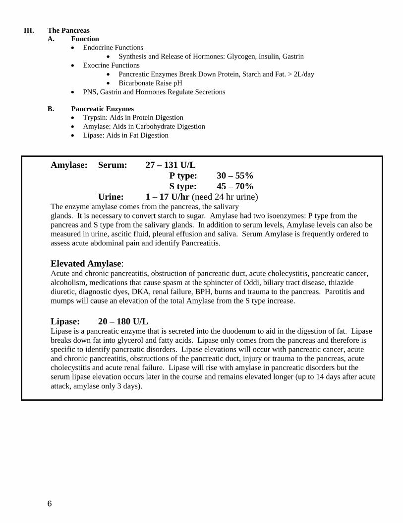
Amylase: Serum: 27 – 131 U/L
P type: 30 – 55%
S type: 45 – 70%
Urine: 1 – 17 U/hr (need 24 hr urine) The enzyme amylase comes from the pancreas, the salivary
glands. It is necessary to convert starch to sugar. Amylase had two isoenzymes: P type from the
pancreas and S type from the salivary glands. In addition to serum levels, Amylase levels can also be
measured in urine, ascitic fluid, pleural effusion and saliva. Serum Amylase is frequently ordered to
assess acute abdominal pain and identify Pancreatitis.
Elevated Amylase: Acute and chronic pancreatitis, obstruction of pancreatic duct, acute cholecystitis, pancreatic cancer,
alcoholism, medications that cause spasm at the sphincter of Oddi, biliary tract disease, thiazide
diuretic, diagnostic dyes, DKA, renal failure, BPH, burns and trauma to the pancreas. Parotitis and
mumps will cause an elevation of the total Amylase from the S type increase.
Lipase: 20 – 180 U/L Lipase is a pancreatic enzyme that is secreted into the duodenum to aid in the digestion of fat. Lipase
breaks down fat into glycerol and fatty acids. Lipase only comes from the pancreas and therefore is
specific to identify pancreatic disorders. Lipase elevations will occur with pancreatic cancer, acute
and chronic pancreatitis, obstructions of the pancreatic duct, injury or trauma to the pancreas, acute
cholecystitis and acute renal failure. Lipase will rise with amylase in pancreatic disorders but the
serum lipase elevation occurs later in the course and remains elevated longer (up to 14 days after acute
attack, amylase only 3 days).
6
C. Acute Pancreatitis
Pathophysiology
Auto Digestion
Tissue Damage
Fat Necrosis
Vascular Damage and Hemorrhage
Increased Capillary Permeability
Hypotension
Forms/Types
Edematous
Hemorrhagic
Classifications
Acute Pancreatitis
Recurrent Acute
Recurrent Chronic
Chronic Pancreatitis
Cause (blocked enzyme release)
Alcoholism
Biliary Stones
Hyperlipidemia
Abd Trauma
Infection (bacterial or viral)
Shock
Drugs (Most Common: Cyclosporine, Acetaminophen, Cimetadine, Steroids, Salicylates,
Furosemide, Thiazides, Estrogens)
Clinical Presentation
Pain
Low Grade Fever
NandV
Distended/Tender/Rigid Abd
Guarding with Rebound Tenderness
Jaundice
Hypoactive Bowel Sounds
Steatorrhea: bulky, pale, foul-smelling stools
? Ascites
Hypovolemic Shock
Labs (MOST diagnostic underlined)
Hypocalcemia (classic sign)
Low Ca, Mg, K
Hyperglycemia
Hyperbilirubinemia
Hypertriglyceridemia
Increased BUN and Creatinine
Ranson’s Criteria
On Admission
Age > 55yr
WBC > 16,000
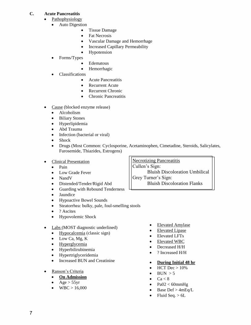
Necrotizing Pancreatitis
Cullen’s Sign:
Bluish Discoloration Umbilical
Grey Turner’s Sign:
Bluish Discoloration Flanks
Elevated Amylase
Elevated Lipase
Elevated LFTs
Elevated WBC
Decreased H/H
? Increased H/H
During Initial 48 hr
HCT Dec > 10%
BUN > 5
Ca < 8
Pa02 < 60mmHg
Base Def > 4mEq/L
Fluid Seq. > 6L
7
Glucose > 200
LDH > 350
AST > 250
Treatment Options
Fluid Resuscitation
Rest the Pancreas: NPO, NGT
Pain Management
Monitor and Replace Electrolytes
Tx Multisystem
Nutritional Support
Surgery
IV. Gastrointestinal Bleeding
A. Lower GI Bleeding: Not Typically Life Threatening
Causes
Diverticulitis
Angiodysplasia
Cancer
Hemorrhoids
Inflammatory Bowel Disease (Ulcerative Colitis; Crohn's Disease)
Bowel Infarction
B. Upper GI Bleeding
Causes
Peptic Ulcer Disease: Duodenal, Gastric and Stomal ulcers account for 50% bleeding episodes
Gastritis or Esophagitis
Esophageal Varices
Mallory -Weiss Syndrome
Clinical Presentation
Hematemesis
Melona
PUD
Distended and Tender Bbd
Hyperactive Bowel Sounds
Hypovolemia
Shock
Treatment
NG Decompression/Lavage – Room Temp vs. Iced
Fluid Resuscitation
Blood Product Admin
Endoscopic Sclerotherapy
Pharmacology
H2 Blockers, Antacids, Proton Pump Inhibitors
Sucralfate
Vasopressin: constricts splanchnic inflow to reduce portal pressure
Somatostatin and Octreotide: vasoconstricts splanchnic vessels to decrease blood flow
Surgery
Vagotomy and Pyloroplasty
Assessment
H and H
Coags and Platelets
Hemoconcentration
Elevated BUN
LFTs
Endoscopy
Angiography
Raionuclide Scans
8
Oversew Ulcer or Tear
Total and Subtotal Gastric Resection
Billroth I: Vagotomy, Antrectomy, AnastomosisStomach and Duodenum
Billroth II: Vagotomy, Antrectomy, Anastomosis Stomach and Jejunum
Whipple: Removal of the Distal 3rd
of Stomach, Entire duodenum, Head of Pancreas,
Gastrojejunotomy
Colon Resection
Bleeding Esophageal Varices
TIPSS: Transjugular Intrahepatic Portosytemic Stent Shunt
Beta Blocker – Decreases Pressure
Blakemore Tube
Portal Caval Shunt
V. Disorders of the Bowel
A. Bowel Infarction
Etiology
Embolic or Thrombotic Occlusion
Typically from the Superior Mesenteric Artery
Clinical Presentation
Severe Epigastric Pain
Rebound Tenderness
Guarding and Rigidity
Stimulated Sympathetic Response from Pain
Treatment Options
Angiography to Identify/Confirm Occlusion
Surgery to Remove Occlusion and Dead Bowel
B. Bowel Obstruction
Etiology
Internal Lumen Obstruction ex. Tumor
External Lumen Obstruction ex. Adhesions
Emboli: no blood flow
Paralytic Ileus
Clinical Presentation
Complete vs. Partial
Distended Edematous Bowel
Fluid and Electrolytes Leaking from Bowel
Elevated WBC
Fever
Small Intestine
Acute Pain with Sudden Onset
N and V (movement on both ends)
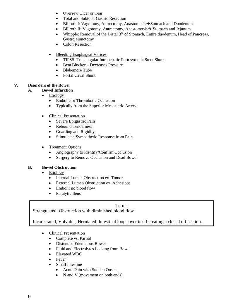
Terms
Strangulated: Obstruction with diminished blood flow
Incarcerated, Volvulus, Herniated: Intestinal loops over itself creating a closed off section.
9
Wave-Like Hyperactive High Pitched Bowel Sounds
May Have Some Gas or Feces
Distention (mild)
Large Intestine
Slow Onset Pain Progression Mild Severe, Lower Abd
No N and V (nothing moving)
No Stool
Low Pitched Bowel Sounds
Distention (large amount)
Treatment Options
Diagnosis Obstruction by Hx, X-Ray, CT, Upper or Lower Barium Radiology Tests
Pain Management
IV Fluids
Decompress w NG, Rectal or Intestinal Tube
Abx
NPO and Time (rest the bowel)
Surgery
C. Perforation/Peritonitis
Etiology
Gastric/Intestinal Contents Leak into Peritoneal Cavity
Ulcer Perforation
Diverticular Rupture
Trauma
Bowel Infarction
Clinical Presentation
Infection/Sepsis (all the SandS)
Sudden Onset of Severe Pain
Rigid Abdomen w Rebound Tenderness
Hypoactive Bowel Sounds No Bowel Sounds
Treatment Options
Surgery to Repair Cause and Clean Up
ABX
Fluids
Tx of Sepsis
Tx of MODS
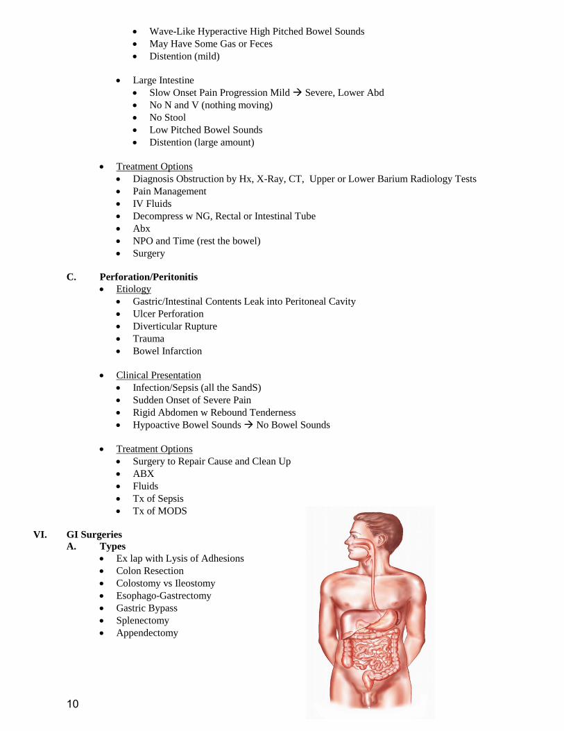
VI. GI Surgeries
A. Types
Ex lap with Lysis of Adhesions
Colon Resection
Colostomy vs Ileostomy
Esophago-Gastrectomy
Gastric Bypass
Splenectomy
Appendectomy
10
B. Care Concerns
Infection - Leaks
Sepsis
Third Spacing/Hypovolemia
Bleeding
Electrolyte Imbalance
Nutrition
Immobility
Pain
Potential for Respiratory Compromise
VII. Abdominal Trauma
A. Mechanism of Injury
Blunt Trauma
MVC
Falls
Assaults
Crush
Sports
Penetrating Trauma
GSW
Stabbings
Impalements
B. Types of Injuries
Organ Contusions
Organ Laceration
Spleen Common Site of Injury
Solid Organs vs Hallow Organs
Crush w Tissue Damage
Vascular Injury
Hypoperfusion
Hemorrhage
C. Assessment
Abd Exam
Pain/Tenderness
Firmness
Discoloration
Bowel Sounds
Abd Sonogram
CT
Diagnostic Peritoneal Lavage
Labs
X-Ray
Cullen’s Sign: Hemorrhagic Patches (bruising) Around the Umbilicus (pancreatitis, GI Hemorrhage,
ruptured ectopic pregnancy)
Grey Turner’s Sign: Bruising Around the Flank Area (Hemorrhagic Pancreatitis, Retroperitoneal
Bleeding)
Kehr’s Sign: Left Shoulder Pain from Irritation to the Diaphragm From Blood as a Result of Splenic
Rupture. Best Elicited with pt Lying Flat or in Trendelenburg’s Position.
11
Abdominal Compartment Syndrome
D. Treatment
Fluid Resuscitation
Diagnose Problem
Plug Holes and/or Repair Lacerations
Support Damaged Organ(s)
Remove Damaged Tissue/Organ(s)
Post Tx Concerns
Infection/Sepsis
Hemodynamic Status
Organ Function
ARDS, ATN, MODS
VIII. Summary
12
Renal Part 1
0
AACN CCRN Review
Renal Part 1
Presenter: Carol Rauen, RN, MS, PCCN, CCRN, CCNS, CEN
13
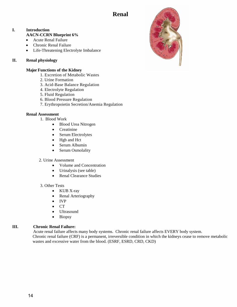
Renal
I. Introduction
AACN-CCRN Blueprint 6%
Acute Renal Failure
Chronic Renal Failure
Life-Threatening Electrolyte Imbalance
II. Renal physiology
Major Functions of the Kidney
1. Excretion of Metabolic Wastes
2. Urine Formation
3. Acid-Base Balance Regulation
4. Electrolyte Regulation
5. Fluid Regulation
6. Blood Pressure Regulation
7. Erythropoietin Secretion/Anemia Regulation
Renal Assessment
1. Blood Work
Blood Urea Nitrogen
Creatinine
Serum Electrolytes
Hgb and Hct
Serum Albumin
Serum Osmolality
2. Urine Assessment
Volume and Concentration
Urinalysis (see table)
Renal Clearance Studies
3. Other Tests
KUB X-ray
Renal Arteriography
IVP
CT
Ultrasound
Biopsy
III. Chronic Renal Failure: Acute renal failure affects many body systems. Chronic renal failure affects EVERY body system.
Chronic renal failure (CRF) is a permanent, irreversible condition in which the kidneys cease to remove metabolic
wastes and excessive water from the blood. (ESRF, ESRD, CRD, CKD)
14
Etiology - more than 100 different diseases can cause RF
Glomerular Disease
Tubular Diseases
Vascular Kidney Diseases
Urinary Tract Disease
Infection (kidney)
Systemic Vascular Diseases
Metabolic Diseases
Connective Tissue Diseases
A. Terms
1. Azotemia – Nitrogenous Waste Products in the Bloodstream
2. Uremic Syndrome – Systemic and Laboratory Manifestations of ESRD
3. Renal Replacement Therapy – Treatment Options
B. Stages of Renal Failure
1. Diminished Renal Reserve
2. Renal Insufficiency
3. End Stage Renal Disease (ESRD) – Affects every system in the body
C. Treatment: Renal Replacement Therapies
Medications
Hemodialysis
Peritoneal Dialysis
Renal Transplant
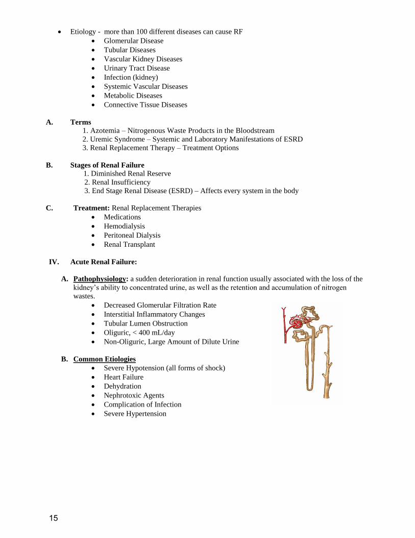
IV. Acute Renal Failure:
A. Pathophysiology: a sudden deterioration in renal function usually associated with the loss of the
kidney’s ability to concentrated urine, as well as the retention and accumulation of nitrogen
wastes.
Decreased Glomerular Filtration Rate
Interstitial Inflammatory Changes
Tubular Lumen Obstruction
Oliguric, < 400 mL/day
Non-Oliguric, Large Amount of Dilute Urine
B. Common Etiologies
Severe Hypotension (all forms of shock)
Heart Failure
Dehydration
Nephrotoxic Agents
Complication of Infection
Severe Hypertension
15
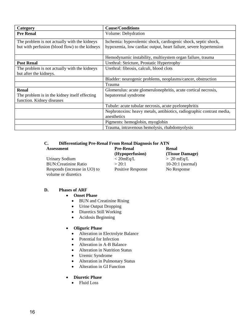
Category Cause/Conditions
Pre Renal Volume: Dehydration
The problem is not actually with the kidneys
but with perfusion (blood flow) to the kidneys
Ischemia: hypovolemic shock, cardiogenic shock, septic shock,
hypoxemia, low cardiac output, heart failure, severe hypertension
Hemodynamic instability, multisystem organ failure, trauma
Post Renal Urethral: Stricture, Prostatic Hypertrophy
The problem is not actually with the kidneys
but after the kidneys.
Urethral: fibrosis, calculi, blood clots
Bladder: neurogenic problems, neoplasms/cancer, obstruction
Trauma
Renal
The problem is in the kidney itself effecting
function. Kidney diseases
Glomerulus: acute glomerulonephritis, acute cortical necrosis,
hepatorenal syndrome
Tubule: acute tubular necrosis, acute pyelonephritis
Nephrotoxins: heavy metals, antibiotics, radiographic contrast media,
anesthetics
Pigments: hemoglobin, myoglobin
Trauma, intravenous hemolysis, rhabdomyolysis
C. Differentiating Pre-Renal From Renal Diagnosis for ATN
Assessment Pre-Renal
(Hypoperfusion)
Renal
(Tissue Damage)
Urinary Sodium < 20mEq/L > 20 mEq/L
BUN:Creatinine Ratio > 20:1 10-20:1 (normal)
Responds (increase in UO) to
volume or diuretics
Positive Response No Response
D. Phases of ARF
Onset Phase
BUN and Creatinine Rising
Urine Output Dropping
Diuretics Still Working
Acidosis Beginning
Oliguric Phase
Alteration in Electrolyte Balance
Potential for Infection
Alteration in A-B Balance
Alteration in Nutrition Status
Uremic Syndrome
Alteration in Pulmonary Status
Alteration in GI Function
Diuretic Phase
Fluid Loss
16
Goal is to maintain adequate fluid balance and regulate electrolytes
Alteration in Electrolytes
Recovery Phase
Goal is Supportive Care
Prevent Further Insults
Assessment of Renal Function
Keep patient well hydrated and free from infection
Prevent Further Insults
E. Systemic Response to Acute Failure
Hypertension
Tachycardia
Decreased UO
Lethargy
Pulmonary Edema
Depends on Type
Very Similar to Chronic RF
F. Nursing Care Needs
Ensure Hydration
Fluid Challenges
Diuretics
Monitor Fluid Status
Weigh Daily and I and O
Monitor Electrolyte Imbalance
Support Renal Function
G. Treatment Options/Alternatives
Drug Therapy
Diet Therapy
Renal Replacement Therapies (CVVH, Hemodialysis, Peritoneal Dialysis)
Renal Transplant
H. Support Therapy for ATN
Pt Problem Treatment
Extracellular Volume Overload
Restrict NaCl and H20
Diuretics
Dialysis
Hyponatremia
Restrict Oral H20
Restrict Hypotonic IV Solutions
Hyperkalemia
Restrict K intake Dialysis
K Binding Resins Glucose/Insulin
Eliminate K Supplements NaBicarb Ca Gluconate
Metabolic Acidosis
Na Bicarb Dialysis
Hyperphosphatemia
Restrict PHO4 Dialysis
Phosphate Binding Agents
Hypocalcemia
Calcium Carbonate Calcium Gluconate
Phosphate Binding Agents Dialysis
17

Hypermagnesemia
D/C Mg Containing Antacids
Dialysis
Nutrition
High Protein
Enteral or Parental Nutrition
Drug Dosage
Adjust Doses Around GFR
Avoid NSAIDS, ACE I, Dye, Nephrotoxic Abx
V. Renal Replacement Therapies
Goal – to remove body waste and fluids in the presence of acute or chronic renal failure
A. Terms –
Diffusion: movement of particles from an area of greater to an area of lesser
concentration. During dialysis diffusion results in the movement of urea,
creatinine, and uric acid from the patient’s blood in the dialysate
Osmosis: the movement of water across a semi-permeable membrane from an
area of lesser to an area of greater concentration (osmolality) of particles. During
dialysis osmosis results in extra fluid from the patient being removed.
Ultrafiltration: the movement of fluid across a semi-permeable membrane as a
result of an artificially created pressure gradient. More efficient than osmosis for
the removal of water.
Dialysis: involves the movement of fluid and particles across a semipermeable
membrane. It is a treatment that can help restore fluid and electrolyte balance,
control acid-base balance, and remove waste and toxic material from the body. It
can sustain life successfully in both acute and chronic situation where
substitution for or augmentation of normal renal function is needed.
B. Insurance Coverage – in 1972 the Congress enacted legislation that provides for people
with ESRD to receive Medicare regardless of age. This is not true in all countries.
Hemodialysis
Goal – involves shunting the patient’s blood from the body through a dialyzer in which
diffusion and ultrafiltration occur and then back into the patient’s circulation. Requires
access to the pt’s blood, a mechanism to transport the blood to and from the dialyzer (where
exchange of fluid, electrolytes, and waste products occur). HD can be used in the treatment
of acute and chronic renal failure
Access – five different types of access can be used
Arteriovenous Fistula
Arteriovenous Graft
External Arteriovenous Shunt
Femoral Vein Catheterization
Subclavian Vein Catheterization
18
Contraindications - Causes rapid fluid shifts
Labile Cardiovascular States
Recent MI
Hypotension
Complications
Hypotension
Air Embolism
Arrhythmias
Infection
Disequilibrium Syndrome -Rapid shifts in osmolality between cerebral spinal
fluid and blood can lead to cerebral edema
Coagulopathies - Heparin used during dialysis to prevent clotting of blood
outside of body
Chronic Care Needs –
Patients are typically hemodialyzed 2-3 times a week for 2-4 hours
Require many medication
Encounter multiple acute and chronic health risks as a result of the renal
failure and dialysis
Have dietary and fluid restrictions
Safety concerns regarding access sites
Assessment requirements for access sites
Peritoneal Dialysis
Goal – The goal is the same as above but a machine is not used to perform the “cleaning of
the blood.” The dialyzing fluid is instilled into the peritoneal cavity, and the peritoneum
becomes the dialyzing membrane. PD is used for acute and chronic renal failure and can be
done in the hospital or at home.
Access – an abdominal catheter is inserted into the peritoneal space. In chronic use this
catheter remains in place permanently and only changed periodically should problems arise.
Procedure – Approximately 2 liters of sterile dialysate is instilled into the peritoneal cavity
and allowed to dwell for a period of time. During this time osmosis and diffusion of particles
takes place. The catheter is then reopened and the fluid is drained from the patient (entire
process is called an exchange). This process is done repeated during a 24 hr period.
Contraindications
Peritonitis
Abdominal Surgery
Abdominal Adhesions
Pregnancy
Complications
Peritonitis
Respiratory Distress
19
Chronic Care Needs – PD can be done independently at home and the individual can lead a
fairly normal schedule. Not as many risks as HD. Most common problem is infection of
abdominal catheter.
Continuous ambulatory peritoneal dialysis (CAPD) – 4 –5 exchanges are done
a day.
Continuous cyclic peritoneal dialysis (CCPD) – exchanges are done with the
use of a machine to control the infusion, dwell and drain times and patients can
set up before going to sleep and have their PD occur automatically whale they
sleep. They are completely independent the rest of the day.
Continuous Renal Replacement Therapy Goal - CRRT provides continuous ultrafiltration of extracellular fluid and clearance of
uremic toxins. Only done in the critical care setting.
Access – Arterial and venous cannulation sites are required or two venous cannulation.
Procedure – the blood leaves the patient and flow through a hemofilter where the
ultrafiltration takes place and removal of water and waste (collected into standard urine bag)
and then the blood is returned to the patient via the venous access. The flow gradient to move
the blood through the filter is the patient’s own blood pressure. There are several types of
processes that are used in the critical care setting for CRRT. Not necessary to learn this year.
It will be covered in your acute care course next fall.
Contraindications:
Inability to tolerate extracorporeal circulation
Hypercoagulability
Inability to tolerate anti-coagulation therapy (heparin)
Fluid, electrolyte and acid-base shifts are less severe than with hemodialysis
and usually better tolerated
Complications
Fluid Imbalance - Hypo/Hypervolemia (Depends on ultrafiltration rate and
intravascular volume requirements)
Electrolyte Imbalance - Hypokalemia, Hyponatremia,
Hypocalcemia, and Hypomagnesaemia
Metabolic Acidosis - Bicarbonate readily removed
Drug removal - Potential for removing most drugs
Hemorrhage - Heparin used as blood leaves body to prevent coagulation
Thrombosis/Infection
Hypo/Hyperthermia
VI. Renal Transplantation
VII. Summary
20
Renal Part 2
0
AACN CCRN Review
Renal Part 2
Presenter: Carol Rauen, RN, MS, PCCN, CCRN, CCNS, CEN
21
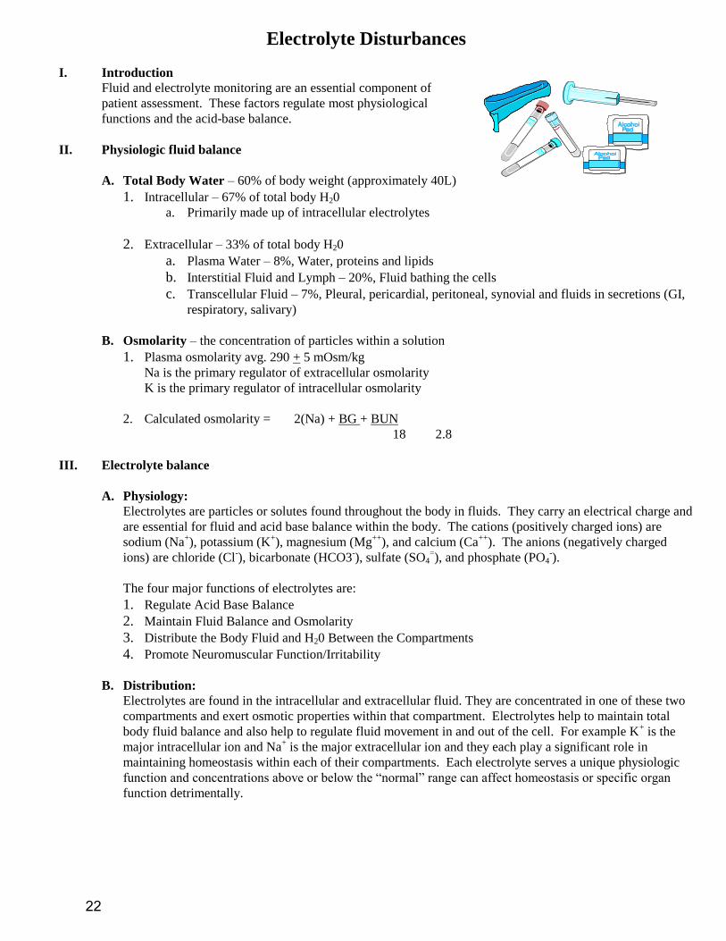
Electrolyte Disturbances
I. Introduction
Fluid and electrolyte monitoring are an essential component of
patient assessment. These factors regulate most physiological
functions and the acid-base balance.
II. Physiologic fluid balance
A. Total Body Water – 60% of body weight (approximately 40L)
1. Intracellular – 67% of total body H20
a. Primarily made up of intracellular electrolytes
2. Extracellular – 33% of total body H20
a. Plasma Water – 8%, Water, proteins and lipids
b. Interstitial Fluid and Lymph – 20%, Fluid bathing the cells
c. Transcellular Fluid – 7%, Pleural, pericardial, peritoneal, synovial and fluids in secretions (GI,
respiratory, salivary)
B. Osmolarity – the concentration of particles within a solution
1. Plasma osmolarity avg. 290 + 5 mOsm/kg
Na is the primary regulator of extracellular osmolarity
K is the primary regulator of intracellular osmolarity
2. Calculated osmolarity = 2(Na) + BG + BUN
18 2.8
III. Electrolyte balance
A. Physiology:
Electrolytes are particles or solutes found throughout the body in fluids. They carry an electrical charge and
are essential for fluid and acid base balance within the body. The cations (positively charged ions) are
sodium (Na+), potassium (K
+), magnesium (Mg
++), and calcium (Ca
++). The anions (negatively charged
ions) are chloride (Cl-), bicarbonate (HCO3
-), sulfate (SO4
=), and phosphate (PO4
-).
The four major functions of electrolytes are:
1. Regulate Acid Base Balance
2. Maintain Fluid Balance and Osmolarity
3. Distribute the Body Fluid and H20 Between the Compartments
4. Promote Neuromuscular Function/Irritability
B. Distribution:
Electrolytes are found in the intracellular and extracellular fluid. They are concentrated in one of these two
compartments and exert osmotic properties within that compartment. Electrolytes help to maintain total
body fluid balance and also help to regulate fluid movement in and out of the cell. For example K+ is the
major intracellular ion and Na+ is the major extracellular ion and they each play a significant role in
maintaining homeostasis within each of their compartments. Each electrolyte serves a unique physiologic
function and concentrations above or below the “normal” range can affect homeostasis or specific organ
function detrimentally.
22
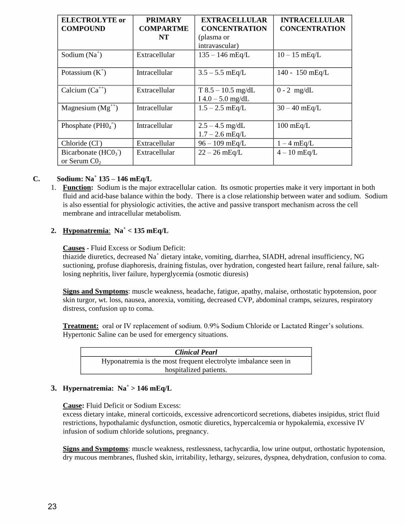
ELECTROLYTE or
COMPOUND
PRIMARY
COMPARTME
NT
EXTRACELLULAR
CONCENTRATION
(plasma or
intravascular)
INTRACELLULAR
CONCENTRATION
Sodium (Na+)
Extracellular 135 – 146 mEq/L 10 – 15 mEq/L
Potassium (K+)
Intracellular 3.5 – 5.5 mEq/L 140 - 150 mEq/L
Calcium (Ca++
) Extracellular T 8.5 – 10.5 mg/dL
I 4.0 – 5.0 mg/dL
0 - 2 mg/dL
Magnesium (Mg++
)
Intracellular 1.5 – 2.5 mEq/L 30 – 40 mEq/L
Phosphate (PH04=) Intracellular 2.5 – 4.5 mg/dL
1.7 – 2.6 mEq/L
100 mEq/L
Chloride (Cl-) Extracellular 96 – 109 mEq/L 1 – 4 mEq/L
Bicarbonate (HC03-)
or Serum C02
Extracellular 22 – 26 mEq/L 4 – 10 mEq/L
C. Sodium: Na+ 135 – 146 mEq/L
1. Function: Sodium is the major extracellular cation. Its osmotic properties make it very important in both
fluid and acid-base balance within the body. There is a close relationship between water and sodium. Sodium
is also essential for physiologic activities, the active and passive transport mechanism across the cell
membrane and intracellular metabolism.
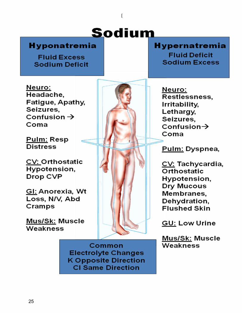
2. Hyponatremia: Na+ < 135 mEq/L
Causes - Fluid Excess or Sodium Deficit:
thiazide diuretics, decreased Na+ dietary intake, vomiting, diarrhea, SIADH, adrenal insufficiency, NG
suctioning, profuse diaphoresis, draining fistulas, over hydration, congested heart failure, renal failure, salt-
losing nephritis, liver failure, hyperglycemia (osmotic diuresis)
Signs and Symptoms: muscle weakness, headache, fatigue, apathy, malaise, orthostatic hypotension, poor
skin turgor, wt. loss, nausea, anorexia, vomiting, decreased CVP, abdominal cramps, seizures, respiratory
distress, confusion up to coma.
Treatment: oral or IV replacement of sodium. 0.9% Sodium Chloride or Lactated Ringer’s solutions.
Hypertonic Saline can be used for emergency situations.
Clinical Pearl
Hyponatremia is the most frequent electrolyte imbalance seen in
hospitalized patients.
3. Hypernatremia: Na+ > 146 mEq/L
Cause: Fluid Deficit or Sodium Excess:
excess dietary intake, mineral corticoids, excessive adrencorticord secretions, diabetes insipidus, strict fluid
restrictions, hypothalamic dysfunction, osmotic diuretics, hypercalcemia or hypokalemia, excessive IV
infusion of sodium chloride solutions, pregnancy.
Signs and Symptoms: muscle weakness, restlessness, tachycardia, low urine output, orthostatic hypotension,
dry mucous membranes, flushed skin, irritability, lethargy, seizures, dyspnea, dehydration, confusion to coma.
23
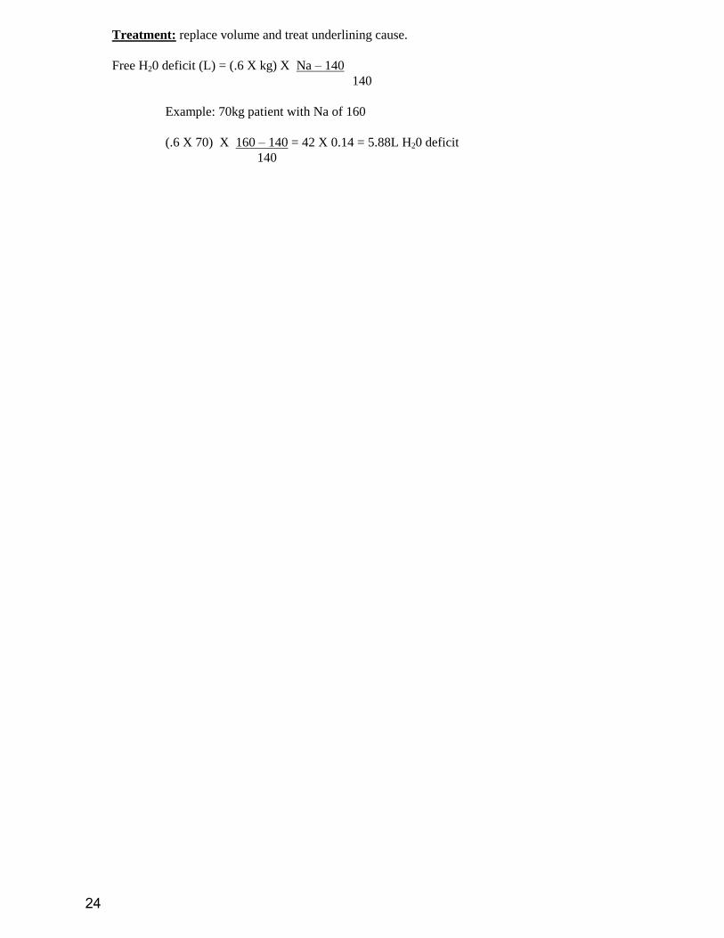
Treatment: replace volume and treat underlining cause.
Free H20 deficit (L) = (.6 X kg) X Na – 140
140
Example: 70kg patient with Na of 160
(.6 X 70) X 160 – 140 = 42 X 0.14 = 5.88L H20 deficit
140
24
[
25
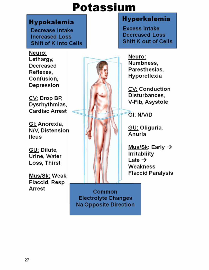
D. Potassium: K+ = 3.5 – 5.5 mEq/L
1. Function: Major intracellular cation contributes to cell homeostasis and function by maintaining its osmolarity and
electro neutrality. Potassium plays a principle role in electrical conductivity by influencing neuromuscular
transmission of nerve impulses and cardiac muscle contractility. Also helps to maintain acid-base balance and
normal kidney function.
2. Hypokalemia: K+ < 3.5 mEq/L
Cause: Decreased Intake, Increase Loss or Shift of K into Cells:
starvation, dehydration, massive fluid infusion lacking in K+, decreased dietary intake, vomiting, diarrhea,
corticosteroids therapy, draining fistula, diuretics, some antibiotics, laxative overuse, NG suctioning, hypernatremia,
metabolic alkalosis (relative hypokalemia), aldosteronism.
Signs and Symptoms: ECG Changes – depressed ST segments, flat or inverted T waves, presence of U waves,
dysrhythmias, cardiac arrest, dilute urine, anorexia, nausea, vomiting, ileus, lethargy, mental depression, paralysis,
confusion, muscle weakness, respiratory arrest, can precipitate digitalis toxicity.
Treatment: Oral or Parenteral Replacement of K+
3. Hyperkalemia: K+ > 5.5 mEq/L
Cause: Excess Intake, Decreased Loss, Shift of K out of Cells: movement of K out of the cells (acidosis, sepsis,
fever, trauma, hyperglycemia, rhabdomyolysis, catecholamines, insulin deficiency, tissue necrosis), excessive
dietary intake, renal failure (decreased excretion), Addison’s disease (adrenal insufficiency), large volume of stored
blood products, potassium sparing diuretics, medications that promote K+
retention (ACE inhibitors, beta blockers,
NSAIDS, heparin), hyperosmolar states, excessive potassium administration.
Signs and Symptoms: ECG changes – tall, peaked, tented T waves, flattened or absent P waves, widening QRS,
asystole, alteration of depolarization/repolarization of cardiac muscle, oliguria, nausea, vomiting, diarrhea, calf pain,
numbness or paresthesia, hyporeflexia up to flaccid paralysis.
Treatment: Three-Part Therapy
1. Cardiac Protect: 10ml of Calcium Chloride or Calcium Gluconate slow IV push. Renders the myocardium
less excitable by decreasing the effects of excess extracellular K+.
2. Shift K+ into the Cell:
1 amp Sodium Bicarbonate
5-10U Regular Insulin
50ml Bolus 50% Dextrose
Albuterol 10 – 20mg inhalation or intravenous (beta2 adrenergic agent – stimulates B2 receptor in
the pancreas to release more insulin).
3. Removal of K+:
Loop Diuretic
Sodium Polystyrene Sulfonate (Kayexalate) a cation exchange resin given orally or by retention
enema. Oral administration is more effective. Each 1gm will lower the K+ 1mEq with orally
administration, and 0.5mEq with rectal administration. Sorbitol prevents constipation.
Dialysis can also be utilized to remove K+ from the body.
26
27
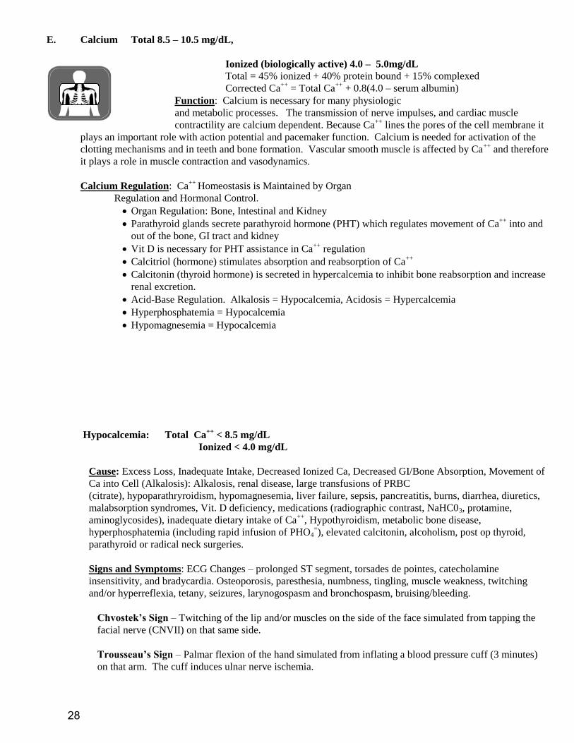
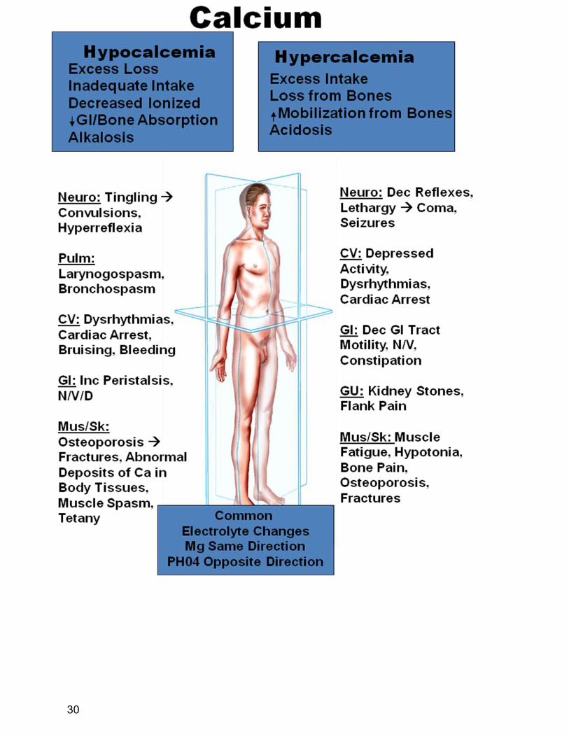
E. Calcium Total 8.5 – 10.5 mg/dL,
Ionized (biologically active) 4.0 – 5.0mg/dL
Total = 45% ionized + 40% protein bound + 15% complexed
Corrected Ca++
= Total Ca++
+ 0.8(4.0 – serum albumin)
Function: Calcium is necessary for many physiologic
and metabolic processes. The transmission of nerve impulses, and cardiac muscle
contractility are calcium dependent. Because Ca++
lines the pores of the cell membrane it
plays an important role with action potential and pacemaker function. Calcium is needed for activation of the
clotting mechanisms and in teeth and bone formation. Vascular smooth muscle is affected by Ca++
and therefore
it plays a role in muscle contraction and vasodynamics.
Calcium Regulation: Ca++
Homeostasis is Maintained by Organ
Regulation and Hormonal Control.
Organ Regulation: Bone, Intestinal and Kidney
Parathyroid glands secrete parathyroid hormone (PHT) which regulates movement of Ca++
into and
out of the bone, GI tract and kidney
Vit D is necessary for PHT assistance in Ca++
regulation
Calcitriol (hormone) stimulates absorption and reabsorption of Ca++
Calcitonin (thyroid hormone) is secreted in hypercalcemia to inhibit bone reabsorption and increase
renal excretion.
Acid-Base Regulation. Alkalosis = Hypocalcemia, Acidosis = Hypercalcemia
Hyperphosphatemia = Hypocalcemia
Hypomagnesemia = Hypocalcemia
Hypocalcemia: Total Ca++
< 8.5 mg/dL
Ionized < 4.0 mg/dL
Cause: Excess Loss, Inadequate Intake, Decreased Ionized Ca, Decreased GI/Bone Absorption, Movement of
Ca into Cell (Alkalosis): Alkalosis, renal disease, large transfusions of PRBC
(citrate), hypoparathryroidism, hypomagnesemia, liver failure, sepsis, pancreatitis, burns, diarrhea, diuretics,
malabsorption syndromes, Vit. D deficiency, medications (radiographic contrast, NaHC03, protamine,
aminoglycosides), inadequate dietary intake of Ca++
, Hypothyroidism, metabolic bone disease,
hyperphosphatemia (including rapid infusion of PHO4=), elevated calcitonin, alcoholism, post op thyroid,
parathyroid or radical neck surgeries.
Signs and Symptoms: ECG Changes – prolonged ST segment, torsades de pointes, catecholamine
insensitivity, and bradycardia. Osteoporosis, paresthesia, numbness, tingling, muscle weakness, twitching
and/or hyperreflexia, tetany, seizures, larynogospasm and bronchospasm, bruising/bleeding.
Chvostek’s Sign – Twitching of the lip and/or muscles on the side of the face simulated from tapping the
facial nerve (CNVII) on that same side.
Trousseau’s Sign – Palmar flexion of the hand simulated from inflating a blood pressure cuff (3 minutes)
on that arm. The cuff induces ulnar nerve ischemia.
28
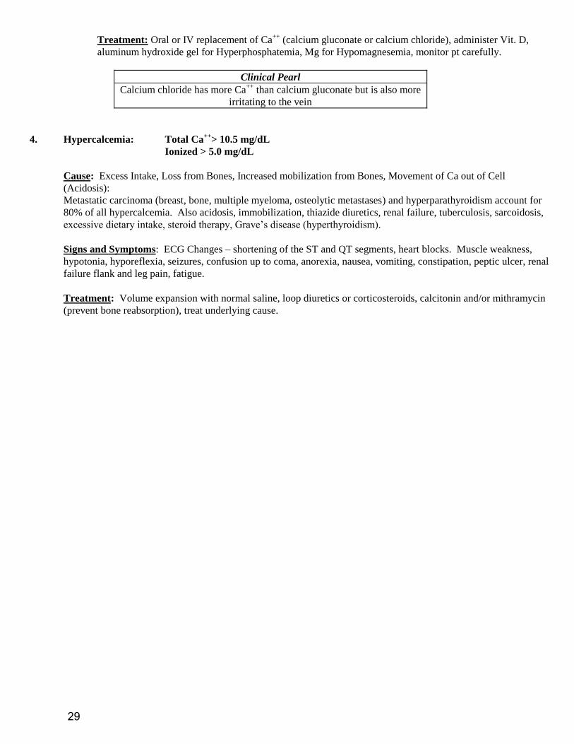
Treatment: Oral or IV replacement of Ca++
(calcium gluconate or calcium chloride), administer Vit. D,
aluminum hydroxide gel for Hyperphosphatemia, Mg for Hypomagnesemia, monitor pt carefully.
Clinical Pearl
Calcium chloride has more Ca++
than calcium gluconate but is also more
irritating to the vein
4. Hypercalcemia: Total Ca++
> 10.5 mg/dL
Ionized > 5.0 mg/dL
Cause: Excess Intake, Loss from Bones, Increased mobilization from Bones, Movement of Ca out of Cell
(Acidosis):
Metastatic carcinoma (breast, bone, multiple myeloma, osteolytic metastases) and hyperparathyroidism account for
80% of all hypercalcemia. Also acidosis, immobilization, thiazide diuretics, renal failure, tuberculosis, sarcoidosis,
excessive dietary intake, steroid therapy, Grave’s disease (hyperthyroidism).
Signs and Symptoms: ECG Changes – shortening of the ST and QT segments, heart blocks. Muscle weakness,
hypotonia, hyporeflexia, seizures, confusion up to coma, anorexia, nausea, vomiting, constipation, peptic ulcer, renal
failure flank and leg pain, fatigue.
Treatment: Volume expansion with normal saline, loop diuretics or corticosteroids, calcitonin and/or mithramycin
(prevent bone reabsorption), treat underlying cause.
29
30
F. Magnesium: Mg++
= 1.5 – 2.5 mEq/L
Function: Magnesium is essential for the production and use of energy, all ATP reactions involve Mg++
. The
Na+/K
+ ATPase pump is dependent on Mg
++, therefore making it an important component in the action potential and
depolarization and repolarization of the cardiac muscle. Mg++
appears to play a role in membrane stabilization
decreasing the likelihood of cardiac cell irritability or ectopy. It also has vasodilating effects, and it influences the
release of neurotransmitters at the neuromuscular junction by stabilizing the nerve axon.
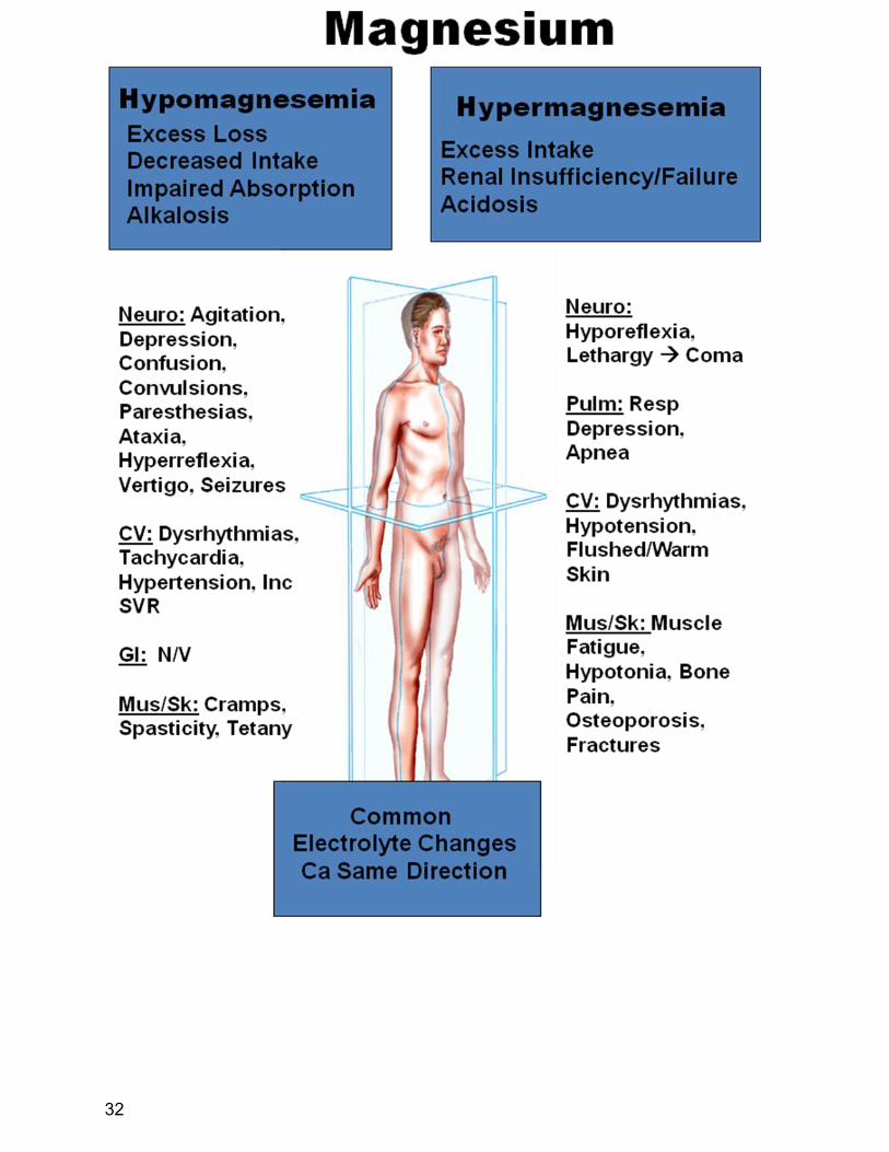
Hypomagnesemia: Mg++
< 1.5 mEq/L
Cause: Excess Loss, Decreased Intake, Impaired Absorption, Movement of Mg into the Cell (Alkalosis):
Excessive diuretic therapy, starvation, malabsorption, medications (digitalis, cyclosporine, cisplatin), endocrine
disorders (DKA, HHNK, hyperaldosteronism, hyperthyroidism), chronic alcoholism, pancreatitis, alkalosis,
vomiting, NG suctioning, citrate-chelation, decreased intake (enteral or parenteral).
Signs and Symptoms: (very similar to hypocalcemia) ECG Changes – flat or inverted T waves, ST segment
depression, prolonged QT interval, supraventricular and/or ventricular ectopy including torsades de pointes and
Vfib. Chvostek’s and Trousseau’s signs, hyperreflexia, vertigo, seizures, confusion, hallucinations, depression up to
coma, increased SVR and hypertension, nausea and vomiting.
Treatment: IV administration of Mg++
with close monitoring. 1–4g MgS04 over 2 minutes to 6 hr (depending on
severity of depletion). Common side effects of MgS04 administration are flushed feeling or sweating, bradycardia,
hypotension and IV site burning.
Clinical Pearl
When low Mg++
and low K+ are both present the patient will be unresponsive to
KCL therapy until the hypomagnesaemia is treated.
Hypermagnesemia: Mg++
> 2.5mEq/L
Cause: Excess Mg++
intake (MgS04, laxatives, antacids), Renal Insufficiency or Failure, Movement of Mg out of
Cell (Acidosis)
Signs and Symptoms: ECG Changes – peaked T waves, shortened QT interval, prolonged PR and QRS intervals,
bradycardia, heart blocks, asystole. Hyporeflexia, respiratory depression to apnea, lethargy to coma, seizure,
hypotension, hypocalcaemia, hyperkalemia, flushed/warm skin.
Treatment: Volume administration, diuretics, decrease Mg++
intake, IV insulin and glucose will drive Mg++
back
into cell, treat acidosis, hemodialysis or CAPD with Mg-free dialysate.
V. Summary
Na Cl Glucose
K CO2
31
32