Embed Size (px)
Citation preview
T h e w o r l d w i d e j o u r n a l f o r t h e c o m p a n i o n a n i m a l v e t e r i n a r i a n
#23.22 0 1 3 - 1 0 $ / 1 0 e
Gastrointestinal issues
Feline triaditis • Digestive issues of working and athletic dogs • How I approach... Constipation in the cat • Canine gastrointestinal microbiome in health and disease • Managing canine inflammatory bowel disease • Epidemiology of canine parvovirus infection in the USA – an update • Feline intestinal tumors • Diseases of the esophagus • A step-wise approach to dogs and cats with chronic diarrhea
Come to hear the latest in VETERINARY INTERNAL MEDICINE!
ACC - LIVERPOOL
23rdECVIM-CA CONGRESS12th - 14th September 2013
w w w. e c v i m c o n g r e s s . o r g
generously supports the ECVIM-CA Congress
Adv_Liverpool.indd 1 10-09-12 12:29
10-31-1668
PEFC recyclépefc-france.org
02
09
14
22
29
37
39
46
54
A point often made to veterinary and medi-cal undergraduates during training is the notion that the digestive system - with a mouth at one end and an anus at the other, both communicating directly with the ex-ternal environment - can be regarded as a long tube which is actually outside the body to which it is attached. The concept does
not, of course, stand up to scrutiny - it takes little medical know-ledge to appreciate that the organs that together make up the digestive tract are well and truly interlinked with an animal’s other structures, and that the system is far from being a stand-alone entity; apart from anything else, without the nutrition that it provides, an animal will very soon cease to exist at all.
Another concept that arises when considering the gastrointestinal tract is how relevant it is to everyday life, in that we use parts of its anatomy or physiological function as metaphors in day-to-day speech – so that we talk about “digesting information” when learning something new, or “having guts” as a phrase to indicate daring – and this surely reflects just how important a healthy gastrointestinal system is to our daily lives. Of course, we all need to eat to survive, and whilst we may not consider, say, our own hepatic function on a daily basis, we are most certainly aware of our digestive system and its functioning several times a day. But whilst all animals eat to live, when a problem arises with the digestive system – and there are many, whether that be acute vomiting or diarrhea, or chronic malabsorption or constipation – it can be the cause of major concerns.
And so the inevitable conclusion from all of the above is that this issue of Veterinary Focus will provide food for thought, with the table of contents acting as a menu to offer the reader with a hunger to learn a true feast of knowledge. Bon appétit!!
Ewan McNeill - Editor in chief
E D I T O R I A L
Editorial committee• Franziska Conrad, DVM, Scientific Communications,
Royal Canin, Germany• Craig Datz, DVM, Dipl. ACVN, Nutrition and Scientific
Affairs Manager, Royal Canin, USA• Pauline Devlin, BSc, PhD, Scientific Communications
and External Affairs, Royal Canin, UK• Laura Diana, DVM, Dipl. FCV, UBA, Scientific
Communications, Royal Canin, Argentina• María Elena Fernández, DVM, Scientific
Communications, Royal Canin, Spain• Joanna Gale, BVetMed, CertLAS ,MRCVS, Science
and Technical Communications Manager, WALTHAM Centre for Pet Nutrition, UK
• Giulio Giannotti, BSc, Product Manager, Royal Canin, Italy
• Hervé Marc, Global Corporate Affairs Manager, Royal Canin, France
• Philippe Marniquet, DVM, Dipl. ESSEC, Veterinary Communication Manager, Royal Canin, France
• Yann Quéau, DVM, Dipl. ACVN, Research Nutritionist, Royal Canin, France
Translation control• Elisabeth Landes, DVM (German)• Clemens Schickling DVM (German)• Noemi Del Castillo, PhD (Spanish)• Giulio Giannotti, BSc (Italian)• Matthias Ma, DVM (Chinese)• Yoshiko Nakamura, DVM (Japanese)• Boris Shulyak, PhD (Russian)
Deputy publisher: Buena Media PlusCEO: Bernardo GallitelliAddress: 85, avenue Pierre Grenier92100 Boulogne - France
Phone: +33 (0) 1 72 44 62 00Editor• Ewan McNeill, BVMS, Cert VR, MRCVS Editorial secretaries• Laurent Cathalan
[email protected]• Pierre Ménard
Printed in the European Union ISSN 1354-0157Circulation: 80,000 copiesLegal deposit: June 2013Cover: Shutterstock
Veterinary Focus is also published in French, German, Chinese, Italian, Polish, Spanish, Japanese & Russian.
The licensing arrangements for thera-peutic agents intended for use in small animal species vary greatly worldwide. In the absence of a specific license, consideration should be given to issu-ing an appropriate cautionary warning prior to administration of any such drug.
Feline triaditisIsabelle Cattin
Digestive issues of working and athletic dogsLaurence Yaguiyan-Colliard and Dominique Grandjean
How I approach... Constipation in the catValérie Freiche
Canine gastrointestinal microbiome in health and diseaseJan Suchodolski and Kenneth Simpson
Managing canine inflammatory bowel diseaseKenneth Simpson
Epidemiology of canine parvovirus infection in the USA – an updateSandi Lefebvre
Feline intestinal tumors Laura Marconato and Giuliano Bettini
Diseases of the esophagusIwan Burgener
Cut-out and keep guide...A step-wise approach to dogs and cats with chronic diarrhea Jörg Steiner
2 / Veterinary Focus / Vol 23 n°2 / 2013
■
Feline triaditis
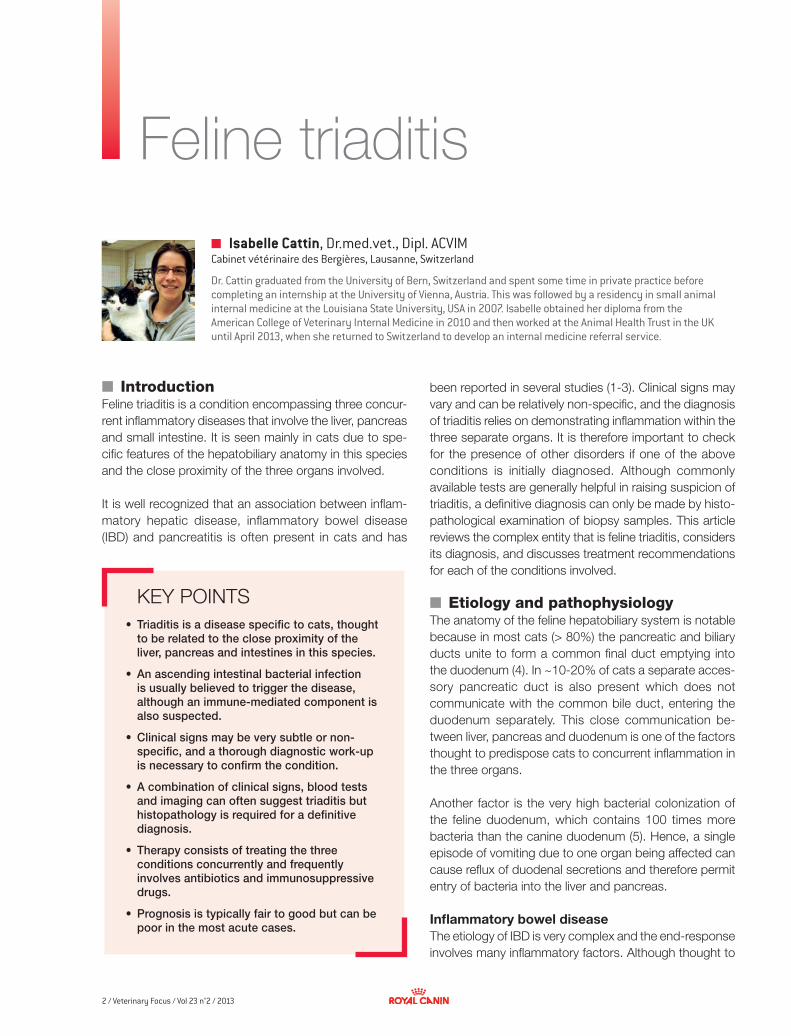
Isabelle Cattin, Dr.med.vet., Dipl. ACVIM Cabinet vétérinaire des Bergières, Lausanne, Switzerland
Dr. Cattin graduated from the University of Bern, Switzerland and spent some time in private practice before completing an internship at the University of Vienna, Austria. This was followed by a residency in small animal internal medicine at the Louisiana State University, USA in 2007. Isabelle obtained her diploma from the American College of Veterinary Internal Medicine in 2010 and then worked at the Animal Health Trust in the UK until April 2013, when she returned to Switzerland to develop an internal medicine referral service.
KEY POINTS• Triaditisisadiseasespecifictocats,thought
toberelatedtothecloseproximityoftheliver,pancreasandintestinesinthisspecies.
• Anascendingintestinalbacterialinfectionisusuallybelievedtotriggerthedisease,althoughanimmune-mediatedcomponentisalsosuspected.
• Clinicalsignsmaybeverysubtleornon-specific,andathoroughdiagnosticwork-upisnecessarytoconfirmthecondition.
• Acombinationofclinicalsigns,bloodtestsandimagingcanoftensuggesttriaditisbuthistopathologyisrequiredforadefinitivediagnosis.
• Therapyconsistsoftreatingthethreeconditionsconcurrentlyandfrequentlyinvolvesantibioticsandimmunosuppressivedrugs.
• Prognosisistypicallyfairtogoodbutcanbepoorinthemostacutecases.
■ IntroductionFeline triaditis is a condition encompassing three concur-rent inflammatory diseases that involve the liver, pancreas and small intestine. It is seen mainly in cats due to spe-cific features of the hepatobiliary anatomy in this species and the close proximity of the three organs involved.
It is well recognized that an association between inflam-matory hepatic disease, inflammatory bowel disease (IBD) and pancreatitis is often present in cats and has
been reported in several studies (1-3). Clinical signs may vary and can be relatively non-specific, and the diagnosis of triaditis relies on demonstrating inflammation within the three separate organs. It is therefore important to check for the presence of other disorders if one of the above conditions is initially diagnosed. Although commonly available tests are generally helpful in raising suspicion of triaditis, a definitive diagnosis can only be made by histo-pathological examination of biopsy samples. This article reviews the complex entity that is feline triaditis, considers its diagnosis, and discusses treatment recommendations for each of the conditions involved.
■ Etiology and pathophysiologyThe anatomy of the feline hepatobiliary system is notable because in most cats (> 80%) the pancreatic and biliary ducts unite to form a common final duct emptying into the duodenum (4). In ~10-20% of cats a separate acces-sory pancreatic duct is also present which does not communicate with the common bile duct, entering the duodenum separately. This close communication be-tween liver, pancreas and duodenum is one of the factors thought to predispose cats to concurrent inflammation in the three organs.
Another factor is the very high bacterial colonization of the feline duodenum, which contains 100 times more bacteria than the canine duodenum (5). Hence, a single episode of vomiting due to one organ being affected can cause reflux of duodenal secretions and therefore permit entry of bacteria into the liver and pancreas.
Inflammatory bowel diseaseThe etiology of IBD is very complex and the end-response involves many inflammatory factors. Although thought to
3 / Veterinary Focus / Vol 23 n°2 / 2013
l I I I I I I I I I I I I I I l I I I I I I I I I I I I I I l I I I I I I I I I I I I I I l I I I I I I I I I I I I I l I I I I I I I I I I I I I I l I I I I I I I I I I I I I I l I I I I I I I I I I I I I I l I I I I I I I I I I I I I I I I I I I I I I l I I I I I I I I I I I I I I l I I I I I I I I I I I I I I l I I I I I I I I I I I I I l I I I I I I I I I I I I I I l I I I I I I I I I I
be a multifactorial disease, the main mechanism in IBD is considered to be an inappropriate immune response to dietary or bacterial antigens presented to the gastroin-testinal mucosa. The resulting cellular infiltration (inflam-mation) creates mucosal changes (e.g. villous blunting/atrophy, crypt hypertrophy) resulting in maldigestion and malabsorption.
CholangitisAlthough the previous terminology for this group of dis-eases was cholangiohepatitis, the WSAVA liver group suggested that the term cholangitis is more appropriate, as it is primarily a biliary tree disease (6). Two main forms of inflammatory liver disease occur; the neutrophilic form (previously also described as suppurative), and the lym-phocytic form (previously lymphoplasmacytic or non-suppurative). The first version is the one usually consid-ered to be part of the triaditis complex, with an infiltration that is, as the name suggests, mostly neutrophilic; it is believed to result from a bacterial infection ascending from the intestinal tract. In the second form, the infiltrate is predominantly lymphocytic with plasma cells; the etio-logy is poorly understood but it is thought to be immune-mediated, or possibly result from a chronic neutrophilic cholangitis.
PancreatitisThe chronic form of pancreatitis is much more commonly seen in cats and is also the form recognized in the triaditis complex. Its etiology is believed to be immune-mediated, although in some instances an ascending bacterial infec-tion may also be causative. The inflammation present in chronic pancreatitis is usually mainly lymphocytic, with fibrosis and acinar atrophy commonly seen.
■ Clinical signsAlthough feline triaditis involves different organs, clinical signs may suggest a single organ disorder, although gastrointestinal signs (vomiting, diarrhea) are often present. Chronic pancreatitis in cats is typically silent or produces very subtle changes, so most cases of feline triaditis will show signs consistent with either IBD or cholangitis (or both concurrently).
Common signs of IBD are chronic vomiting and diarrhea, often accompanied by weight loss. Typically, middle-aged to older cats are affected but cats as young as one-year-old have been diagnosed, so age alone should not be used to rule out the disease (7). Signs can be mild to severe, and an acute presentation is also possible but less frequent.
While cats with cholangitis can present with the same signs as IBD, jaundice is a hallmark of the disease and it is often the reason for presentation to the veterinarian. The clinical signs may vary between the neutrophilic and the lymphocytic forms, although they often overlap (Table 1).
As noted, chronic pancreatitis in cats is usually a silent disease or presents with very mild or non-specific signs (anorexia, lethargy). It is important to remember that (contrary to the situation in dogs) vomiting is not the most common clinical finding with feline pancreatitis, with only about one third of cats presenting with this sign. Chronic pancreatitis can lead to exocrine pancreatic insufficiency (EPI) so that voluminous feces, weight loss and a ravenous appetite may also be noted.
In summary, triaditis can include any of the clinical signs described above and it should always be considered as a
Table 1. Differences in signalment, etiology and clinical signs between neutrophilic and lymphocytic cholangitis in cats.
Neutrophilic Lymphocytic
Age Older (> 10-year-old) Young (< 4-year-old)
Breed predisposition No breed predisposition Persians?
Etiology Bacterial infection Immune-mediated
Course of disease Acute - marked illness Chronic - variable signs
Appetite Decreased Decreased, normal or polyphagia
Jaundice Yes +/- fever Yes +/- fever
Ascites No Possible
Weight loss Common Possible
4 / Veterinary Focus / Vol 23 n°2 / 2013
l I I I I I I I I I I I I I I l I I I I I I I I I I I I I I l I I I I I I I I I I I I I I l I I I I I I I I I I I I I l I I I I I I I I I I I I I I l I I I I I I I I I I I I I I l I I I I I I I I I I I I I I l I I I I I I I I I I I I I I I I I I I I I I l I I I I I I I I I I I I I I l I I I I I I I I I I I I I I l I I I I I I I I I I I I I l Il I I I I I I I I I I I I I I l I I I I
© D
r I C
attin
F E L I N E T R I A D I T I S
potential differential diagnosis when a cat presents with chronic weight loss, vomiting, diarrhea or jaundice.
■ DiagnosisAs noted above, the identification of any one of the three disorders within the triaditis complex should lead to in-vestigation for concurrent illnesses. IBD diagnosis is essentially a diagnosis of exclusion and necessitates the elimination of other causes of chronic gastrointestinal disease (typically endoparasitosis, food or antibiotic res-ponsive diarrhea, protozoal or bacterial intestinal infec-tions, neoplastic disease, etc.).
Physical examinationPhysical examination can be fairly non-specific but may reveal poor body condition, unkempt coat and dehydration. More specific findings can include thickened intestinal loops (IBD), jaundice, an enlarged, hard liver (more com-mon in lymphocytic cholangitis) and abdominal pain, although the latter can be difficult to establish in cats. Physical examination can be unremarkable in milder forms of the disease.
Blood, urine and feces testsInitial screening should consist of hematology, bioche-mistry, urinalysis and a fecal examination. More specific tests include folate and cobalamin measurements, feline pancreatic lipase immunoreactivity (fPLI), clotting para-meters (prothrombin time (PT) and partial thromboplastin time (PTT)), as well as feline trypsin-like immunoreactivity (fTLI) in some cases.
• HematologyA mild non-regenerative anemia is not uncommon and usually represents anemia from chronic disease. A micro-cytic hypochromic anemia suggest chronic blood loss
and iron deficiency, sometimes seen with both IBD and cholangitis. A neutrophilia may be noted (and can be marked in some cases) but is not always present.
• BiochemistryPanhypoproteinemia is seen much less commonly in feline IBD than in dogs and is, in the author’s experience, usually associated with more advanced disease. Liver enzymes are commonly elevated with extrahepatic dis-ease (i.e. pancreatitis or IBD) and usually more so in hepatobiliary disease (cholangitis). An elevation in alanine aminotransferase (ALT) and alkaline phosphatase (AlkP) is usually present. It is sometimes also useful to evaluate gamma-glutamyl transferase (GGT) levels, as this enzyme tends to rise before AlkP in cats and is therefore a more sensitive indicator of cholestasis. A mild increase in bili-rubin may be noted in anorexic cats, although a marked rise is usually consistent with hepatobiliary disease. Amy-lase and lipase are very unreliable indicators of feline pan-creatitis and are not useful for diagnosis.
• UrinalysisUrine should be examined, essentially to exclude any concurrent disease (diabetes mellitus, urinary infection), and to check for the presence of bilirubin, which is always an abnormal finding in cats.
• Fecal examinationFecal examination is part of the process to rule out other potential diseases and should be performed when eva-luating any cat presenting with chronic weight loss or diarrhea. Tests should include egg count, Giardia exami-nation and possibly a fecal culture. In cats with signs of large bowel diarrhea, a PCR test for Tritrichomonas fetus infection should be considered.
• Folate and cobalaminMalabsorption of these two vitamins can occur with IBD and EPI, and measurement of serum levels (on a fasted sample) is important, as deficiency can lead to anemia and immune dysfunction.
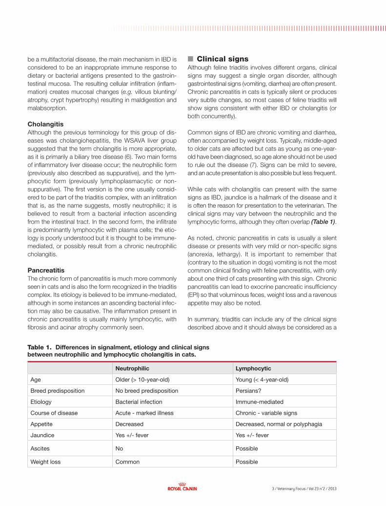
• fPLIfPLI is a much more sensitive and specific marker for pan-creatitis than the previously used fTLI. A quantitative test is now widely available from many laboratories. A semi-quantitative commercial benchtop in-house version (Figure 1) is also available and has shown good correlation with the laboratory test. While the sensitivity of the test in general (i.e. its ability to detect the disease) is excellent (100%) in severe to moderate forms of pancreatitis, it is useful to
Figure 1. The semi-quantitative in-house benchtop test has a very good correlation to the quantitative laboratory fPLI blood test.
5 / Veterinary Focus / Vol 23 n°2 / 2013
l I I I I I I I I I I I I I I l I I I I I I I I I I I I I I l I I I I I I I I I I I I I I l I I I I I I I I I I I I I l I I I I I I I I I I I I I I l I I I I I I I I I I I I I I l I I I I I I I I I I I I I I l I I I I I I I I I I I I I I I I I I I I I I l I I I I I I I I I I I I I I l I I I I I I I I I I I I I I l I I I I I I I I I I I I I l I©
Ani
mal
Hea
lth T
rust
© A
nim
al H
ealth
Tru
st
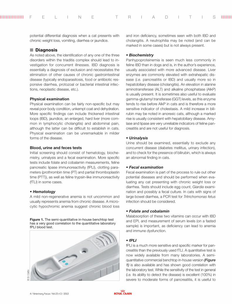
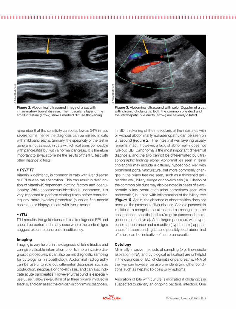
In IBD, thickening of the muscularis of the intestines with or without abdominal lymphadenopathy can be seen on ultrasound (Figure 2). The intestinal wall layering usually remains intact. However, a lack of abnormality does not rule out IBD. Lymphoma is the most important differential diagnosis, and the two cannot be differentiated by ultra-sonographic findings alone. Abnormalities seen in feline cholangitis may include a diffusely hypoechoic liver with prominent portal vasculature, but more commonly chan-ges in the biliary tree are seen, such as a thickened gall-bladder wall, biliary sludge or cholelithiasis (8). Dilation of the common bile duct may also be noted in cases of extra-hepatic biliary obstruction (also sometimes seen with pancreatitis) but also with inflammation of the biliary tree (Figure 3). Again, the absence of abnormalities does not preclude the presence of liver disease. Chronic pancreatitis is difficult to recognize on ultrasound as changes can be absent or non-specific (nodular/irregular pancreas, hetero-geneous parenchyma). An enlarged pancreas, with hypo-echoic appearance and a reactive (hyperechoic) appear-ance of the surrounding fat, and possibly focal abdominal effusion, can be indicative of acute pancreatitis.
CytologyMinimally invasive methods of sampling (e.g. fine-needle aspiration (FNA) and cytological evaluation) are unhelpful in the diagnosis of IBD, cholangitis or pancreatitis. FNA of the liver can however be useful in identifying other condi-tions such as hepatic lipidosis or lymphoma.
Aspiration of bile with culture is indicated if cholangitis is suspected to identify an ongoing bacterial infection. One
remember that the sensitivity can be as low as 54% in less severe forms, hence the diagnosis can be missed in cats with mild pancreatitis. Similarly, the specificity of the test in general is not as good in cats with clinical signs compatible with pancreatitis but with a normal pancreas. It is therefore important to always correlate the results of the fPLI test with other diagnostic tests.
• PT/PTTVitamin K deficiency is common in cats with liver disease or EPI due to malabsorption. This can result in dysfunc-tion of vitamin-K dependent clotting factors and coagu-lopathy. While spontaneous bleeding is uncommon, it is very important to perform clotting times before consider-ing any more invasive procedure (such as fine-needle aspiration or biopsy) in cats with liver disease.
• fTLIfTLI remains the gold standard test to diagnose EPI and should be performed in any case where the clinical signs suggest exocrine pancreatic insufficiency.
ImagingImaging is very helpful in the diagnosis of feline triaditis and can give valuable information prior to more invasive dia-gnostic procedures; it can also permit diagnostic sampling for cytology or histopathology. Abdominal radiography can be useful to rule out differential diagnoses such as obstruction, neoplasia or cholelithiasis, and can also indi-cate acute pancreatitis. However ultrasound is especially useful, as it allows evaluation of all three organs involved in triaditis, and can assist the clinician in confirming diagnosis.
Figure 2. Abdominal ultrasound image of a cat with inflammatory bowel disease. The muscularis layer of the small intestine (arrow) shows marked diffuse thickening.
Figure 3. Abdominal ultrasound with color Doppler of a cat with chronic cholangitis. Both the common bile duct and the intrahepatic bile ducts (arrow) are severely dilated.

6 / Veterinary Focus / Vol 23 n°2 / 2013
l I I I I I I I I I I I I I I l I I I I I I I I I I I I I I l I I I I I I I I I I I I I I l I I I I I I I I I I I I I l I I I I I I I I I I I I I I l I I I I I I I I I I I I I I l I I I I I I I I I I I I I I l I I I I I I I I I I I I I I I I I I I I I I l I I I I I I I I I I I I I I l I I I I I I I I I I I I I I l I I I I I I I I I I I I I l Il I I I I I I I I I I I I I I l I I I I
© D
r. I C
attin
PULSE OX IMETRY AND CAPNOGRAPHY IN EMERGENCY AND INTENSIVE CAREF E L I N E T R I A D I T I S
study demonstrated that bile culture resulted in a much higher chance of positive results than liver culture for the same patient, so bile culture is preferred over liver culture whenever possible (9).
HistopathologyFor all three conditions that make up feline triaditis a definitive diagnosis can only be confirmed histopatho-logically. Different ways of collecting samples are avail-able, and the clinician should be aware of their advan-tages and limitations (Table 2).
Typically the inflammatory pattern seen in IBD is lympho-plasmacytic; however a granulomatous or eosinophilic form can also be found. A suppurative inflammation is seen in cases with an infectious etiology. In the first type of inflammatory infiltrate, differentiation from lymphoma can be difficult, and usually more advanced diagnostic tests such as immunohistochemistry are needed.
Microscopic evaluation of liver samples is useful to make the diagnosis of cholangitis. Although often possible, a clear differentiation between the neutrophilic and lympho-cytic form is sometimes not easy where a mixed inflam-matory pattern is seen (Figure 4).
The diagnosis of pancreatitis is made via histopathology, but the clinical significance of the changes seen is not always clear and caution should be taken when interpreting the findings.
■ TreatmentSupportive care is initially needed for cats with more
severe signs, including intravenous fluid therapy, analgesia, anti-emetics, antiacids, and correction of electrolyte abnormalities. More specific treatments are aimed at controlling each of the three conditions of the triaditis complex (Table 3).
DietEnteral feeding support (i.e. via naso-esophageal or eso-phagostomy tube) is necessary in cats with intractable anorexia to prevent development of hepatic lipidosis. A
Table 2. Advantages and disadvantages of different sampling methods for evaluation of triaditis.
Advantages Disadvantages
EndoscopyLow risk. Visualization of the mucosa, direct sampling. Often sufficient samples for evaluation of IBD. Treatment can be initiated rapidly.
Mucosal samples only. Gastric and duodenal samples only (+/- ileum).
LaparoscopyLow risk. Also allows access to liver and pancreas.
Experience and equipment needed. Risks associated with full-thickness biopsy (dehiscence/peritonitis). May delay treatment (steroids).
Surgery Can inspect all organs. Full thickness biopsies.Increased risks. Risks associated with full-thickness biopsy (dehiscence/peritonitis). May delay treatment (steroids).
Figure 4. Histopathology of a cat’s liver with lymphocytic cholangitis. Note the marked infiltration of small lymphocytes in the portal area and concurrent biliary proliferation.
7 / Veterinary Focus / Vol 23 n°2 / 2013
l I I I I I I I I I I I I I I l I I I I I I I I I I I I I I l I I I I I I I I I I I I I I l I I I I I I I I I I I I I l I I I I I I I I I I I I I I l I I I I I I I I I I I I I I l I I I I I I I I I I I I I I l I I I I I I I I I I I I I I I I I I I I I I l I I I I I I I I I I I I I I l I I I I I I I I I I I I I I l I I I I I I I I I I I I I l I
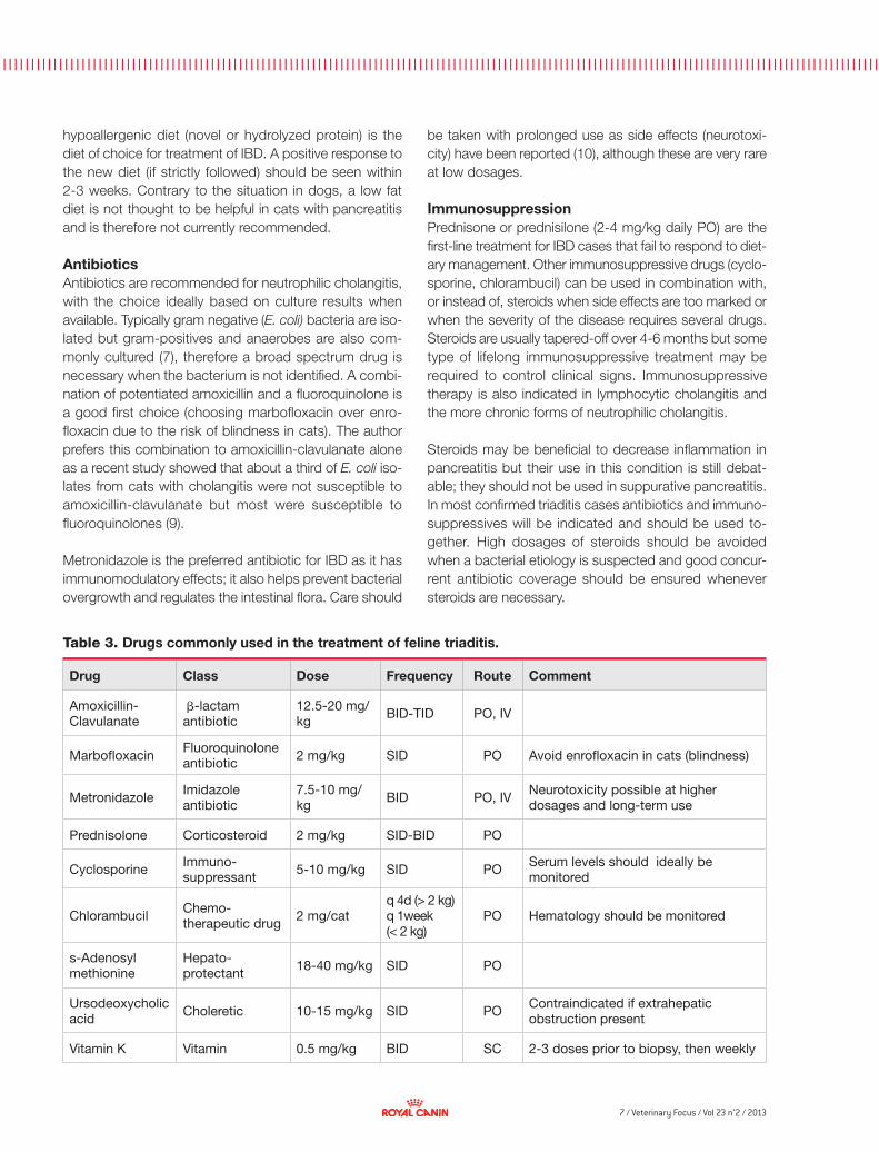
Table 3. Drugs commonly used in the treatment of feline triaditis.
Drug Class Dose Frequency Route Comment
Amoxicillin-Clavulanate
βb-lactam antibiotic
12.5-20 mg/kg BID-TID PO, IV
Marbofloxacin Fluoroquinolone antibiotic 2 mg/kg SID PO Avoid enrofloxacin in cats (blindness)
Metronidazole Imidazole antibiotic
7.5-10 mg/kg BID PO, IV Neurotoxicity possible at higher
dosages and long-term use
Prednisolone Corticosteroid 2 mg/kg SID-BID PO
Cyclosporine Immuno-suppressant 5-10 mg/kg SID PO Serum levels should ideally be
monitored
Chlorambucil Chemo-therapeutic drug 2 mg/cat
q 4d (> 2 kg)q 1week (< 2 kg)
PO Hematology should be monitored
s-Adenosyl methionine
Hepato-protectant 18-40 mg/kg SID PO
Ursodeoxycholic acid Choleretic 10-15 mg/kg SID PO Contraindicated if extrahepatic
obstruction present
Vitamin K Vitamin 0.5 mg/kg BID SC 2-3 doses prior to biopsy, then weekly
hypoallergenic diet (novel or hydrolyzed protein) is the diet of choice for treatment of IBD. A positive response to the new diet (if strictly followed) should be seen within 2-3 weeks. Contrary to the situation in dogs, a low fat diet is not thought to be helpful in cats with pancreatitis and is therefore not currently recommended.
AntibioticsAntibiotics are recommended for neutrophilic cholangitis, with the choice ideally based on culture results when available. Typically gram negative (E. coli) bacteria are iso-lated but gram-positives and anaerobes are also com-monly cultured (7), therefore a broad spectrum drug is necessary when the bacterium is not identified. A combi-nation of potentiated amoxicillin and a fluoroquinolone is a good first choice (choosing marbofloxacin over enro-floxacin due to the risk of blindness in cats). The author prefers this combination to amoxicillin-clavulanate alone as a recent study showed that about a third of E. coli iso-lates from cats with cholangitis were not susceptible to amoxicillin-clavulanate but most were susceptible to fluoroquinolones (9).
Metronidazole is the preferred antibiotic for IBD as it has immunomodulatory effects; it also helps prevent bacterial overgrowth and regulates the intestinal flora. Care should
be taken with prolonged use as side effects (neurotoxi-city) have been reported (10), although these are very rare at low dosages.
ImmunosuppressionPrednisone or prednisilone (2-4 mg/kg daily PO) are the first-line treatment for IBD cases that fail to respond to diet-ary management. Other immunosuppressive drugs (cyclo-sporine, chlorambucil) can be used in combination with, or instead of, steroids when side effects are too marked or when the severity of the disease requires several drugs. Steroids are usually tapered-off over 4-6 months but some type of lifelong immunosuppressive treatment may be required to control clinical signs. Immunosuppressive therapy is also indicated in lymphocytic cholangitis and the more chronic forms of neutrophilic cholangitis.
Steroids may be beneficial to decrease inflammation in pancreatitis but their use in this condition is still debat-able; they should not be used in suppurative pancreatitis. In most confirmed triaditis cases antibiotics and immuno-suppressives will be indicated and should be used to-gether. High dosages of steroids should be avoided when a bacterial etiology is suspected and good concur-rent antibiotic coverage should be ensured whenever steroids are necessary.
8 / Veterinary Focus / Vol 23 n°2 / 2013
l I I I I I I I I I I I I I I l I I I I
1. Hirsch VM, Doige CE. Suppurative cholangitis in cats. J Am Vet Med Assoc 1983;182:1223-1226.
2. Kelly DF, Baggott DG, Gaskell CJ. Jaundice in the cat associated with inflammation of the biliary tract and pancreas. J Small Anim Pract 1975;16:163-172.
3. Center SA, Rowland PH. The cholangitis/cholangiohepatitis complex in the cat. In Proceedings. 12th Am Col Vet Intern Med 1994;766-771.
4. Zawie DA, Garvey MS. Feline hepatic disease. Vet Clin North Am Small Anim Pract 1984;2:1201-1230.
5. Johnston KL, Shift NC, Forster-van Hijfte M, et al. Comparison of the bacterial flora of the duodenum in healthy cats and cats with signs of gastrointestinal tract disease. J Am Vet Med Assoc 2001;218:48-51.
6. Van den Ingh TSGAM, Cullen JM, Twedt DC, et al. Morphological classification of biliary disorders of the canine and feline liver. In: WSAVA Liver
Standardization Group; Standards for Clinical and Histological Diagnosis of Canine and Feline Liver Diseases. Saunders 2006;61-76.
7. Dennis JS, Kruger JM, Mullaney TP. Lymphocytic/plasmacytic gastroenteritis in cats: 14 cases (1985-1990). J Am Vet Med Assoc 1992;200:1712-1718.
8. Newell SM, Selcer BA, Girard E, et al. Correlations between ultrasonographic findings and specific hepatic diseases in cats: 72 cases (1985-1997). J Am Vet Med Assoc 1998;213:94-98.
9. Wagner KA, Hartmann FA, Trepanier LA. Bacterial culture results from liver, gallbladder, or bile in 248 dogs and cats evaluated for hepatobiliary disease: 1998-2003. J Vet Intern Med 2007;21:417-424.
10. Caylor KB, Cassimatis MK. Metronidazole neurotoxicosis in two cats. J Am Anim Hosp Assoc 2001;37(3):258-62.
11. http://vetmed.tamu.edu/gilab/research/cobalamin-information#dosing. Accessed 9th Nov 2012.
F E L I N E T R I A D I T I S
Other treatmentsFolate and cobalamin should be supplemented when deficiency is diagnosed; supplementation is usually tem-porary until the disease is controlled. Guidelines for sup-plementation of cobalamin in dogs and cats have been proposed (11). Liver support medications and cholere-tics (s-adenosyl methionine, ursodeoxycholic acid (UDA)) can be useful for cholangitis and are recommended whenever administration is possible. UDA is contraindi-cated in cases of extrahepatic biliary obstruction. Pan-creatic enzyme supplementation is sometimes of benefit for pancreatitis with concurrent EPI and should be con-sidered in cases refractory to other treatments. Vitamin K
should be supplemented if clotting abnormalities are demonstrated and is especially important prior to any biopsy.
■ ConclusionFeline triaditis is a complex disease and should always be considered in cats with clinical signs suggestive of any of the three conditions, or in cats diagnosed with either IBD, cholangitis or pancreatitis. Treatment consists of address-ing each of the conditions and knowledge of the individ-ual pathophysiology of the three diseases is mandatory. Prognosis is usually good but some patients remain refrac-tory to treatment or can relapse.
References
9 / Veterinary Focus / Vol 23 n°2 / 2013
■
■
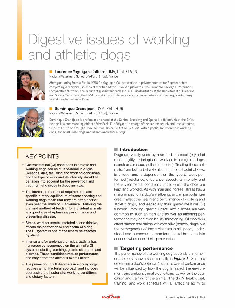
Digestive issues of working and athletic dogs
Laurence Yaguiyan-Colliard, DMV, Dipl. ECVCN National Veterinary School of Alfort (ENVA), France
After graduating from Alfort in 1998 Dr. Yaguiyan-Colliard worked in private practice for 5 years before completing a residency in clinical nutrition at the ENVA. A diplomate of the European College of Veterinary Comparative Nutrition, she is currently assistant professor in Clinical Nutrition at the Department of Breeding and Sports Medicine at the ENVA. She also sees referral cases in clinical nutrition at the Frégis Veterinary Hospital in Arcueil, near Paris.
Dominique Grandjean, DVM, PhD, HDRNational Veterinary School of Alfort (ENVA), France
Dominique Grandjean is professor and head of the Canine Breeding and Sports Medicine Unit at the ENVA. He also is a commanding officer of the Paris Fire Brigade, in charge of the canine search and rescue teams. Since 1981 he has taught Small Animal Clinical Nutrition in Alfort, with a particular interest in working dogs, especially sled dogs and search and rescue dogs.
■ IntroductionDogs are widely used by man for both sport (e.g. sled races, agility, skijoring) and work activities (guide dogs, search and rescue, police units, etc.). Treating these ani-mals, from both a behavioral and nutritional point of view, is unique, and is dependent on the type of work per-formed (resistance, endurance, speed), its intensity, and the environmental conditions under which the dogs are kept and worked. As with man and horses, stress has a major impact on a dog's wellbeing, and in particular can greatly affect the health and performance of working and athletic dogs, and especially their gastrointestinal (GI) function. Vomiting, gastric ulcers, and diarrhea are very common in such animals and as well as affecting per-formance they can even be life-threatening. GI disorders affect human and animal athletes alike (horses, dogs) but the pathogenesis of these diseases is still poorly under-stood and numerous parameters should be taken into account when considering prevention.
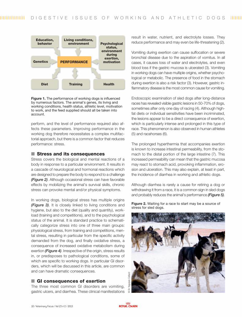
■ Targeting performanceThe performance of the working dog depends on numer-ous factors, shown schematically in Figure 1. Genetics determine a dog's potential (1), but its overall performance will be influenced by how the dog is reared, the environ-ment, and ambient climatic conditions, as well as the edu-cation and training of the animal. The dog's health, diet, training, and work schedule will all affect its ability to
KEY POINTS• Gastrointestinal(GI)conditionsinathleticand
workingdogscanbemultifactorialinorigin.Genetics,diet,thelivingandworkingconditions,andthetypeofworkanditsintensityshouldallbetakenintoaccountforthepreventionandtreatmentofdiseaseintheseanimals.
• TheincreasednutritionalrequirementsandspecificdietaryequilibriumofsomesportingandworkingdogsmeanthattheyareoftennearorevenpastthelimitsofGItolerance.Tailoringthedietandmethodoffeedingforindividualanimalsisagoodwayofoptimizingperformanceandpreventingdisease.
• Stress,whethermental,metabolic,oroxidative,affectstheperformanceandhealthofadog.TheGIsystemisoneofthefirsttobeaffectedbystress.
• Intenseand/orprolongedphysicalactivityhasnumerousconsequencesontheanimal'sGIsystemincludingvomiting,gastriculcerationanddiarrhea.Theseconditionsreduceperformanceandmayaffecttheanimal'soverallhealth.
• ThepreventionofGIdisordersinworkingdogsrequiresamultifactorialapproachandincludesaddressingthehusbandry,workingconditionsanddietaryfactors.
10 / Veterinary Focus / Vol 23 n°2 / 2013
l I I I I I I I I I I I I I I l I I I I l I I I I I I I I I I I I I I l I I I I I I I I I I I I I I l I I I I I I I I I I I I I I l I I I I I I I I I I I I I l I I I I I I I I I I I I I I l I I I I I I I I I I I I I I l I I I I I I I I I I I I I I l I I I I I I I I I I I I I I I I I I I I I I l I I I I I I I I I I I I I I l I I I I I I I I I I I I I I l I I I I I I I I I I I I I l I
© D
r. Ya
guiy
an-C
ollia
rd
D I G E S T I V E I S S U E S O F W O R K I N G A N D A T H L E T I C D O G S
perform, and the level of performance required also af-fects these parameters. Improving performance in the working dog therefore necessitates a complex multifac-torial approach, but there is a common factor that reduces performance: stress.
■ Stress and its consequences Stress covers the biological and mental reactions of a body in response to a particular environment. It results in a cascade of neurological and hormonal reactions which are designed to prepare the body to respond to a challenge (Figure 2). Although occasional stress can have favorable effects by mobilizing the animal's survival skills, chronic stress can provoke mental and/or physical symptoms.
In working dogs, biological stress has multiple origins (Figure 3). It is closely linked to living conditions and hygiene, but also to the diet (quality and quantity), work-load (training and competitions), and to the psychological status of the animal. It is standard practice to schemati-cally categorize stress into one of three main groups: physiological stress, from training and competitions, men-tal stress, resulting in particular from the specific activity demanded from the dog, and finally oxidative stress, a consequence of increased oxidative metabolism during exertion (Figure 4). Irrespective of the origin, stress results in, or predisposes to pathological conditions, some of which are specific to working dogs. In particular GI disor-ders, which will be discussed in this article, are common and can have dramatic consequences.
■ GI consequences of exertion The three most common GI disorders are vomiting, gastric ulcers, and diarrhea. These clinical manifestations
result in water, nutrient, and electrolyte losses. They reduce performance and may even be life-threatening (2).
Vomiting during exertion can cause suffocation or severe bronchial disease due to the aspiration of vomitus. In all cases, it causes loss of water and electrolytes, and even blood loss if the gastric mucosa is ulcerated (3). Vomiting in working dogs can have multiple origins, whether psycho-logical or metabolic. The presence of food in the stomach during exertion is also a risk factor (3). However, gastric in-flammatory disease is the most common cause for vomiting.
Endoscopic examination of sled dogs after long-distance races has revealed visible gastric lesions in 50-70% of dogs, sometimes after only one day of racing (4). Although high-fat diets or individual sensitivities have been incriminated, the lesions appear to be a direct consequence of exertion, which is particularly intense and prolonged in this type of race. This phenomenon is also observed in human athletes (5) and racehorses (6).
The prolonged hyperthermia that accompanies exertion is known to increase intestinal permeability, from the sto-mach to the distal portion of the large intestine (7). This increased permeability can mean that the gastric mucosa may react to stomach acid, provoking inflammation, ero-sion and ulceration. This may also explain, at least in part, the incidence of diarrhea in working and athletic dogs.
Although diarrhea is rarely a cause for retiring a dog or withdrawing it from a race, it is a common sign in sled dogs and probably reduces the animal's performance (Figure 5).
Figure 2. Waiting for a race to start may be a source of stress for sled dogs.
Genetics
Diet Health
Psychological status,
environment during
exertion, motivation
Education, behavior
Living conditions, environment
Training
Figure 1. The performance of working dogs is influenced by numerous factors. The animal's genes, its living and working conditions, health status, athletic level, motivation to work, and the feed supplied should all be taken into account.
PERFORMANCE

11 / Veterinary Focus / Vol 23 n°2 / 2013
l I I I I I I I I I I I I I I l I I I I I I I I I I I I I I l I I I I I I I I I I I I I I l I I I I I I I I I I I I I l I I I I I I I I I I I I I I l I I I I I I I I I I I I I I l I I I I I I I I I I I I I I l I I I I I I I I I I I I I I I I I I I I I I l I I I I I I I I I I I I I I l I I I I I I I I I I I I I I l I I I I I I I I I I I I I l I
Other than parasitic and infectious problems, (which will not be covered here), the diet can be a source of GI dis-orders. Especially for dogs that work in extreme condi-tions and for prolonged periods (e.g. sled dogs, search and rescue dogs), a very high quality diet is required to provide the necessary energy. For example, the mainte-nance energy requirement (MER) for a Siberian husky weighing 25 kg in a temperate region is around 1,200 kcal of metabolizable energy (ME)/day, whereas a dog of the same breed participating in the 1,600 km Yukon Quest sled race at an external temperature of between -20 and -50°C will require over 9,500 kcal ME/day (8). To cover these requirements, and to ensure a sufficient energy supply to the cells during prolonged exertion, diet rations for sled dogs are particularly rich in lipids (9). This high-fat content means that the dog's digestive capacities may be exceed-ed, resulting in maldigestion and malabsorption. The undi-gested particles are fermented or putrefied by colonic bac-teria. As well as disrupting the normal bacterial flora, these degradation products cause inflammation of the intestinal mucosa and an osmotic effect that causes liquefaction of the stools. The involvement of pathogenic GI microbes such as Clostridium and Salmonella does not, by itself, explain the prevalence of diarrhea in sled dogs (10).
Although the pathogenesis of GI lesions in both human and non-human athletes is still poorly understood, it would seem that reduction in splanchnic blood flow has an important role (11) and the effects on the GI tract can last well beyond the actual period of exertion, since reperfusion of the tract after ischemia can itself cause vasomotor and inflammatory disorders. Other causes have been suggested, although they have yet to be proven: ischemia of the GI mucosa during exertion, intestinal dys-biosis, or simply the mechanical effect of the intestinal contents on the mucosa and peristalsis, known as the "cecal slap syndrome" (12). These phenomena contribute to the development of oxidative stress.
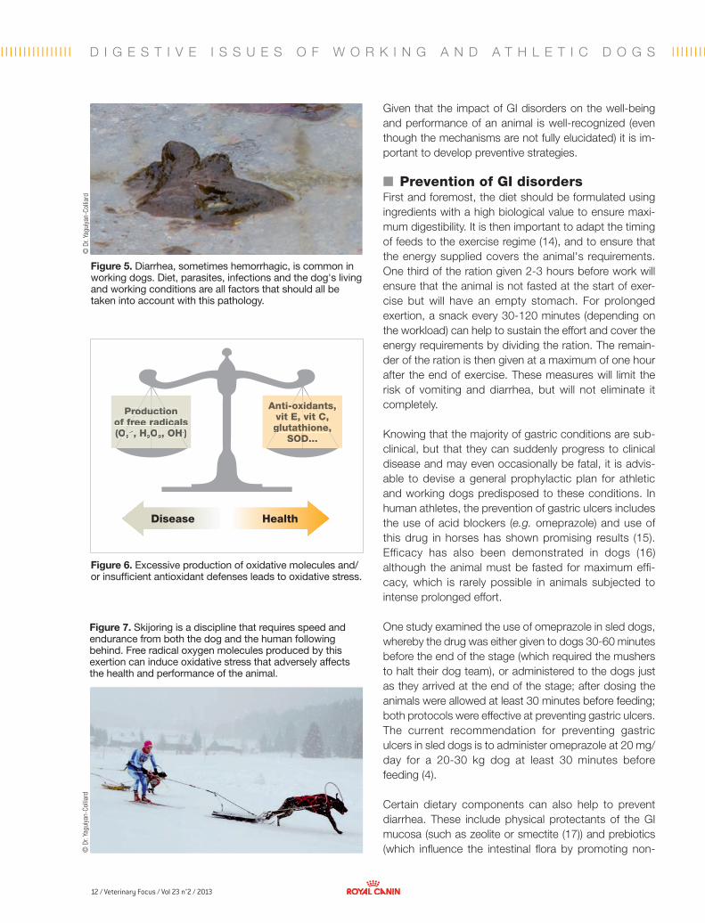
Oxidative stress is defined as an imbalance between the production of reactive molecules (free radicals, and oxy-gen, nitrogen, or chlorine ions) and the body's defenses (Figure 6). It is not a disease in itself but a pathophysio-logical mechanism that promotes disease or is respon-sible for accelerated ageing of the body. Factors such as the environment (stress, temperature, pollutants, etc.), ischemia/reperfusion syndrome, injuries, organ disease (causing inflammation, ulceration and/or necrosis), and oxidative metabolism (exertion) can all lead to the production of oxidative molecules in the body. These induce various molecular modifications on saturated and unsaturated fatty
acids, pigments, amino acids, proteins, and even nucleic acids. These modifications directly affect the cells’ integrity, leading to cell death. Physical exercise induces the pro-duction of reactive oxygen ions; the longer and more intense the exercise, the greater the production (Figure 7). The body has methods to neutralize oxidative molecules, including an enzymatic system (superoxide dismutase (SOD), glutathione peroxidase, etc.) and non-enzymatic chemical methods (albumin, vitamin C, vitamin E, carote-noids, etc.). Following repeated, intensive, or prolonged physical exercise, the antioxidant capacities of the body can be insufficient, resulting in inflammatory lesions or damage to vital organs. Oxidative stress has been demon-strated in working and athletic dogs (13).
Figure 3. There are multiple sources of stress, from both a physiological and environmental point of view. It is advisable to assess the impact of each parameter to reduce the animal's overall stress.
Genetics
Training intensity and frequency of exertion
Diet Parasites and infections
STRESSLiving
conditions
Psychological status
Figure 4. Schematically, stress has 3 main origins: metabolic (following exertion), mental (depending on the environment and the working conditions), and oxidative. Stress, irrespective of its origin, causes pathological conditions, some of which are specific to working and athletic dogs. GI repercussions are the most common and can be dramatic.
Gastrointestinal conditions
Specific pathological conditions
Exertion Environment Oxidative metabolism
Metabolic stress Mental stress Oxidative
cellular stress
12 / Veterinary Focus / Vol 23 n°2 / 2013
l I I I I I I I I I I I I I I l I I I I I I I I I I I I I I l I I I I I I I I I I I I I I l I I I I I I I I I I I I I l I I I I I I I I I I I I I I l I I I I I I I I I I I I I I l I I I I I I I I I I I I I I l I I I I I I I I I I I I I I I I I I I I I I l I I I I I I I I I I I I I I l I I I I I I I I I I I I I I l I I I I I I I I I I I I I l Il I I I I I I I I I I I I I I l I I I I
© D
r. Ya
guiy
an-C
ollia
rd©
Dr.
Yagu
iyan
-Col
liard
D I G E S T I V E I S S U E S O F W O R K I N G A N D A T H L E T I C D O G S
Given that the impact of GI disorders on the well-being and performance of an animal is well-recognized (even though the mechanisms are not fully elucidated) it is im-portant to develop preventive strategies.
■ Prevention of GI disordersFirst and foremost, the diet should be formulated using ingredients with a high biological value to ensure maxi-mum digestibility. It is then important to adapt the timing of feeds to the exercise regime (14), and to ensure that the energy supplied covers the animal's requirements. One third of the ration given 2-3 hours before work will ensure that the animal is not fasted at the start of exer-cise but will have an empty stomach. For prolonged exertion, a snack every 30-120 minutes (depending on the workload) can help to sustain the effort and cover the energy requirements by dividing the ration. The remain-der of the ration is then given at a maximum of one hour after the end of exercise. These measures will limit the risk of vomiting and diarrhea, but will not eliminate it completely.
Knowing that the majority of gastric conditions are sub-clinical, but that they can suddenly progress to clinical disease and may even occasionally be fatal, it is advis-able to devise a general prophylactic plan for athletic and working dogs predisposed to these conditions. In human athletes, the prevention of gastric ulcers includes the use of acid blockers (e.g. omeprazole) and use of this drug in horses has shown promising results (15). Efficacy has also been demonstrated in dogs (16) although the animal must be fasted for maximum effi-cacy, which is rarely possible in animals subjected to intense prolonged effort.
One study examined the use of omeprazole in sled dogs, whereby the drug was either given to dogs 30-60 minutes before the end of the stage (which required the mushers to halt their dog team), or administered to the dogs just as they arrived at the end of the stage; after dosing the animals were allowed at least 30 minutes before feeding; both protocols were effective at preventing gastric ulcers. The current recommendation for preventing gastric ulcers in sled dogs is to administer omeprazole at 20 mg/day for a 20-30 kg dog at least 30 minutes before feeding (4).
Certain dietary components can also help to prevent diarrhea. These include physical protectants of the GI mucosa (such as zeolite or smectite (17)) and prebiotics (which influence the intestinal flora by promoting non-
Figure 5. Diarrhea, sometimes hemorrhagic, is common in working dogs. Diet, parasites, infections and the dog's living and working conditions are all factors that should all be taken into account with this pathology.
Figure 7. Skijoring is a discipline that requires speed and endurance from both the dog and the human following behind. Free radical oxygen molecules produced by this exertion can induce oxidative stress that adversely affects the health and performance of the animal.
Figure 6. Excessive production of oxidative molecules and/or insufficient antioxidant defenses leads to oxidative stress.
HealthDisease
Production Production of free radicals of free radicals (O(O2
.-, H2OO2, OH, OH.)
Anti-oxidants, Anti-oxidants, vit E, vit C, vit E, vit C, glutathione, glutathione,
SOD...SOD...
13 / Veterinary Focus / Vol 23 n°2 / 2013
l I I I I I I I I I I I I I I l I I I I I I I I I I I I I I l I I I I I I I I I I I I I I l I I I I I I I I I I I I I l I I I I I I I I I I I I I I l I I I I I I I I I I I I I I l I I I I I I I I I I I I I I l I I I I I I I I I I I I I I I I I I I I I I l I I I I I I I I I I I I I I l I I I I I I I I I I I I I I l I I I I I I I I I I I I I l I
1. Huson HJ, Ostrander EA, Ruvinsky A. Genetic aspects of performance in working dogs. In The genetics of the dog, eds. Ostrander EA and Ruvinsky A, 2nd Ed: Oxford, CABI Publishing, 2012; 477-484.
2 Dennis MM, Nelson SN, Cantor GH, et al. Assessment of necropsy findings in sled dogs that died during Iditarod Trail sled dog races: 23 cases (1994-2006). J Am Vet Med Assoc 2008;232:564-573.
3. Davis MS, Willard MD, Nelson SL, et al. Prevalence of gastric lesions in racing Alaskan sled dogs. J Vet Intern Med 2003;17:311-314.
4. Davis MS. Gastritis/gastric ulcers in canine athletes. In Proceedings, ISDVMA 11th Biennial Meeting, Banff 2012;54-56.
5. Michel H, Larrey D, Blanc P. Hepato-digestive disorders in athletic practice [in French]. Presse Med 1994;23:479-484.
6. Murray MJ, Schusser GF, Pipers FS, et al. Factors associated with gastric lesions in thoroughbred racehorses. Equ Vet J 1996;28:368-374.
7. Davis MS, Willard M, Williamson K, et al. Temporal relationship between gastrointestinal protein loss, gastric ulceration or erosion, and strenuous exercise in racing Alaskan dogs. J Vet Intern Med 2006;20:835-839.
8. Yazwinski M. Assessment of serum myokines and markers of inflammation associated with exercice in sled dogs; and dietary analysis and kilocalories fed during the Yukon Quest. In Proceedings, ISDVMA 11th Biennial Meeting, Banff 2012;51-53.
9. Reynolds AJ, Fuhrer L, Dunlap HL, et al. Lipid metabolite responses to diet and training in sled dogs. J Nutr 1994;124:2754S-2759S.
10. MacKenzie E, Riehl J, Banse H, et al. Prevalence of diarrhea and enteropathogens in racing sled dogs. J Vet Intern Med 2010;24:97-103.
11. Steege RWFT and Kolkman JJK. Review article: the physiopathology and
management of gastrointestinal symptoms during physical exercise, and the role of splanchnic blood flow. Aliment Pharmal Ther 2012;35(5):516-28.
12. Sanchez LD, Tracy JA, Berkoff D, et al. Ischemic colitis in marathon runners: A case-based review. J Emerg Med 2006;30:321-326.
13. Baskin CR, Hinchcliff KW, DiSylvestro RA, et al. Effect of dietary antioxidant supplementation on oxidative damage and resistance to oxidative damage during prolonged exercise in sled dogs. Am J Vet Res 2000;61:886-891.
14. Kronfeld DS and Downey RL. Nutritional strategies for stamina in dogs and horses. Proc Nutr Soc Aust 1981;6:21-29.
15. Andrews FM, Sifferman RL, Bernard W, et al. Efficacy of omeprazole paste in the treatment and prevention of gastric ulcers in horses. Equ Vet J Suppl 1999:29;81-86.
16. Jenkins CC, DeNovo RC, Patton CS, et al. Comparison of effects of cimetidine and omeprazole on mechanically created gastric ulceration and on aspirin-induced gastritis in dogs. Am J Vet Res 1991;52:658-661.
17. Grandjean D, Crépin F, Paragon BM. The interest of smectite in acute diarrhea in sled dogs [in French]. Recueil de Médecine Vétérinaire 1992;168(5):323-329.
18. Swanson KS, Grieshop CM, Flickinger EA, et al. Supplemental fructooligosaccharides and mannanoligosaccharides influence immune function, ileal and total tract nutrient digestibilities, microbial populations and concentrations of protein catabolites in the large bowel of dogs. J Nutr 2002;132(5):980-989.
19. Mickelborough TD. Omega-3 polyunsaturated fatty acids in physical performance optimization. Int J Sport Nutr Exerc Metab (in press).
pathogenic bacteria). Fructooligosaccharides (a prebiotic) and foodstuffs such as sugarbeet pulp (which is rich in pre-biotics) can therefore be included in the daily ration of work-ing dogs. Mannanoligosaccharides (MOS) both help to pre-vent pathogenic bacteria adhering to the intestinal mucosa and stimulate the local production of immunoglobulin A (18). With a more indirect effect on metabolism, fish oils, rich in polyunsaturated fatty acids from the omega-3 family, have a proven action against inflammation and oxidative stress (19). Similarly, the use of antioxidants has been shown to have a beneficial effect on a dog’s performance (13).
■ ConclusionThe use of the dog for sport or work, as with human ath-letes, imposes psychological and physical constraints that the animal must overcome. The ability to respond to stress and overcome it will, of course, depend on the ani-mal's genetic composition and training. However, factors such as the living conditions, preventive healthcare, diet, and warm-up prior to exercise and recovery after exer-cise must all be optimized by the vet and others that care for working dogs to ensure they attain and retain the best possible health status.
References
14 / Veterinary Focus / Vol 23 n°2 / 2013
■
Constipation in the cat Valérie Freiche, DMV, Dipl. ESVClinique Vétérinaire Alliance, Bordeaux, France
Dr. Freiche graduated from the National Veterinary School of Alfort, France in 1988. After an internship in Internal Medicine at Alfort, she was appointed head of the Gastroenterology department and served there for 14 years. She currently practices in Bordeaux where she treats referred cases in internal medicine, with a particular interest in gastroenterology and interventional endoscopy. Co-author of a recently-published gastroenterology textbook, she also presents numerous CPD programs for veterinarians and holds the French specialist diploma in Internal Medicine.
■ IntroductionConstipation is defined as “an absence or reduction in the frequency of defecation” and is much more common in the cat than in the dog; this article offers a comprehensive overview of the diagnosis and management of the condi-tion in the cat. Feline constipation may be caused by a wide range of disorders (and indeed the etiology often differs from the causes of constipation in the dog) which include anatomical, metabolic and functional problems. Prolonged stasis of feces in the colonic segment results in progressive dehydration of fecal matter, which becomes very dry, hard, and difficult to pass (1). Megacolon (defined
KEY POINTS
• Obesity,inactivity,andalow-fiberdietarepredisposingfactorsinfelineconstipation.
• Oneofthemostcommoncausesoffelineconstipationisidiopathicmegacolon.
• Theetiologycanusuallybeestablishedafterobtainingathoroughhistoryandclinicalexamination.Repeateduseoflaxativeswithoutanetiologicaldiagnosisisnotadvisable.
• Furtherinvestigationsshouldalwaysbeginwithradiographsoftheabdomenandpelvis.
• Inthemajorityofcases,goodnutritionalmanagementandsupplementationwithpsylliumwillhelppreventtheneedforrepeatedcoloniclavagesorsurgery,evenwithmegacolon.
as “a generalized distension of the colon combined with loss of motility”) is also often present and can be primary in origin or secondary to recurrent episodes of fecal retention from varying causes. There are two main reasons for a cat to be presented at consultation:
• A cat which presents with recurrent, chronic constipa-tion, resulting in the intermittent emission of small, dry stools. The animal’s health status is typically good but the cat regularly requires brief hospitalization to empty the colon under sedation.
• A cat which presents as an emergency such that hospi-talization and immediate intensive care with fluid therapy, and a rapid etiological diagnosis, are required.
Dyschezia (tenesmus with difficulty in passing feces) may also be present, as can obstipation (fecal impaction which prevents defecation). Before going further a brief physiological and etiological overview is worthwhile.
■ Colon physiologyThe cat's colon has an average length of 30 cm. Its two main physiological functions are:
• The absorption of water and electrolytes from the lumen, which occurs in the proximal colon.
• The storage and periodical elimination of stools, which occurs in the distal colon.
Colonic longitudinal and circular smooth muscles provide motility and tone. This motility is regulated by gastrointes-tinal hormones and the intrinsic and extrinsic colonic ner-vous system. There are two types of colonic movement:
HOW I APPROACH…

15 / Veterinary Focus / Vol 23 n°2 / 2013
l I I I I I I I I I I I I I I l I I I I I I I I I I I I I I l I I I I I I I I I I I I I I l I I I I I I I I I I I I I l I I I I I I I I I I I I I I l I I I I I I I I I I I I I I l I I I I I I I I I I I I I I l I I I I I I I I I I I I I I I I I I I I I I l I I I I I I I I I I I I I I l I I I I I I I I I I I I I I l I I I I I I I I I I I I I l I I I I I I I I I I I I I I l I I I I I I I I I I
© D
r. V.
Fre
iche
segmental, rhythmic “stirring” contractions, and peristaltic waves which propel fecal matter caudally.
Although colonic contractions are passive (the sympathe-tic nervous system regulates the segmental contractions, whilst the parasympathetic system generates the peri-staltic contractions), defecation is a voluntary act control-led by the central nervous system.
The bacterial concentration within the colon is very high: 1010 microbes per gram of fecal matter (the distal small intestine has only 104 microbes/g) and is primarily com-posed of anaerobic Enterobacteria, Lactobacilli, and Streptococci that form a balanced ecosystem involved in various enzymatic reactions. By fermenting ingested car-bohydrates and fiber, they promote the production of short-chain fatty acids, water, hydrogen, methane, and CO2. Volatile fatty acids represent the best source of energy for the colonocytes and constitute a substrate for lipid synthesis; their production induces a localized reduction in pH, which reduces the ionization of long-chain fatty acids and bile acids, which are known to be irritant and deleterious to the colonic mucosa. The colonic bacteria also increase the concentration of ammonium ions eliminated in the feces.
The mean physiological transit time from ingestion to elimination varies from 12-24 hours, but this may be pro-longed without adversely affecting the animal.
■ EtiologyThe absence or slowing of fecal progression along the colon, or difficulty in passing feces, can be caused by a variety of lesions. Table 1 summarizes the main causes of intermittent or chronic constipation in the cat; the most common being chronic coprostasis (fecal impaction) and megacolon (congenital, acquired, post-traumatic, or idio-pathic). Obstructive or stenotic endoluminal lesions are rarer; colonic tumors are often obstructive by the time they are diagnosed and typically result in constipation (Figure 1) (1).
Obesity, combined with inactivity or lack of dietary fiber, is a known predisposing factor. During hospitalization, envi-ronmental changes can also cause a temporary and reversible constipation: this is increased by dehydration and hypokalemia, which themselves are commonly found with constipation! Congenital anomalies (e.g. aganglionosis (2) or anal perforation with rectovaginal fis-tula) are rare and the clinical signs become apparent soon after weaning. Bony lesions and lumbosacral or pelvic
neurological disorders can also cause constipation from pain and/or modification of the pelvic canal.
Post-inflammatory rectal stenosis can occur at any age, and may follow acute diarrhea which can damage the anal sphincter.
■ PresentationA cat with sudden onset constipation, in the absence of dietary or environmental changes, warrants a detailed clinical examination. Constipation may be chronic or intermittent and can go unnoticed for a long period if the animal goes outside and does not have a litter tray in the house. The clinical signs may be moderate (e.g. with chronic coprostasis) or severe (e.g. with distal colonic obstruction) and, depending on the causative disease, other clinical signs may be noted, including prostration, vomiting (very common in this species, and in my expe-rience this can be the only presenting sign), weight loss (due to persistent anorexia or obstructive neoplasia), dehydration, anorexia, tenesmus, agitation, expulsion of non-fecal matter (mucous, fresh blood), abdominal pain (1), behavioral abnormalities, abdominal distension, peri-neal deformity, and anal atony.
■ DiagnosisThe following points are key to history-taking and clinical examination:
• History This can be used to identify trigger factors: e.g. ingestion of bones, previous history of pelvic trauma (Figures 2 and 3),
Figure 1. Colonoscopy performed on an 11-year-old female cat presenting with vomiting, anorexia, and tenesmus. An intralumenal, proliferative stenotic lesion can be seen ventrally; histology confirmed the presence of a colorectal carcinoma with a poor prognosis.
16 / Veterinary Focus / Vol 23 n°2 / 2013
l I I I I I I I I I I I I I I l I I I I l I I I I I I I I I I I I I I l I I I I I I I I I I I I I I l I I I I I I I I I I I I I I l I I I I I I I I I I I I I l I I I I I I I I I I I I I I l I I I I I I I I I I I I I I l I I I I I I I I I I I I I I l I I I I I I I I I I I I I I I I I I I I I I l I I I I I I I I I I I I I I l I I I I I I I I I I I I I I l I I I I I I I I I I I I I l IC O N S T I P A T I O N I N T H E C A T
type of diet, altered behavior, dysorexia, locomotor disor-ders (3,4).
Constipation usually affects middle-aged or older cats (4) and environmental factors (number of animals in contact, introduction of a new cat, etc.) can play an important role. Owners often describe seeing the cat attempting to defecate for some time without managing to produce anything, or the cat may defecate outside the tray due to behavioral disturbances caused by the constipation.
• Clinical examinationExamination should be comprehensive and include a thorough assessment of the abdomen: this is not a problem for most cats but obesity can make it hard to identify
the internal organs. Lymph nodes should be carefully evaluated and the anal area should be inspected thoroughly. Palpation of the colon (2) is essential and its diameter should be estimated; is the colon simply filled with compacted feces or is it distended by stools with a diameter greater than that of the pelvic canal?
In cases of coprostasis, or marked impaction, it may be possible to trace the outline of the colon from the ileoceco-colic junction to the rectum. It is important to determine whether the coprostasis is generalized or whether fecal retention is proximal to a specific section of the colon (in which case a localized endoluminal mass or extrinsic com-pressive lesion may be responsible). The thyroids should always be palpated, especially in cats over 8 years of age.
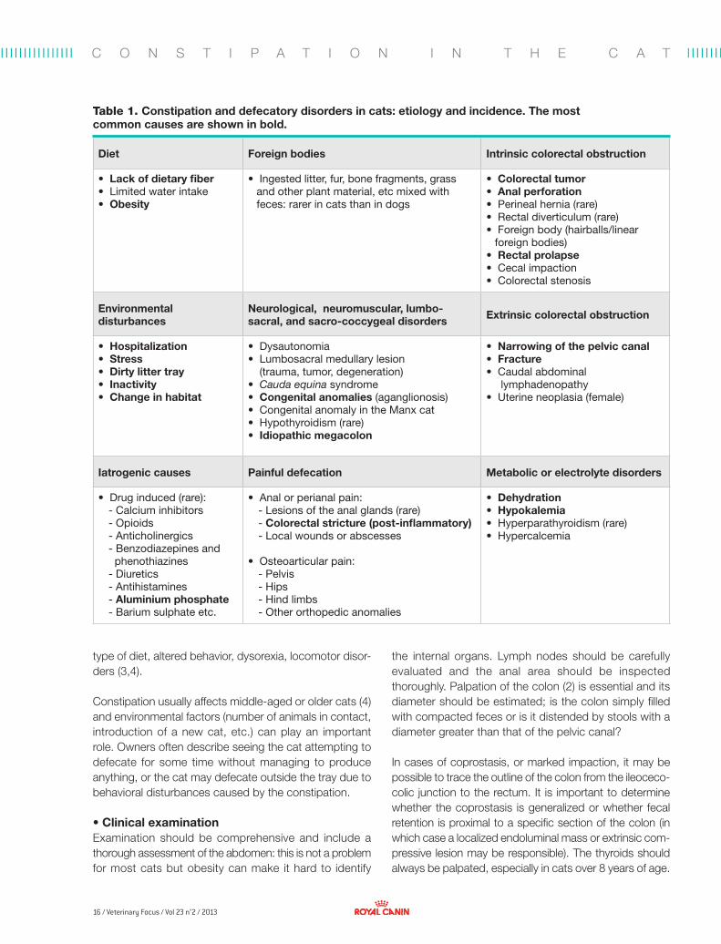
Table 1. Constipation and defecatory disorders in cats: etiology and incidence. The most common causes are shown in bold.
Diet Foreign bodies Intrinsic colorectal obstruction
• Lack of dietary fiber • Limited water intake• Obesity
• Ingested litter, fur, bone fragments, grass and other plant material, etc mixed with feces: rarer in cats than in dogs
• Colorectal tumor• Anal perforation• Perineal hernia (rare)• Rectal diverticulum (rare)• Foreign body (hairballs/linear
foreign bodies)• Rectal prolapse• Cecal impaction• Colorectal stenosis
Environmental disturbances
Neurological, neuromuscular, lumbo-sacral, and sacro-coccygeal disorders Extrinsic colorectal obstruction
• Hospitalization• Stress• Dirty litter tray• Inactivity• Change in habitat
• Dysautonomia• Lumbosacral medullary lesion
(trauma, tumor, degeneration)• Cauda equina syndrome• Congenital anomalies (aganglionosis)• Congenital anomaly in the Manx cat• Hypothyroidism (rare)• Idiopathic megacolon
• Narrowing of the pelvic canal • Fracture• Caudal abdominal
lymphadenopathy• Uterine neoplasia (female)
Iatrogenic causes Painful defecation Metabolic or electrolyte disorders
• Drug induced (rare):- Calcium inhibitors- Opioids- Anticholinergics- Benzodiazepines and phenothiazines- Diuretics- Antihistamines- Aluminium phosphate- Barium sulphate etc.
• Anal or perianal pain:- Lesions of the anal glands (rare)- Colorectal stricture (post-inflammatory)- Local wounds or abscesses
• Osteoarticular pain:- Pelvis- Hips- Hind limbs- Other orthopedic anomalies
• Dehydration• Hypokalemia• Hyperparathyroidism (rare) • Hypercalcemia
17 / Veterinary Focus / Vol 23 n°2 / 2013
l I I I I I I I I I I I I I I l I I I I I I I I I I I I I I l I I I I I I I I I I I I I I l I I I I I I I I I I I I I l I I I I I I I I I I I I I I l I I I I I I I I I I I I I I l I I I I I I I I I I I I I I l I I I I I I I I I I I I I I I I I I I I I I l I I I I I I I I I I I I I I l I I I I I I I I I I I I I I l I I I I I I I I I I I I I l I
3
5
2
4
© D
r. L.
Cou
turie
r AZU
RVET
© D
r. V.
Fre
iche
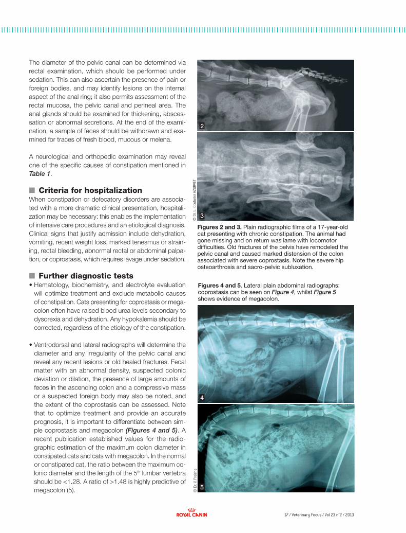
Figures 2 and 3. Plain radiographic films of a 17-year-old cat presenting with chronic constipation. The animal had gone missing and on return was lame with locomotor difficulties. Old fractures of the pelvis have remodeled the pelvic canal and caused marked distension of the colon associated with severe coprostasis. Note the severe hip osteoarthrosis and sacro-pelvic subluxation.
The diameter of the pelvic canal can be determined via rectal examination, which should be performed under sedation. This can also ascertain the presence of pain or foreign bodies, and may identify lesions on the internal aspect of the anal ring; it also permits assessment of the rectal mucosa, the pelvic canal and perineal area. The anal glands should be examined for thickening, absces-sation or abnormal secretions. At the end of the exami-nation, a sample of feces should be withdrawn and exa-mined for traces of fresh blood, mucous or melena.
A neurological and orthopedic examination may reveal one of the specific causes of constipation mentioned in Table 1.
■ Criteria for hospitalizationWhen constipation or defecatory disorders are associa-ted with a more dramatic clinical presentation, hospitali-zation may be necessary: this enables the implementation of intensive care procedures and an etiological diagnosis. Clinical signs that justify admission include dehydration, vomiting, recent weight loss, marked tenesmus or strain-ing, rectal bleeding, abnormal rectal or abdominal palpa-tion, or coprostasis, which requires lavage under sedation.
■ Further diagnostic tests • Hematology, biochemistry, and electrolyte evaluation
will optimize treatment and exclude metabolic causes of constipation. Cats presenting for coprostasis or mega-colon often have raised blood urea levels secondary to dysorexia and dehydration. Any hypokalemia should be corrected, regardless of the etiology of the constipation.
• Ventrodorsal and lateral radiographs will determine the diameter and any irregularity of the pelvic canal and reveal any recent lesions or old healed fractures. Fecal matter with an abnormal density, suspected colonic deviation or dilation, the presence of large amounts of feces in the ascending colon and a compressive mass or a suspected foreign body may also be noted, and the extent of the coprostasis can be assessed. Note that to optimize treatment and provide an accurate prognosis, it is important to differentiate between sim-ple coprostasis and megacolon (Figures 4 and 5). A recent publication established values for the radio-graphic estimation of the maximum colon diameter in constipated cats and cats with megacolon. In the normal or constipated cat, the ratio between the maximum co-lonic diameter and the length of the 5th lumbar vertebra should be <1.28. A ratio of >1.48 is highly predictive of megacolon (5).
Figures 4 and 5. Lateral plain abdominal radiographs: coprostasis can be seen on Figure 4, whilst Figure 5 shows evidence of megacolon.
18 / Veterinary Focus / Vol 23 n°2 / 2013
l I I I I I I I I I I I I I I l I I I I I I I I I I I I I I l I I I I I I I I I I I I I I l I I I I I I I I I I I I I l I I I I I I I I I I I I I I l I I I I I I I I I I I I I I l I I I I I I I I I I I I I I l I I I I I I I I I I I I I I I I I I I I I I l I I I I I I I I I I I I I I l I I I I I I I I I I I I I I l I I I I I I I I I I I I I l Il I I I I I I I I I I I I I I l I I I I
© D
r. V.
Fre
iche
C O N S T I P A T I O N I N T H E C A T
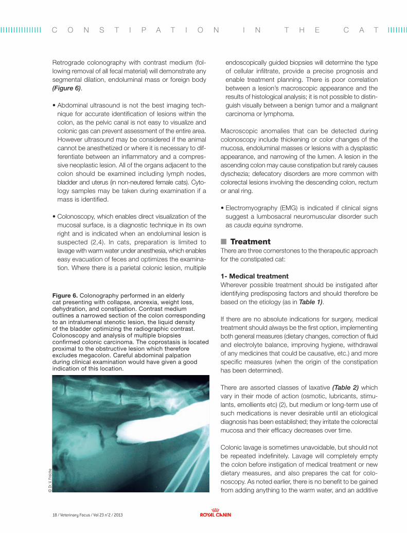
Retrograde colonography with contrast medium (fol-lowing removal of all fecal material) will demonstrate any segmental dilation, endoluminal mass or foreign body (Figure 6).
• Abdominal ultrasound is not the best imaging tech-nique for accurate identification of lesions within the colon, as the pelvic canal is not easy to visualize and colonic gas can prevent assessment of the entire area. However ultrasound may be considered if the animal cannot be anesthetized or where it is necessary to dif-ferentiate between an inflammatory and a compres-sive neoplastic lesion. All of the organs adjacent to the colon should be examined including lymph nodes, bladder and uterus (in non-neutered female cats). Cyto-logy samples may be taken during examination if a mass is identified.
• Colonoscopy, which enables direct visualization of the
mucosal surface, is a diagnostic technique in its own right and is indicated when an endoluminal lesion is suspected (2,4). In cats, preparation is limited to lavage with warm water under anesthesia, which enables easy evacuation of feces and optimizes the examina-tion. Where there is a parietal colonic lesion, multiple
Figure 6. Colonography performed in an elderly cat presenting with collapse, anorexia, weight loss, dehydration, and constipation. Contrast medium outlines a narrowed section of the colon corresponding to an intralumenal stenotic lesion, the liquid density of the bladder optimizing the radiographic contrast. Colonoscopy and analysis of multiple biopsies confirmed colonic carcinoma. The coprostasis is located proximal to the obstructive lesion which therefore excludes megacolon. Careful abdominal palpation during clinical examination would have given a good indication of this location.
endoscopically guided biopsies will determine the type of cellular infiltrate, provide a precise prognosis and enable treatment planning. There is poor correlation between a lesion’s macroscopic appearance and the results of histological analysis; it is not possible to distin-guish visually between a benign tumor and a malignant carcinoma or lymphoma.
Macroscopic anomalies that can be detected during colonoscopy include thickening or color changes of the mucosa, endoluminal masses or lesions with a dysplastic appearance, and narrowing of the lumen. A lesion in the ascending colon may cause constipation but rarely causes dyschezia; defecatory disorders are more common with colorectal lesions involving the descending colon, rectum or anal ring.
• Electromyography (EMG) is indicated if clinical signs suggest a lumbosacral neuromuscular disorder such as cauda equina syndrome.
■ TreatmentThere are three cornerstones to the therapeutic approach for the constipated cat:
1- Medical treatment Wherever possible treatment should be instigated after identifying predisposing factors and should therefore be based on the etiology (as in Table 1).
If there are no absolute indications for surgery, medical treatment should always be the first option, implementing both general measures (dietary changes, correction of fluid and electrolyte balance, improving hygiene, withdrawal of any medicines that could be causative, etc.) and more specific measures (when the origin of the constipation has been determined).
There are assorted classes of laxative (Table 2) which vary in their mode of action (osmotic, lubricants, stimu-lants, emollients etc) (2), but medium or long-term use of such medications is never desirable until an etiological diagnosis has been established; they irritate the colorectal mucosa and their efficacy decreases over time.
Colonic lavage is sometimes unavoidable, but should not be repeated indefinitely. Lavage will completely empty the colon before instigation of medical treatment or new dietary measures, and also prepares the cat for colo-noscopy. As noted earlier, there is no benefit to be gained from adding anything to the warm water, and an additive

19 / Veterinary Focus / Vol 23 n°2 / 2013
l I I I I I I I I I I I I I I l I I I I I I I I I I I I I I l I I I I I I I I I I I I I I l I I I I I I I I I I I I I l I I I I I I I I I I I I I I l I I I I I I I I I I I I I I l I I I I I I I I I I I I I I l I I I I I I I I I I I I I I I I I I I I I I l I I I I I I I I I I I I I I l I I I I I I I I I I I I I I l I I I I I I I I I I I I I l I
may even irritate the already-inflamed mucosa. Lavages should be administered with a flexible tube by an experi-enced practitioner on an anesthetized and intubated cat.
Prokinetic agents stimulate the colonic smooth muscle but are not effective where motility is impaired and are contraindicated when there is partial occlusion of the bowel. The only drug with proven efficacy in the cat is cisapride (1); unfortunately this is no longer marketed in many countries, but if available may be given at 0.1-1 mg/kg PO every 8-12 hours. Prucalopride, a new drug cur-rently being tested in man, is unlicensed for cats but has been used at 0.64 mg/kg in this species, resulting in in-creased defecation within the first hour of administration without affecting fecal consistency.
Note that a transient constipation due to an inappropriate diet does not justify intensive therapeutic intervention; symptomatic treatment will allow a rapid return to normal function.
2- Surgical treatmentSurgery is indicated when there are obstructive lesions of the pelvic canal or non-parietal compressive lesions, or when all medical or dietary measures have failed (6). Sur-gery may be necessary if there has been a pelvic fracture with malunion (requiring osteotomy of the pubic symphysis), to remove foreign bodies, and to treat compressive extra-colonic lesions (e.g. caudal abdominal tumors, adhesions).
Rarely colotomy may be required if the indurated colonic contents cannot be evacuated by lavage.
Subtotal colectomy for an endoluminal mass or mega-colon (in the rare cases that are unresponsive to medical treatment and dietary measures) involves preservation of the ileocecocolic junction (1,7) and the prognosis is generally good – other than the risk of dehiscence, which is high given the high bacterial concentrations at the surgical site.
The antibiotic of choice for the immediate post-operative period is a combination of metronidazole and cephalexin, and food should be reintroduced as early as possible. The frequency of defecation increases over the first few weeks post-operatively and softer stools are often produced, although fecal continence remains normal.
3- Dietary measuresEnriching the diet with fiber helps to regulate transit in constipated cats and is often essential to avoid the need for colectomy.
There are two main forms of fiber:
• Insoluble fiber stimulates colonic motility and is poorly degraded or unaffected by enteric bacteria. Cellulose and other insoluble fiber can absorb large quantities of water which increases fecal volume. Note that this type
Table 2. Laxatives and emollients for use in the cat.
Lubricants Osmotics Emollients
• Liquid paraffin: 2 mL PO twice daily (NB. never administer to a cat with a syringe as this can lead to lipid pneumonia)
• Maltose dextrin, soya lecithin, sugars, animal and vegetable fats, plus vitamin E: oral gel, once or twice a day
• Lactulose: 0.5-1 mL/kg/day divided into two doses
• Wheat bran • Parapsyllium powder (1/2-
1 teaspoonful twice daily)• Parapsyllium seeds (compliance
may be difficult)
Stimulants Lavages Local action
• Bisacodyl 5 mg: ½ - 1 tablet daily • Use lukewarm water (NB. avoid addition of povidone iodine or use of a soapy solution, this irritates the colonic mucosa)
• Sorbitol (E420), sodium citrate (E331), sodium lauryl sulfoacetate: these agents act locally on the rectum via osmosis and are given via rectal pipette

20 / Veterinary Focus / Vol 23 n°2 / 2013
2
3
4
1
© D
r. V.
Fre
iche
© D
r. V.
Fre
iche
l I I I I I I I I I I I I I I l I I I I I I I I I I I I I I l I I I I I I I I I I I I I I l I I I I I I I I I I I I I l I I I I I I I I I I I I I I l I I I I I I I I I I I I I I l I I I I I I I I I I I I I I l I I I I I I I I I I I I I I I I I I I I I I l I I I I I I I I I I I I I I l I I I I I I I I I I I I I I l I I I I I I I I I I I I I l Il I I I I I I I I I I I I I I l I I I I
The constipated cat: clinical case study
HistoryChestnut was found injured in a dustbin at an estimated age of 4 weeks and was noted to have difficulty walking during the first few weeks after adoption. No other cats lived in the household. In the months prior to presentation the owner had noted vomiting and dyschezia three times and had frequently administered laxatives (sorbitol and sodium citrate via rectal pipette).
The vomiting (gastric fluid and bile several times per day, with no trace of blood) was unrelated to food intake. Chestnut had free access to outdoors, was negative for FeLV and FIV and was up to date with her vacci-nations. Her recent diet consisted of tuna fish, rice, and green beans.
Clinical examination• General condition was satisfactory
(BCS=3/5)• Dull coat• Rectal temperature: 38.8° C• Dehydration estimated at 5%• Normal colored mucous membranes• Capillary refill time < 2 seconds• Normal cardiopulmonary auscultation• Abdominal organs were easily identifiable
on palpation, no anomaly was detected other than marked distal colonic dilation and the presence of very hard, compacted stools extending for more than 20 cm along the colon
Differential diagnosis• Occlusive or subocclusive syndrome
(secondary to coprostasis in the broad sense of the term)
• Metabolic disease and/or hyperthyroidism (vomiting)
• Fluid and electrolyte imbalance (secondary to vomiting)
• Dysautonomia (unlikely due to the isolated coprostasis)
Further diagnostic tests and results• General biochemistry profile: moderate
prerenal azotemia (urea = 13.35 mmol/L; creatinine = 153 mmol/L)
• Electrolyte profile: mild hypokalemia (3.3 mmol/L) compensated by administer-ing 7.5 mEq of KCl in 250 mL of lactated Ringer’s solution
• Basal total thyroxine levels: within normal limits (22 nmol/L)
• Plain radiographs of the pelvis (Figure 2) demonstrated generalized colonic dilation; there was no visible bony callus but the pelvic canal was deformed (thought to be due to an old acetabular fracture)
• Rectal examination under sedation after rehydration and colonic lavage with warm water to evacuate all fecal material (Figure 3): the residual diameter of the pelvic canal was < 2.5 cm.
• Abdominal ultrasound was planned if other investigations did not yield a diagnosis
DiagnosisMegacolon secondary to pelvic canal trauma; this concurred with the previous history of severe locomotory difficulties at the time of adoption
Treatment• Rehydration• The reduced pelvic diameter suggested
that medical treatment alone was unlikely to be successful: a subtotal colectomy and corrective symphysiotomy were suggested as the preferred option but the owner declined surgery
• Addition of psyllium granules to the diet: 1 teaspoon daily in the food
• Lactulose: 2 mL BID by mouth
Therapeutic follow-up and long-term progress • The addition of psyllium granules to the food
posed problems in terms of appetence, and a commercial fiber-based kibble* (2.9% crude fiber) was chosen.
• After a few days, Chestnut managed to expel her feces spontaneously (Figure 4) and 3 years later is in excellent general health, no longer vomits and produces feces which are long, narrow but normal in consistency. There has been no recurrence of the coprostasis.
Chestnut, a 2-year-old neutered female domestic shorthaired tabby (Figure 1) presented with chronic vomiting and dyschezia of several months duration. In the week prior to pre-sentation she had vomited twice daily and had a reduced appetite.
Figure 2. Lateral abdominal radiograph showing the disparity between the pelvic canal diameter and the diameter of the full colon (arrowed).
Figure 3. Feces produced during hospitalization.
Figure 4. Feces produced 3 weeks after instigating a fiber-based diet. Note the improved consistency and increased volume of feces.
* Royal Canin GI Fibre Response
C O N S T I P A T I O N I N T H E C A T

21 / Veterinary Focus / Vol 23 n°2 / 2013
1. Little S. Constipation in cats. In Proceedings WSAVA-FASAWA World Congress Korea 2011;669-671.
2. Washabau RJ and Holt DE. Diseases of the large intestine. In: Ettinger SJ and Feldman EC, eds. Textbook of Veterinary Internal Medicine. 6th ed. Philadelphia: Elsevier Saunders, 2005:1378-1408.
3. Harkin KR. Constipation, tenesmus, dyschezia and fecal incontinence. In: Ettinger SJ and Feldman EC, eds. Textbook of Veterinary Internal Medicine. 6th ed. Philadelphia: Elsevier Saunders, 2005:144-147.
4. Jergens AE and Zoran DL. Diseases of the colon and rectum. In: Hall EJ, Simpson JW and Williams DA, eds. BSAVA Manual of Canine and Feline Gastroenterology. 2nd ed. Gloucester, BSAVA, 2005:203-212.
5. Trevail T, Gunn-Moore D, Carrera I, et al. Radiographic diameter of the colon in normal and constipated cats and in cats with megacolon. Vet Radiol Ultrasound 2011;52(5), 516-520.
6. White RN. Surgical management of constipation. J Feline Med Surg 2002;4:129-138.
7. Bright RM. Idiopathic megacolon in the cat: Subtotal colectomy with preservation of the ileocolic valve. Vet Med Rep 1991;3:183, 186-187.
8. Freiche VG, Houston D, Weese H, et al. Uncontrolled study assessing the impact of a psyllium-enriched extruded dry diet on fecal consistency in cats with constipation. J Feline Med Surg 2011;13:903-911.
l I I I I I I I I I I I I I I l I I I I I I I I I I I I I I l I I I I I I I I I I I I I I l I I I I I I I I I I I I I l I I I I I I I I I I I I I I l I I I I I I I I I I I I I I l I I I I I I I I I I I I I I l I I I I I I I I I I I I I I I I I I I I I I l I I I I I I I I I I I I I I l I I I I I I I I I I I I I I l I I I I I I I I I I I I I l I
of fiber reduces the overall digestibility of the ration and should therefore not be used indiscriminately.
• Soluble fiber, such as sugar beet pulp, fruit pectins, psyllium, and guar gum, has the ability to retain water and is easily fermented by small intestinal bacteria. Pow-dered psyllium (as granules or incorporated into a dry feed) is extremely useful for the treatment of consti-pated cats. Bacterial activity and fermentation have a highly beneficial positive effect on the colonic mucosa by liberating short-chain fatty acids that represent a source
of energy for the colonocytes and are also involved in the regulation of colonic motility. Note that if administered in excessive quantities, soluble fiber can soften the stools.
Individual cats can vary significantly in their response to fiber and it is important to adjust the amount fed for each case. One recent study reported successful long-term management of 15 cats using a dry diet enriched with psyllium; some cats had suffered with megacolon and recurrent episodes of constipation which had not respond-ed to other medical treatments (8).
References
22 / Veterinary Focus / Vol 23 n°2 / 2013
■
■
Canine gastrointestinal microbiome in health and disease
Jan Suchodolski, Dr. med. vet., PhD, Dipl. ACVMDepartment of Small Animal Clinical Sciences, Texas A&M University, USA
Dr. Suchodolski graduated with a veterinary degree from the University of Veterinary Medicine in Vienna, Austria. He received his PhD in veterinary microbiology from Texas A&M University for his work on molecular markers for the assessment of the canine intestinal microbiome. He is board certified in immunology by the American College of Veterinary Microbiologists (ACVM) and currently serves as Clinical Assistant Professor and Associate Director of the Gastrointestinal Laboratory at Texas A&M University. His research is focused on gastrointestinal function testing, gastrointestinal pathogens, and intestinal microbial ecology with an emphasis on probiotics and prebiotics and how intestinal pathogens lead to disturbances in the intestinal microbiota.
Kenneth Simpson, BVM&S, PhD, Dipl. ACVIM, Dipl. ECVIM-CACollege of Veterinary Medicine, Cornell University, New York State, USA
Dr. Simpson qualified from the Royal (Dick) School of Veterinary Studies, University of Edinburgh, in 1984 before gaining his PhD from the University of Leicester. He then lectured at the Royal Veterinary College in London before moving to Cornell University in 1995; he was appointed Professor of Medicine at Cornell in 2007. His main interest is the interplay between bacteria and host that leads to chronic inflammatory disease and cancer, aiming to effectively translate laboratory-based studies into better disease detection, therapy and ultimately prophylaxis for both animals and humans.
KEY POINTS• Advancesinmicrobiologyhaverevealeda
muchmoreabundant,diverse,andcomplexgastrointestinalmicrobiotathanwaspreviouslyappreciatedusingculture-basedmethods.
• Contemporaryculture–independentmicrobiology,basedonthedetectionofmolecularsignaturesofbacteriasuchas16Sand23SrRNAgenes,enablesin-depthevaluationofthepresenceandlocalizationofbacteriainthegut.
• Theintestinalmicrobiotahasakeyroleinmaintaininghealthandimmunity.
• Dysbiosis,orimbalancesintheintestinalmicrobiota,areincreasinglyassociatedwithinflammatoryboweldisease(IBD).
• Culture-independentmethodshaveenabledthediscoveryofmucosallyinvasivebacteriaindogswithgranulomatouscolitis.
• Acombinationofdysbiosisandhostsusceptibilitymayinfluencetheresponsetoantibioticsseenindogswithantibiotic-responsiveenteropathies.
• Elucidatingthefactorsthatshapetheintestinalmicrobiomewillprovidenovelopportunitiesforprophylaxisandtherapeuticintervention.
■ IntroductionThe intestinal microbiota is defined as the aggregate of all living micro-organisms (bacteria, fungi, protozoa, and viruses) that inhabit the gastrointestinal (GI) tract. The word microflora is often used in older textbooks, but microbiota (from the word bios in ancient Greek meaning "life") is the more appropriate term.
Until a few years ago, culture was the principal method used to identify bacteria inhabiting the canine GI tract, and this technique still yields useful result when employed for detection of specific enteropathogens (e.g. Salmo-nella, Campylobacter jejuni). However, it is now well re-cognized that the vast majority of intestinal microbes present in the GI tract remain undetected using culture-based methods (1). A new molecular method, known as 16S rRNA sequencing, allows bacteria to be identified in a much more reliable way using a culture-independent approach. Bacterial DNA is extracted from an intestinal sample and the 16S rRNA gene is amplified and proces-sed via PCR using a high-throughput sequencer, allowing a more comprehensive identification of the bacteria present in the sample (Figure 1). Such molecular studies have revealed that the canine GI tract is home to a highly com-plex microbial ecosystem, referred to as the intestinal mi-crobiome, consisting of several hundred different bacterial genera and probably more than a thousand bacterial

23 / Veterinary Focus / Vol 23 n°2 / 2013
l I I I I I I I I I I I I I I l I I I I I I I I I I I I I I l I I I I I I I I I I I I I I l I I I I I I I I I I I I I l I I I I I I I I I I I I I I l I I I I I I I I I I I I I I l I I I I I I I I I I I I I I l I I I I I I I I I I I I I I I I I I I I I I l I I I I I I I I I I I I I I l I I I I I I I I I I I I I I l I I I I I I I I I I I I I l I I I I I I I I I I I I I I l I I I I I I I I I I
phylotypes (2). It has been estimated that the intestinal mi-crobiome consists of approximately 10 times more micro-bial cells (1012-1014) than the number of host cells, and the microbial gene pool is 100-fold larger compared to the host gene pool. It is emerging that this highly complex microbial ecosystem plays a crucial role in regulation of host health and immunity, as demonstrated in various studies in hu-mans, animal models, and (most recently) dogs and cats (1).
The microbial metabolites produced by the resident microbiome are thought to be one of the most important driving forces behind the co-evolution of GI microbiota with their host (Table 1). Gut microbes benefit the host in several ways; they act as a defensive barrier against tran-sient pathogens, aid in nutrient breakdown and energy harvest from the diet, provide nutritional metabolites for enterocytes, and play a crucial role in the regulation of the host immune system. In contrast, various GI disorders have been associated with alterations in the composition of the intestinal microbiota (dysbiosis) in dogs, such as chro-nic enteropathies and granulomatous colitis in boxer dogs.
■ GI microbiota of healthy dogs As noted above, molecular-phylogenetic analysis of the bacterial 16S rRNA gene has created a more detailed inventory of the bacterial groups present in the GI tract and has revolutionized our understanding of the complex gut ecology. Aerobic bacteria occur in relatively higher proportions in the small intestine, while the large intestine harbors almost exclusively anaerobic or facultative anae-robic bacteria. The bacterial phyla Firmicutes, Bacteroi-detes, Proteobacteria, Actinobacteria, and Fusobacteria constitute approximately 99% of all gut microbiota in dogs (2,3). Those phyla can be subdivided phylogeneti-cally into several bacterial families and genera (Figure 1). Helicobacter spp. are the major group found in the canine stomach; the small intestine harbors predominantly Clos-tridia, Lactobacillales, and Proteobacteria; and Clostri-diales, Bacteroides, Prevotella, and Fusobacteria domi-nate in the large intestine. The phylum Firmicutes comprises many phylogenetically distinct bacterial groups, the so-called Clostridium clusters. These groups (e.g. Rumino-coccus spp., Faecalibacterium spp., Dorea spp.), together with Bacteroidetes and Actinobacteria (Bifidobacterium spp.) are believed to be important producers of metabo-lites (e.g. short-chain fatty acids, indole) that have a direct beneficial impact on host health (Table 1).
Of special note is that each animal harbors a very unique and individual microbial profile. These differences in bac-terial composition between individual animals may explain,
Figure 1. Predominant bacterial genera observed in fecal samples of five healthy dogs. Data was derived from high-throughput sequencing of the 16S rRNA gene (22). Note how the type and number of bacterial groups vary between individual dogs.
Dog 1 Dog 2 Dog 3 Dog 4 Dog 5
Other
Anaerotruncus
Caloranaerobacter
Lactococcus
Pseudomonas
Leucobacter
Mucispirillum
Anaerofilum
Bulleidia
Selenomonas
Olsenella
Rothia
Coprococcus
Campylobacter
Parabacteroides
Sporobacter
% o
f se
qu
en
ce
Bacillus
Papillibacter
Lactobacillus
Slackia
Butyricicoccus
Coprobacillus
Subdoligranulum
Anaerobiospirillum
Paludibacter
Succinivibrio
Parasutterella
Eubacterium
Peptococcus
Anaeroplasma
Escherichia
Phascolarctobacterium
Bifidobacterium
Eubacterium
Sutterella
Catenibacterium
Faecalibacterium
Enterococcus
Turicibacter
Allobaculum
Blautia
Ruminococcus
Fusobacterium
Collinsella
Megamonas
Clostridium
Bacteroides
Prevotella

24 / Veterinary Focus / Vol 23 n°2 / 2013
l I I I I I I I I I I I I I I l I I I I I I I I I I I I I I l I I I I I I I I I I I I I I l I I I I I I I I I I I I I l I I I I I I I I I I I I I I l I I I I I I I I I I I I I I l I I I I I I I I I I I I I I l I I I I I I I I I I I I I I I I I I I I I I l I I I I I I I I I I I I I I l I I I I I I I I I I I I I I l I I I I I I I I I I I I I l Il I I I I I I I I I I I I I I l I I I I C A N I N E G A S T R O I N T E S T I N A L M I C R O B I O M E I N H E A LT H A N D D I S E A S E
in part, the highly individualized response observed to therapeutic approaches that are designed to modulate intestinal microbiota.
In addition to knowing the inventory of GI bacteria it is important to consider their distribution within the intesti-nal lumen and the mucosa. The regional and spatial dis-tribution of bacteria within the GI tract can be analyzed at the molecular level using fluorescence in situ hybridiza-tion (FISH) assays. The normal canine stomach typically harbors abundant Helicobacter species that colonize the superficial mucosa, gastric glands, and parietal cells (Figure 2) (4,5). The mucosa of the large intestine is also home to a large number of mucosa-associated bacteria including Helicobacter species, whereas very few bacte-ria are seen in association with the small intestinal mucosa (Figure 2). Apart from the stomach, where intra-mucosal Helicobacter are frequently visualized, muco-sally invasive bacteria are absent in the healthy small and large intestines.
While the recent literature has started to provide a solid overview of the composition and spatial distribution of the
canine GI microbiota, further studies are needed to un-ravel disease associations and functional changes in health and disease.
■ Microbiota in immunity and health A balanced microbial ecosystem is crucial for optimal health. The physiologic microbiota provides stimuli for the immune system, helps in the defense against invad-ing enteropathogens, and affords nutritional benefits to the host (Table 1). The resident microbiota is important in the development of the physiological gut structure. For example, germ-free animals exhibit an altered mucosal architecture (e.g. decreased number of lymphoid follicles, smaller villi). The microbiome in early life is crucial for establishing oral tolerance in order to prevent onset of inappropriate immune responses against bacterial and food antigens, which have been associated with chronic GI inflammation.
There is a constant “cross-talk” between intestinal bacteria and the host immune system that is believed to be media-ted through a combination of microbial metabolites and surface molecules that activate innate immune receptors
Metabolic end products Metabolic activities of intestinal microbiota Effect on host health
Propionate, acetate, butyrate Carbohydrate fermentationAnti-inflammatory, energy source of enterocytes, regulation of intestinal motility, amelioration of leaky gut barrier
Retinoic acid (Vitamin A derivate) Vitamin synthesisImportant for generation of peripheral regulatory T-cells
Vitamin K2, B12, biotin, folate Vitamin synthesisImportant co-factors for various metabolic pathways
CeramideInduces degradation of sphingomyelin via alkaline sphingomyelinase
Significant role in apoptosis and in the prevention of intestinal epithelial dysplasia and tumorigenesis
Indole Degradation of the amino acid tryptophan
Increases epithelial-cell tight-junction resistance and attenuates indicators of inflammation
Secondary bile acids (cholate/deoxycholate)
Deconjugation/dehydroxylation of bile acids Intestinal fat absorption
Taurine Bacterial deconjugation of bile acids Facilitates fat absorption from the GI tract, important for liver metabolism
Oxalyl CoA decarboxylase Degradation of oxalate through oxalyl CoA decarboxylase
Decreases in oxalate degrading enzyme associated with increased risk for calcium oxalate urolithiasis
Ammonia Decarboxylation, deamination of amino acids Increases associated with encephalopathy
D-lactate Carbohydrate fermentation Increases associated with encephalopathy
Table 1. Microbial-derived metabolites in the GI tract.
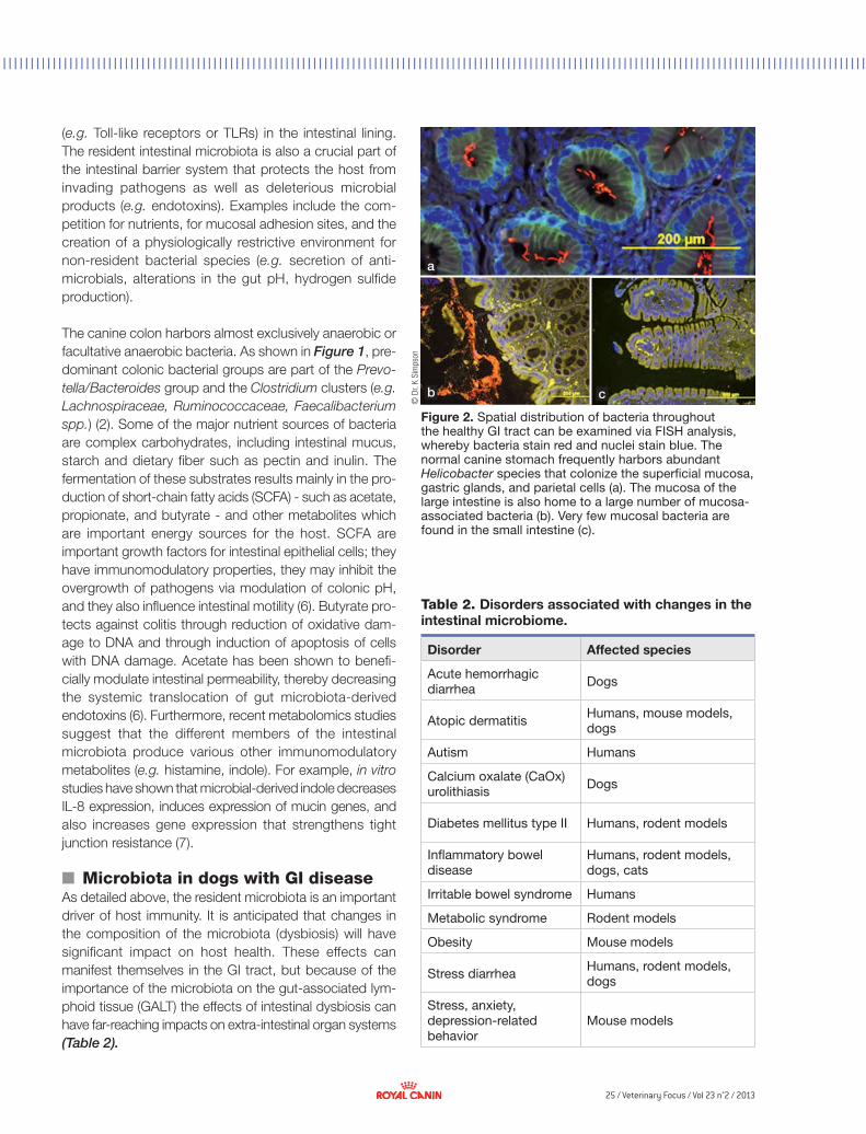
25 / Veterinary Focus / Vol 23 n°2 / 2013
l I I I I I I I I I I I I I I l I I I I I I I I I I I I I I l I I I I I I I I I I I I I I l I I I I I I I I I I I I I l I I I I I I I I I I I I I I l I I I I I I I I I I I I I I l I I I I I I I I I I I I I I l I I I I I I I I I I I I I I I I I I I I I I l I I I I I I I I I I I I I I l I I I I I I I I I I I I I I l I I I I I I I I I I I I I l I
a
b c©
Dr.
K Si
mps
on
(e.g. Toll-like receptors or TLRs) in the intestinal lining. The resident intestinal microbiota is also a crucial part of the intestinal barrier system that protects the host from invading pathogens as well as deleterious microbial products (e.g. endotoxins). Examples include the com-petition for nutrients, for mucosal adhesion sites, and the creation of a physiologically restrictive environment for non-resident bacterial species (e.g. secretion of anti-microbials, alterations in the gut pH, hydrogen sulfide production).
The canine colon harbors almost exclusively anaerobic or facultative anaerobic bacteria. As shown in Figure 1, pre-dominant colonic bacterial groups are part of the Prevo-tella/Bacteroides group and the Clostridium clusters (e.g. Lachnospiraceae, Ruminococcaceae, Faecalibacterium spp.) (2). Some of the major nutrient sources of bacteria are complex carbohydrates, including intestinal mucus, starch and dietary fiber such as pectin and inulin. The fermentation of these substrates results mainly in the pro-duction of short-chain fatty acids (SCFA) - such as acetate, propionate, and butyrate - and other metabolites which are important energy sources for the host. SCFA are important growth factors for intestinal epithelial cells; they have immunomodulatory properties, they may inhibit the overgrowth of pathogens via modulation of colonic pH, and they also influence intestinal motility (6). Butyrate pro-tects against colitis through reduction of oxidative dam-age to DNA and through induction of apoptosis of cells with DNA damage. Acetate has been shown to benefi-cially modulate intestinal permeability, thereby decreasing the systemic translocation of gut microbiota-derived endotoxins (6). Furthermore, recent metabolomics studies suggest that the different members of the intestinal microbiota produce various other immunomodulatory metabolites (e.g. histamine, indole). For example, in vitro studies have shown that microbial-derived indole decreases IL-8 expression, induces expression of mucin genes, and also increases gene expression that strengthens tight junction resistance (7).
■ Microbiota in dogs with GI diseaseAs detailed above, the resident microbiota is an important driver of host immunity. It is anticipated that changes in the composition of the microbiota (dysbiosis) will have significant impact on host health. These effects can manifest themselves in the GI tract, but because of the importance of the microbiota on the gut-associated lym-phoid tissue (GALT) the effects of intestinal dysbiosis can have far-reaching impacts on extra-intestinal organ systems (Table 2).
Figure 2. Spatial distribution of bacteria throughout the healthy GI tract can be examined via FISH analysis, whereby bacteria stain red and nuclei stain blue. The normal canine stomach frequently harbors abundant Helicobacter species that colonize the superficial mucosa, gastric glands, and parietal cells (a). The mucosa of the large intestine is also home to a large number of mucosa-associated bacteria (b). Very few mucosal bacteria are found in the small intestine (c).
Disorder Affected species
Acute hemorrhagic diarrhea Dogs
Atopic dermatitis Humans, mouse models, dogs
Autism Humans
Calcium oxalate (CaOx) urolithiasis Dogs
Diabetes mellitus type II Humans, rodent models
Inflammatory bowel disease
Humans, rodent models, dogs, cats
Irritable bowel syndrome Humans
Metabolic syndrome Rodent models
Obesity Mouse models
Stress diarrhea Humans, rodent models, dogs
Stress, anxiety, depression-related behavior
Mouse models
Table 2. Disorders associated with changes in the intestinal microbiome.
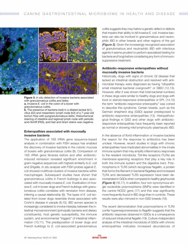
26 / Veterinary Focus / Vol 23 n°2 / 2013
l I I I I I I I I I I I I I I l I I I I I I I I I I I I I I l I I I I I I I I I I I I I I l I I I I I I I I I I I I I l I I I I I I I I I I I I I I l I I I I I I I I I I I I I I l I I I I I I I I I I I I I I l I I I I I I I I I I I I I I I I I I I I I I l I I I I I I I I I I I I I I l I I I I I I I I I I I I I I l I I I I I I I I I I I I I l Il I I I I I I I I I I I I I I l I I I I
a b2
b1 b3
© D
r. K
Sim
pson
C A N I N E G A S T R O I N T E S T I N A L M I C R O B I O M E I N H E A LT H A N D D I S E A S E
Figure 3. In situ detection of invasive bacteria associated with granulomatous colitis and ileitis: a. Invasive E. coli in the colon of a boxer with granulomatous colitis. b. The presence of bacteria (red) in a dilated lacteal (b1), villus (b2) and mesenteric lymph node (b3) of a 7-year-old bichon frise with pyogranulomatous ileitis. Histochemical staining of intestine and regional lymph node with periodic acid-Schiff (PAS), acid fast and Gram stains was negative.
colitis suggests they may harbor a genetic defect or defects that impairs their ability to kill invasive E. coli. Invasive bac-teria can also be involved in granulomatous and neutro-philic IBD in other breeds and other regions of the gut (Figure 3). Given the increasingly recognized association of granulomatous and neutrophilic IBD with infectious agents it seems prudent to perform specialized testing for bacteria and fungi before considering any form of immuno-suppressive treatment.
Antibiotic-responsive enteropathies without mucosally invasive bacteriaHistorically, dogs with signs of chronic GI disease that lacked an intestinal obstruction and resolved with anti-microbial therapy were diagnosed as having “idiopathic small intestinal bacterial overgrowth” or SIBO (12,13). However, after it was shown that total bacterial numbers in these dogs were similar to healthy dogs and dogs with food or steroid-responsive enteropathies or EPI, (14,15) the term “antibiotic-responsive enteropathy” was coined to describe this syndrome. Certain breeds, such as the German shepherd dog (GSD) appear predisposed to antibiotic-responsive enteropathies (13). Histopatholo-gical findings in GSD and other dogs with antibiotic-responsive enteropathies have frequently been reported as normal or showing mild lymphocytic-plasmacytic IBD.
In the absence of florid inflammation or invasive bacteria the reason for the response to antibiotics has been unclear. However, recent studies in dogs with chronic enteropathies have implicated abnormalities in the innate immune system that may amplify inflammatory responses to the resident microbiota. Toll-like receptors (TLRs) are membrane-spanning receptors that play a key role in both the immune system and the digestive tract. Poly-morphisms in TLR5 (which recognizes flagellin, a protein that forms the filament in bacterial flagellae) and increased TLR4 and decreased TLR5 expression have been de-monstrated in GSDs when compared to healthy greyhounds (Figure 4) (16,17). In addition, four non-synonymous sin-gle nucleotide polymorphisms (SNPs) were identified in the canine NOD2 gene (17) and this was significantly more frequently found in IBD dogs than in controls. These results were also mirrored in non-GSD breeds (18).
The recent demonstration that polymorphisms in TLR5 confer hyperresponsiveness to flagellin suggests that the antibiotic response observed in GSDs is a consequence of reduced intraluminal flagellin (19). Culture-independent analysis of the intestinal microbiota of GSDs with chronic enteropathies indicates increased abundances of
Enteropathies associated with mucosally invasive bacteriaThe application of 16S rRNA gene sequence-based analysis in combination with FISH assays has enabled the discovery of invasive bacteria in the colonic mucosa of boxers with granulomatous colitis (8). Comparison of 16S rRNA gene libraries before and after antibiotic-induced remission revealed significant enrichment in gram-negative sequences with highest similarity to E. coli and Shigella. In situ analysis with FISH probes against E. coli showed multifocal clusters of invasive bacteria within macrophages. Subsequent studies have shown that granulomatous colitis in French bulldogs is also asso-ciated with mucosally invasive E. coli. Eradication of inva-sive E. coli in boxer dogs and French bulldogs with granu-lomatous colitis correlates with remission from disease, inferring a causal relationship (9). The types of E. coli iso-lated from boxer dogs resemble those associated with Crohn’s disease in people (8,10). IBD across species is increasingly considered to involve interplay between the in-testinal microenvironment (principally bacteria and dietary constituents), host genetic susceptibility, the immune system, and environmental “triggers” of intestinal inflam-mation (10,11). The predisposition of boxer dogs and French bulldogs to E. coli-associated granulomatous
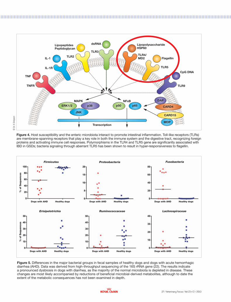
27 / Veterinary Focus / Vol 23 n°2 / 2013
l I I I I I I I I I I I I I I l I I I I I I I I I I I I I I l I I I I I I I I I I I I I I l I I I I I I I I I I I I I l I I I I I I I I I I I I I I l I I I I I I I I I I I I I I l I I I I I I I I I I I I I I l I I I I I I I I I I I I I I I I I I I I I I l I I I I I I I I I I I I I I l I I I I I I I I I I I I I I l I I I I I I I I I I I I I l I©
Dr.
K Si
mps
on
Figure 4. Host susceptibility and the enteric microbiota interact to promote intestinal inflammation. Toll-like receptors (TLRs) are membrane-spanning receptors that play a key role in both the immune system and the digestive tract, recognizing foreign proteins and activating immune cell responses. Polymorphisms in the TLR4 and TLR5 gene are significantly associated with IBD in GSDs; bacteria signaling through aberrant TLR5 has been shown to result in hyper-responsiveness to flagellin.
TNF
TNFR
IL-1
IL-1R
TLR2
TLR3
dsRNA
TLR5
TLR9
CpG DNA
Flagellin
LipopolysaccharideHSP60
LipopeptidesPeptidoglycan
MAPK NFxB
Transcription
p50 p65
DAP
CARD4
CARD15
MDP
ERK1/2 p38
JNK
TLR4/MD2
Figure 5. Differences in the major bacterial groups in fecal samples of healthy dogs and dogs with acute hemorrhagic diarrhea (AHD). Data was derived from high-throughput sequencing of the 16S rRNA gene (22). The results indicate a pronounced dysbiosis in dogs with diarrhea, as the majority of the normal microbiota is depleted in disease. These changes are most likely accompanied by reductions of beneficial microbial-derived metabolites, although to date the extent of the metabolic consequences has not been examined in depth.
Firmicutes
% o
f S
eq
uen
ces
% o
f S
eq
uen
ces
Dogs with AHD Dogs with AHD Dogs with AHD
Dogs with AHDDogs with AHDDogs with AHD
Healthy dogs Healthy dogs Healthy dogs
Healthy dogsHealthy dogsHealthy dogs
Proteobacteria Fusobacteria
Erisipelotrichia Ruminococcaceae Lachnospiraceae
28 / Veterinary Focus / Vol 23 n°2 / 2013
l I I I I I I I I I I I I I I l I I I I
1. Suchodolski JS. Intestinal microbiota of dogs and cats: a bigger world than we thought. Vet Clin North Am Small Anim Pract 2011;41:261-272.
2. Handl S, Dowd SE, Garcia-Mazcorro JF, et al. Massive parallel 16S rRNA gene pyrosequencing reveals highly diverse fecal bacterial and fungal communities in healthy dogs and cats. FEMS Microbial Ecol 2011;76:301-310.
3. Chaban B, Links MG, Hill JE. A molecular enrichment strategy based on cpn60 for detection of epsilon-proteobacteria in the dog fecal microbiome. Microb Ecol 2012;63:348-357.
4. Recordati C, Gualdi V, Craven M, et al. Spatial distribution of Helicobacter spp. in the gastrointestinal tract of dogs. Helicobacter 2009;14:180-191.
5. Priestnall SL, Wiinberg B, Spohr A, et al. Evaluation of "Helicobacter heilmannii" subtypes in the gastric mucosas of cats and dogs. J Clin Microbiol 2004;42:2144-2151.
6. Fukuda S, Toh H, Taylor TD, et al. Acetate-producing bifidobacteria protect the host from enteropathogenic infection via carbohydrate transporters. Gut Microbes 2012;3:449-454.
7. Bansal T, Alaniz RC, Wood TK, et al. The bacterial signal indole increases epithelial-cell tight-junction resistance and attenuates indicators of inflammation. Proc Natl Acad Sci USA 2010;107:228-233.
8. Simpson KW, Dogan B, Rishniw M, et al. Adherent and invasive Escherichia coli is associated with granulomatous colitis in boxer dogs. Infection and Immunity 2006;74:4778-4792.
9. Manchester AC, Hill S, Sabatino B, et al. Association between granulomatous colitis in French bulldogs and invasive Escherichia coli and response to fluoroquinolone antimicrobials. J Vet Intern Med 2013;27:56-61.
10. Packey CD, Sartor RB. Commensal bacteria, traditional and opportunistic pathogens, dysbiosis and bacterial killing in inflammatory bowel diseases. Curr Op Infect Dis 2009;22:292-301.
11. Simpson KW, Jergens AE. Pitfalls and progress in the diagnosis and management of canine inflammatory bowel disease. Vet Clin North Am Small Anim Pract 2011;41:381-398.
12. Batt RM, Carter MW, Peters TJ. Biochemical changes in the jejunal mucosa of dogs with a naturally occurring enteropathy associated with bacterial overgrowth. Gut 1984;25:816-823.
13. Batt RM, Needham JR, Carter MW. Bacterial overgrowth associated with a naturally occurring enteropathy in the German shepherd dog. Res Vet Sci 1983;35:42-46.
14. German AJ, Day MJ, Ruaux CG, et al. Comparison of direct and indirect tests for small intestinal bacterial overgrowth and antibiotic-responsive diarrhea in dogs. J Vet Intern Med 2003;17:33-43.
15. Johnston KL. Small intestinal bacterial overgrowth. Vet Clin North Am Small Anim Pract 1999;29:523-550.
16. Allenspach K, House A, Smith K, et al. Evaluation of mucosal bacteria and histopathology, clinical disease activity and expression of Toll-like receptors in German shepherd dogs with chronic enteropathies. Vet Microbiol 2010;146:326-335.
17. Kathrani A, House A, Catchpole B, et al. Polymorphisms in the Tlr4 and Tlr5 gene are significantly associated with inflammatory bowel disease in German shepherd dogs. PloS ONE 2010;5:1-10.
18. Kathrani A, House A, Catchpole B, et al. Breed-independent toll-like receptor 5 polymorphisms show association with canine inflammatory bowel disease. Tissue Antigens 2011;78:94-101.
19. Kathrani A, Holder A, Catchpole B, et al. TLR5 risk-associated haplotype for canine inflammatory bowel disease confers hyper-responsiveness to flagellin. PloS ONE 2012;7:e30117.
20. Westermarck E, Skrzypczak T, Harmoinen J, et al. Tylosin-responsive chronic diarrhea in dogs. J Vet Int Med 2005;19:177-186.
21. Suchodolski JS, Dowd SE, Westermarck E, et al. The effect of the macrolide antibiotic tylosin on microbial diversity in the canine small intestine as demonstrated by massive parallel 16S rRNA gene sequencing. BMC Microbiol 2009;9:210.
22. Suchodolski JS, Markel ME, Garcia-Mazcorro JF, et al. The fecal microbiome in dogs with acute diarrhea and idiopathic inflammatory bowel disease. PloS ONE 2012;7:e51907.
23. Suchodolski JS, Dowd SE, Wilke V, et al. 16S rRNA gene pyrosequencing reveals bacterial dysbiosis in the duodenum of dogs with idiopathic inflammatory bowel disease. PloS ONE 2012;7:e39333.
24. Sokol H, Pigneur B, Watterlot L, et al. Faecalibacterium prausnitzii is an anti-inflammatory commensal bacterium identified by gut microbiota analysis of Crohn disease patients. Proc Natl Acad Sci USA 2008;105:16731-16736.
25. Craven M, Egan CE, Dowd SE, et al. Inflammation drives dysbiosis and bacterial invasion in murine models of ileal Crohn's disease. PloS ONE 2012;7:e41594.
C A N I N E G A S T R O I N T E S T I N A L M I C R O B I O M E I N H E A LT H A N D D I S E A S E
Lactobacillales compared to healthy greyhounds (16). The relationship between dysbiosis, clinical disease, and enhanced inflammatory responses remains to be clarified.
Positive clinical responses to the macrolide antibiotic tylosin have also been consistently reported in subsets of dogs with chronic enteropathies (20). Recently, the small intestinal microbiota has been analyzed in dogs following tylosin administration, providing potential clues on the effect of this antibiotic on intestinal microbes (21).
■ ConclusionTaken as a whole, the microbial alterations documented in dogs with chronic GI disease are comparable to those observed across species where a shift in the microbiome from gram-positive Firmicutes (e.g. Clostridales) to gram-negative bacteria, predominantly Proteobacteria (includ-ing Enterobacteriaceae), correlates with intestinal inflam-mation (10,22-24). This depletion of commensal groups may impair the ability of the host to down-regulate the aberrant intestinal immune response, as several of these
bacterial groups secrete metabolites that have direct anti-inflammatory properties (24). However, at this time the rela-tionship between microbial alterations and inflammation is not well understood. Is dysbiosis a cause or a conse-quence of inflammation? Acute enteritis in dogs is asso-ciated with dysbiosis, especially depletions of bacterial groups that are important producers of SCFA and other microbial metabolites (Figure 5) (22), suggesting that bacterial changes are a consequence of the inflammatory response, but they may influence inflammation in gene-tically susceptible hosts. Recent experimental studies have shown that acute inflammation, triggered by proto-zoan infection and NSAID administration, can induce dysbiosis that parallels the shifts observed in Crohn’s dis-ease, and that host genetics may impact the threshold and the magnitude of dysbiosis (25). Clearly we are only just beginning to unravel the complex interrelationships between the enteric microbiota, health and disease. Elu-cidating the factors that shape the intestinal microbiome will provide novel opportunities for prophylaxis and thera-peutic intervention in dogs with IBD.
References
29 / Veterinary Focus / Vol 23 n°2 / 2013
■
Managing canine inflammatory bowel disease
■ IntroductionInflammatory bowel disease (IBD) is the term applied to a group of chronic intestinal diseases (enteropathies) characterized by persistent or recurrent gastrointestinal (GI) signs and inflammation of the GI tract. IBD involves a complex interaction between host genetics, the intes-tinal microenvironment (principally bacteria and dietary
KEY POINTS• ThetermIBDisappliedtoagroupofchronic
intestinaldisordersthatareincreasinglyconsideredtobeaconsequenceofgeneticsusceptibilityandaberrantresponsetotheintestinalmicroenvironment.
• TreatmentisinfluencedbythetypeofIBD,andisguidedbybreedpredisposition,theseverityofclinicalandclinicopathologicalfindings,andthegrossandhistopathologicalappearanceoftheintestines.
• Infectiousagentssuchasbacteriaandfungishouldbeactivelyexcludedindogswithneutrophilicorgranulomatousenteritis.
• Themajorityofdogswithminimal-changeenteropathyor“lymphocyticplasmacyticenteritis”,accompaniedbynormalserumalbumin,respondtodietorantimicrobialtherapy.
• Immunosuppressionisreservedfordogswithrefractoryorsevere“lymphocyticplasmacyticenteritis”orlymphangiectasia.
• Dogswithaprotein-losingenteropathyandlowserumalbuminhaveapoorprognosis.
constituents), the immune system, and environmental “triggers” of intestinal inflammation (1). The specific steps that lead to IBD, and the basis for phenotypic variation and unpredictable responses to treatment, are unknown.
This article will focus on the treatment of dogs with IBD, but in turn treatment is influenced by the type of IBD and is guided by breed predisposition, the severity of clinical and clinicopathological findings, and the gross and histo-pathological appearance of the intestines.
■ Diagnosis and phenotyping of IBDA diagnosis of IBD usually involves careful integration of signalment, home environment, history, physical findings, clinicopathological testing, diagnostic imaging and histo-pathology of intestinal biopsies.
Breed predispositionThe predisposition of certain dog breeds to IBD strongly supports a role for host genetics (Table 1) although to date the causal genetic defects have not been identified. This breed predisposition, (e.g. boxers, French bulldogs and German shepherds) along with clinical response to antibiotics points to an interaction of host susceptibility and microbiota (2-5). In boxers and French bulldogs with granulomatous colitis, lasting remission correlates with the eradication of mucosally invasive E. coli that are similar to strains isolated from Crohn’s disease (4). Studies in German shepherd dogs have identified polymorphisms in innate immunity factors, namely Toll-like receptors (TLRs), that segregate with disease; these dogs have been shown to have increased TLR2 and decreased TLR5 expression relative to healthy greyhounds (6).
An interaction of genetics and diet in dogs is supported by the finding that gluten-sensitive enteropathy in Irish
Kenneth Simpson, BVM&S, PhD, Dipl. ACVIM, Dipl. ECVIM-CACollege of Veterinary Medicine, Cornell University, New York State, USA
Dr. Simpson qualified from the Royal (Dick) School of Veterinary Studies, University of Edinburgh, in 1984 before gaining his PhD from the University of Leicester. He then lectured at the Royal Veterinary College in London before moving to Cornell University in 1995; he was appointed Professor of Medicine at Cornell in 2007. His main interest is the interplay between bacteria and host that leads to chronic inflammatory disease and cancer, aiming to effectively translate laboratory-based studies into better disease detection, therapy and ultimately prophylaxis for both animals and humans.
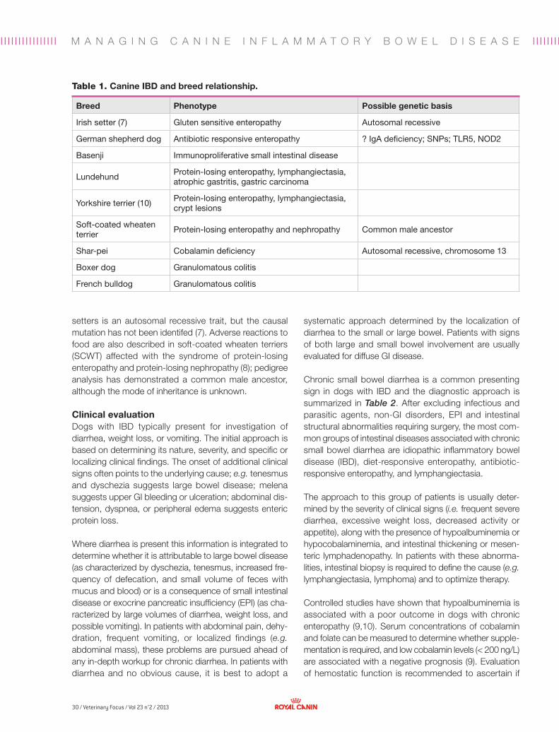
30 / Veterinary Focus / Vol 23 n°2 / 2013
l I I I I I I I I I I I I I I l I I I I I I I I I I I I I I l I I I I I I I I I I I I I I l I I I I I I I I I I I I I l I I I I I I I I I I I I I I l I I I I I I I I I I I I I I l I I I I I I I I I I I I I I l I I I I I I I I I I I I I I I I I I I I I I l I I I I I I I I I I I I I I l I I I I I I I I I I I I I I l I I I I I I I I I I I I I l Il I I I I I I I I I I I I I I l I I I I M A N A G I N G C A N I N E I N F L A M M A T O R Y B O W E L D I S E A S E
setters is an autosomal recessive trait, but the causal mutation has not been identifed (7). Adverse reactions to food are also described in soft-coated wheaten terriers (SCWT) affected with the syndrome of protein-losing enteropathy and protein-losing nephropathy (8); pedigree analysis has demonstrated a common male ancestor, although the mode of inheritance is unknown.
Clinical evaluationDogs with IBD typically present for investigation of diarrhea, weight loss, or vomiting. The initial approach is based on determining its nature, severity, and specific or localizing clinical findings. The onset of additional clinical signs often points to the underlying cause; e.g. tenesmus and dyschezia suggests large bowel disease; melena suggests upper GI bleeding or ulceration; abdominal dis-tension, dyspnea, or peripheral edema suggests enteric protein loss. Where diarrhea is present this information is integrated to determine whether it is attributable to large bowel disease (as characterized by dyschezia, tenesmus, increased fre-quency of defecation, and small volume of feces with mucus and blood) or is a consequence of small intestinal disease or exocrine pancreatic insufficiency (EPI) (as cha-racterized by large volumes of diarrhea, weight loss, and possible vomiting). In patients with abdominal pain, dehy-dration, frequent vomiting, or localized findings (e.g. abdominal mass), these problems are pursued ahead of any in-depth workup for chronic diarrhea. In patients with diarrhea and no obvious cause, it is best to adopt a
systematic approach determined by the localization of diarrhea to the small or large bowel. Patients with signs of both large and small bowel involvement are usually evaluated for diffuse GI disease.
Chronic small bowel diarrhea is a common presenting sign in dogs with IBD and the diagnostic approach is summarized in Table 2. After excluding infectious and parasitic agents, non-GI disorders, EPI and intestinal structural abnormalities requiring surgery, the most com-mon groups of intestinal diseases associated with chronic small bowel diarrhea are idiopathic inflammatory bowel disease (IBD), diet-responsive enteropathy, antibiotic-responsive enteropathy, and lymphangiectasia.
The approach to this group of patients is usually deter-mined by the severity of clinical signs (i.e. frequent severe diarrhea, excessive weight loss, decreased activity or appetite), along with the presence of hypoalbuminemia or hypocobalaminemia, and intestinal thickening or mesen-teric lymphadenopathy. In patients with these abnorma-lities, intestinal biopsy is required to define the cause (e.g. lymphangiectasia, lymphoma) and to optimize therapy. Controlled studies have shown that hypoalbuminemia is associated with a poor outcome in dogs with chronic enteropathy (9,10). Serum concentrations of cobalamin and folate can be measured to determine whether supple-mentation is required, and low cobalamin levels (< 200 ng/L) are associated with a negative prognosis (9). Evaluation of hemostatic function is recommended to ascertain if
Table 1. Canine IBD and breed relationship.
Breed Phenotype Possible genetic basis
Irish setter (7) Gluten sensitive enteropathy Autosomal recessive
German shepherd dog Antibiotic responsive enteropathy ? IgA deficiency; SNPs; TLR5, NOD2
Basenji Immunoproliferative small intestinal disease
Lundehund Protein-losing enteropathy, lymphangiectasia, atrophic gastritis, gastric carcinoma
Yorkshire terrier (10) Protein-losing enteropathy, lymphangiectasia, crypt lesions
Soft-coated wheaten terrier
Protein-losing enteropathy and nephropathy Common male ancestor
Shar-pei Cobalamin deficiency Autosomal recessive, chromosome 13
Boxer dog Granulomatous colitis
French bulldog Granulomatous colitis
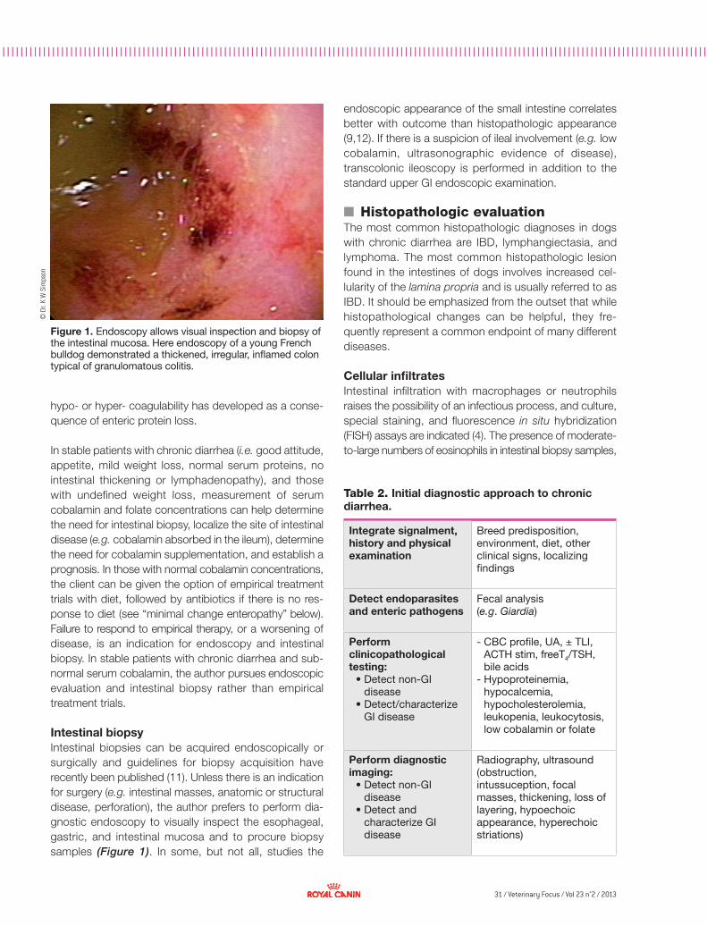
31 / Veterinary Focus / Vol 23 n°2 / 2013
l I I I I I I I I I I I I I I l I I I I I I I I I I I I I I l I I I I I I I I I I I I I I l I I I I I I I I I I I I I l I I I I I I I I I I I I I I l I I I I I I I I I I I I I I l I I I I I I I I I I I I I I l I I I I I I I I I I I I I I I I I I I I I I l I I I I I I I I I I I I I I l I I I I I I I I I I I I I I l I I I I I I I I I I I I I l I©
Dr.
K W
Sim
pson
hypo- or hyper- coagulability has developed as a conse-quence of enteric protein loss.
In stable patients with chronic diarrhea (i.e. good attitude, appetite, mild weight loss, normal serum proteins, no intestinal thickening or lymphadenopathy), and those with undefined weight loss, measurement of serum cobalamin and folate concentrations can help determine the need for intestinal biopsy, localize the site of intestinal disease (e.g. cobalamin absorbed in the ileum), determine the need for cobalamin supplementation, and establish a prognosis. In those with normal cobalamin concentrations, the client can be given the option of empirical treatment trials with diet, followed by antibiotics if there is no res-ponse to diet (see “minimal change enteropathy” below). Failure to respond to empirical therapy, or a worsening of disease, is an indication for endoscopy and intestinal biopsy. In stable patients with chronic diarrhea and sub-normal serum cobalamin, the author pursues endoscopic evaluation and intestinal biopsy rather than empirical treatment trials.
Intestinal biopsy Intestinal biopsies can be acquired endoscopically or surgically and guidelines for biopsy acquisition have recently been published (11). Unless there is an indication for surgery (e.g. intestinal masses, anatomic or structural disease, perforation), the author prefers to perform dia-gnostic endoscopy to visually inspect the esophageal, gastric, and intestinal mucosa and to procure biopsy samples (Figure 1). In some, but not all, studies the
Integrate signalment, history and physical examination
Breed predisposition, environment, diet, other clinical signs, localizing findings
Detect endoparasites and enteric pathogens
Fecal analysis(e.g. Giardia)
Perform clinicopathological testing:
• Detect non-GI disease
• Detect/characterize GI disease
- CBC profile, UA, ± TLI, ACTH stim, freeT4/TSH, bile acids
- Hypoproteinemia, hypocalcemia, hypocholesterolemia, leukopenia, leukocytosis, low cobalamin or folate
Perform diagnostic imaging:
• Detect non-GI disease
• Detect and characterize GI disease
Radiography, ultrasound (obstruction, intussuception, focal masses, thickening, loss of layering, hypoechoic appearance, hyperechoic striations)
Table 2. Initial diagnostic approach to chronic diarrhea.
Figure 1. Endoscopy allows visual inspection and biopsy of the intestinal mucosa. Here endoscopy of a young French bulldog demonstrated a thickened, irregular, inflamed colon typical of granulomatous colitis.
endoscopic appearance of the small intestine correlates better with outcome than histopathologic appearance (9,12). If there is a suspicion of ileal involvement (e.g. low cobalamin, ultrasonographic evidence of disease), transcolonic ileoscopy is performed in addition to the standard upper GI endoscopic examination.
■ Histopathologic evaluationThe most common histopathologic diagnoses in dogs with chronic diarrhea are IBD, lymphangiectasia, and lymphoma. The most common histopathologic lesion found in the intestines of dogs involves increased cel-lularity of the lamina propria and is usually referred to as IBD. It should be emphasized from the outset that while histopathological changes can be helpful, they fre-quently represent a common endpoint of many different diseases.
Cellular infiltratesIntestinal infiltration with macrophages or neutrophils raises the possibility of an infectious process, and culture, special staining, and fluorescence in situ hybridization (FISH) assays are indicated (4). The presence of moderate-to-large numbers of eosinophils in intestinal biopsy samples,

32 / Veterinary Focus / Vol 23 n°2 / 2013
l I I I I I I I I I I I I I I l I I I I I I I I I I I I I I l I I I I I I I I I I I I I I l I I I I I I I I I I I I I l I I I I I I I I I I I I I I l I I I I I I I I I I I I I I l I I I I I I I I I I I I I I l I I I I I I I I I I I I I I I I I I I I I I l I I I I I I I I I I I I I I l I I I I I I I I I I I I I I l I I I I I I I I I I I I I l Il I I I I I I I I I I I I I I l I I I I M A N A G I N G C A N I N E I N F L A M M A T O R Y B O W E L D I S E A S E
often accompanied by circulating eosinophilia, suggests possible parasitic infestation or adverse dietary reactions (13).
Increased numbers of lymphocytes and plasma cells, so-called “lymphoplasmacytic enteritis”, is the most frequently reported form of IBD. Moderate-to-severe lymphoplasmacytic enteritis is often described in asso-ciation with a protein-losing enteropathy (14) and predis-posed breeds include the basenji, lundehund, and shar-pei (15-17). However, the appropriateness and clinical relevance of the term lymphoplasmacytic enteritis is contentious, particularly with the small intestine: it has been shown that dogs have similar numbers of duodenal CD3-positive T cells before and after clinical remission induced by diet or steroids (18), and cats with and without signs of intestinal disease have similar numbers of lympho-cytes and plasma cells (19).
Mucosal architectureChanges in mucosal architecture, such as villous mor-phology, lymphatic dilatation, goblet cell mucus content, and crypt lesions are related to the presence and severity of GI disease (7,14). Dilation of lymphatics and the pre-sence of crypt abscesses and cysts are most frequently encountered in dogs with protein-losing enteropathies and are often accompanied by lymphoplasmacytic inflam-mation of varying severity (10,14).
Standardized gradingThe interpretation of GI histopathology varies considerably
among pathologists (20). To address this problem, a scheme to standardize the evaluation of intestinal histo-pathology has been published (21) but unfortunately, like previous standardized photographic schemes, it has poor agreement among pathologists and is not correlated with presence of disease or its outcome.
■ Therapeutic approaches to IBDThe therapeutic approach to IBD is influenced by suspicion of a breed-related problem; severity of disease as characterized by clinical signs, albumin and cobalamin concentrations, and endoscopic appearance, the type of cellular infiltrate, the presence of bacteria or fungi, and the presence of architectural changes such as atrophy, ulceration, lymphangiectasia and/or crypt cysts. Thera-peutic intervention is directed at correcting nutritional deficiencies (e.g. cobalamin) and counteracting inflam-mation and dysbiosis. Minimal change enteropathy Minimal change enteropathy is characterized by low clini-cal disease activity, normal serum albumin, normal cobalamin, and normal diagnostic imaging and intestinal histopathology. Empirically, treatment for Giardia and endoparasitic infection can be given (typically fenbenda-zole at 50 mg/kg PO for 5 days) and options for dietary trials are outlined in Table 3. A positive response sug-gests “diet-responsive enteropathy”, a term that includes both dietary allergy and intolerance. In dogs with GI signs related to diet a clinical response is usually observed within 1-2 weeks of changing the diet; if the response is good, the diet should be continued. Re-challenge with the original diet is required to confirm clinical signs are related to the diet, but few owners are willing to consent to this. Challenge with single dietary ingredients is neces-sary to define the specific components eliciting an adverse response. If dietary trials with two different diets are unsuccessful the next step is usually an antibiotic trial.
An antibiotic trial typically involves treatment with tylosin (10-15 mg/kg PO TID), oxytetracycline (20 mg/kg PO TID), or metronidazole (10 mg/kg PO BID) (2,3,5). A positive response suggests “antibiotic-responsive enteropathy” – previously known as “small intestinal bacterial over-growth”, despite the absence of increase in total bacteria (2,3,22) – and the dog is typically maintained on antibiotics for 28 days. If signs recur after treatment chronic maintain-ence therapy with tylosin at 5 mg/kg PO SID can be used in dogs that are tylosin-responsive (23). If the response is poor, the patient should be carefully reappraised before considering other treatment options.
Table 3. Strategies for dietary trials.
Global modification• Switch to a different diet or different manufacturer
Optimize assimilation• Highly digestible (usually rice-based)• Restricted fat• Easy-to-digest fats (e.g. MCT* oil)• Restricted fiber
Antigenic modification• Antigen restricted/novel protein source• Protein hydrolysate
Immunomodulation• Altered fatty acid profile (e.g. omega-3)• Prebiotics (e.g. FOS**, inulin)
*MCT = medium-chain triglyceride; **FOS = fructooligosaccharides
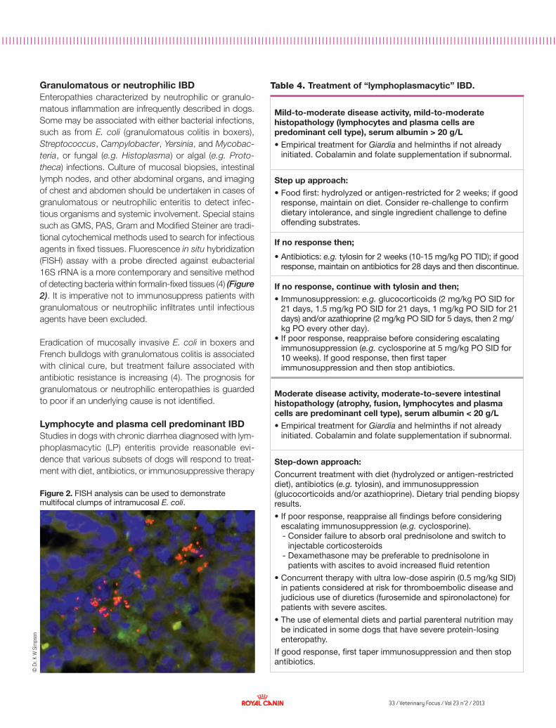
33 / Veterinary Focus / Vol 23 n°2 / 2013
l I I I I I I I I I I I I I I l I I I I I I I I I I I I I I l I I I I I I I I I I I I I I l I I I I I I I I I I I I I l I I I I I I I I I I I I I I l I I I I I I I I I I I I I I l I I I I I I I I I I I I I I l I I I I I I I I I I I I I I I I I I I I I I l I I I I I I I I I I I I I I l I I I I I I I I I I I I I I l I I I I I I I I I I I I I l I©
Dr.
K W
Sim
pson
Granulomatous or neutrophilic IBDEnteropathies characterized by neutrophilic or granulo-matous inflammation are infrequently described in dogs. Some may be associated with either bacterial infections, such as from E. coli (granulomatous colitis in boxers), Streptococcus, Campylobacter, Yersinia, and Mycobac-teria, or fungal (e.g. Histoplasma) or algal (e.g. Proto-theca) infections. Culture of mucosal biopsies, intestinal lymph nodes, and other abdominal organs, and imaging of chest and abdomen should be undertaken in cases of granulomatous or neutrophilic enteritis to detect infec-tious organisms and systemic involvement. Special stains such as GMS, PAS, Gram and Modified Steiner are tradi-tional cytochemical methods used to search for infectious agents in fixed tissues. Fluorescence in situ hybridization (FISH) assay with a probe directed against eubacterial 16S rRNA is a more contemporary and sensitive method of detecting bacteria within formalin-fixed tissues (4) (Figure 2). It is imperative not to immunosuppress patients with granulomatous or neutrophilic infiltrates until infectious agents have been excluded.
Eradication of mucosally invasive E. coli in boxers and French bulldogs with granulomatous colitis is associated with clinical cure, but treatment failure associated with antibiotic resistance is increasing (4). The prognosis for granulomatous or neutrophilic enteropathies is guarded to poor if an underlying cause is not identified.
Lymphocyte and plasma cell predominant IBDStudies in dogs with chronic diarrhea diagnosed with lym-phoplasmacytic (LP) enteritis provide reasonable evi-dence that various subsets of dogs will respond to treat-ment with diet, antibiotics, or immunosuppressive therapy
Figure 2. FISH analysis can be used to demonstrate multifocal clumps of intramucosal E. coli.
Table 4. Treatment of “lymphoplasmacytic” IBD.
Mild-to-moderate disease activity, mild-to-moderate histopathology (lymphocytes and plasma cells are predominant cell type), serum albumin > 20 g/L• Empirical treatment for Giardia and helminths if not already
initiated. Cobalamin and folate supplementation if subnormal.
Step up approach:• Food first: hydrolyzed or antigen-restricted for 2 weeks; if good
response, maintain on diet. Consider re-challenge to confirm dietary intolerance, and single ingredient challenge to define offending substrates.
If no response then;
• Antibiotics: e.g. tylosin for 2 weeks (10-15 mg/kg PO TID); if good response, maintain on antibiotics for 28 days and then discontinue.
If no response, continue with tylosin and then;• Immunosuppression: e.g. glucocorticoids (2 mg/kg PO SID for
21 days, 1.5 mg/kg PO SID for 21 days, 1 mg/kg PO SID for 21 days) and/or azathioprine (2 mg/kg PO SID for 5 days, then 2 mg/kg PO every other day).
• If poor response, reappraise before considering escalating immunosuppression (e.g. cyclosporine at 5 mg/kg PO SID for 10 weeks). If good response, then first taper immunosuppression and then stop antibiotics.
Moderate disease activity, moderate-to-severe intestinal histopathology (atrophy, fusion, lymphocytes and plasma cells are predominant cell type), serum albumin < 20 g/L • Empirical treatment for Giardia and helminths if not already
initiated. Cobalamin and folate supplementation if subnormal.
Step-down approach:Concurrent treatment with diet (hydrolyzed or antigen-restricted diet), antibiotics (e.g. tylosin), and immunosuppression (glucocorticoids and/or azathioprine). Dietary trial pending biopsy results.
• If poor response, reappraise all findings before considering escalating immunosuppression (e.g. cyclosporine).- Consider failure to absorb oral prednisolone and switch to
injectable corticosteroids- Dexamethasone may be preferable to prednisolone in
patients with ascites to avoid increased fluid retention
• Concurrent therapy with ultra low-dose aspirin (0.5 mg/kg SID) in patients considered at risk for thromboembolic disease and judicious use of diuretics (furosemide and spironolactone) for patients with severe ascites.
• The use of elemental diets and partial parenteral nutrition may be indicated in some dogs that have severe protein-losing enteropathy.
If good response, first taper immunosuppression and then stop antibiotics.
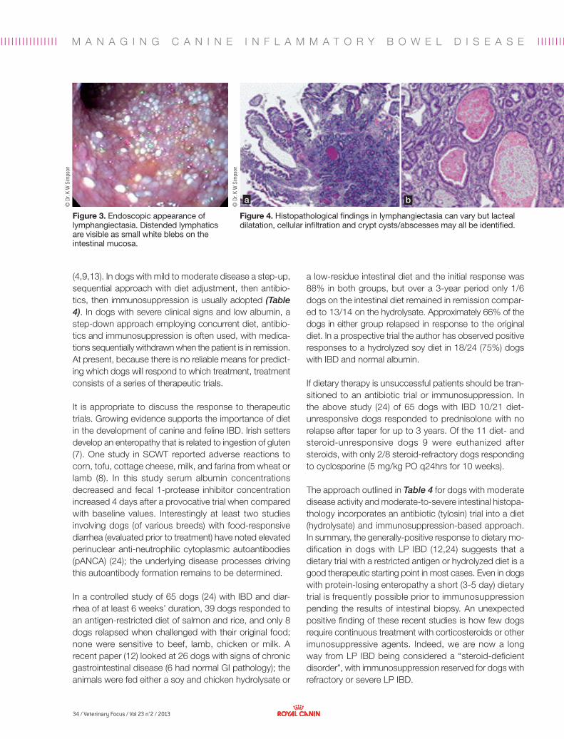
34 / Veterinary Focus / Vol 23 n°2 / 2013
l I I I I I I I I I I I I I I l I I I I l I I I I I I I I I I I I I I l I I I I I I I I I I I I I I l I I I I I I I I I I I I I I l I I I I I I I I I I I I I l I I I I I I I I I I I I I I l I I I I I I I I I I I I I I l I I I I I I I I I I I I I I l I I I I I I I I I I I I I I I I I I I I I I l I I I I I I I I I I I I I I l I I I I I I I I I I I I I I l I I I I I I I I I I I I I l I
a b
© D
r. K
W S
imps
on
© D
r. K
W S
imps
on
M A N A G I N G C A N I N E I N F L A M M A T O R Y B O W E L D I S E A S E
(4,9,13). In dogs with mild to moderate disease a step-up, sequential approach with diet adjustment, then antibio-tics, then immunosuppression is usually adopted (Table 4). In dogs with severe clinical signs and low albumin, a step-down approach employing concurrent diet, antibio-tics and immunosuppression is often used, with medica-tions sequentially withdrawn when the patient is in remission. At present, because there is no reliable means for predict-ing which dogs will respond to which treatment, treatment consists of a series of therapeutic trials.
It is appropriate to discuss the response to therapeutic trials. Growing evidence supports the importance of diet in the development of canine and feline IBD. Irish setters develop an enteropathy that is related to ingestion of gluten (7). One study in SCWT reported adverse reactions to corn, tofu, cottage cheese, milk, and farina from wheat or lamb (8). In this study serum albumin concentrations decreased and fecal 1-protease inhibitor concentration increased 4 days after a provocative trial when compared with baseline values. Interestingly at least two studies involving dogs (of various breeds) with food-responsive diarrhea (evaluated prior to treatment) have noted elevated perinuclear anti-neutrophilic cytoplasmic autoantibodies (pANCA) (24); the underlying disease processes driving this autoantibody formation remains to be determined.
In a controlled study of 65 dogs (24) with IBD and diar-rhea of at least 6 weeks’ duration, 39 dogs responded to an antigen-restricted diet of salmon and rice, and only 8 dogs relapsed when challenged with their original food; none were sensitive to beef, lamb, chicken or milk. A recent paper (12) looked at 26 dogs with signs of chronic gastrointestinal disease (6 had normal GI pathology); the animals were fed either a soy and chicken hydrolysate or
a low-residue intestinal diet and the initial response was 88% in both groups, but over a 3-year period only 1/6 dogs on the intestinal diet remained in remission compar-ed to 13/14 on the hydrolysate. Approximately 66% of the dogs in either group relapsed in response to the original diet. In a prospective trial the author has observed positive responses to a hydrolyzed soy diet in 18/24 (75%) dogs with IBD and normal albumin.
If dietary therapy is unsuccessful patients should be tran-sitioned to an antibiotic trial or immunosuppression. In the above study (24) of 65 dogs with IBD 10/21 diet-unresponsive dogs responded to prednisolone with no relapse after taper for up to 3 years. Of the 11 diet- and steroid-unresponsive dogs 9 were euthanized after steroids, with only 2/8 steroid-refractory dogs responding to cyclosporine (5 mg/kg PO q24hrs for 10 weeks).
The approach outlined in Table 4 for dogs with moderate disease activity and moderate-to-severe intestinal histopa-thology incorporates an antibiotic (tylosin) trial into a diet (hydrolysate) and immunosuppression-based approach. In summary, the generally-positive response to dietary mo-dification in dogs with LP IBD (12,24) suggests that a dietary trial with a restricted antigen or hydrolyzed diet is a good therapeutic starting point in most cases. Even in dogs with protein-losing enteropathy a short (3-5 day) dietary trial is frequently possible prior to immunosuppression pending the results of intestinal biopsy. An unexpected positive finding of these recent studies is how few dogs require continuous treatment with corticosteroids or other imunosuppressive agents. Indeed, we are now a long way from LP IBD being considered a “steroid-deficient disorder”, with immunosuppression reserved for dogs with refractory or severe LP IBD.
Figure 3. Endoscopic appearance of lymphangiectasia. Distended lymphatics are visible as small white blebs on the intestinal mucosa.
Figure 4. Histopathological findings in lymphangiectasia can vary but lacteal dilatation, cellular infiltration and crypt cysts/abscesses may all be identified.
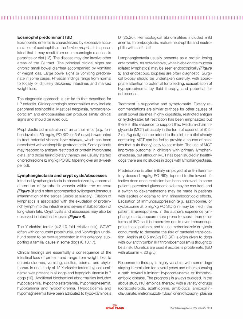
35 / Veterinary Focus / Vol 23 n°2 / 2013
l I I I I I I I I I I I I I I l I I I I I I I I I I I I I I l I I I I I I I I I I I I I I l I I I I I I I I I I I I I l I I I I I I I I I I I I I I l I I I I I I I I I I I I I I l I I I I I I I I I I I I I I l I I I I I I I I I I I I I I I I I I I I I I l I I I I I I I I I I I I I I l I I I I I I I I I I I I I I l I I I I I I I I I I I I I l I
Eosinophil predominant IBDEosinophilic enteritis is characterized by excessive accu-mulation of eosinophils in the lamina propria. It is specu-lated that it may result from an immunologic reaction to parasites or diet (13). The disease may also involve other areas of the GI tract. The principal clinical signs are chronic small bowel diarrhea accompanied by vomiting or weight loss. Large bowel signs or vomiting predomi-nate in some cases. Physical findings range from normal to focally or diffusely thickened intestines and marked weight loss.
The diagnostic approach is similar to that described for LP enteritis. Clinicopathologic abnormalities may include peripheral eosinophilia. Mast cell neoplasia, hypoadreno-corticism and endoparasites can produce similar clinical signs and should be ruled out.
Prophylactic administration of an anthelmintic (e.g. fen-bendazole at 50 mg/kg PO SID for 3-5 days) is warranted to treat potential visceral larva migrans, which has been associated with eosinophilic gastroenteritis. Some patients may respond to antigen-restricted or protein hydrolysate diets, and those failing dietary therapy are usually started on prednisolone (2 mg/kg PO SID tapering over an 8-week period).
Lymphangiectasia and crypt cysts/abscessesIntestinal lymphangiectasia is characterized by abnormal distention of lymphatic vessels within the mucosa (Figure 3) and is often accompanied by lipogranulomatous inflammation of the serosa (visible at surgery). Dilation of lymphatics is associated with the exudation of protein-rich lymph into the intestine and severe malabsorption of long-chain fats. Crypt cysts and abscesses may also be observed in intestinal biopsies (Figure 4).
The Yorkshire terrier (4.2-10-fold relative risk), SCWT (often with concurrent proteinuria), and Norwegian lunde-hund seem to be over-represented in this category, sup-porting a familial cause in some dogs (8,10,17).
Clinical findings are essentially a consequence of the intestinal loss of protein, and range from weight loss to chronic diarrhea, vomiting, ascites, edema, and chylo-thorax. In one study of 12 Yorkshire terriers hypoalbumi-nemia was present in all dogs and hypoglobulinemia in 7 dogs (10). Additional biochemical abnormalities included hypocalcemia, hypocholesterolemia, hypomagnesemia, hypokalemia and hypochloremia. Hypocalcemia and hypomagnesemia have been attributed to hypovitaminosis
D (25,26). Hematological abnormalities included mild anemia, thrombocytosis, mature neutrophilia and neutro-philia with a left shift.
Lymphangiectasia usually presents as a protein-losing enteropathy. As noted above, white blebs on the mucosa (dilated lymphatics) may be seen endoscopically (Figure 3) and endoscopic biopsies are often diagnostic. Surgi-cal biopsy should be undertaken carefully, with appro-priate attention to potential for bleeding, exacerbation of hypoproteinemia by fluid therapy, and potential for dehiscence.
Treatment is supportive and symptomatic. Dietary re-commendations are similar to those for other causes of small bowel diarrhea (highly digestible, restricted antigen or hydrolysate); fat restriction has been emphasized but there is little evidence to support this. Medium-chain tri-glyceride (MCT) oil usually in the form of coconut oil (0.5- 2 mL/kg daily) can be added to the diet, or a diet already containing MCT can be fed to provide a source of calo-ries that is (in theory) easy to assimilate. The use of MCT improves outcome in children with primary lymphan-ghiectasia, but although MCT has been studied in healthy dogs there are no studies in dogs with lymphangiectasia.
Prednisolone is often initially employed at anti-inflamma-tory doses (1 mg/kg PO BID), tapered to the lowest ef-fective dose once remission has been achieved. In some patients parenteral glucocorticoids may be required, and a switch to dexamethasone may be made in patients with ascites or edema to limit mineralocorticoid effects. Escalation of immunosuppression (e.g. azathioprine, or cyclosporine at 5 mg/kg PO SID (27)) may be tried if the patient is unresponsive. In the author’s experience lym-phangiectasia appears more prone to sepsis than other forms of IBD so it is imperative not to over-immunosup-press these patients, and to use metronidazole or tylosin concurrently to decrease the risk of bacterial transloca-tion. Aspirin at 0.5 mg/kg PO SID is often given to dogs with low antithrombin III if thromboembolism is thought to be a risk. Diuretics are used if ascites is problematic (IBD with albumin < 20 g/L).
Response to therapy is highly variable, with some dogs staying in remission for several years and others pursuing a path toward fulminant hypoproteinemia or thrombo-embolic disease. The prognosis is always guarded. In the above study (10) empirical therapy, with a variety of drugs (corticosteroids, azathioprine, antibiotics (amoxicillin-clavulanate, metronidazole, tylosin or enrofloxacin)), plasma
36 / Veterinary Focus / Vol 23 n°2 / 2013
l I I I I I I I I I I I I I I l I I I I
1. Simpson KW, Jergens AE. Pitfalls and progress in the diagnosis and management of canine inflammatory bowel disease. Vet Clin North Am Small Anim Pract 2011;41(2):381-398.
2. Batt RM, McLean L, Riley JE. Response of the jejunal mucosa of dogs with aerobic and anaerobic bacterial overgrowth to antibiotic therapy. Gut 1988;29(4):473-482.
3. German AJ, Day MJ, Ruaux CG, et al. Comparison of direct and indirect tests for small intestinal bacterial overgrowth and antibiotic-responsive diarrhea in dogs. J Vet Intern Med 2003;17(1):33-43.
4. Simpson KW, Dogan B, Rishniw M, et al. Adherent and invasive Escherichia coli is associated with granulomatous colitis in boxer dogs. Infect Immun 2006;74(8):4778-4792.
5. Westermarck E, Skrzypczak T, Harmoinen J, et al. Tylosin-responsive chronic diarrhea in dogs. J Vet Intern Med 2005;19(2):177-186.
6. Allenspach K, House A, Smith K, et al. Evaluation of mucosal bacteria and histopathology, clinical disease activity and expression of Toll-like receptors in German shepherd dogs with chronic enteropathies. Vet Microbiol 2010; 146:326-335.
7. Garden OA, Pidduck H, Lakhani KH, et al. Inheritance of gluten-sensitive enteropathy in Irish Setters. Am J Vet Res 2000;61(4):462-468.
8. Vaden SL, Hammerberg B, Davenport DJ, et al. Food hypersensitivity reactions in Soft Coated Wheaten Terriers with protein-losing enteropathy or protein-losing nephropathy or both: gastroscopic food sensitivity testing, dietary provocation, and fecal immunoglobulin E. J Vet Intern Med 2000;14(1):60-67.
9. Allenspach K, Wieland B, Gröne A, et al. Chronic enteropathies in dogs: evaluation of risk factors for negative outcome. J Vet Intern Med 2007;21(4):700-708.
10. Craven M, Simpson JW, Ridyard AE, et al. Canine inflammatory bowel disease: retrospective analysis of diagnosis and outcome in 80 cases (1995-2002). J Small Anim Pract 2004;45(7):336-342.
11. Willard MD, Mansell J, Fosgate GT, et al. Effect of sample quality on the sensitivity of endoscopic biopsy for detecting gastric and duodenal lesions in dogs and cats. J Vet Intern Med 2008;22(5):1084-1089.
12. Mandigers PJ, Biourge V, van den Ingh TS, et al. A randomized, open-label, positively-controlled field trial of a hydrolyzed protein diet in dogs with chronic small bowel enteropathy. J Vet Intern Med 2010;24(6):1350-1357.
13. Kleinschmidt S, Meneses F, Nolte I, et al. Characterization of mast cell numbers and subtypes in biopsies from the gastrointestinal tract of dogs with lymphocytic-plasmacytic or eosinophilic gastroenterocolitis 2007;120(3-4):80-92. Epub 2007 Jul 17.
14. Peterson PB, Willard MD. Protein-losing enteropathies. Vet Clin North Am Small Anim Pract 2003;33(5):1061-1082.
15. Breitschwerdt EB, Ochoa R, Barta M, et al. Clinical and laboratory characterization of Basenjis with immunoproliferative small intestinal disease. Am J Vet Res 1984;45(2):267-273.
16. Grützner N, Bishop MA, Suchodolski JS, et al. Association study of cobalamin deficiency in the Chinese Shar Pei. J Hered 2010;101(2):211-217. Epub 2009 Nov 19.
17. Kolbjørnsen O, Press CM, Landsverk T. Gastropathies in the Lundehund. I. Gastritis and gastric neoplasia associated with intestinal lymphangiectasia. APMIS 1994;102(9):647-661.
18. Schreiner NM, Gaschen F, Gröne A, et al. Clinical signs, histology, and CD3-positive cells before and after treatment of dogs with chronic enteropathies. J Vet Intern Med 2008;22(5):1079-1083.
19. Waly NE, Stokes CR, Gruffydd-Jones TJ, et al. Immune cell populations in the duodenal mucosa of cats with inflammatory bowel disease. J Vet Intern Med 2004;18(6):816-825.
20. Willard MD, Jergens AE, Duncan RB, et al. Interobserver variation among histopathologic evaluations of intestinal tissues from dogs and cats. J Am Vet Med Assoc 2002;220(8):1177-1182.
21. Day MJ, Bilzer T, Mansell J, et al. Histopathological standards for the diagnosis of gastrointestinal inflammation in endoscopic biopsy samples from the dog and cat: a report from the WSAVA Gastrointestinal Standardization Group. J Comp Pathol 2008;138(Suppl)1:S1-S43.
22. Simpson KW. Small intestinal bacterial overgrowth. J Am Vet Med Assoc 1994;205(3):405-407.
23. Westermarck E. Personal communication 2010.24. Luckschander N, Allenspach K, Hall J, et al. Perinuclear antineutrophilic
cytoplasmic antibody and response to treatment in diarrheic dogs with food responsive disease or inflammatory bowel disease. J Vet Intern Med 2006;20(2):221-227.
25. Bush WW, Kimmel SE, Wosar MA, et al. Secondary hypoparathyroidism attributed to hypomagnesemia in a dog with protein-losing enteropathy. J Am Vet Med Assoc 2001;219(12):1732-1734, 1708.
26. Mellanby RJ, Mellor PJ, Roulois A, et al. Hypocalcaemia associated with low serum vitamin D metabolite concentrations in two dogs with protein-losing enteropathies. J Small Anim Pract 2005;46(7):345-351.
27. Allenspach K, Rüfenacht S, Sauter S, et al. Pharmacokinetics and clinical efficacy of cyclosporine treatment of dogs with steroid-refractory inflammatory bowel disease. J Vet Intern Med 2006;20(2):239-244.
M A N A G I N G C A N I N E I N F L A M M A T O R Y B O W E L D I S E A S E
and diuretics was associated with a poor outcome; 7/12 cases died or were euthanized within 3 months of diagno-sis, thromboembolism being suspected in 3/7 dogs.
■ ConclusionTreatment for IBD is guided by breed predisposition, the severity of clinical and clinicopathological findings, and
the gross and histopathological appearance of the intes-tines. Each case should be subject to a systematic work-up but the prognosis can be variable. However there can be no doubt that over the next few years continued re-search will deliver better knowledge and more precise treatment regimes for the many animals that present with IBD-like signs.
References

37 / Veterinary Focus / Vol 23 n°2 / 2013
■
Epidemiology of canine parvovirus infection in the USA – an update
Sandi Lefebvre, DVM, PhD Banfield Pet Hospital, Portland, Oregon, USA
Dr. Lefebvre joined Banfield in 2011 as Associate Medical Advisor – Research for the Banfield Applied Research and Knowledge (BARK) team. A 2003 graduate of Ontario Veterinary College, she obtained her PhD in epidemiology through research and development of guidelines for pet visitation in human hospitals. Her most recent professional role was as scientific editor for JAVMA and AJVR.
■ IntroductionCanine parvovirus type-2 is included among the core vaccines advocated by the American Animal Hospital Association for routine administration in all pet dogs (1). Despite this recommendation, parvovirus infection remains a problem in clinical practice. Indeed, evidence exists that the virus has mutated since it first emerged in the late 1970s as an important cause of hemorrhagic enteritis in dogs (2). However, most studies (3,4) of the epidemiology of the disease were performed decades ago, and it is possible that risk factors have changed over time.
■ Method of analysisThe medical records of all canine patients seen at Ban-field from January 1, 2010 through December 31, 2011 were examined to identify dogs that had a diagnosis of parvovirus infection as confirmed via laboratory testing of blood or feces. To evaluate potential risk factors for confirmed parvovirus infection, the medical records were also reviewed for gender, reproductive status, age cate-gory (≤ 6 months or > 6 months), and breed. Chi square tests were performed when sample sizes exceeded 1,000 dogs to yield odds ratios (ORs) as an estimate of relative risk and 95% confidence intervals (CIs). The p value was set at < 0.05.
■ ResultsDuring 2010, 7,734 of 1,942,137 dogs seen at Banfield hospitals had a confirmed diagnosis of parvovirus infec-tion (40 cases/10,000 dogs); in 2011, the numbers were
slightly smaller at 7,708 of 2,021,929 dogs seen (38 cases/10,000 dogs). Females were at significantly lower odds (likelihood) of infection, compared with males (OR, 0.80; 95% CI, 0.79-0.82). No significant association between neuter status and infection was evident when analyses were adjusted for age. Dogs ≤ 6 months of age were 11.40 times as likely as older dogs to have the dia-gnosis (95% CI, 11.05-11.75).
Five breeds were identified as being at significantly greater odds of confirmed parvovirus infection relative to mixed breed dogs, namely the pit bull terrier, Chihuahua, Rottweiler, American Staffordshire terrier, and Lancashire heeler (Table 1). With data stratified by age, pit bulls, Rottweilers, and Lancashire heelers were at greater odds when ≤ 6 months of age than when they were older (Table 2). On the other hand, Chihuahuas and American Staffordshire terriers were at greater odds when > 6 months.
■ Discussion and conclusionComparison of these findings with those of older studies (3,4) which involved considerably smaller samples of teach-ing hospital data reveals some interesting differences in breed risk. In those studies, doberman pinschers, border collies, German shepherd dogs, and Pomeranians were identified as being at increased odds of having parvovirus infection relative to other pure or mixed breed dogs, whereas in the present study, they were at reduced odds (0.64 [95% CI, 0.52-0.79], 0.37 [0.31-0.45], 0.92 [0.85-0.99], and 0.37 [0.33-0.42], respectively), and this seem-ing protective effect persisted when data were stratified

38 / Veterinary Focus / Vol 23 n°2 / 2013
l I I I I I I I I I I I I I I l I I I I
1. Welborn LV, DeVries JG, Ford R, et al. 2011 AAHA canine vaccination guidelines. J Am Anim Hosp Assoc 2011;47:1-42.
2. Decaro N, Buonavoglia C. Canine parvovirus - a review of epidemiological and diagnostic aspects, with emphasis on type 2c. Vet Microbiol 2012;155:1-12.
3. Glickman LT, Domanski LM, Patronek GJ, et al. Breed-related risk factors for
canine parvovirus enteritis. J Am Vet Med Assoc 1985;187:589-594.4. Houston DM, Ribble CS, Head LL. Risk factors associated with parvovirus
enteritis in dogs: 283 cases (1982-1991). J Am Vet Med Assoc 1996;208:542-546.
by age. One possibility for the differences between time periods is that identification of high-risk breeds in the past might have led to a greater proportion of those breeds being vaccinated against parvovirus infection. Differences in vacci-nation status might also explain the observed increased
odds of infection in dogs > 6 months (versus ≤ 6 months) for Chihuahuas and American Staffordshire terriers. Addi-tional studies are needed to determine whether the epide-miology of parvoviral enteritis is indeed changing and to identify the factors contributing to that change.
Breed No. of dogs affected % of breed seen OR 95% CI
Pit bull (n = 199,440) 5,113 2.56 2.88 2.70-3.06
Chihuahua (n = 232,477) 3,774 1.62 1.80 1.69-1.92
Rottweiler (n = 48,394) 578 1.19 1.32 1.19-1.46
American Staffordshire terrier (n = 5,399)
78 1.44 1.60 1.26-2.02
Lancashire heeler (n = 1,004) 22 2.19 2.40 1.48-3.66
* 1,247 of 136,284 mixed-breed dogs seen at Banfield hospitals during the study period had a confirmed parvovirus infection, representing 92 cases/10,000 mixed-breed dogs seen.
Age ≤ 6 months Age > 6 months
Breed OR 95% CI OR 95% CI
Pit bull 3.01 2.80-3.25 2.45 2.16-2.79
Chihuahua 1.58 1.46-1.71 2.46 2.17-2.79
Rottweiler 1.45 1.29-1.62 0.95* 0.75-1.19
American Staffordshire terrier 1.43 1.05-1.95 2.11 1.36-3.16
Lancashire heeler 2.62 1.51-4.28 1.72* 0.47-4.48
*Value is not significant (i.e., P > 0.05).
Table 1. Significant breed-specific ratios for the odds of confirmed parvovirus infection in various pure canine breeds versus mixed breeds*.
Table 2. Breed-specific ratios for the odds of confirmed parvovirus infection in various dog breeds by age category.
EPIDEMIOLOGY OF CANINE PARVOVIRUS INFECTION IN THE USA – AN UPDATE
References
39 / Veterinary Focus / Vol 23 n°2 / 2013
■
■
Feline intestinal tumors Laura Marconato, DVM, Dipl. ECVIM-CA (Oncology) Centro Oncologico Veterinario, Sasso Marconi, Italy
Dr. Marconato completed her veterinary studies at the University of Milan in 1999. Since 2000 her work has focused on medical oncology, with a special interest in lymphoma. In 2008 she received her diploma from the ECVIM and she is currently President of the Italian Society of Veterinary Oncology.
Giuliano Bettini, DVM Department of Veterinary Medical Sciences, University of Bologna, Italy
Dr. Bettini obtained his degree in Veterinary Medicine at the University of Bologna in 1988, and since 2001 has been Associate Professor of Veterinary Pathology at the University of Bologna. His research activities are related mainly to comparative oncology, particularly the characterization of molecular prognosticators of animal tumors and the investigation of environmental carcinogenesis.
KEY POINTS• Lymphomaisthemostfrequentintestinal
tumorincats,followedbycarcinomaandmastocytoma.
• Lymphomacanpresentinvariousformsandmaybecharacterizedbyitsbiologicalbehavior,responsetotreatment,andprognosis.
• Clinicalfindingsduringtheinitialstagesofintestinaltumorscanbevague;itisoftenonlylaterinthediseaseprocessthatsymptomsdevelopwhichcanbeattributedtotheintestinaltractand/orpossiblemetastasis.
• Thediagnosticprocessrequireslaboratorytestsandimaging,withbiopsyofneoplastictissuerequiredtoallowatreatmentplan.
• Treatmentandprognosisvarywiththetumortype,clinicalstagingandlocation.
■ IntroductionIntestinal tumors are quite rare in cats, representing < 10% of all neoplasia in this species (1). However they represent an important area within feline gastroentero-logy, not least because the clinical signs and symptoms of neoplastic and non-neoplastic conditions can overlap.
Intestinal neoplasms generally originate in the small intes-tine, with lymphoma being by far the most frequent tumor (55%), followed by carcinoma (32%), and mastocytoma (4%). Intestinal sarcomas are extremely rare (2). The etio-logy is unknown, but generally there is a greater predispo-sition amongst males, and Siamese appear predisposed to lymphoma (1).
■ Intestinal lymphomaLymphoma is the most frequent tumor of the intestinal tract, and intestinal lymphoma is also the most frequent form of lymphoma found in cats (3-5). It can present as either focal or diffuse forms (where the thickening is trans-mural and the lumen is preserved) and can originate from either T or B lymphocytes. To arrive at a diagnosis, an assessment needs to be made of the clinical, histological and immunophenotype criteria, and these factors also have prognostic relevance (6-8).
Lymphoma originating from MALT (mucosal associated lymphoid tissue) has a T phenotype and is the most com-mon form (Figure 1); it is typically made up of small cells (low-grade) and usually involves the small intestine (7-9). Initially the tumor tends to be indolent, but will become progressively more aggressive, both locally and systemi-cally (high grade). Extension of the tumor through the wall can result in perforation and peritonitis. Clinically, diffuse thickening of the bowel may be noted (3). Differential dia-gnosis in the early stages includes IBD (inflammatory bowel disease) (10) and indeed T-intestinal lymphoma can
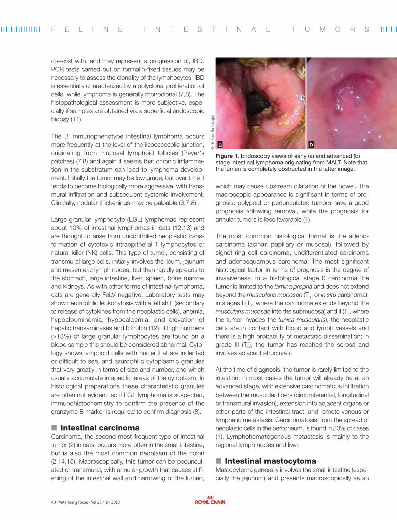
40 / Veterinary Focus / Vol 23 n°2 / 2013
l I I I I I I I I I I I I I I l I I I I I I I I I I I I I I l I I I I I I I I I I I I I I l I I I I I I I I I I I I I l I I I I I I I I I I I I I I l I I I I I I I I I I I I I I l I I I I I I I I I I I I I I l I I I I I I I I I I I I I I I I I I I I I I l I I I I I I I I I I I I I I l I I I I I I I I I I I I I I l I I I I I I I I I I I I I l Il I I I I I I I I I I I I I I l I I I I
a b
© D
r. Ro
ssel
la T
erra
gni
co-exist with, and may represent a progression of, IBD. PCR tests carried out on formalin-fixed tissues may be necessary to assess the clonality of the lymphocytes; IBD is essentially characterized by a polyclonal proliferation of cells, while lymphoma is generally monoclonal (7,8). The histopathological assessment is more subjective, espe-cially if samples are obtained via a superficial endoscopic biopsy (11).
The B immunophenotype intestinal lymphoma occurs more frequently at the level of the ileocecocolic junction, originating from mucosal lymphoid follicles (Peyer's patches) (7,8) and again it seems that chronic inflamma-tion in the substratum can lead to lymphoma develop-ment. Initially the tumor may be low grade, but over time it tends to become biologically more aggressive, with trans-mural infiltration and subsequent systemic involvement. Clinically, nodular thickenings may be palpable (3,7,8).
Large granular lymphocyte (LGL) lymphomas represent about 10% of intestinal lymphomas in cats (12,13) and are thought to arise from uncontrolled neoplastic trans-formation of cytotoxic intraepithelial T lymphocytes or natural killer (NK) cells. This type of tumor, consisting of transmural large cells, initially involves the ileum, jejunum and mesenteric lymph nodes, but then rapidly spreads to the stomach, large intestine, liver, spleen, bone marrow and kidneys. As with other forms of intestinal lymphoma, cats are generally FeLV negative. Laboratory tests may show neutrophilic leukocytosis with a left shift (secondary to release of cytokines from the neoplastic cells), anemia, hypoalbuminemia, hypocalcemia, and elevation of hepatic transaminases and bilirubin (12). If high numbers (>13%) of large granular lymphocytes are found on a blood sample this should be considered abnormal. Cyto-logy shows lymphoid cells with nuclei that are indented or difficult to see, and azurophilic cytoplasmic granules that vary greatly in terms of size and number, and which usually accumulate in specific areas of the cytoplasm. In histological preparations these characteristic granules are often not evident, so if LGL lymphoma is suspected, immunohistochemistry to confirm the presence of the granzyme B marker is required to confirm diagnosis (8).
■ Intestinal carcinoma Carcinoma, the second most frequent type of intestinal tumor (2) in cats, occurs more often in the small intestine, but is also the most common neoplasm of the colon (2,14,15). Macroscopically, this tumor can be peduncul-ated or transmural, with annular growth that causes stiff-ening of the intestinal wall and narrowing of the lumen,
which may cause upstream dilatation of the bowel. The macroscopic appearance is significant in terms of pro-gnosis: polypoid or pedunculated tumors have a good prognosis following removal, while the prognosis for annular tumors is less favorable (1).
The most common histological format is the adeno- carcinoma (acinar, papillary or mucosal), followed by signet-ring cell carcinoma, undifferentiated carcinoma and adenosquamous carcinoma. The most significant histological factor in terms of prognosis is the degree of invasiveness. In a histological stage 0 carcinoma the tumor is limited to the lamina propria and does not extend beyond the muscularis mucosae (Tis, or in situ carcinoma); in stages I (T1, where the carcinoma extends beyond the muscularis mucosae into the submucosa) and II (T2, where the tumor invades the tunica muscularis), the neoplastic cells are in contact with blood and lymph vessels and there is a high probability of metastatic dissemination; in grade III (T3), the tumor has reached the serosa and involves adjacent structures.
At the time of diagnosis, the tumor is rarely limited to the intestine; in most cases the tumor will already be at an advanced stage, with extensive carcinomatous infiltration between the muscular fibers (circumferential, longitudinal or transmural invasion), extension into adjacent organs or other parts of the intestinal tract, and remote venous or lymphatic metastasis. Carcinomatosis, from the spread of neoplastic cells in the peritoneum, is found in 30% of cases (1). Lymphohematogenous metastasis is mainly to the regional lymph nodes and liver.
■ Intestinal mastocytoma Mastocytoma generally involves the small intestine (espe-cially the jejunum) and presents macroscopically as an
F E L I N E I N T E S T I N A L T U M O R S
Figure 1. Endoscopy views of early (a) and advanced (b) stage intestinal lymphoma originating from MALT. Note that the lumen is completely obstructed in the latter image.

41 / Veterinary Focus / Vol 23 n°2 / 2013
l I I I I I I I I I I I I I I l I I I I I I I I I I I I I I l I I I I I I I I I I I I I I l I I I I I I I I I I I I I l I I I I I I I I I I I I I I l I I I I I I I I I I I I I I l I I I I I I I I I I I I I I l I I I I I I I I I I I I I I I I I I I I I I l I I I I I I I I I I I I I I l I I I I I I I I I I I I I I l I I I I I I I I I I I I I l I
intramural mass of varying size which can form an intus-susception and eventually intestinal perforation. Ulcers are often apparent within thickened areas of the intestinal walls, due both to direct infiltration of the tumor, and the release of histamine from mast cell degranulation. In the sclerosing form the neoplastic cells are interspersed with abundant fibrous tissue. Marked infiltration of eosinophils, deposition of fibrous tissue and the presence of necrosis can make it difficult to differentiate mastocytoma from eosinophilic enteritis (1,16,17). The biological behavior is aggressive, with high metastatic potential and a low survival rate.
■ Clinical pictureClinical signs and symptoms are often initially vague and may be deceptive, and can vary according to the area involved: if the tumor is situated in the small intestine, there may be intermittent vomiting, hematemesis, dehy-dration, weight loss and anorexia. If situated in the large intestine, tenesmus, hematochezia and dyschezia are evident. In the case of peritoneal carcinomatosis ascites can cause secondary abdominal distension (1).
If the tumor is particularly large or is located in a narrow area of the bowel, partial or complete intestinal occlusion may occur, with secondary perforation and peritonitis. In this situation the cat will demonstrate abdominal pain, vomiting and fever.
Cats with intestinal lymphoma may not present with vomiting and/or diarrhea, but only anorexia and/or weight loss. Clinically, such cases often have poor body condi-tion or are emaciated. In rare cases, the clinical presenta-tion is more acute due to secondary intussusception, obstruction and/or perforation with septic peritonitis.
In general, abdominal palpation will reveal thickened intestinal loops (especially with low-grade lymphoma) or an abdominal mass (especially in high-grade lymphoma) (3).
■ Diagnostic procedure and stagingDiagnosis may be achieved via various laboratory tests and diagnostic imaging. The hemochromocytometric test can reveal microcytic anemia (due to occult blood loss in the feces) and leukocytosis with secondary toxic neutrophils (from peritonitis or tumor necrosis). A leuko-cytosis can also be paraneoplastic as a result of neoplas-tic cells releasing granulocytic colony-stimulating factor or granulocytic-macrophage colony-stimulating factor (1). As noted above, cats with LGL lymphoma may have
large granular lymphocytes evident in the peripheral bloodstream (12,13).
Biochemical tests may show hypoproteinemia (25-30% of cases), elevated hepatic enzymes, hypercholestero-lemia (especially with non-lymphoma neoplasia), and elevated urea (10-30%) (1). Hypoalbuminemia is unusual in low-grade lymphoma because the intestinal wall tends to remain intact even in the more advanced stages. Again in cats with low-grade lymphoma, it is common to find depleted levels of cobalamin, as this is largely absorbed at the level of the ileum, an area frequently involved with this type of lymphoma (1,3).
Abdominal radiography sometimes reveals an abdominal mass or signs of partial or complete occlusion of the bowel. Ascites may be noted due to secondary peritonitis from a neoplastic perforation. However, the widespread use of ultrasound has significantly decreased the need for contrast studies; ultrasound allows for a detailed exa-mination of the intestinal wall, assessing its thickness, stratification, content, and peristalsis, and makes it pos-sible to view the surrounding peritoneum and regional lymph nodes.
Ultrasound findings with intestinal neoplasia include wall thickening and a loss of the normal stratification (1). Lym-phoma often causes diffuse thickening of the intestinal wall, with reduced echogenicity, loss of normal motility and regional lymphoadenomegaly (3). Sometimes, hypo-echogenic nodules in the intestinal wall can be observed. Intestinal carcinomas can have ultrasound findings that overlap with those of lymphoma, but they tend to be more asymmetrical and localized, and more frequently present with obstruction of the lumen; again signs of in-testinal perforation may be apparent. In the differential diagnosis of intestinal neoplasia inflammatory disorders must always be considered as they can cause focal gra-nulomata which may be indistinguishable from neoplasia; ultrasound allows examination of the entire abdomen and may detect metastasis to other organs and/or the pre-sence of abdominal effusion. It also allows biopsy of spe-cific intestinal lesions and lymph nodes for cytologic and/or histopathological examination. Finally note that if an intestinal neoplasm is suspected, the chest should be surveyed via radiography or CT.
A recent study showed that cytology from needle aspi-rates gave an accurate diagnosis in many cases, and this technique should therefore be offered for all patients where an exploratory laparotomy or laparoscopy is not possible
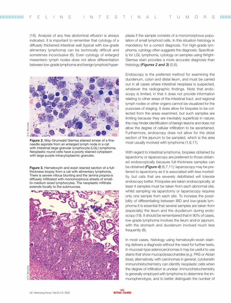
42 / Veterinary Focus / Vol 23 n°2 / 2013
l I I I I I I I I I I I I I I l I I I I I I I I I I I I I I l I I I I I I I I I I I I I I l I I I I I I I I I I I I I l I I I I I I I I I I I I I I l I I I I I I I I I I I I I I l I I I I I I I I I I I I I I l I I I I I I I I I I I I I I I I I I I I I I l I I I I I I I I I I I I I I l I I I I I I I I I I I I I I l I I I I I I I I I I I I I l Il I I I I I I I I I I I I I I l I I I I
© D
r. M
arco
nato
© D
r. M
arco
nato
F E L I N E I N T E S T I N A L T U M O R S
plasia if the sample consists of a monomorphous popu-lation of small lymphoid cells. In this situation histology is mandatory for a correct diagnosis. For high-grade lym-phoma, cytology often suggests the diagnosis. Specifical-ly for LGL lymphoma, cytology on samples using Wright-Giemsa stain provides a more accurate diagnosis than histology (Figures 2 and 3) (5,6).
Endoscopy is the preferred method for examining the duodenum, colon and distal ileum, and must be carried out in all cases where intestinal neoplasia is suspected, whatever the radiographic findings. Note that endo-scopy is limited, in that it does not provide information relating to other areas of the intestinal tract, and regional lymph nodes or other organs cannot be visualized for the purposes of staging. It does allow for biopsies to be col-lected from the areas examined, but such samples are limiting because they are inevitably superficial in nature; this may hinder identification of benign lesions and does not allow the degree of cellular infiltration to be ascertained. Furthermore, endoscopy does not allow for the distal section of the jejunum to be sampled, which is the area most usually involved with lymphoma (1,6,11).
With regard to intestinal lymphoma, biopsies obtained by laparotomy or laparoscopy are preferred to those obtain-ed endoscopically because full-thickness samples can be obtained (Figure 4) (6,7,11); laparoscopy may be pre-ferred to laparotomy as it is associated with less morbid-ity, but cats that are severely debilitated will tolerate endoscopy better. If biopsies are taken endoscopically, at least 4 samples must be taken from each abnormal site, whilst sampling via laparotomy or laparoscopy requires only one sample from each site. To increase the possi-bility of differentiating between IBD and low-grade lym-phoma it is essential that several samples are taken from (especially) the ileum and the duodenum during endo-scopy (19). It should be remembered that in 90% of cases, low-grade lymphoma involves the ileum and/or jejunum, with the stomach and duodenum involved much less frequently (8).
In most cases, histology using hematoxylin-eosin stain-ing delivers a diagnosis without the need for further tests. In mucosal-type adenocarcinomas it may be useful to use stains that show mucopolysaccharides (e.g. PAS or Alcian blue); alternatively, with carcinomas in general, cytokeratin immunohistochemistry can identify neoplastic cells when the degree of infiltration is unclear. Immunohistochemistry is generally employed with lymphoma to determine the im-munophenotype, and to better distinguish the number of
Figure 2. May-Grunwald Giemsa stained smear of a fine- needle aspirate from an enlarged lymph node in a cat with intestinal large granular lymphocyte (LGL) lymphoma. Neoplastic round cells have a poorly stained cytoplasm with large purple intracytoplasmic granules.
Figure 3. Hematoxylin and eosin stained section of a full-thickness biopsy from a cat with alimentary lymphoma. There is severe villous blunting and the lamina propria is diffusely infiltrated with monomorphous sheets of small-to-medium sized lymphocytes. The neoplastic infiltrate extends focally to the submucosa.
(18). Analysis of any free abdominal effusion is always indicated. It is important to remember that cytology of a diffusely thickened intestinal wall (typical with low-grade alimentary lymphoma) can be technically difficult and sometimes inconclusive (6). Even cytology of enlarged mesenteric lymph nodes does not allow differentiation between low-grade lymphoma and benign lymphoid hyper-
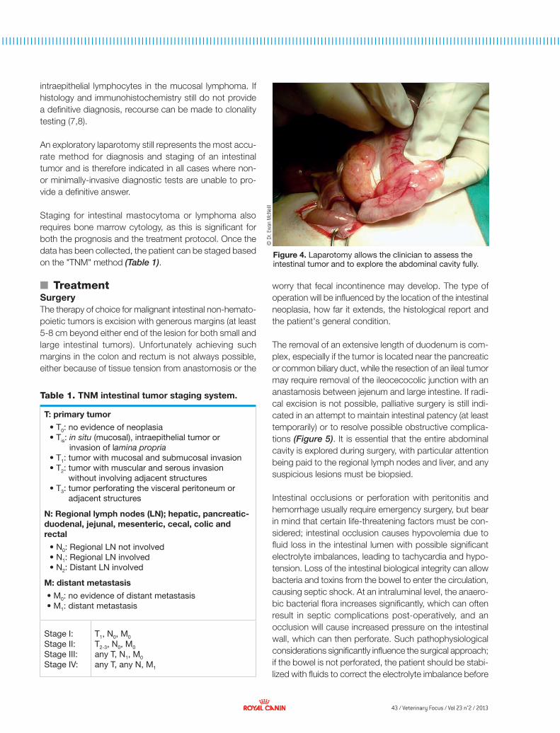
43 / Veterinary Focus / Vol 23 n°2 / 2013
l I I I I I I I I I I I I I I l I I I I I I I I I I I I I I l I I I I I I I I I I I I I I l I I I I I I I I I I I I I l I I I I I I I I I I I I I I l I I I I I I I I I I I I I I l I I I I I I I I I I I I I I l I I I I I I I I I I I I I I I I I I I I I I l I I I I I I I I I I I I I I l I I I I I I I I I I I I I I l I I I I I I I I I I I I I l I
© D
r. Ew
an M
cNei
ll
intraepithelial lymphocytes in the mucosal lymphoma. If histology and immunohistochemistry still do not provide a definitive diagnosis, recourse can be made to clonality testing (7,8).
An exploratory laparotomy still represents the most accu-rate method for diagnosis and staging of an intestinal tumor and is therefore indicated in all cases where non- or minimally-invasive diagnostic tests are unable to pro-vide a definitive answer.
Staging for intestinal mastocytoma or lymphoma also requires bone marrow cytology, as this is significant for both the prognosis and the treatment protocol. Once the data has been collected, the patient can be staged based on the "TNM" method (Table 1).
■ TreatmentSurgeryThe therapy of choice for malignant intestinal non-hemato-poietic tumors is excision with generous margins (at least 5-8 cm beyond either end of the lesion for both small and large intestinal tumors). Unfortunately achieving such margins in the colon and rectum is not always possible, either because of tissue tension from anastomosis or the
worry that fecal incontinence may develop. The type of operation will be influenced by the location of the intestinal neoplasia, how far it extends, the histological report and the patient's general condition.
The removal of an extensive length of duodenum is com-plex, especially if the tumor is located near the pancreatic or common biliary duct, while the resection of an ileal tumor may require removal of the ileocecocolic junction with an anastamosis between jejenum and large intestine. If radi-cal excision is not possible, palliative surgery is still indi-cated in an attempt to maintain intestinal patency (at least temporarily) or to resolve possible obstructive complica-tions (Figure 5). It is essential that the entire abdominal cavity is explored during surgery, with particular attention being paid to the regional lymph nodes and liver, and any suspicious lesions must be biopsied.
Intestinal occlusions or perforation with peritonitis and hemorrhage usually require emergency surgery, but bear in mind that certain life-threatening factors must be con-sidered; intestinal occlusion causes hypovolemia due to fluid loss in the intestinal lumen with possible significant electrolyte imbalances, leading to tachycardia and hypo-tension. Loss of the intestinal biological integrity can allow bacteria and toxins from the bowel to enter the circulation, causing septic shock. At an intraluminal level, the anaero-bic bacterial flora increases significantly, which can often result in septic complications post-operatively, and an occlusion will cause increased pressure on the intestinal wall, which can then perforate. Such pathophysiological considerations significantly influence the surgical approach; if the bowel is not perforated, the patient should be stabi-lized with fluids to correct the electrolyte imbalance before
T: primary tumor
• T0: no evidence of neoplasia• Tis: in situ (mucosal), intraepithelial tumor or
invasion of lamina propria• T1: tumor with mucosal and submucosal invasion• T2: tumor with muscular and serous invasion
without involving adjacent structures• T3: tumor perforating the visceral peritoneum or
adjacent structures
N: Regional lymph nodes (LN); hepatic, pancreatic-duodenal, jejunal, mesenteric, cecal, colic and rectal
• N0: Regional LN not involved• N1: Regional LN involved• N2: Distant LN involved
M: distant metastasis
• M0: no evidence of distant metastasis• M1: distant metastasis
Stage I: Stage II: Stage III: Stage IV:
T1, N0, M0 T2-3, N0, M0 any T, N1, M0 any T, any N, M1
Table 1. TNM intestinal tumor staging system.
Figure 4. Laparotomy allows the clinician to assess the intestinal tumor and to explore the abdominal cavity fully.
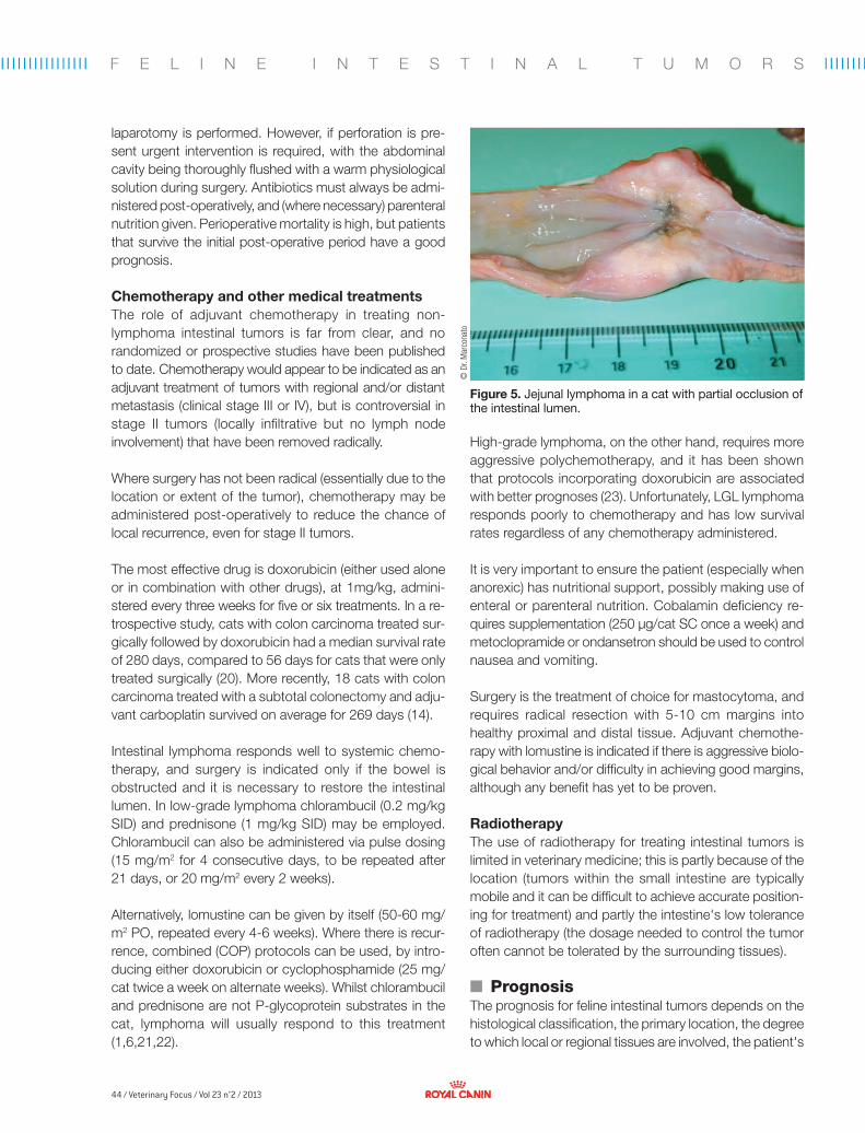
44 / Veterinary Focus / Vol 23 n°2 / 2013
l I I I I I I I I I I I I I I l I I I I l I I I I I I I I I I I I I I l I I I I I I I I I I I I I I l I I I I I I I I I I I I I I l I I I I I I I I I I I I I l I I I I I I I I I I I I I I l I I I I I I I I I I I I I I l I I I I I I I I I I I I I I l I I I I I I I I I I I I I I I I I I I I I I l I I I I I I I I I I I I I I l I I I I I I I I I I I I I I l I I I I I I I I I I I I I l I
© D
r. M
arco
nato
F E L I N E I N T E S T I N A L T U M O R S
laparotomy is performed. However, if perforation is pre-sent urgent intervention is required, with the abdominal cavity being thoroughly flushed with a warm physiological solution during surgery. Antibiotics must always be admi-nistered post-operatively, and (where necessary) parenteral nutrition given. Perioperative mortality is high, but patients that survive the initial post-operative period have a good prognosis.
Chemotherapy and other medical treatmentsThe role of adjuvant chemotherapy in treating non- lymphoma intestinal tumors is far from clear, and no randomized or prospective studies have been published to date. Chemotherapy would appear to be indicated as an adjuvant treatment of tumors with regional and/or distant metastasis (clinical stage III or IV), but is controversial in stage II tumors (locally infiltrative but no lymph node involvement) that have been removed radically.
Where surgery has not been radical (essentially due to the location or extent of the tumor), chemotherapy may be administered post-operatively to reduce the chance of local recurrence, even for stage II tumors.
The most effective drug is doxorubicin (either used alone or in combination with other drugs), at 1mg/kg, admini-stered every three weeks for five or six treatments. In a re-trospective study, cats with colon carcinoma treated sur-gically followed by doxorubicin had a median survival rate of 280 days, compared to 56 days for cats that were only treated surgically (20). More recently, 18 cats with colon carcinoma treated with a subtotal colonectomy and adju-vant carboplatin survived on average for 269 days (14).
Intestinal lymphoma responds well to systemic chemo-therapy, and surgery is indicated only if the bowel is obstructed and it is necessary to restore the intestinal lumen. In low-grade lymphoma chlorambucil (0.2 mg/kg SID) and prednisone (1 mg/kg SID) may be employed. Chlorambucil can also be administered via pulse dosing (15 mg/m2 for 4 consecutive days, to be repeated after 21 days, or 20 mg/m2 every 2 weeks).
Alternatively, lomustine can be given by itself (50-60 mg/m2 PO, repeated every 4-6 weeks). Where there is recur-rence, combined (COP) protocols can be used, by intro-ducing either doxorubicin or cyclophosphamide (25 mg/cat twice a week on alternate weeks). Whilst chlorambucil and prednisone are not P-glycoprotein substrates in the cat, lymphoma will usually respond to this treatment (1,6,21,22).
Figure 5. Jejunal lymphoma in a cat with partial occlusion of the intestinal lumen.
High-grade lymphoma, on the other hand, requires more aggressive polychemotherapy, and it has been shown that protocols incorporating doxorubicin are associated with better prognoses (23). Unfortunately, LGL lymphoma responds poorly to chemotherapy and has low survival rates regardless of any chemotherapy administered.
It is very important to ensure the patient (especially when anorexic) has nutritional support, possibly making use of enteral or parenteral nutrition. Cobalamin deficiency re-quires supplementation (250 µg/cat SC once a week) and metoclopramide or ondansetron should be used to control nausea and vomiting.
Surgery is the treatment of choice for mastocytoma, and requires radical resection with 5-10 cm margins into healthy proximal and distal tissue. Adjuvant chemothe-rapy with lomustine is indicated if there is aggressive biolo-gical behavior and/or difficulty in achieving good margins, although any benefit has yet to be proven.
RadiotherapyThe use of radiotherapy for treating intestinal tumors is limited in veterinary medicine; this is partly because of the location (tumors within the small intestine are typically mobile and it can be difficult to achieve accurate position-ing for treatment) and partly the intestine's low tolerance of radiotherapy (the dosage needed to control the tumor often cannot be tolerated by the surrounding tissues).
■ PrognosisThe prognosis for feline intestinal tumors depends on the histological classification, the primary location, the degree to which local or regional tissues are involved, the patient's

45 / Veterinary Focus / Vol 23 n°2 / 2013
l I I I I I I I I I I I I I I l I I I I I I I I I I I I I I l I I I I I I I I I I I I I I l I I I I I I I I I I I I I l I I I I I I I I I I I I I I l I I I I I I I I I I I I I I l I I I I I I I I I I I I I I l I I I I I I I I I I I I I I I I I I I I I I l I I I I I I I I I I I I I I l I I I I I I I I I I I I I I l I I I I I I I I I I I I I l I
1. Selting KA. Intestinal tumors. In: Withrow SJ, Vail DM, Page RL eds, Withrow & MacEwen’s Small Animal Clinical Oncology. 5th ed. Philadelphia: WB Saunders Co, 2012;412-423.
2. Rissetto K, Villamil JA, Selting KA, et al. Recent trends in feline intestinal neoplasia: an epidemiologic study of 1,129 cases in the veterinary medical database from 1964 to 2004. J Am Anim Hosp Assoc 2011;47:28-36.
3. Barrs V, Beatty J. Feline alimentary lymphoma: 1. Classification, risk factors, clinical signs and non-invasive diagnostics. J Feline Med Surg 2012;14:182-90.
4. Russell KJ, Beatty JA, Dhand NT, et al. Feline low-grade alimentary lymphoma: how common is it? J Feline Med Surg 2012;14:910-912.
5. Vezzali E, Parodi AL, Marcato PS, et al. Histopathologic classification of 171 cases of canine and feline non-Hodgkin lymphoma according to the WHO. Vet Comp Oncol 2010;8:38-49.
6. Barrs V, Beatty J. Feline alimentary lymphoma: 2. Further diagnostics, therapy and prognosis. J Feline Med Surg 2012;14:191-201.
7. Kiupel M, Smedley RC, Pfent C, et al. Diagnostic algorithm to differentiate lymphoma from inflammation in feline small intestinal biopsy samples. Vet Pathol 2011;48:212-222.
8. Moore PF, Rodriguez-Bertos A, Kass PH. Feline gastrointestinal lymphoma: mucosal architecture, immunophenotype, and molecular clonality. Vet Pathol 2012;49:658-668.
9. Cesari A, Bettini G, Vezzali E. Feline intestinal T-cell lymphoma: assessment of morphologic and kinetic features in 30 cases. J Vet Diagn Invest 2009;21:277-279.
10. Briscoe KA, Krockenberger M, Beatty JA, et al. Histopathological and immunohistochemical evaluation of 53 cases of feline lymphoplasmacytic enteritis and low-grade alimentary lymphoma. J Comp Path 2011;145:187-198.
11. Evans SE, Bonczynski JJ, Broussard JD, et al. Comparison of endoscopic and full-thickness biopsy specimens for diagnosis of inflammatory bowel disease and alimentary tract lymphoma in cats. J Am Vet Med Assoc 2006;229:1447-1450.
12. Krick EL, Little L, Patel R, et al. Description of clinical and pathological findings, treatment and outcome of feline large granular lymphocyte lymphoma (1996-2004). Vet Comp Oncol 2008;6:102-110.
13. Roccabianca P, Vernau W, Caniatti M, et al. Feline large granular lymphocyte (LGL) lymphoma with secondary leukemia: primary intestinal origin with predominance of a CD3/CD8 phenotype. Vet Pathol 2006;43:15-28.
14. Arteaga TA, McKnight J, Bergman PJ. A review of 18 cases of feline colonic adenocarcinoma treated with subtotal colectomies and adjuvant carboplatin. J Am Anim Hosp Assoc 2012;48:399-404.
15. Cribb AE. Feline gastrointestinal adenocarcinoma: a review and retrospective study. Can Vet J 1988;29:709-712.
16. Craig LE, Hardam EE, Hertzke DM, et al. Feline gastrointestinal eosinophilic sclerosing fibroplasia. Vet Pathol 2009;46:63-70.
17. Halsey CHC, Powers BE, Kamstock DA: Feline intestinal sclerosing mast cell tumour: 50 cases (1997-2008). Vet Comp Oncol 2010;8:72-79.
18. Bonfanti U, Bertazzolo W, Bottero E, et al. Diagnostic value of cytologic examination of gastrointestinal tract tumors in dogs and cats: 83 cases (2001-2004). J Am Vet Med Assoc, 2006;229:1130-1133.
19. Scott KD, Zoran DL, Mansell J, et al. Utility of endoscopic biopsies of the duodenum and ileum for diagnosis of inflammatory bowel disease and small cell lymphoma in cats. J Vet Intern Med 2011;25:1253-1257.
20. Slawienski MJ, Mauldin GE, Mauldin GN, et al. Malignant colonic neoplasia in cats: 46 cases (1990-1996). J Am Vet Med Assoc 1997;211:878-881.
21. Stein TJ, Pellin M, Steinberg H, et al. Treatment of feline gastrointestinal small-cell lymphoma with chlorambucil and glucocorticoids. J Am Anim Hosp Assoc 2010;46:413-417.
22. Zwahlen CH, Lucroy MD, Kraegel SA, et al. Results of chemotherapy for cats with alimentary malignant lymphoma: 21 cases (1993-1997). J Am Vet Med Assoc 1998;213:1144-1149.
23. Moore AS, Cotter SM, Frimberger AE, et al. A comparison of doxorubicin and COP for maintenance of remission in cats with lymphoma. J Vet Intern Med 1996;10:372–375.
24. Kiselow MA, Rassnick KM, McDonough SP, et al. Outcome of cats with low-grade lymphocytic lymphoma: 41 cases (1995-2005). J Am Vet Med Assoc 2008;232:405-410.
25. Fondacaro JV, Richter KP, Carpenter JL, et al. Feline gastrointestinal lymphoma: 67 cases (1988–1996). Eur J Comp Gastroenterol 1999;4:5-11.
general condition, the presence of metastasis, and whe-ther surgery is elective or emergency in nature. Typically, long-term survivors eventually die from local recurrence of the tumor.
Cats with small intestine adenocarcinoma are high-risk perioperatively, but nonetheless if they survive beyond two weeks after surgery the long-term prognosis is rea-sonable. The treatment of colonic adenocarcinoma with adjuvant chemotherapy (doxorubicin) may prolong sur-vival (20). High-grade lymphoma carries a poor prognosis; if treated with chemotherapy, such cases survive on
average 3 months, with 18% of cases showing complete remission (23).
The median survival increases by 7-10 months if the che-motherapy protocols include doxorubicin, with 38-87% attaining remission (23). Low-grade lymphomas are, on the other hand, less aggressive, with longer survival rates (up to 2-3 years) and higher rates of total remission (56-96%) (21,24,25). LGL lymphomas have a short survival rate (average 17 days), with a remission rate < 5% (1,6, 21,22). Finally, the prognosis for benign intestinal tumors is excellent and surgical removal is curative.
References

46 / Veterinary Focus / Vol 23 n°2 / 2013
■
Diseases of the esophagus Iwan Burgener, Prof. Dr. med. vet., PhD, Dr. habil, Dipl. ACVIM (SA-IM), Dipl. ECVIM-CA Small Animal Clinic, Faculty of Veterinary Medicine, University of Leipzig, Germany
Dr. Burgener studied veterinary medicine at the University of Bern and graduated in 1996. After a doctoral thesis in neuroimmunology and an internship, he completed a residency in Small Animal Internal Medicine in Bern and at the Louisiana State University in Baton Rouge, USA between 1999-2003. He achieved his PhD and a habilitation in the field of gastroenterology at Bern University, and is currently professor and head of division for Internal Medicine (with a special interest in gastroenterology and endocrinology) at the University of Leipzig.
KEY POINTS• Unlikecatsandhumans,dogshavestriated
musclealongtheentirelengthoftheesophagus.Asaresult,diseasesofstriatedmuscle(e.g.myastheniagravis)mayinvolvetheesophagus.
• Whateverthecause,theclinicalsymptomsofesophagealdiseaseoftenincluderegurgitation,salivationanddysphagia.
• Regurgitationisapassiveprocessandmustbedistinguishedfromvomiting.
• Foreignbodiesintheesophaguscaninmostcasesberemovedbyendoscopyorunderfluoroscopy.Complicationsaremorelikelytooccurinsmalldogs,iftheforeignbodyisabone,orifithasbeenlodgedforseveraldaysbeforeremoval.
• Anesophagealstricturemayoccurasaresultofforeignbodies,esophagitis,gastroesophagealrefluxoraftertheadministrationofcertaindrugs,andshouldbetreatedbyballoondilatation.
• Megaesophagusisthemostcommoncauseofregurgitation,andmaybecongenitaloracquired.
• Acetylcholinereceptorantibodylevelsshouldbemeasuredtocheckformyastheniagravisineveryanimalwithmegaesophagus.
■ IntroductionThe esophagus, or gullet, is a long tube in which food and water are transported from the mouth to the stom-ach. In order to ensure quick and complete transport of food the muscles contract rhythmically during swallow-ing. Structurally, the esophageal mucosa is normally well protected by squamous epithelium with tight junctions and mucus containing bicarbonate ions. Note that the
musculature of the canine esophagus is striated along its entire length, whereas in cats (and humans), the bot-tom 30-50% of the esophagus is composed of smooth muscle. This means that some diseases, such as the focal form of myasthenia gravis, can cause esophageal signs in dogs.
Regurgitation is often the only clinical symptom that is noted in dogs and cats with esophageal problems. Because regurgitation is a very important clue for local-izing the problem to the esophagus, it is important to distinguish between dysphagia (difficulty in swallowing) and true vomiting (see Table 1). Dysphagia is often accompanied by retching, salivation, and food or fluid loss, and this is suggestive of an oral or pharyngeal cause. If an animal is vomiting, which is an active dis-charge of food or fluid from the stomach or the proximal duodenum, there will be a visible abdominal effort, and af-fected animals often show nausea, salivation and retching before vomiting. Regurgitation, however, is a passive retrograde passage of undigested food or liquid without abdominal effort.
■ History and clinical examinationThe history is very important in distinguishing between vomiting and regurgitation. The clinician should establish if there are abnormalities in food and water intake, the time of "vomiting" after eating, the appearance of the vomit (degree of digestion, odor, the presence or absence of mucus, bile or blood), and whether or not there is pain when swallowing. In particular the absence of choking, nausea and abdominal effort during "vomit-ing" should be interpreted as a clear indication of regur-gitation and thus suggestive of an esophageal problem. The time of "vomiting" after food intake can unfortunately offer no reliable differentiation between regurgitation and vomiting.
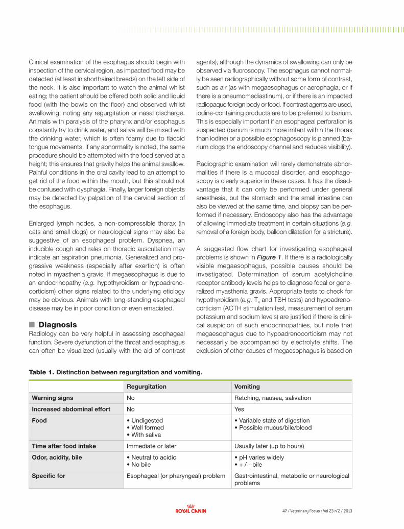
47 / Veterinary Focus / Vol 23 n°2 / 2013
l I I I I I I I I I I I I I I l I I I I I I I I I I I I I I l I I I I I I I I I I I I I I l I I I I I I I I I I I I I l I I I I I I I I I I I I I I l I I I I I I I I I I I I I I l I I I I I I I I I I I I I I l I I I I I I I I I I I I I I I I I I I I I I l I I I I I I I I I I I I I I l I I I I I I I I I I I I I I l I I I I I I I I I I I I I l I I I I I I I I I I I I I I l I I I I I I I I I I
Clinical examination of the esophagus should begin with inspection of the cervical region, as impacted food may be detected (at least in shorthaired breeds) on the left side of the neck. It is also important to watch the animal whilst eating; the patient should be offered both solid and liquid food (with the bowls on the floor) and observed whilst swallowing, noting any regurgitation or nasal discharge. Animals with paralysis of the pharynx and/or esophagus constantly try to drink water, and saliva will be mixed with the drinking water, which is often foamy due to flaccid tongue movements. If any abnormality is noted, the same procedure should be attempted with the food served at a height; this ensures that gravity helps the animal swallow. Painful conditions in the oral cavity lead to an attempt to get rid of the food within the mouth, but this should not be confused with dysphagia. Finally, larger foreign objects may be detected by palpation of the cervical section of the esophagus.
Enlarged lymph nodes, a non-compressible thorax (in cats and small dogs) or neurological signs may also be suggestive of an esophageal problem. Dyspnea, an inducible cough and rales on thoracic auscultation may indicate an aspiration pneumonia. Generalized and pro-gressive weakness (especially after exertion) is often noted in myasthenia gravis. If megaesophagus is due to an endocrinopathy (e.g. hypothyroidism or hypoadreno-corticism) other signs related to the underlying etiology may be obvious. Animals with long-standing esophageal disease may be in poor condition or even emaciated.
■ DiagnosisRadiology can be very helpful in assessing esophageal function. Severe dysfunction of the throat and esophagus can often be visualized (usually with the aid of contrast
agents), although the dynamics of swallowing can only be observed via fluoroscopy. The esophagus cannot normal-ly be seen radiographically without some form of contrast, such as air (as with megaesophagus or aerophagia, or if there is a pneumomediastinum), or if there is an impacted radiopaque foreign body or food. If contrast agents are used, iodine-containing products are to be preferred to barium. This is especially important if an esophageal perforation is suspected (barium is much more irritant within the thorax than iodine) or a possible esophagoscopy is planned (ba-rium clogs the endoscopy channel and reduces visibility).
Radiographic examination will rarely demonstrate abnor-malities if there is a mucosal disorder, and esophago-scopy is clearly superior in these cases. It has the disad-vantage that it can only be performed under general anesthesia, but the stomach and the small intestine can also be viewed at the same time, and biopsy can be per-formed if necessary. Endoscopy also has the advantage of allowing immediate treatment in certain situations (e.g. removal of a foreign body, balloon dilatation for a stricture).
A suggested flow chart for investigating esophageal problems is shown in Figure 1. If there is a radiologically visible megaesophagus, possible causes should be investigated. Determination of serum acetylcholine receptor antibody levels helps to diagnose focal or gene-ralized myasthenia gravis. Appropriate tests to check for hypothyroidism (e.g. T4 and TSH tests) and hypoadreno-corticism (ACTH stimulation test, measurement of serum potassium and sodium levels) are justified if there is clini-cal suspicion of such endocrinopathies, but note that megaesophagus due to hypoadrenocorticism may not necessarily be accompanied by electrolyte shifts. The exclusion of other causes of megaesophagus is based on
Regurgitation Vomiting
Warning signs No Retching, nausea, salivation
Increased abdominal effort No Yes
Food • Undigested• Well formed• With saliva
• Variable state of digestion• Possible mucus/bile/blood
Time after food intake Immediate or later Usually later (up to hours)
Odor, acidity, bile • Neutral to acidic• No bile
• pH varies widely• + / - bile
Specific for Esophageal (or pharyngeal) problem Gastrointestinal, metabolic or neurological problems
Table 1. Distinction between regurgitation and vomiting.
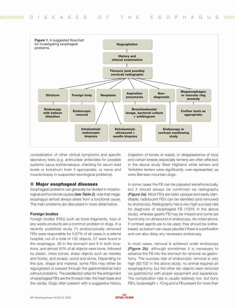
48 / Veterinary Focus / Vol 23 n°2 / 2013
l I I I I I I I I I I I I I I l I I I I I I I I I I I I I I l I I I I I I I I I I I I I I l I I I I I I I I I I I I I l I I I I I I I I I I I I I I l I I I I I I I I I I I I I I l I I I I I I I I I I I I I I l I I I I I I I I I I I I I I I I I I I I I I l I I I I I I I I I I I I I I l I I I I I I I I I I I I I I l I I I I I I I I I I I I I l Il I I I I I I I I I I I I I I l I I I I
Regurgitation
History andclinical examination
Thoracic (and possibly cervical) radiographs
Stricture Foreign body Neoplasia Aspiration pneumonia
Non-diagnostic
Megaesophagus or vascular ring
anomaly
Bronchoalveolarlavage, bacterial culture
+ antibiogram
Further tests as appropriate
Endoscopic removal
Endoscopy with balloon
dilatation
Endoscopy or contrast swallowing
study
Intraluminal:endoscopic
biopsies
Extraluminal:ultrasound +
needle biopsies
Figure 1. A suggested flowchart for investigating esophageal problems.
consideration of other clinical symptoms and specific laboratory tests (e.g. antinuclear antibodies for possible systemic lupus erythematosus, checking for serum lead levels or botulinum toxin if appropriate, or nerve and muscle biopsy in suspected neurological problems).
■ Major esophageal diseasesEsophageal problems can generally be divided in morpho-logical and functional causes (see Table 2); note that mega-esophagus almost always arises from a functional cause. The main problems are discussed in more detail below.
Foreign bodiesForeign bodies (FB’s) such as bone fragments, toys or any waste products are a common problem in dogs. In a recently published study (1) endoscopically removed FB’s were responsible for 0.67% of all cases in a referral hospital; out of a total of 102 objects, 57 were found in the esophagus, 36 in the stomach and 9 in both loca-tions, and almost 50% of all objects were bone, followed by plastic, chew bones, sharp objects such as needles and hooks, and scraps, wood and stone. Depending on the size, shape and material, some FB’s may either be regurgitated or passed through the gastrointestinal tract without problems. The predilection sites for the entrapment of esophageal FB’s are the thoracic inlet, the heart base and the cardia. Dogs often present with a suggestive history
(ingestion of bones or waste, or disappearance of toys) and certain breeds (especially terriers) are often affected; in the above study West Highland white terriers and Yorkshire terriers were significantly over-represented, as were Bernese mountain dogs.
In some cases the FB can be palpated extrathoracically, but it should always be confirmed via radiography (Figure 2a). Most FB’s are radio-opaque and easily iden-tifiable; radiolucent FB’s can be identified (and removed) by endoscopy. Radiography has a very high success rate for diagnosis of esophageal FB (100% in the above study), whereas gastric FB may be missed and some are found only on ultrasound or endoscopy. As noted above, if contrast agents are to be used, they should be iodine-based, as barium can cause pleuritis if there is a perforation and can also delay any necessary endoscopy.
In most cases, removal is achieved under endoscopy (Figure 2b), although sometimes it is necessary to advance the FB into the stomach for removal via gastro-tomy. The success rate of endoscopic removal is very high (92/102 in the above study; no animal required an esophagotomy, but the other ten objects were removed via gastrotomy) with proper equipment and experience. The complication rate is usually relatively low, but bony FB’s, bodyweight < 10 kg and a FB present for more than
D I S E A S E S O F T H E E S O P H A G U S
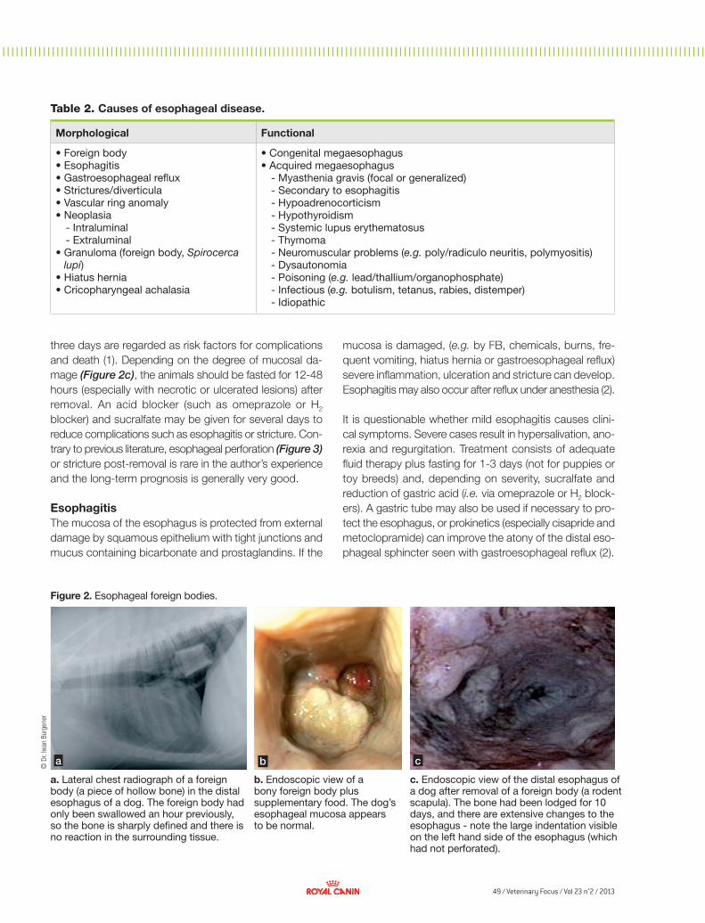
49 / Veterinary Focus / Vol 23 n°2 / 2013
l I I I I I I I I I I I I I I l I I I I I I I I I I I I I I l I I I I I I I I I I I I I I l I I I I I I I I I I I I I l I I I I I I I I I I I I I I l I I I I I I I I I I I I I I l I I I I I I I I I I I I I I l I I I I I I I I I I I I I I I I I I I I I I l I I I I I I I I I I I I I I l I I I I I I I I I I I I I I l I I I I I I I I I I I I I l I
a b c
© D
r. Iw
an B
urge
ner
three days are regarded as risk factors for complications and death (1). Depending on the degree of mucosal da-mage (Figure 2c), the animals should be fasted for 12-48 hours (especially with necrotic or ulcerated lesions) after removal. An acid blocker (such as omeprazole or H2 blocker) and sucralfate may be given for several days to reduce complications such as esophagitis or stricture. Con-trary to previous literature, esophageal perforation (Figure 3) or stricture post-removal is rare in the author’s experience and the long-term prognosis is generally very good.
EsophagitisThe mucosa of the esophagus is protected from external damage by squamous epithelium with tight junctions and mucus containing bicarbonate and prostaglandins. If the
Table 2. Causes of esophageal disease.
Morphological Functional
• Foreign body• Esophagitis• Gastroesophageal reflux• Strictures/diverticula• Vascular ring anomaly• Neoplasia
- Intraluminal- Extraluminal
• Granuloma (foreign body, Spirocerca lupi)
• Hiatus hernia • Cricopharyngeal achalasia
• Congenital megaesophagus • Acquired megaesophagus
- Myasthenia gravis (focal or generalized)- Secondary to esophagitis- Hypoadrenocorticism- Hypothyroidism- Systemic lupus erythematosus- Thymoma- Neuromuscular problems (e.g. poly/radiculo neuritis, polymyositis)- Dysautonomia - Poisoning (e.g. lead/thallium/organophosphate) - Infectious (e.g. botulism, tetanus, rabies, distemper)- Idiopathic
Figure 2. Esophageal foreign bodies.
a. Lateral chest radiograph of a foreign body (a piece of hollow bone) in the distal esophagus of a dog. The foreign body had only been swallowed an hour previously, so the bone is sharply defined and there is no reaction in the surrounding tissue.
b. Endoscopic view of a bony foreign body plus supplementary food. The dog’s esophageal mucosa appears to be normal.
c. Endoscopic view of the distal esophagus of a dog after removal of a foreign body (a rodent scapula). The bone had been lodged for 10 days, and there are extensive changes to the esophagus - note the large indentation visible on the left hand side of the esophagus (which had not perforated).
mucosa is damaged, (e.g. by FB, chemicals, burns, fre-quent vomiting, hiatus hernia or gastroesophageal reflux) severe inflammation, ulceration and stricture can develop. Esophagitis may also occur after reflux under anesthesia (2).
It is questionable whether mild esophagitis causes clini-cal symptoms. Severe cases result in hypersalivation, ano-rexia and regurgitation. Treatment consists of adequate fluid therapy plus fasting for 1-3 days (not for puppies or toy breeds) and, depending on severity, sucralfate and reduction of gastric acid (i.e. via omeprazole or H2 block-ers). A gastric tube may also be used if necessary to pro-tect the esophagus, or prokinetics (especially cisapride and metoclopramide) can improve the atony of the distal eso-phageal sphincter seen with gastroesophageal reflux (2).
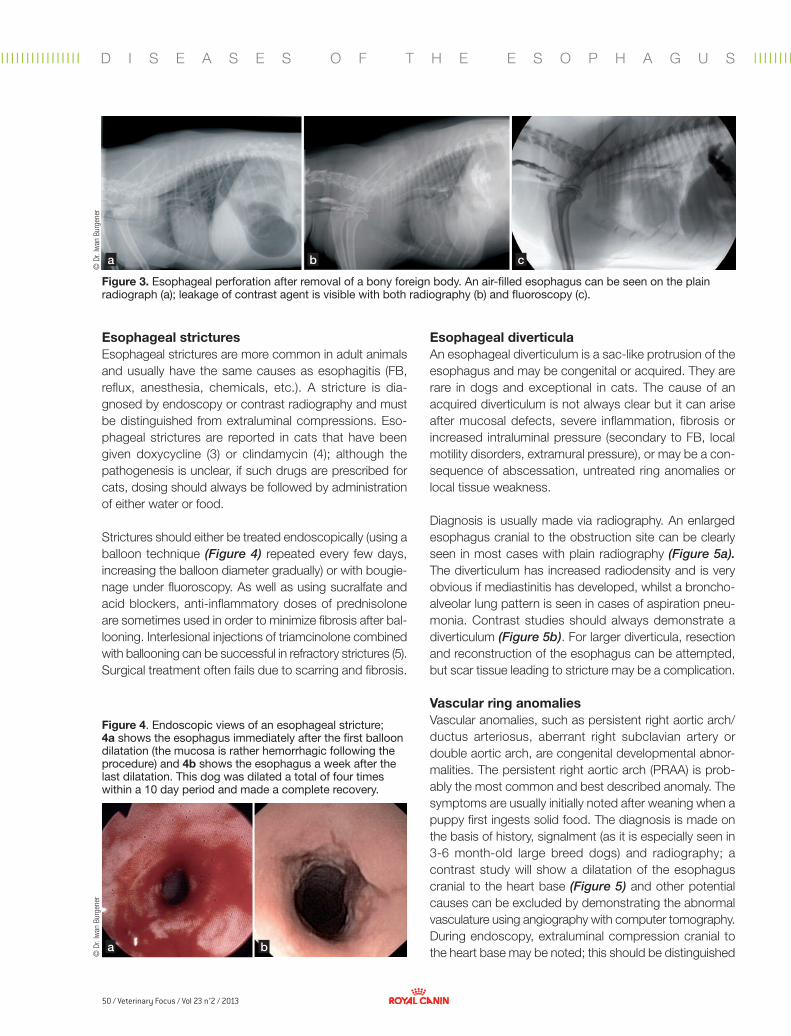
50 / Veterinary Focus / Vol 23 n°2 / 2013
l I I I I I I I I I I I I I I l I I I I I I I I I I I I I I l I I I I I I I I I I I I I I l I I I I I I I I I I I I I l I I I I I I I I I I I I I I l I I I I I I I I I I I I I I l I I I I I I I I I I I I I I l I I I I I I I I I I I I I I I I I I I I I I l I I I I I I I I I I I I I I l I I I I I I I I I I I I I I l I I I I I I I I I I I I I l Il I I I I I I I I I I I I I I l I I I I
a b c
a b
© D
r. Iw
an B
urge
ner
© D
r. Iw
an B
urge
ner
Esophageal stricturesEsophageal strictures are more common in adult animals and usually have the same causes as esophagitis (FB, reflux, anesthesia, chemicals, etc.). A stricture is dia-gnosed by endoscopy or contrast radiography and must be distinguished from extraluminal compressions. Eso-phageal strictures are reported in cats that have been given doxycycline (3) or clindamycin (4); although the pathogenesis is unclear, if such drugs are prescribed for cats, dosing should always be followed by administration of either water or food.
Strictures should either be treated endoscopically (using a balloon technique (Figure 4) repeated every few days, increasing the balloon diameter gradually) or with bougie-nage under fluoroscopy. As well as using sucralfate and acid blockers, anti-inflammatory doses of prednisolone are sometimes used in order to minimize fibrosis after bal-looning. Interlesional injections of triamcinolone combined with ballooning can be successful in refractory strictures (5). Surgical treatment often fails due to scarring and fibrosis.
Esophageal diverticulaAn esophageal diverticulum is a sac-like protrusion of the esophagus and may be congenital or acquired. They are rare in dogs and exceptional in cats. The cause of an acquired diverticulum is not always clear but it can arise after mucosal defects, severe inflammation, fibrosis or increased intraluminal pressure (secondary to FB, local motility disorders, extramural pressure), or may be a con-sequence of abscessation, untreated ring anomalies or local tissue weakness.
Diagnosis is usually made via radiography. An enlarged esophagus cranial to the obstruction site can be clearly seen in most cases with plain radiography (Figure 5a). The diverticulum has increased radiodensity and is very obvious if mediastinitis has developed, whilst a broncho-alveolar lung pattern is seen in cases of aspiration pneu-monia. Contrast studies should always demonstrate a diverticulum (Figure 5b). For larger diverticula, resection and reconstruction of the esophagus can be attempted, but scar tissue leading to stricture may be a complication.
Vascular ring anomaliesVascular anomalies, such as persistent right aortic arch/ductus arteriosus, aberrant right subclavian artery or double aortic arch, are congenital developmental abnor-malities. The persistent right aortic arch (PRAA) is prob-ably the most common and best described anomaly. The symptoms are usually initially noted after weaning when a puppy first ingests solid food. The diagnosis is made on the basis of history, signalment (as it is especially seen in 3-6 month-old large breed dogs) and radiography; a contrast study will show a dilatation of the esophagus cranial to the heart base (Figure 5) and other potential causes can be excluded by demonstrating the abnormal vasculature using angiography with computer tomography. During endoscopy, extraluminal compression cranial to the heart base may be noted; this should be distinguished
Figure 3. Esophageal perforation after removal of a bony foreign body. An air-filled esophagus can be seen on the plain radiograph (a); leakage of contrast agent is visible with both radiography (b) and fluoroscopy (c).
Figure 4. Endoscopic views of an esophageal stricture; 4a shows the esophagus immediately after the first balloon dilatation (the mucosa is rather hemorrhagic following the procedure) and 4b shows the esophagus a week after the last dilatation. This dog was dilated a total of four times within a 10 day period and made a complete recovery.
D I S E A S E S O F T H E E S O P H A G U S
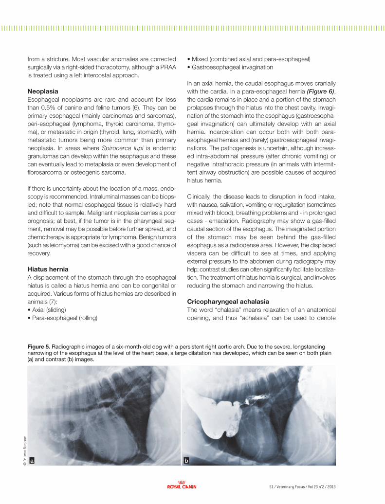
51 / Veterinary Focus / Vol 23 n°2 / 2013
l I I I I I I I I I I I I I I l I I I I I I I I I I I I I I l I I I I I I I I I I I I I I l I I I I I I I I I I I I I l I I I I I I I I I I I I I I l I I I I I I I I I I I I I I l I I I I I I I I I I I I I I l I I I I I I I I I I I I I I I I I I I I I I l I I I I I I I I I I I I I I l I I I I I I I I I I I I I I l I I I I I I I I I I I I I l I
a b
© D
r. Iw
an B
urge
ner
from a stricture. Most vascular anomalies are corrected surgically via a right-sided thoracotomy, although a PRAA is treated using a left intercostal approach.
NeoplasiaEsophageal neoplasms are rare and account for less than 0.5% of canine and feline tumors (6). They can be primary esophageal (mainly carcinomas and sarcomas), peri-esophageal (lymphoma, thyroid carcinoma, thymo-ma), or metastatic in origin (thyroid, lung, stomach), with metastatic tumors being more common than primary neoplasia. In areas where Spirocerca lupi is endemic granulomas can develop within the esophagus and these can eventually lead to metaplasia or even development of fibrosarcoma or osteogenic sarcoma.
If there is uncertainty about the location of a mass, endo-scopy is recommended. Intraluminal masses can be biops-ied; note that normal esophageal tissue is relatively hard and difficult to sample. Malignant neoplasia carries a poor prognosis; at best, if the tumor is in the pharyngeal seg-ment, removal may be possible before further spread, and chemotherapy is appropriate for lymphoma. Benign tumors (such as leiomyoma) can be excised with a good chance of recovery.
Hiatus herniaA displacement of the stomach through the esophageal hiatus is called a hiatus hernia and can be congenital or acquired. Various forms of hiatus hernias are described in animals (7):• Axial (sliding) • Para-esophageal (rolling)
Figure 5. Radiographic images of a six-month-old dog with a persistent right aortic arch. Due to the severe, longstanding narrowing of the esophagus at the level of the heart base, a large dilatation has developed, which can be seen on both plain (a) and contrast (b) images.
• Mixed (combined axial and para-esophageal)• Gastroesophageal invagination
In an axial hernia, the caudal esophagus moves cranially with the cardia. In a para-esophageal hernia (Figure 6), the cardia remains in place and a portion of the stomach prolapses through the hiatus into the chest cavity. Invagi-nation of the stomach into the esophagus (gastroesopha-geal invagination) can ultimately develop with an axial hernia. Incarceration can occur both with both para-esophageal hernias and (rarely) gastroesophageal invagi-nations. The pathogenesis is uncertain, although increas-ed intra-abdominal pressure (after chronic vomiting) or negative intrathoracic pressure (in animals with intermit-tent airway obstruction) are possible causes of acquired hiatus hernia.
Clinically, the disease leads to disruption in food intake, with nausea, salivation, vomiting or regurgitation (sometimes mixed with blood), breathing problems and - in prolonged cases - emaciation. Radiography may show a gas-filled caudal section of the esophagus. The invaginated portion of the stomach may be seen behind the gas-filled esophagus as a radiodense area. However, the displaced viscera can be difficult to see at times, and applying external pressure to the abdomen during radiography may help; contrast studies can often significantly facilitate localiza-tion. The treatment of hiatus hernia is surgical, and involves reducing the stomach and narrowing the hiatus.
Cricopharyngeal achalasiaThe word “chalasia” means relaxation of an anatomical opening, and thus “achalasia” can be used to denote
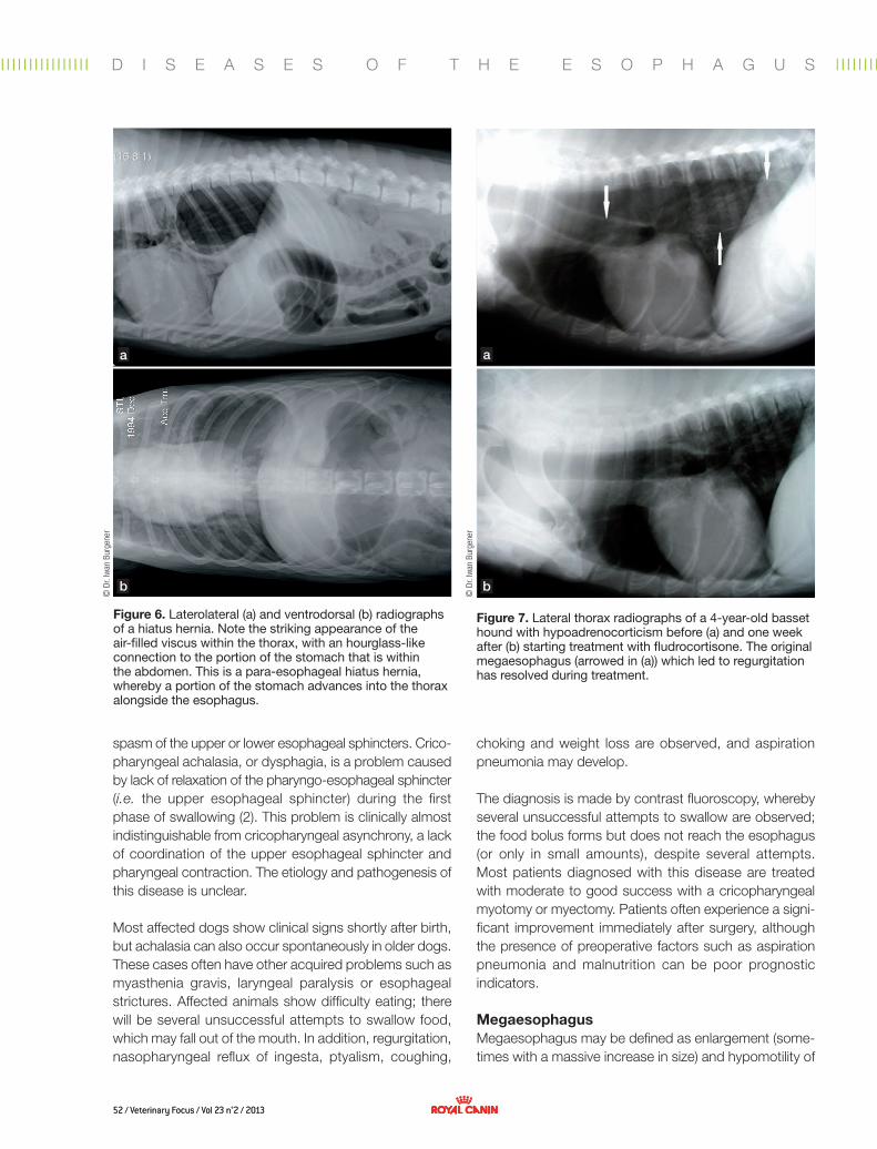
52 / Veterinary Focus / Vol 23 n°2 / 201352 / Veterinary Focus / Vol 23 n°2 / 2013
l I I I I I I I I I I I I I I l I I I I I I I I I I I I I I l I I I I I I I I I I I I I I l I I I I I I I I I I I I I l I I I I I I I I I I I I I I l I I I I I I I I I I I I I I l I I I I I I I I I I I I I I l I I I I I I I I I I I I I I I I I I I I I I l I I I I I I I I I I I I I I l I I I I I I I I I I I I I I l I I I I I I I I I I I I I l Il I I I I I I I I I I I I I I l I I I I
a a
b b
© D
r. Iw
an B
urge
ner
© D
r. Iw
an B
urge
ner
spasm of the upper or lower esophageal sphincters. Crico-pharyngeal achalasia, or dysphagia, is a problem caused by lack of relaxation of the pharyngo-esophageal sphincter (i.e. the upper esophageal sphincter) during the first phase of swallowing (2). This problem is clinically almost indistinguishable from cricopharyngeal asynchrony, a lack of coordination of the upper esophageal sphincter and pharyngeal contraction. The etiology and pathogenesis of this disease is unclear.
Most affected dogs show clinical signs shortly after birth, but achalasia can also occur spontaneously in older dogs. These cases often have other acquired problems such as myasthenia gravis, laryngeal paralysis or esophageal strictures. Affected animals show difficulty eating; there will be several unsuccessful attempts to swallow food, which may fall out of the mouth. In addition, regurgitation, nasopharyngeal reflux of ingesta, ptyalism, coughing,
choking and weight loss are observed, and aspiration pneumonia may develop.
The diagnosis is made by contrast fluoroscopy, whereby several unsuccessful attempts to swallow are observed; the food bolus forms but does not reach the esophagus (or only in small amounts), despite several attempts. Most patients diagnosed with this disease are treated with moderate to good success with a cricopharyngeal myotomy or myectomy. Patients often experience a signi-ficant improvement immediately after surgery, although the presence of preoperative factors such as aspiration pneumonia and malnutrition can be poor prognostic indicators.
MegaesophagusMegaesophagus may be defined as enlargement (some-times with a massive increase in size) and hypomotility of
Figure 7. Lateral thorax radiographs of a 4-year-old basset hound with hypoadrenocorticism before (a) and one week after (b) starting treatment with fludrocortisone. The original megaesophagus (arrowed in (a)) which led to regurgitation has resolved during treatment.
Figure 6. Laterolateral (a) and ventrodorsal (b) radiographs of a hiatus hernia. Note the striking appearance of the air-filled viscus within the thorax, with an hourglass-like connection to the portion of the stomach that is within the abdomen. This is a para-esophageal hiatus hernia, whereby a portion of the stomach advances into the thorax alongside the esophagus.
D I S E A S E S O F T H E E S O P H A G U S
53 / Veterinary Focus / Vol 23 n°2 / 2013 53 / Veterinary Focus / Vol 23 n°2 / 2013
1. Gianella P, Pfammatter NS, Burgener IA. Oesophageal and gastric endoscopic foreign body removal: complications and follow-up of 102 dogs. J Small Anim Pract 2009;50:649–654.
2. Moore LE. The esophagus. In: Steiner JM, ed. Small Animal Gastroenterology. Hannover, Schlütersche, 2008;139-150.
3. German AJ, Cannon MJ, Dye C, et al. Oesophageal strictures in cats associated with doxycycline therapy. J Feline Med Surg 2005;7:33-41.
4. Beatty JA, Swift N, Foster DJ, et al. Suspected clindamycin-associated oesophageal injury in cats: five cases. J Feline Med Surg 2006;8(6):412-9.
5. Fraune C, Gaschen F, Ryan K. Intralesional corticosteroid injection in addition
to endoscopic balloon dilation in a dog with benign oesophageal strictures. J Small Anim Pract 2009;50:550-553.
6. Hohenhaus AE. Neoplastic conditions of the esophagus. In: Steiner JM, ed. Small Animal Gastroenterology. Hannover, Schlütersche, 2008;151-153.
7. Hedlund CS. Surgery of the digestive system. In: Fossum TW, ed. Small Animal Surgery. St. Louis, Mosby, 2007;396-400.
8. Johnson BM, DeNovo RC, Mears EA. Canine megaesophagus. In: Bonagura JD, Twedt DC, eds. Kirk’s Current Veterinary Therapy XIV. St. Louis, Saunders Elsevier 2009;486-492.
l I I I I I I I I I I I I I I l I I I I I I I I I I I I I I l I I I I I I I I I I I I I I l I I I I I I I I I I I I I l I I I I I I I I I I I I I I l I I I I I I I I I I I I I I l I I I I I I I I I I I I I I l I I I I I I I I I I I I I I I I I I I I I I l I I I I I I I I I I I I I I l I I I I I I I I I I I I I I l I I I I I I I I I I I I I l I
the esophagus, and is a functional disorder that usually affects the entire length of the esophagus. Swallowed food and water remain in the esophagus and do not pass into the stomach. The disorder can be congenital (inhe-rent) or acquired. As noted above, since the dog is excep-tional in having striated muscle along the entire length of the esophagus, this species is particularly vulnerable to megaesophagus and consequently this can develop with diseases of striated muscle such as myasthenia gravis. Focal or generalized myasthenia, esophagitis, hypothy- roidism, hypoadrenocorticism (Figure 7) or thymomas are the most common causes of megaesophagus (see above), but often no cause is found (i.e. idiopathic). In the conge-nital form, signs usually appear whilst the animal is still growing. Secondary aspiration pneumonia can develop, and signs such as coughing, tachy/dyspnea, fever and general ill-health may also be noted in addition to any symptoms of the underlying disease.
Megaesophagus can most easily be diagnosed via radio-graphy (Figure 7a): the dorsal and ventral walls of the esophagus are normally invisible but can be identified within the mediastinum if the esophagus fills with either air, water or food, and the esophagus is often most easily visualized caudal to the heart. The dorsal border of the trachea is often visible as a very sharp, well defined line that contrasts with air in the esophagus. In cases of doubt, it helps to use a contrast agent, although this
increases the risk of aspiration pneumonia. Endoscopic examinations are generally not required but can be use-ful, if inflammation, tumor, hiatus hernia or a foreign body need to be excluded.
When presented with an animal that has megaesophagus, the objectives are to identify and treat the underlying cause (Figure 7b), reduce the incidence of regurgitation and esophageal enlargement, ensure adequate nutrition, and treat/prevent complications such as esophagitis or aspiration pneumonia (8). Prokinetic drugs (e.g. cisapride) are helpful in cats, where they work best at the level of the smooth muscle, but are of little use in dogs. If the cause is not identified and cannot be specifically treated, the long-term prognosis is usually poor. Special feeding management (feeding at a height, offering small pellets of food, etc.) can improve the long-term prognosis a little, especially by reducing the risk of aspiration pneumonia.
■ ConclusionThe esophagus is prone to various disorders and the clinician should adopt a standard approach to the exami-nation and diagnosis of such cases. The prognosis can be very variable depending on the underlying cause, and for some conditions treatment may be required long-term. Megaesophagus in particular can be challenging to treat and every effort should be made to reach a definitive dia-gnosis in all cases.
References
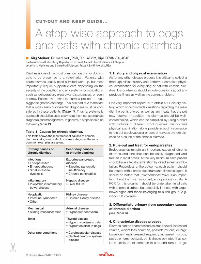
54 / Veterinary Focus / Vol 23 n°2 / 2013
■
✂✂
A step-wise approach to dogs and cats with chronic diarrhea
Jörg Steiner, Dr. med. vet., PhD, Dipl. ACVIM, Dipl. ECVIM-CA, AGAF Gastrointestinal Laboratory, Department of Small Animal Clinical Sciences, College of Veterinary Medicine and Biomedical Sciences, Texas A&M University, USA
Diarrhea is one of the most common reasons for dogs or cats to be presented to a veterinarian. Patients with acute diarrhea usually need a limited work-up, but most importantly require supportive care depending on the severity of the condition and any systemic complications, such as dehydration, electrolyte disturbances, or even anemia. Patients with chronic diarrhea present a much bigger diagnostic challenge. This is in part due to the fact that a wide variety of differential diagnoses must be con-sidered in these patients (Table 1). Thus, a systematic approach should be used to arrive at the most appropriate diagnosis and management. In general, 6 steps should be followed (Table 2).
Table 1. Causes for chronic diarrhea.
Primary causes of chronic diarrhea
Secondary causes of chronic diarrhea
Infectious • Endoparasites• Enteropathogens• Small intestinal
dysbiosis
Exocrinepancreaticdisease• Exocrine pancreatic
insufficiency• Chronic pancreatitis
Inflammatory• Idiopathic inflammatory
bowel disease
Hepaticdisease• Liver failure
Neoplastic• Intestinal lymphoma• Other
Kidneydisease• Chronic kidney disease
Mechanical• Sliding intussusception
Adrenaldisease• Hypoadrenocorticism
Toxic Thyroiddisease• Hyperthyroidism in cats• Hypothyroidism in dogs
Otherrareconditions • Cardiovasculardisease• Centralnervoussystem
disease
This table shows the most frequent causes of chronic diarrhea in dogs and cats. For some categories the most common examples are given.
1. History and physical examinationAs for any other disease process it is critical to collect a thorough clinical history and perform a complete physi-cal examination for every dog or cat with chronic diar-rhea. History-taking should include questions about any previous illness as well as the current problem.
One very important aspect is to obtain a full dietary his-tory, which should include questions regarding the main diet the pet is offered as well as any treats that the pet may receive. In addition the diarrhea should be well-characterized, which can be simplified by using a chart with pictures of different stool qualities. History and physical examination alone provide enough information to rule out cardiovascular or central nervous system dis-ease as a cause of the chronic diarrhea.
2. Rule-out and treat for endoparasitesEndoparasites remain an important cause of chronic diarrhea and one that can be easily diagnosed and treated in most cases. At the very minimum each patient should have a fecal examination by direct smear and flo-tation. Regardless of the outcome, each patient should be treated with a broad-spectrum anthelminthic agent. It should be noted that Tritrichomonas fetus is an impor-tant, if not the most important, endoparasite in cats. A PCR for this organism should be undertaken in all cats with chronic diarrhea, but especially in those with large-bowel signs and those belonging to a risk group (e.g. indoor cat colonies).
3. Differentiate primary from secondary causes of chronic diarrhea (see Table 3)
4. Characterize disease processDiarrhea can be characterized as small bowel (increased volume, weight loss common, possible melena) or large bowel diarrhea (increased frequency, increased mucous, possible hematochezia), but it should be noted that iso-lated colitis is not common in cats and rare in dogs.
CUT-OUT AND KEEP GUIDE...

55 / Veterinary Focus / Vol 23 n°2 / 2013
l I I I I I I I I I I I I I I l I I I I I I I I I I I I I I l I I I I I I I I I I I I I I l I I I I I I I I I I I I I l I I I I I I I I I I I I I I l I I I I I I I I I I I I I I l I I I I I I I I I I I I I I l I I I I I I I I I I I I I I I I I I I I I I l I I I I I I I I I I I I I I l I I I I I I I I I I I I I I l I I I I I I I I I I I I I l I I I I I I I I I I I I I I l I I I I I I I I I I
Table 2. Systematic work-up for dogs and cats with chronic diarrhea.
Step # Diagnostic step
1 History and physical examination
2 Rule-out and treat for endoparasites
3 Differentiate primary from secondary causes of chronic diarrhea
4 Characterize disease process
5 Therapeutic trial (if not contraindicated)
6 Histopathologic evaluation of biopsies
Table 3. Rule-out secondary causes of chronic diarrhea in dogs and cats.
Condition Diagnostic test
Exocrine pancreatic insufficiency
Decreased serum TLI concentration
Chronic pancreatitis
Increased serum PLI concentration (as measured by Spec cPL and Spec fPL assay)
Liver failure
Decreased serum albumin, cholesterol, BUN, and/or glucose concentrations; increased serum bilirubin concentration
Chronic kidney disease
Increased serum creatinine and BUN concentration; decreased urine specific gravity
Hypoadreno-corticism
Decreased serum sodium and increased serum potassium osmolality and lack of a stress-leukogram (> 2,500 lymphocytes/mL or > 500 eosinophils/mL); baseline cortisol concentration if any suspicion; confirmation with ACTH stimulation test
Hyperthyroidism in cats
Total T4, if not increased then free T4; thyroid scan may be needed if unable to rule out hyperthyroidism
Hypothyroidism in dogs
Total T4, if low then free T4; further diagnostics may be needed if thyroid status is still unclear
Another way to further characterize the disease process is by measuring serum cobalamin and folate concentrations.
Serum folate concentration can be decreased in patients with severe and long-standing proximal or dif-fuse small intestinal disease and can be increased in patients with small intestinal dysbiosis. Serum cobala-min concentration can be decreased with severe and long-standing distal or diffuse small intestinal disease, EPI, or small intestinal dysbiosis. 5. Therapeutic trials (if not contraindicated)It should first be noted that a therapeutic trial is con-traindicated in patients that are emaciated, have severe hypoalbuminemia, or some other systemic complication.
If the patient is cobalamin deficient, cobalamin should be supplemented by parenteral application of pure vitamin B12 (see protocol at http://www.vetmed.tamu.edu/gilab/research/cobalamin-information#dosing). Dietary trials are effective in up to 60% of dogs and cats with chronic diarrhea. Various diet types may be employed for these patients (Table 4), but at the cur-rent time only limited data is available concerning the use of high-fiber or low-carbohydrate diets.
Over the last decade many experimental and clinical studies have been presented that would suggest that the microbiota (the collection of all microorganisms in the GI tract) play a role in the pathogenesis of chronic gastrointestinal disease. Thus, one strategy for a thera-peutic trial in dogs and cats with chronic diarrhea is the manipulation of the gastrointestinal microbiota (Table 5).
Anti-inflammatory and immunosuppressive agents could also be employed for a therapeutic trial. One should always strive to make a definitive diagnosis before use of these agents. However, when the client declines this option, a therapeutic trial with such an agent is reasonable (Table 6).
6. Histopathologic evaluation of biopsiesBiopsy samples for histopathological evaluation can be collected by endoscopy (least invasive), laparoscopy (least desirable), or exploratory laparotomy (most inva-sive). Regardless of the route of biopsy collection, mul-tiple high-quality biopsies should be collected from each bowel segment and submitted for evaluation. However,
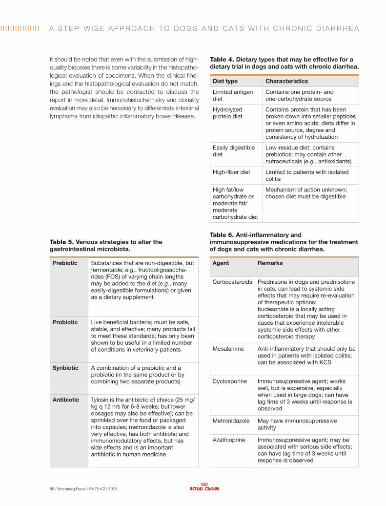
56 / Veterinary Focus / Vol 23 n°2 / 2013
l I I I I I I I I I I I I I I l I I I I
Diet type Characteristics
Limited antigen diet
Contains one protein- and one-carbohydrate source
Hydrolyzed protein diet
Contains protein that has been broken down into smaller peptides or even amino acids; diets differ in protein source, degree and consistency of hydrolization
Easily digestible diet
Low-residue diet; contains prebiotics; may contain other nutraceuticals (e.g., antioxidants)
High-fiber diet Limited to patients with isolated colitis
High fat/low carbohydrate or moderate fat/ moderate carbohydrate diet
Mechanism of action unknown; chosen diet must be digestible
Table 4. Dietary types that may be effective for a dietary trial in dogs and cats with chronic diarrhea.
Agent Remarks
Corticosteroids Prednisone in dogs and prednisolone in cats; can lead to systemic side effects that may require re-evaluation of therapeutic options;budesonide is a locally acting corticosteroid that may be used in cases that experience intolerable systemic side effects with other corticosteroid therapy
Mesalamine Anti-inflammatory that should only be used in patients with isolated colitis; can be associated with KCS
Cyclosporine Immunosuppressive agent; works well, but is expensive, especially when used in large dogs; can have lag time of 3 weeks until response is observed
Metronidazole May have immunosuppressive activity
Azathioprine Immunosuppressive agent; may be associated with serious side effects; can have lag time of 3 weeks until response is observed
Table 5. Various strategies to alter the gastrointestinal microbiota.
Table 6. Anti-inflammatory and immunosuppressive medications for the treatment of dogs and cats with chronic diarrhea.
it should be noted that even with the submission of high-quality biopsies there is some variability in the histopatho-logical evaluation of specimens. When the clinical find-ings and the histopathological evaluation do not match, the pathologist should be contacted to discuss the report in more detail. Immunohistochemistry and clonality evaluation may also be necessary to differentiate intestinal lymphoma from idiopathic inflammatory bowel disease.
Prebiotic Substances that are non-digestible, but fermentable; e.g., fructooligosaccha-rides (FOS) of varying chain lengths may be added to the diet (e.g., many easily-digestible formulations) or given as a dietary supplement
Probiotic Live beneficial bacteria; must be safe, stable, and effective; many products fail to meet these standards; has only been shown to be useful in a limited number of conditions in veterinary patients
Synbiotic A combination of a prebiotic and a probiotic (in the same product or by combining two separate products)
Antibiotic Tylosin is the antibiotic of choice (25 mg/kg q 12 hrs for 6-8 weeks; but lower dosages may also be effective); can be sprinkled over the food or packaged into capsules; metronidazole is also very effective, has both antibiotic and immunomodulatory effects, but has side effects and is an important antibiotic in human medicine
A S T E P - W I S E A P P R O A C H T O D O G S A N D C AT S W I T H C H R O N I C D I A R R H E A
We welcome offers to write ideas for papers and suggestions for topics and authors, which should be directed to the editor. Veterinary Focus is fully covered by copyright. No part of this publication may be reproduced, copied or transmitted in any form or by any means (including graphic, electronic or mechanical), without the written consent of the publishers © Royal Canin 2013. Proprietary names (trademarks) have not been specially identified. It cannot, however, be conducted from the omission of such information that they are non-proprietary names and as such can be used by everyone. The publishers cannot take any responsibility for information provided on dosages and methods of application. Details of this kind must be checked for correctness by the individual user in the appropriate literature. While every effort has been made by the translators to ensure the accuracy of their translations, no responsibility for the correctness of the original articles and thus no resulting claims against professional negligence can be accepted in this connection. Views expressed by authors or contributors do not necessarily reflect the views of the publishers, editors or editorial advisors.
ANNUAL CONGRESSOF THE ESVD-ECVD26th
SAVE THE DATE
18-21 SEPTEMBER 2013 VALENCIA - SPAIN
For more information about the Programme, Speakers, Registration, Venue, visit www.esvd-ecvd2013.com
ESVD – European Society of Veterinary Dermatology
ECVD – European College of Veterinary Dermatology Long Term Partners
• Scientifi c and continuing education programme• Free communications and posters
President ESVD: Luc Beco, BelgiumPresident ECVD: Richard Harvey, United Kingdom President Scientifi c Organizing Committee: Thierry Olivry, USA
Conference topics: • Skin barrier: from biology to allergic pathology in humans and animals• Topical therapy: from pharmacology to antiallergic use • Demodicosis: diagnosis and therapy• Malassezia dermatitis: diagnosis and therapy• Surgical treatment of chronic pododermatitis• Immunomodulators in Veterinary Dermatology• Autoimmune skin diseases and their mimickers• Lasers in Veterinary Dermatology
Valencia Conference Centre www.palcongres-vlc.com