Embed Size (px)
Citation preview
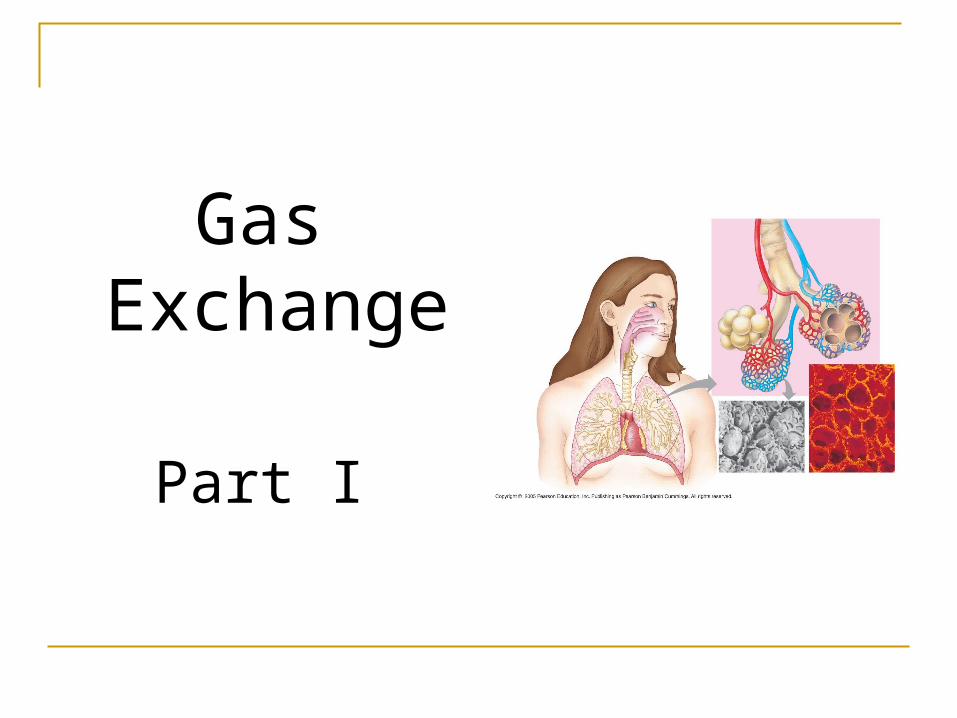
Gas Exchange
Part I
Respiration –
taking up O2 giving up CO2
Photosynthesis –
taking up CO2, giving up O2.
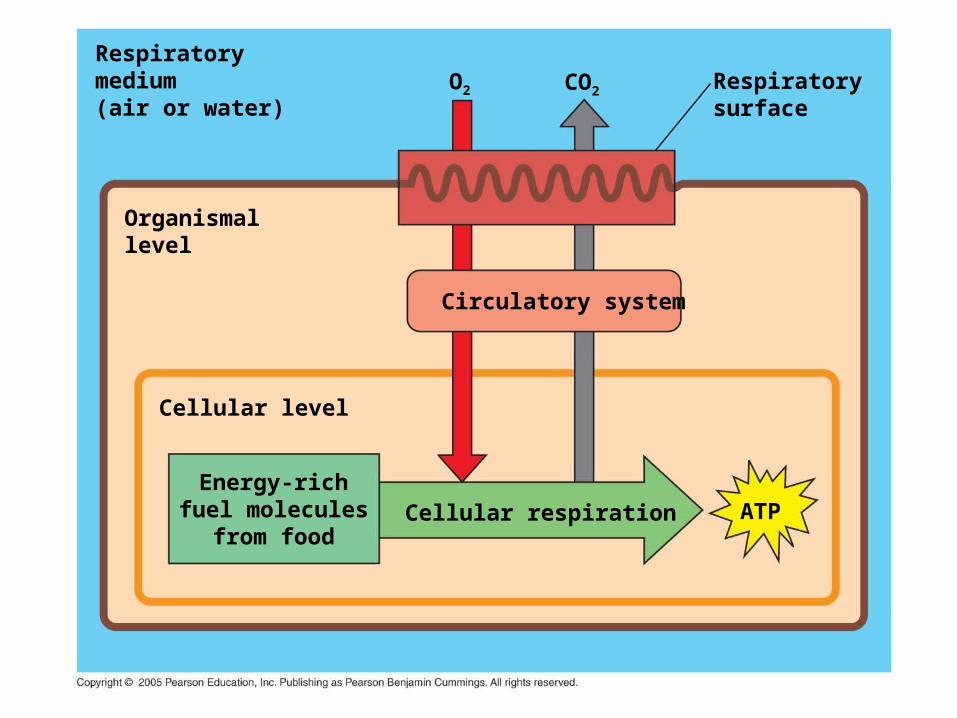
Respiratorymedium(air or water)
Organismallevel
Cellular level
Energy-richfuel molecules
from food
Respiratorysurface
Circulatory system
Cellular respiration
CO2O2
ATP
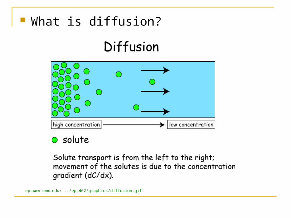
What is diffusion?
epswww.unm.edu/.../eps462/graphics/diffusion.gif
Depends on partial pressure, surface area
A gas always diffuses from an area of high partial pressure to low partial pressure.
What is equilibrium?
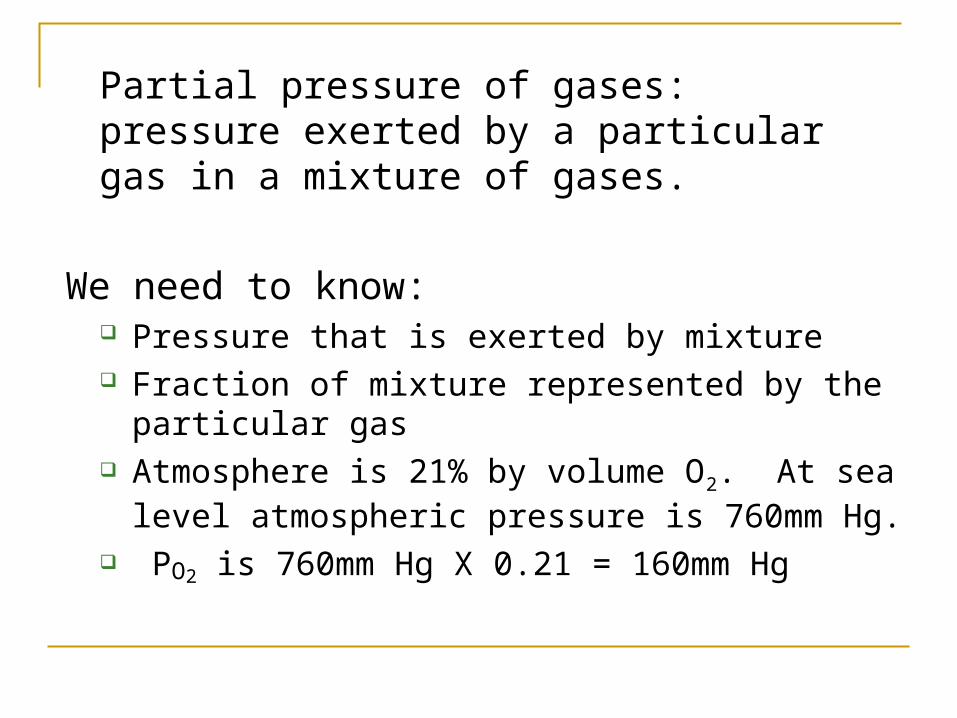
Partial pressure of gases: pressure exerted by a particular gas in a mixture of gases.
We need to know: Pressure that is exerted by mixture Fraction of mixture represented by the particular
gas Atmosphere is 21% by volume O2. At sea level
atmospheric pressure is 760mm Hg. PO2 is 760mm Hg X 0.21 = 160mm Hg
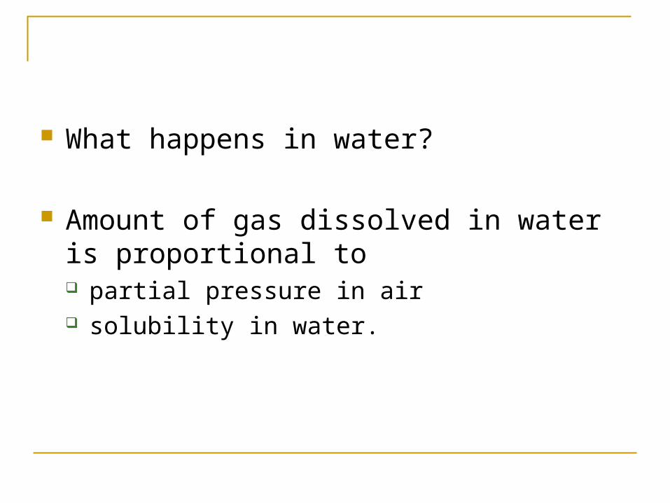
What happens in water?
Amount of gas dissolved in water is proportional to partial pressure in air solubility in water.
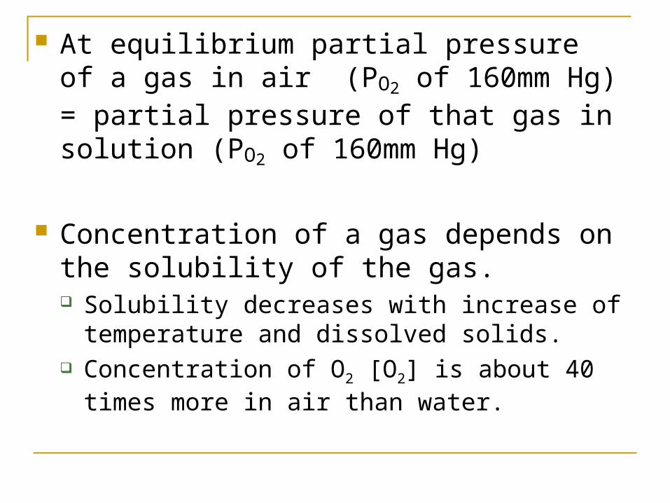
At equilibrium partial pressure of a gas in air (PO2 of 160mm Hg) = partial pressure of that gas in solution (PO2 of 160mm Hg)
Concentration of a gas depends on the solubility of the gas. Solubility decreases with increase of temperature
and dissolved solids. Concentration of O2 [O2] is about 40 times more in
air than water.
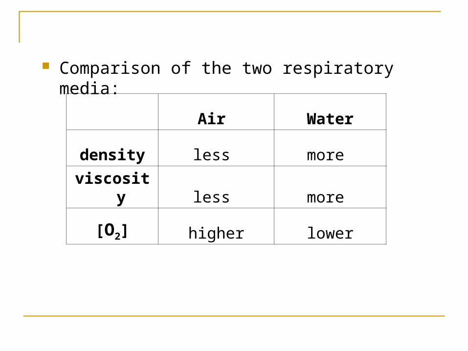
Comparison of the two respiratory media:
Air Water
density less more
viscosity less more
[O2] higher lower
Aquatic animals have had to evolve very effective and efficient gas exchange strategies.
Respiratory surfaces are plasma membranes which must be moist. Gas exchange takes place by diffusion.
Rate of diffusion is directly proportional to the surface area across
which it occurs inversely proportional to the square of the
distance the molecules have to travel. To speed up the rate of diffusion, respiratory
surfaces have to be LARGE and THIN.
In unicellular and simple animals diffusion occurs between all cells and environment.
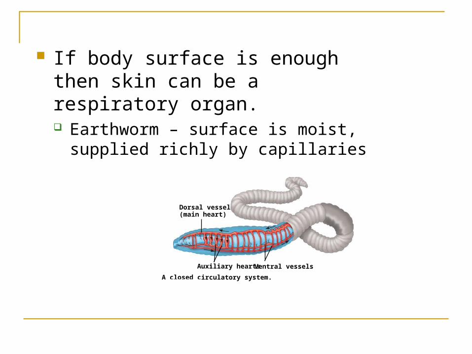
If body surface is enough then skin can be a respiratory organ. Earthworm – surface is moist, supplied
richly by capillaries
LE 42-3
A closed circulatory system.
Auxiliary hearts Ventral vessels
Dorsal vessel(main heart)
If body surface area is insufficient – need for specialized respiratory organs
Larger animals have respiratory organs consisting of respiratory surfaces and other structures.
Size of respiratory surface depends on Size of organism Metabolic demands
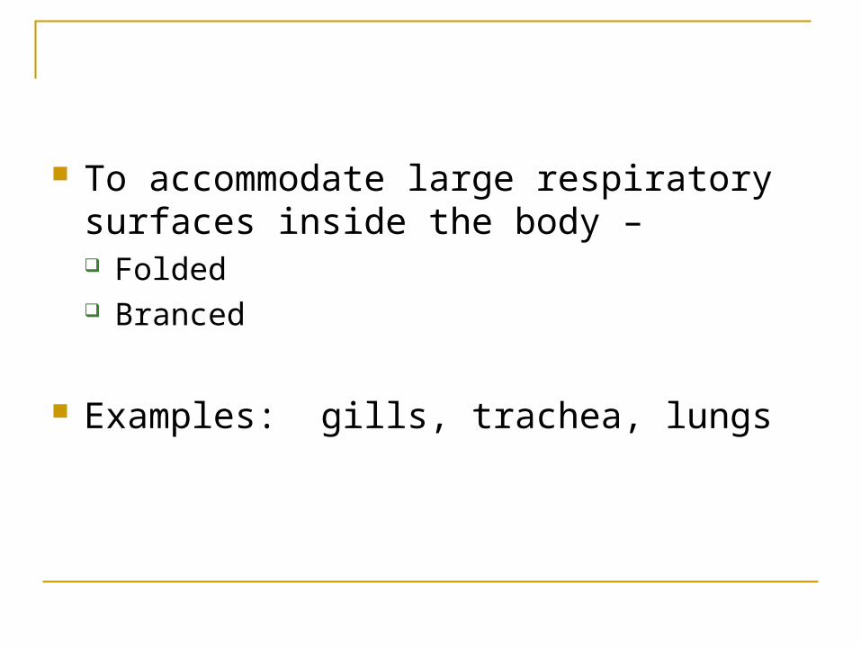
To accommodate large respiratory surfaces inside the body – Folded Branced
Examples: gills, trachea, lungs
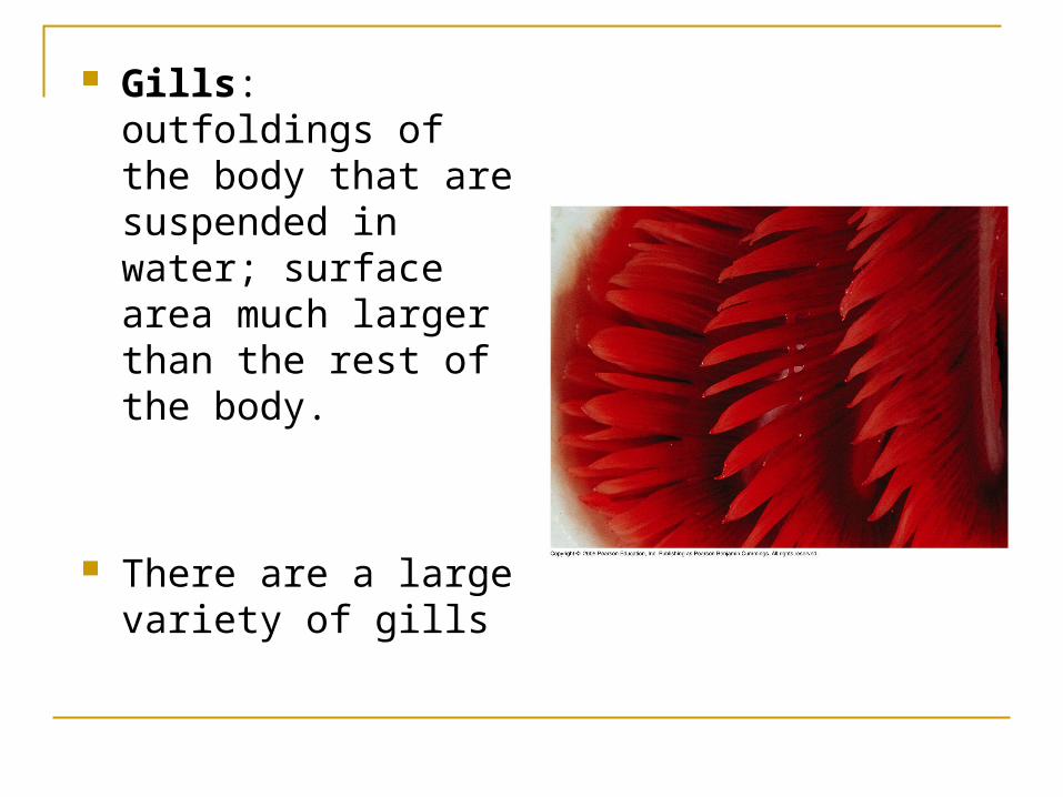
Gills: outfoldings of the body that are suspended in water; surface area much larger than the rest of the body.
There are a large variety of gills

Gill
Parapodia
Marine wormMarine Worm
LE 42-20d
Gills
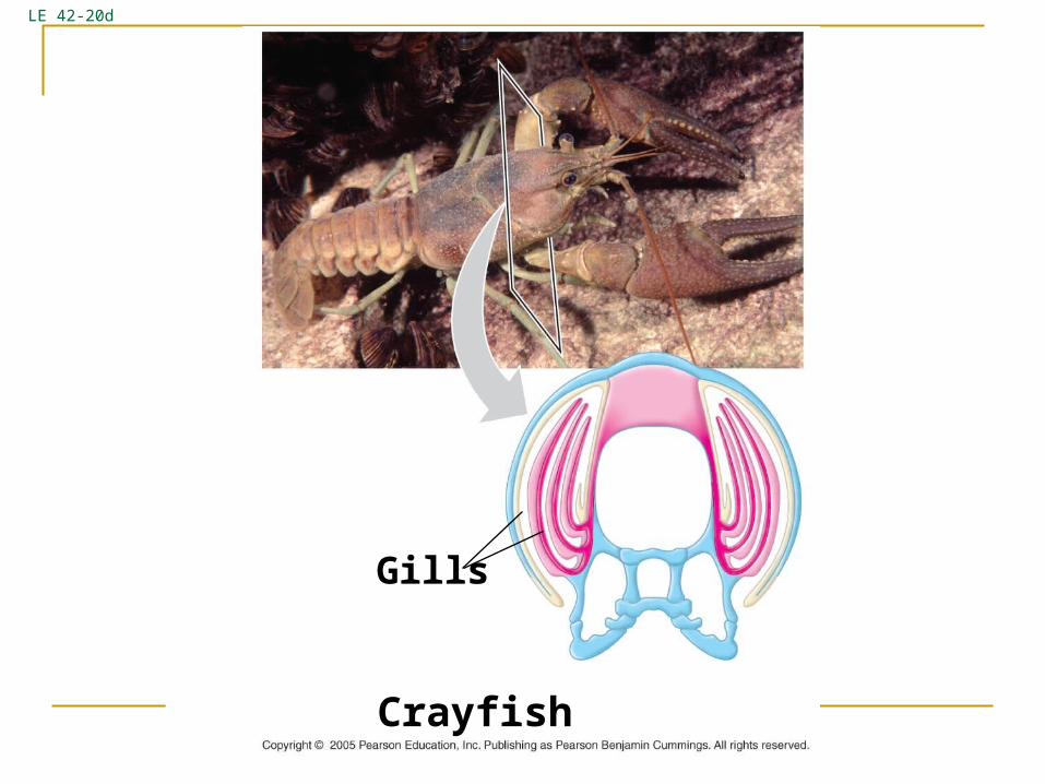
CrayfishCrayfish
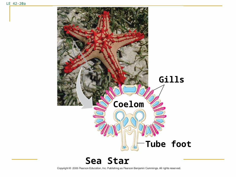
LE 42-20a
Gills
Coelom
Tube foot
Sea starSea Star
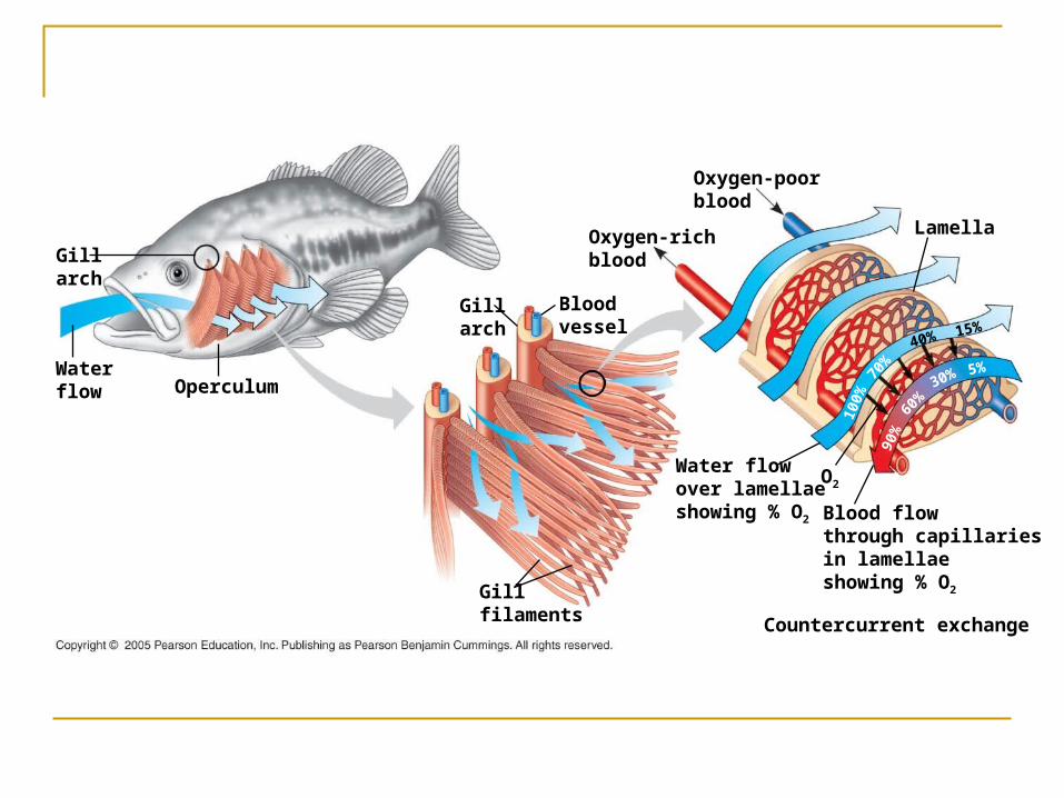
Gillarch
Waterflow Operculum
Gillarch
Bloodvessel
Oxygen-richblood
Water flowover lamellaeshowing % O2
Gillfilaments
O2
Oxygen-poorblood
Lamella
15%40%
70%
100%
90%
60%
30% 5%
Blood flowthrough capillariesin lamellaeshowing % O2
Countercurrent exchange
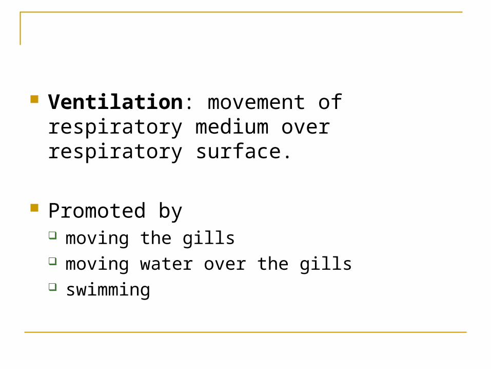
Ventilation: movement of respiratory medium over respiratory surface.
Promoted by moving the gills moving water over the gills swimming
Countercurrent exchange: exchange of substance between two fluids (blood and water) flowing in opposite directions and thereby maximizing gas exchange efficiency (about 80%)
Gills are unsuitable for land: water supports the filaments and keep them
separate gills would dry up
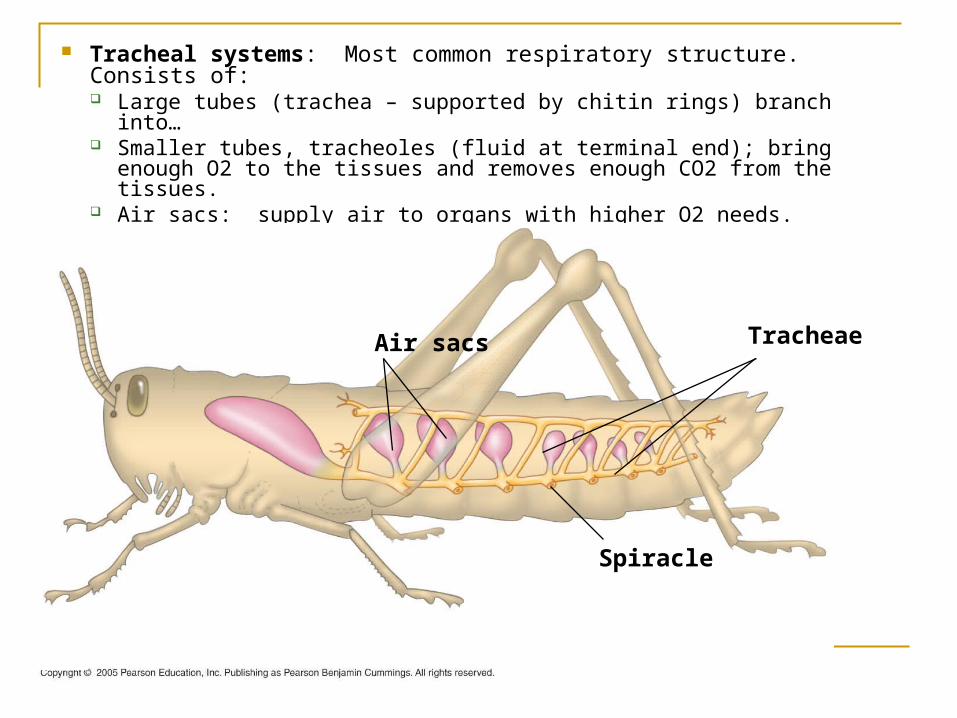
Tracheal systems: Most common respiratory structure. Consists of: Large tubes (trachea – supported by chitin rings) branch into… Smaller tubes, tracheoles (fluid at terminal end); bring enough O2
to the tissues and removes enough CO2 from the tissues. Air sacs: supply air to organs with higher O2 needs.
Air sacs Tracheae
Spiracle
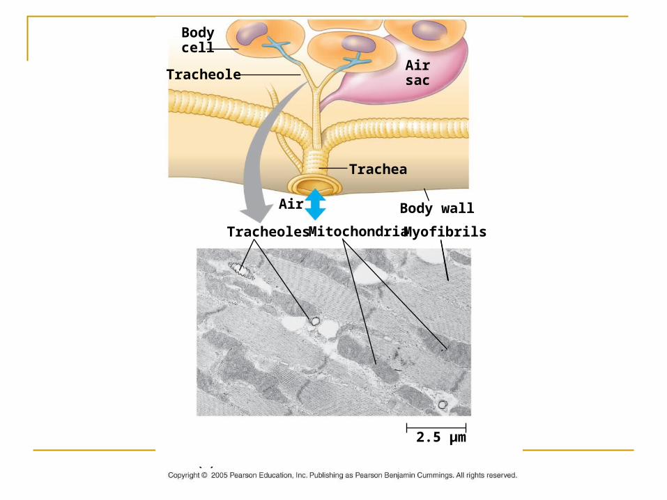
Bodycell
TracheoleAirsac
Trachea
Air Body wall
MyofibrilsTracheoles Mitochondria
2.5 µm
O2 demand can go up during flight by up to 200X.
The demand is met by: Contraction and relaxation of the flight muscles
pumps air through the tracheal system
Flight muscles rich in mitochondria.
Withdrawal of fluid from tracheole into body increases surface area.
Lungs: localized respiratory organs; inflodings of the body surface separated
consisting of numerous small pockets.
Circulatory system transports O2 to the body from the lungs and CO2 from the body to the lungs
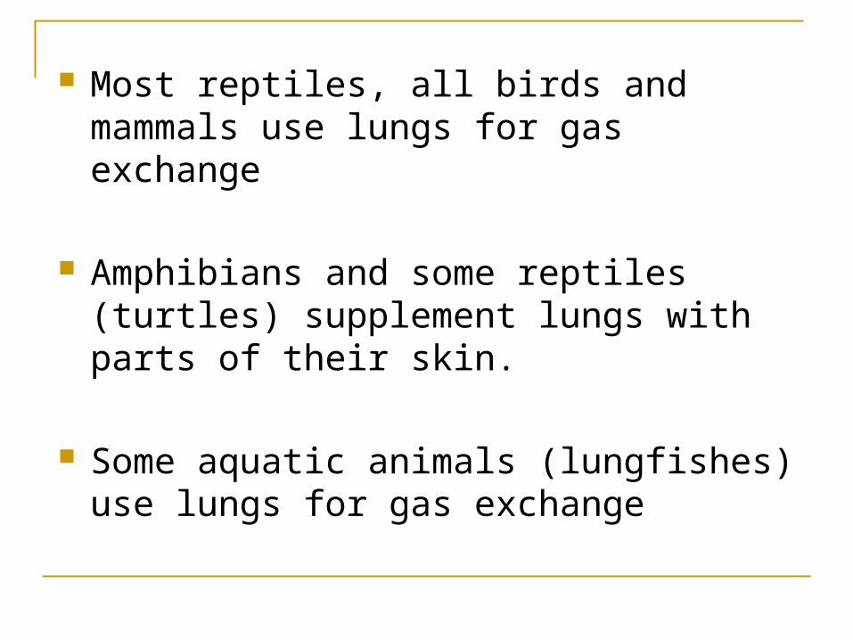
Most reptiles, all birds and mammals use lungs for gas exchange
Amphibians and some reptiles (turtles) supplement lungs with parts of their skin.
Some aquatic animals (lungfishes) use lungs for gas exchange
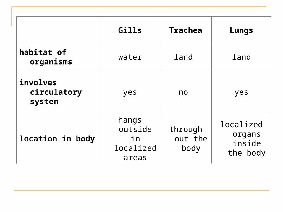
Gills Trachea Lungs
habitat of organisms water land land
involves circulatory system
yes no yes
location in bodyhangs outside
in localized areas
through out the body
localized organs
inside the body
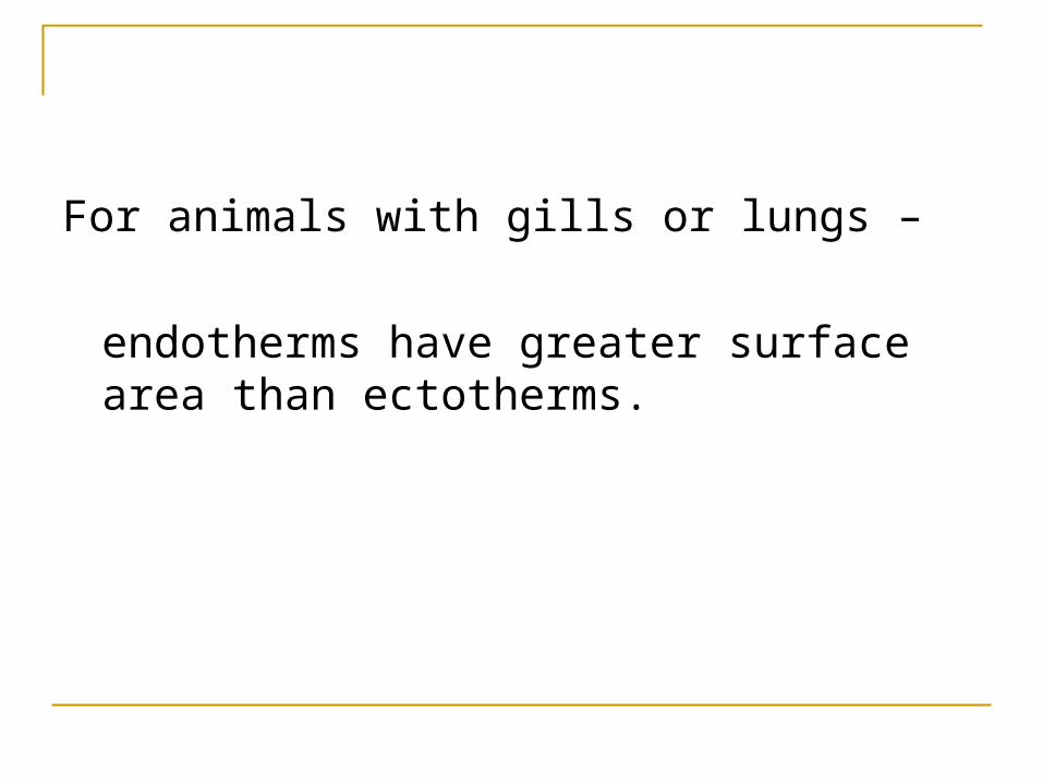
For animals with gills or lungs –
endotherms have greater surface area than ectotherms.

Gas Exchange
Part II
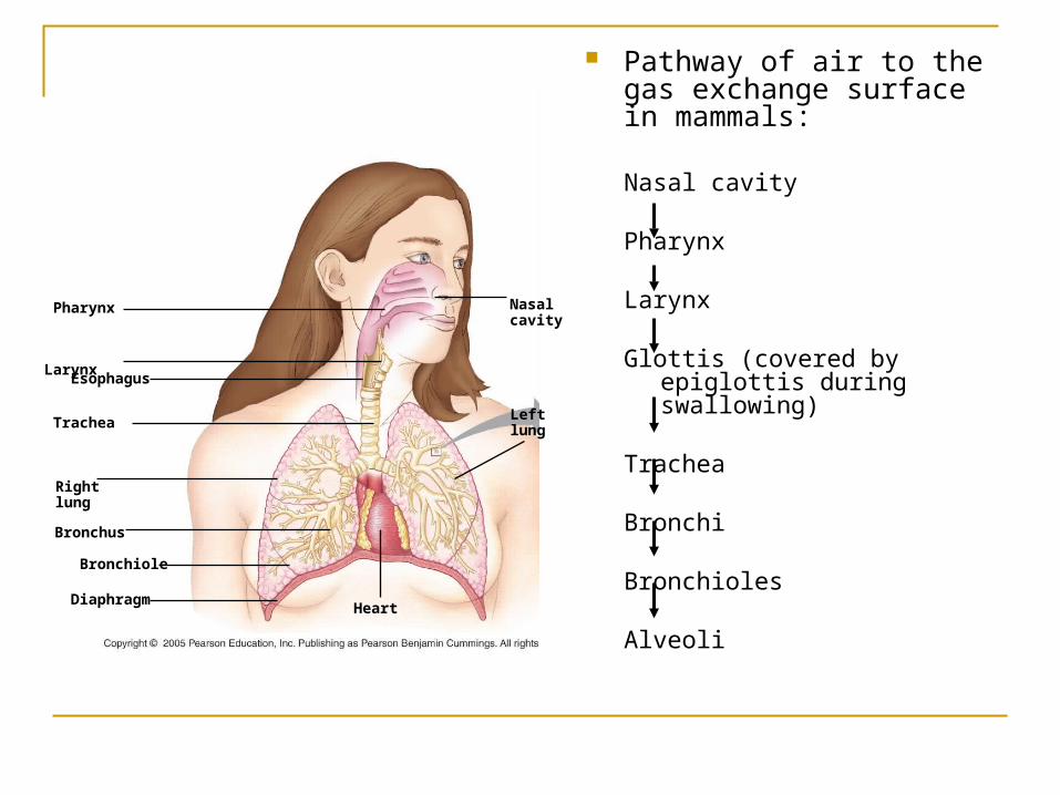
Pathway of air to the gas exchange surface in mammals:
Nasal cavity
Pharynx
Larynx
Glottis (covered by epiglottis during swallowing)
Trachea
Bronchi
Bronchioles Alveoli
Nasalcavity
Leftlung
Heart
Larynx
Pharynx
Esophagus
Trachea
Rightlung
Bronchus
Bronchiole
Diaphragm
Mucus traps dust, beating cilia move the mucus to esophagus
Millions of alveoli in lungs, total area about 100 m2.
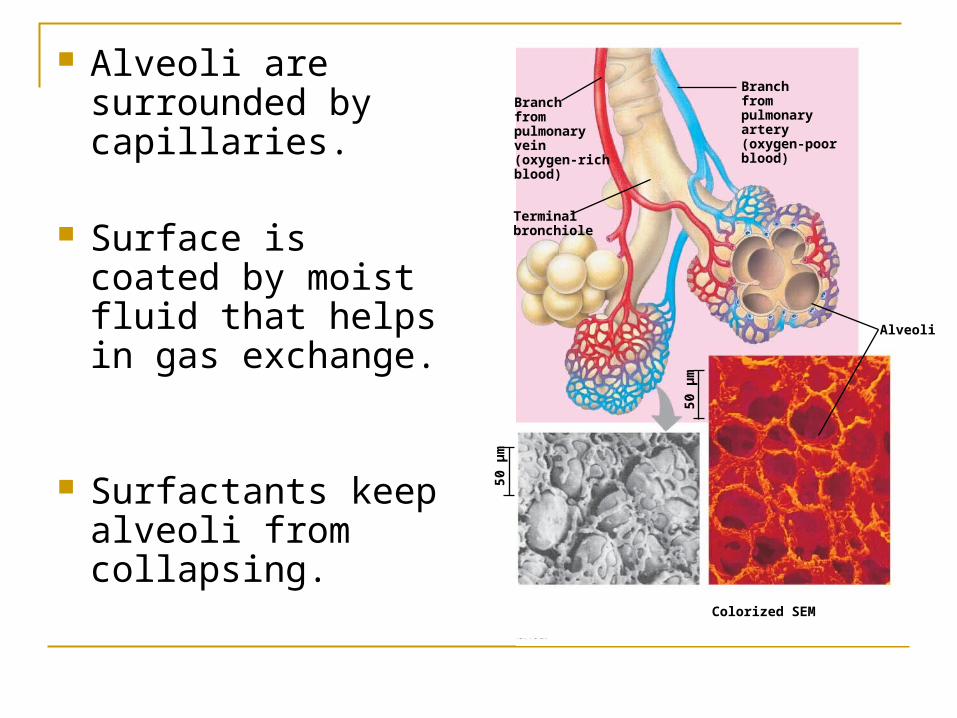
Alveoli are surrounded by capillaries.
Surface is coated by moist fluid that helps in gas exchange.
Surfactants keep alveoli from collapsing.
Branchfrompulmonaryvein(oxygen-richblood)
Terminalbronchiole
Branchfrompulmonaryartery(oxygen-poorblood)
Alveoli
50 µ
m
Colorized SEMSEM
50 µ
m
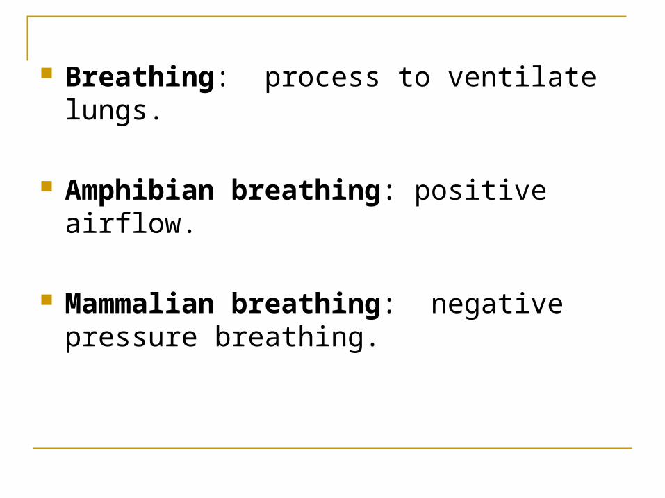
Breathing: process to ventilate lungs.
Amphibian breathing: positive airflow.
Mammalian breathing: negative pressure breathing.
Mammalian breathing During inhalation - expand thoracic cavity, causes
lower air pressure in thoracic chamber, air rushes in; opposite process for exhalation.
Rib muscles, diaphragm, double layered membrane between lungs and thoracic cavity participate.
During exercise muscles of neck, back and chest are also involved.
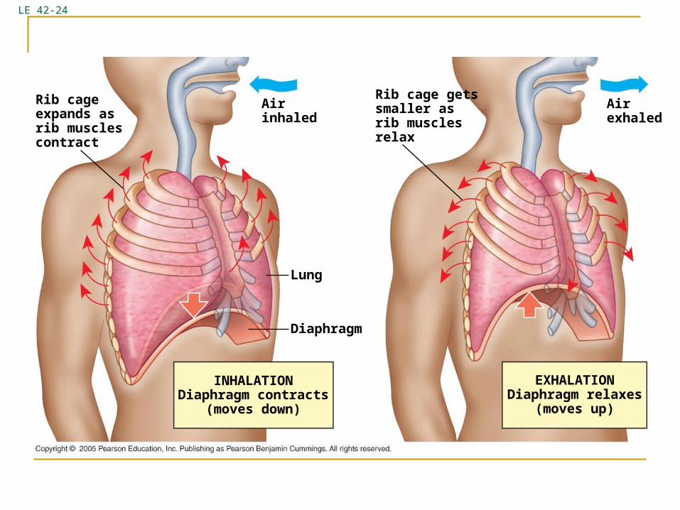
LE 42-24
Rib cageexpands asrib musclescontract
Airinhaled
Lung
Diaphragm
INHALATIONDiaphragm contracts
(moves down)
Rib cage getssmaller asrib musclesrelax
Airexhaled
EXHALATIONDiaphragm relaxes
(moves up)
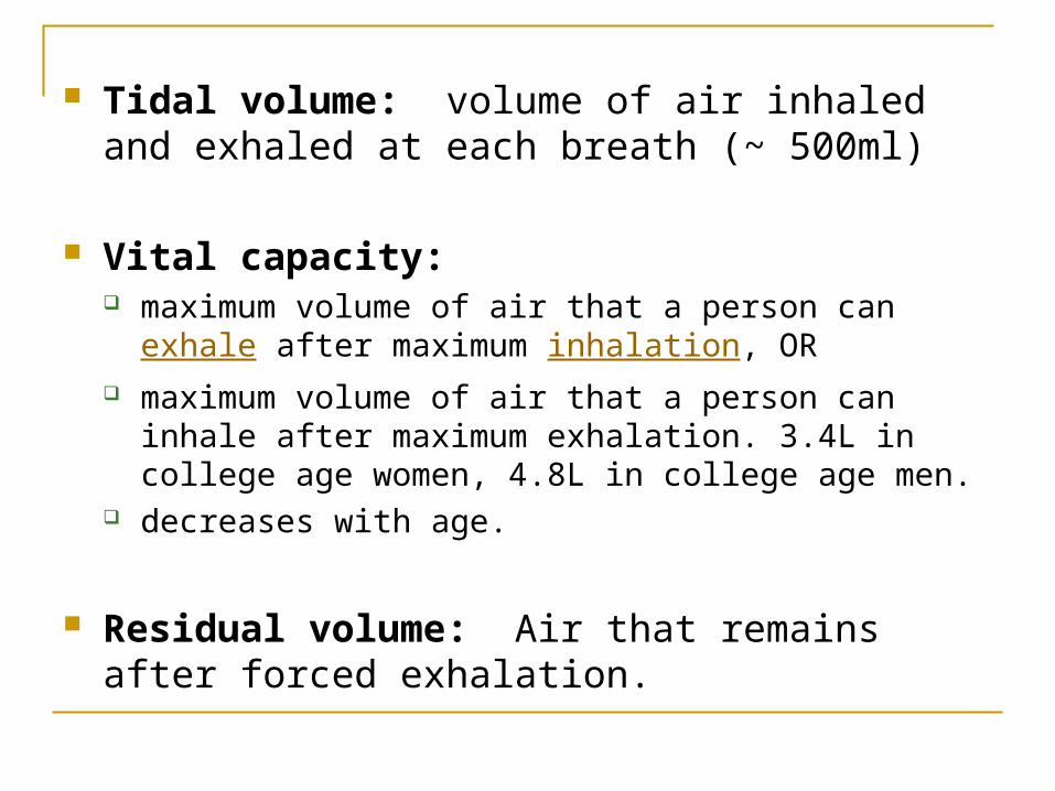
Tidal volume: volume of air inhaled and exhaled at each breath (~ 500ml)
Vital capacity: maximum volume of air that a person can exhale after
maximum inhalation, OR maximum volume of air that a person can inhale after
maximum exhalation. 3.4L in college age women, 4.8L in college age men.
decreases with age.
Residual volume: Air that remains after forced exhalation.
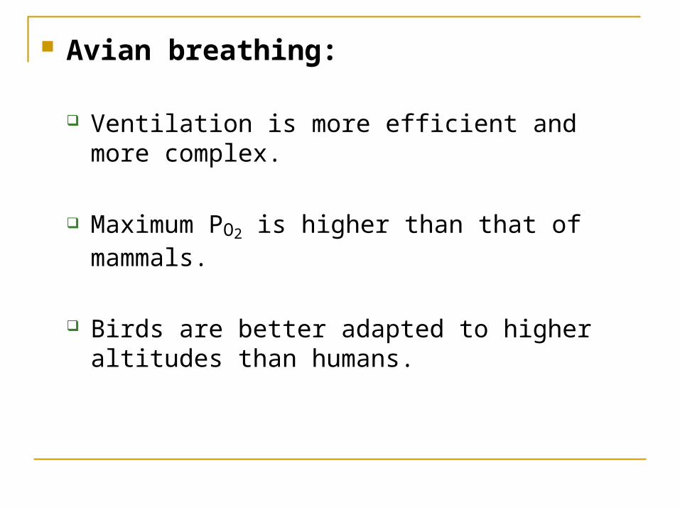
Avian breathing:
Ventilation is more efficient and more complex.
Maximum PO2 is higher than that of mammals.
Birds are better adapted to higher altitudes than humans.
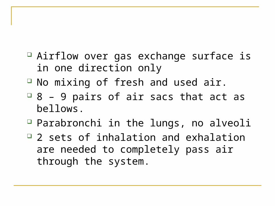
Airflow over gas exchange surface is in one direction only
No mixing of fresh and used air. 8 – 9 pairs of air sacs that act as bellows. Parabronchi in the lungs, no alveoli 2 sets of inhalation and exhalation are needed to
completely pass air through the system.
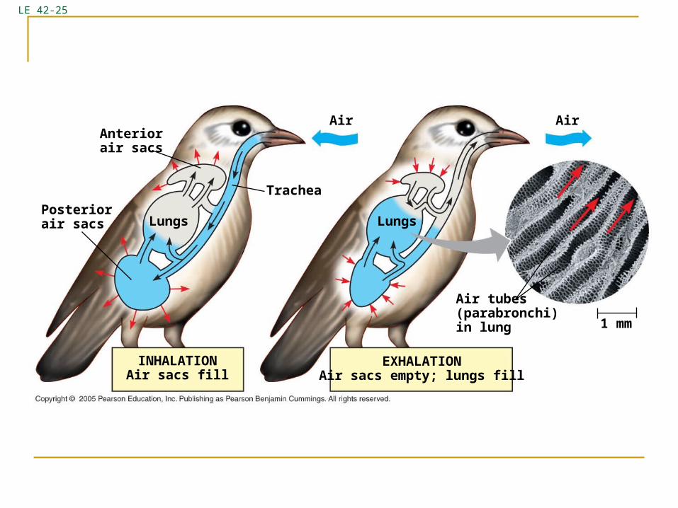
LE 42-25
Anteriorair sacs
LungsPosteriorair sacs
Trachea
Air
Lungs
Air
Air tubes(parabronchi)in lung 1 mm
EXHALATIONAir sacs empty; lungs fill
INHALATIONAir sacs fill
Breathing is controlled (involuntarily) to ensure
Gas exchange coordinates with circulation
Metabolic needs are met
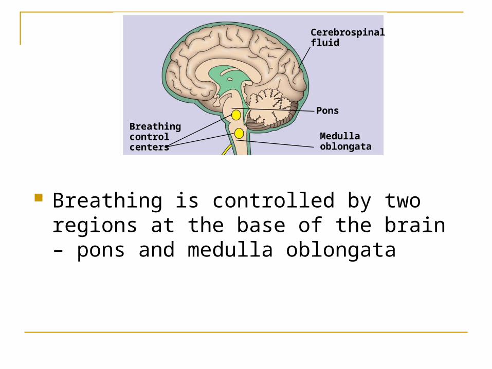
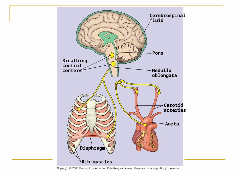
Breathing is controlled by two regions at the base of the brain – pons and medulla oblongata
Breathingcontrolcenters
Cerebrospinalfluid
Medullaoblongata
Pons
During respiration cells produce CO2.
CO2 concentration in blood goes up.
CO2 diffuses from blood to cerebrospinal fluid (CSF).
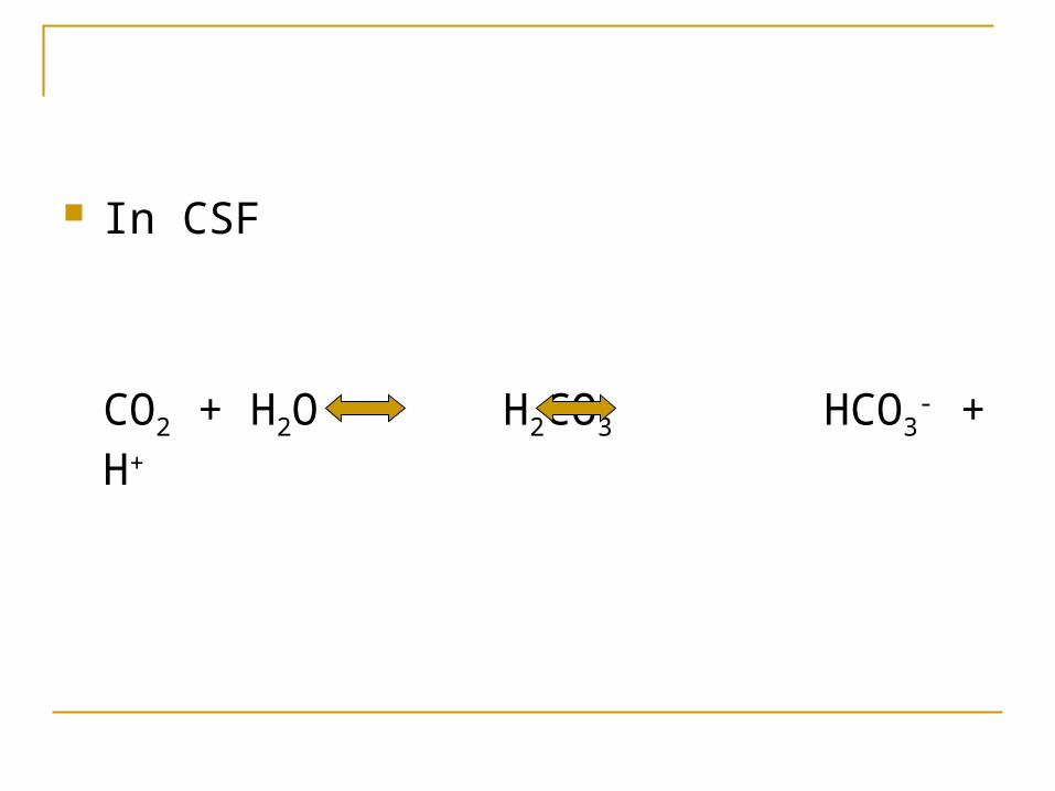
In CSF
CO2 + H2O H2CO3 HCO3- + H+
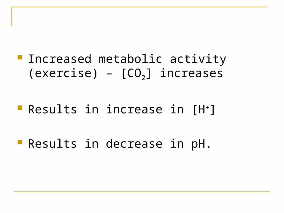
Increased metabolic activity (exercise) – [CO2] increases
Results in increase in [H+]
Results in decrease in pH.
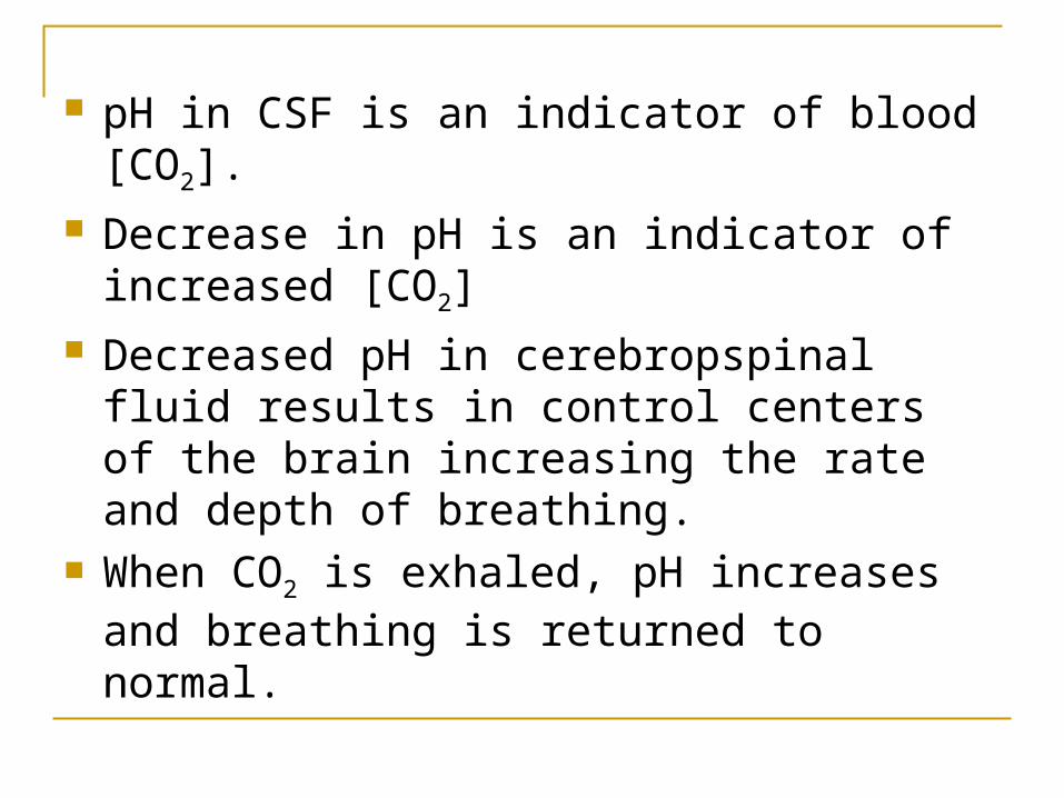
pH in CSF is an indicator of blood [CO2]. Decrease in pH is an indicator of increased
[CO2] Decreased pH in cerebropspinal fluid results
in control centers of the brain increasing the rate and depth of breathing.
When CO2 is exhaled, pH increases and breathing is returned to normal.
Breathingcontrolcenters
Cerebrospinalfluid
Medullaoblongata
Pons
Carotidarteries
Aorta
Diaphragm
Rib muscles
CO2 concentration is primarily used to control breathing
O2 concentration influences breathing only when it is very low. Aorta and carotid arteries have O2 sensors which
signal the brain ti increases breathing Increased breathing is always coupled with
increased cardiac output.
Coordination of circulation and gas exchange.
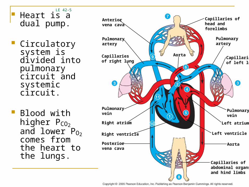
Heart is a dual pump.
Circulatory system is divided into pulmonary circuit and systemic circuit.
Blood with higher PCO2 and lower PO2 comes from the heart to the lungs.
LE 42-5
Anteriorvena cava
Pulmonaryartery
Capillariesof right lung
Aorta
Pulmonaryvein
Right atrium
Right ventricle
Posteriorvena cava
Capillaries ofabdominal organsand hind limbs
Pulmonaryvein
Left ventricle
Left atrium
Aorta
Pulmonaryartery
Capillaries ofhead andforelimbs
Capillariesof left lung
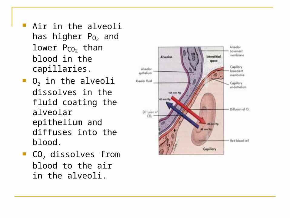
Air in the alveoli has higher PO2 and lower PCO2 than blood in the capillaries.
O2 in the alveoli dissolves in the fluid coating the alveolar epithelium and diffuses into the blood.
CO2 dissolves from blood to the air in the alveoli.
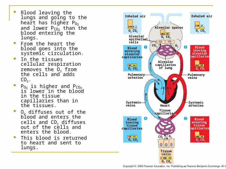
Blood leaving the lungs and going to the heart has higher PO2 and lower PCO2 than the blood entering the lungs.
From the heart the blood goes into the systemic circulation.
In the tissues cellular respiration removes the O2 from the cells and adds CO2.
PO2 is higher and PCO2 is lower in the blood in the tissue capillaries than in the tissues.
O2 diffuses out of the blood and enters the cells and CO2 diffuses out of the cells and enters the blood.
This blood is returned to heart and sent to lungs.
Inhaled air
Bloodenteringalveolar
capillaries
Alveolarepithelialcells
Alveolar spaces
Alveolarcapillaries
of lung
Exhaled air
Bloodleavingalveolar
capillaries
Pulmonaryveins
Pulmonaryarteries
Tissuecapillaries
HeartSystemicveins
Systemicarteries
Bloodleavingtissue
capillaries
Bloodenteringtissue
capillaries
Tissuecells
CO2O2
CO2O2
O2 CO2
CO2O2
< 40 > 45
40 45
CO2O2
100 40
CO2
O 2
CO2O2
40 45
CO2O2
104 40
O2
CO 2
CO2O2
CO2O2
CO2 O2
104 40
120 27160 0.2
Diffusion of O2 in the blood alone is inadequate for meeting metabolic needs.
O2 transport is done to a large degree by respiratory pigments.
During exercise cardiac output is
12.5L of blood per minute with respiratory pigment
555L without the pigment
Respiratory pigments: protein bound to metal, have distinctive color
Hemoglobin: protein and iron (vertebrates)
Hemocyanin: protein and copper (some arthropods and molluscs)
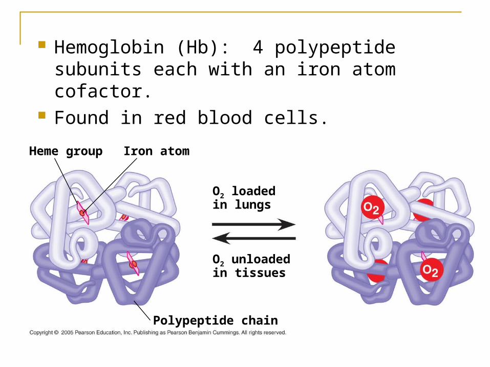
Hemoglobin (Hb): 4 polypeptide subunits each with an iron atom cofactor.
Found in red blood cells.
Polypeptide chain
O2 unloadedin tissues
O2 loadedin lungs
Iron atomHeme group
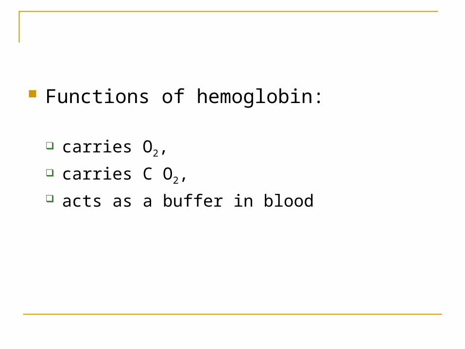
Functions of hemoglobin:
carries O2,
carries C O2, acts as a buffer in blood
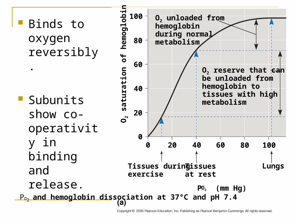
Binds to oxygen reversibly.
Subunits show co-operativity in binding and release.
O2 unloaded fromhemoglobinduring normalmetabolism
O2 reserve that canbe unloaded fromhemoglobin totissues with highmetabolism
PO2 and hemoglobin dissociation at 37°C and pH 7.4P (mm Hg)O2
Tissues duringexercise
Tissues at rest
Lungs
1008060402000
20
40
60
80
100
O2
satu
rati
on
of
hem
og
lob
in (
%)
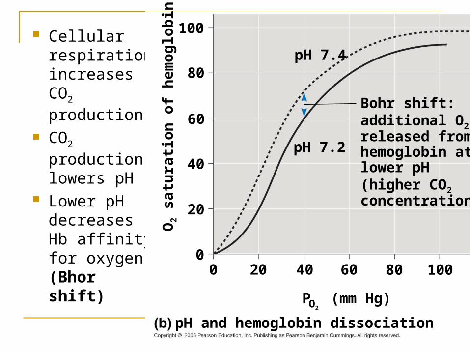
Cellular respiration increases CO2 production.
CO2 production lowers pH
Lower pH decreases Hb affinity for oxygen. (Bhor shift)
Bohr shift:additional O2 released fromhemoglobin atlower pH(higher CO2
concentration)
pH and hemoglobin dissociation
P (mm Hg)O2
1008060402000
20
40
60
80
100
O2
satu
rati
on
of
hem
og
lob
in (
%)
pH 7.2
pH 7.4
When cellular respiration is higher Hb releases more O2.
CO2 transport.
In solution (7%) Bound to Hb (23%) Bicarbonate (HCO3
-) 70%
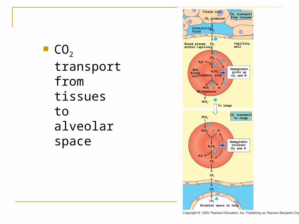
CO2 transport from tissues to alveolar space
CO2 transportfrom tissuesCO2 produced
Tissue cell
CO2
CO2
CO2
Interstitialfluid
Blood plasmawithin capillary
Capillarywall
Hemoglobinpicks up
CO2 and H+
CO2 transportto lungs
To lungs
H2CO3
Carbonic acid
H2O
Hb
HCO3–
Bicarbonate
Redbloodcell
H++
HCO3–
HCO3–
Hemoglobinreleases
CO2 and H+
H++HCO3–
CO2
H2CO3
H2O
CO2
CO2
CO2
Hb
Alveolar space in lung
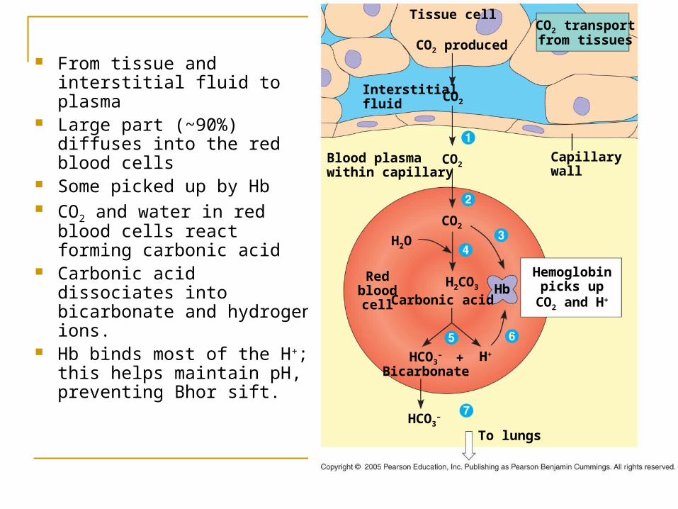
From tissue and interstitial fluid to plasma
Large part (~90%) diffuses into the red blood cells
Some picked up by Hb CO2 and water in red blood
cells react forming carbonic acid
Carbonic acid dissociates into bicarbonate and hydrogen ions.
Hb binds most of the H+; this helps maintain pH, preventing Bhor sift.
CO2 transportfrom tissuesCO2 produced
Tissue cell
CO2
CO2
CO2
Interstitialfluid
Blood plasmawithin capillary
Capillarywall
Hemoglobinpicks up
CO2 and H+
To lungs
H2CO3
Carbonic acid
H2O
Hb
HCO3–
Bicarbonate
Redbloodcell
H++
HCO3–
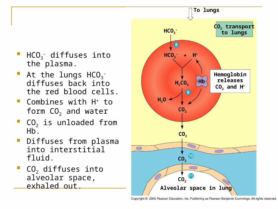
HCO3- diffuses into the
plasma. At the lungs HCO3
- diffuses back into the red blood cells.
Combines with H+ to form CO2 and water
CO2 is unloaded from Hb. Diffuses from plasma into
interstitial fluid. CO2 diffuses into alveolar
space, exhaled out.
CO2 transportto lungs
To lungs
HCO3–
Hemoglobinreleases
CO2 and H+
H++HCO3–
CO2
H2CO3
H2O
CO2
CO2
CO2
Hb
Alveolar space in lung
Animals like cheetah, pronghorned antelope have been selected enhancement normal physiological mechanisms at every stage of O2 metabolism.
Diving mammals have myoglobin that have higher affinity for O2 than human myoglobin.