Embed Size (px)
Citation preview
GAMMA-HYDROXYBUTYRATE / BUTYRIC ACID Latest Revision: May 16, 2005
OOH
O
gamma-hydroxybutyrate
OHOH
O
gamma-hydroxybutyric acid
1. SYNONYMS
CFR: Gamma-Hydroxybutyric acid
CAS #: Sodium: 502-85-2
Other Names: Sodium oxybate
Sodium gamma-hydroxybutyrate
4-Hydroxy butyrate, sodium
4-Hydroxybutanoic acid monosodium salt
GHB
Anetamin
Somsanit
Gamma OH
Somatomax PM
2. CHEMICAL AND PHYSICAL DATA
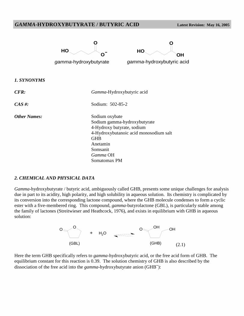
Gamma-hydroxybutyrate / butyric acid, ambiguously called GHB, presents some unique challenges for analysis
due in part to its acidity, high polarity, and high solubility in aqueous solution. Its chemistry is complicated by
its conversion into the corresponding lactone compound, where the GHB molecule condenses to form a cyclic
ester with a five-membered ring. This compound, gamma-butyrolactone (GBL), is particularly stable among
the family of lactones (Streitwieser and Heathcock, 1976), and exists in equilibrium with GHB in aqueous
solution:
OOH
OHH2O
(GBL)
OO
+
(GHB) (2.1)
Here the term GHB specifically refers to gamma-hydroxybutyric acid, or the free acid form of GHB. The
equilibrium constant for this reaction is 0.39. The solution chemistry of GHB is also described by the
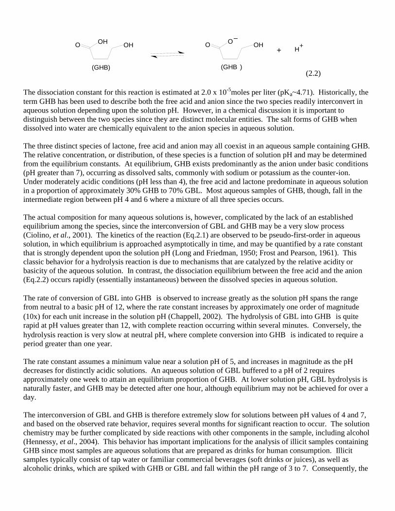
dissociation of the free acid into the gamma-hydroxybutyrate anion (GHB ):
OOH
OH
(GHB)
OO
OH
(GHB )
H++
(2.2)
The dissociation constant for this reaction is estimated at 2.0 x 10⁻⁵
moles per liter (pKa~4.71). Historically, the
term GHB has been used to describe both the free acid and anion since the two species readily interconvert in
aqueous solution depending upon the solution pH. However, in a chemical discussion it is important to
distinguish between the two species since they are distinct molecular entities. The salt forms of GHB when
dissolved into water are chemically equivalent to the anion species in aqueous solution.
The three distinct species of lactone, free acid and anion may all coexist in an aqueous sample containing GHB.
The relative concentration, or distribution, of these species is a function of solution pH and may be determined
from the equilibrium constants. At equilibrium, GHB exists predominantly as the anion under basic conditions
(pH greater than 7), occurring as dissolved salts, commonly with sodium or potassium as the counter-ion.
Under moderately acidic conditions (pH less than 4), the free acid and lactone predominate in aqueous solution
in a proportion of approximately 30% GHB to 70% GBL. Most aqueous samples of GHB, though, fall in the
intermediate region between pH 4 and 6 where a mixture of all three species occurs.
The actual composition for many aqueous solutions is, however, complicated by the lack of an established
equilibrium among the species, since the interconversion of GBL and GHB may be a very slow process
(Ciolino, et al., 2001). The kinetics of the reaction (Eq.2.1) are observed to be pseudo-first-order in aqueous
solution, in which equilibrium is approached asymptotically in time, and may be quantified by a rate constant
that is strongly dependent upon the solution pH (Long and Friedman, 1950; Frost and Pearson, 1961). This
classic behavior for a hydrolysis reaction is due to mechanisms that are catalyzed by the relative acidity or
basicity of the aqueous solution. In contrast, the dissociation equilibrium between the free acid and the anion
(Eq.2.2) occurs rapidly (essentially instantaneous) between the dissolved species in aqueous solution.
The rate of conversion of GBL into GHB is observed to increase greatly as the solution pH spans the range
from neutral to a basic pH of 12, where the rate constant increases by approximately one order of magnitude
(10x) for each unit increase in the solution pH (Chappell, 2002). The hydrolysis of GBL into GHB is quite
rapid at pH values greater than 12, with complete reaction occurring within several minutes. Conversely, the
hydrolysis reaction is very slow at neutral pH, where complete conversion into GHB is indicated to require a
period greater than one year.
The rate constant assumes a minimum value near a solution pH of 5, and increases in magnitude as the pH
decreases for distinctly acidic solutions. An aqueous solution of GBL buffered to a pH of 2 requires
approximately one week to attain an equilibrium proportion of GHB. At lower solution pH, GBL hydrolysis is
naturally faster, and GHB may be detected after one hour, although equilibrium may not be achieved for over a
day.
The interconversion of GBL and GHB is therefore extremely slow for solutions between pH values of 4 and 7,
and based on the observed rate behavior, requires several months for significant reaction to occur. The solution
chemistry may be further complicated by side reactions with other components in the sample, including alcohol
(Hennessy, et al., 2004). This behavior has important implications for the analysis of illicit samples containing
GHB since most samples are aqueous solutions that are prepared as drinks for human consumption. Illicit
samples typically consist of tap water or familiar commercial beverages (soft drinks or juices), as well as
alcoholic drinks, which are spiked with GHB or GBL and fall within the pH range of 3 to 7. Consequently, the
composition of most aqueous samples of GHB is not likely represented by an equilibrium distribution, but is
dependent upon the pH, buffering capacity and other components of the solution, as well as its age. An analysis
should therefore determine the solution pH and whether GBL is present in addition to GHB. Fortunately, the
lactone and the free acid may be readily extracted from aqueous solutions for their separate identification.
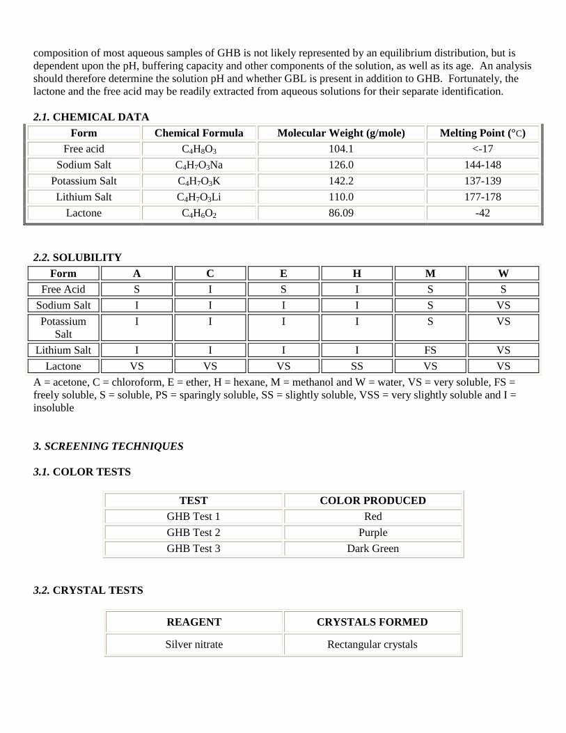
2.1. CHEMICAL DATA
Form Chemical Formula Molecular Weight (g/mole) Melting Point (°C)
Free acid C4H8O3 104.1 <-17
Sodium Salt C4H7O3Na 126.0 144-148
Potassium Salt C4H7O3K 142.2 137-139
Lithium Salt C4H7O3Li 110.0 177-178
Lactone C4H6O2 86.09 -42
2.2. SOLUBILITY
Form A C E H M W
Free Acid S I S I S S
Sodium Salt I I I I S VS
Potassium
Salt
I I I I S VS
Lithium Salt I I I I FS VS
Lactone VS VS VS SS VS VS
A = acetone, C = chloroform, E = ether, H = hexane, M = methanol and W = water, VS = very soluble, FS =
freely soluble, S = soluble, PS = sparingly soluble, SS = slightly soluble, VSS = very slightly soluble and I =
insoluble
3. SCREENING TECHNIQUES
3.1. COLOR TESTS
TEST COLOR PRODUCED
GHB Test 1 Red
GHB Test 2 Purple
GHB Test 3 Dark Green
3.2. CRYSTAL TESTS
REAGENT CRYSTALS FORMED
Silver nitrate Rectangular crystals
3.3. GAS CHROMATOGRAPHY
Method GHB-GCS1
GHB is thermally unstable and may convert into GBL in the gas chromatograph injection port. Reaction with
N,O-bis(trimethylsilyl)trifluoroacetamide (BSTFA) allows for the analysis of the trimethylsilyl (TMS)
derivative. GC/MS permits identification, and GC/FID is also amenable using a similar temperature program.
Although it is possible to simultaneously detect GBL, possible formation from excess GHB warrants caution in
interpreting data. Instead, GBL should be isolated for a separate analysis (see Section 4, Separation
Techniques).
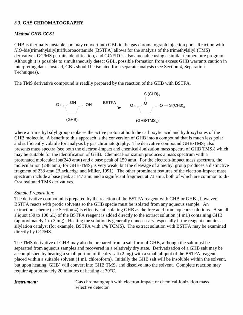
The TMS derivative compound is readily prepared by the reaction of the GHB with BSTFA,
Si(CH3)3
BSTFAO
OHOH O
OO Si(CH3)3
(GHB) (GHB·TMS2)
where a trimethyl silyl group replaces the active proton at both the carboxylic acid and hydroxyl sites of the
GHB molecule. A benefit to this approach is the conversion of GHB into a compound that is much less polar
and sufficiently volatile for analysis by gas chromatography. The derivative compound GHB·TMS2 also
presents mass spectra (see both the electron-impact and chemical-ionization mass spectra of GHB·TMS2) which
may be suitable for the identification of GHB. Chemical-ionization produces a mass spectrum with a
protonated molecular ion(249 amu) and a base peak of 159 amu. For the electron-impact mass spectrum, the
molecular ion (248 amu) for GHB·TMS2 is very weak, but the cleavage of a methyl group produces a distinctive
fragment of 233 amu (Blackledge and Miller, 1991). The other prominent features of the electron-impact mass
spectrum include a base peak at 147 amu and a significant fragment at 73 amu, both of which are common to di-
O-substituted TMS derivatives.
Sample Preparation:
The derivative compound is prepared by the reaction of the BSTFA reagent with GHB or GHB , however,
BSTFA reacts with protic solvents so the GHB specie must be isolated from any aqueous sample. An
extraction scheme (see Section 4) is effective at isolating GHB as the free acid from aqueous solutions. A small
aliquot (50 to 100 L) of the BSTFA reagent is added directly to the extract solution (1 mL) containing GHB
(approximately 1 to 3 mg). Heating the solution is generally unnecessary, especially if the reagent contains a
silylation catalyst (for example, BSTFA with 1% TCMS). The extract solution with BSTFA may be examined
directly by GC/MS.
The TMS derivative of GHB may also be prepared from a salt form of GHB, although the salt must be
separated from aqueous samples and recovered in a relatively dry state. Derivatization of a GHB salt may be
accomplished by heating a small portion of the dry salt (2 mg) with a small aliquot of the BSTFA reagent
placed within a suitable solvent (1 mL chloroform). Initially the GHB salt will be insoluble within the solvent,
but upon heating, GHB will convert into GHB·TMS2 and dissolve into the solvent. Complete reaction may
require approximately 20 minutes of heating at 70 C.
Instrument: Gas chromatograph with electron-impact or chemical-ionization mass
selective detector
Column: 100% polydimethylsiloxane, 12.0 m x 0.20 mm x 0.33µm film thickness
Carrier gas: Helium at 1.0 mL/min
Temperatures: Injector: 250°C
Transfer line: 280°C
Oven program:
70°C initial temperature for 1.20 min
Ramp to 280°C at 15°C/min
Hold final temperature for 5.00 min
Injection parameters: Split Ratio = 50:1, 1 µL injected
COMPOUND RRT
GHB·TMS2 1.00
GBL 0.33
3.4. HIGH PERFORMANCE LIQUID CHROMATOGRAPHY
Method GHB-LCS1
Sample Preparation:
Dissolve or dilute (if necessary) in mobile phase and filter (0.45 µm).
Instrument: High performance liquid chromatograph with diode array detector
Column: 5 µm ODS Hypersil, 4.6 mm x 100 mm
Detector: UV, 215 nm
Flow: 0.75 mL/min
Injection Volume: 5 µL
Buffer: 10 mM NaH2PO4 adjusted to pH 3 with H3PO4
Mobile Phase: Buffer:methanol (80:20)
COMPOUND RRT
GHB 1.000
GBL 1.082
Method GHB-LCS2
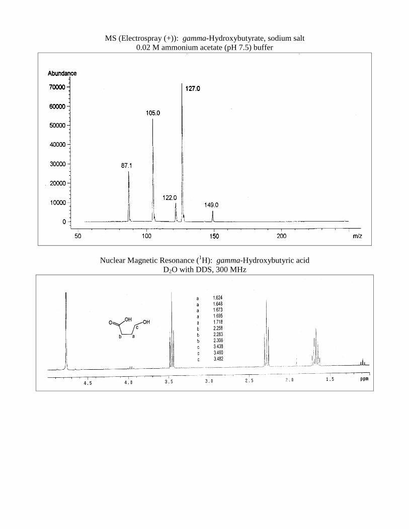
GHB, GBL, and 1,4-butanediol can be identified in drinking water solutions by LC/MS (see the electrospray
mass spectrum of the GHB sodium salt). The electrospray (+) mass spectrum is characterized by several
protonated (M+1) species, including the sodium salt (127 amu), the free acid (105 amu) and the lactone (87
amu). The spectrum also displays a weaker peak for the protonated ammonium salt (122 amu) due to the
presence of ammonium ions in the mobile phase, as well as a di-sodium GHB species (149 amu). Negative ion
detection can be substituted for the GHB analysis, but comparatively poor sensitivity towards GBL and 1,4-
butanediol is observed. Note that GHB (as GHB ) shows no column retention with this buffer system.
Standard Solution Preparation:
Prepare a mixed standard of GHB sodium salt (1-10 mg per mL), GBL (5-10 mg/mL), and 1,4-butanediol (1-10
mg/mL) in methanol.
Instrument: High performance liquid chromatograph with atmospheric
pressure ionization electrospray mass selective detector
Column: 5 µm Aqua C18, 100 mm x 4.6 mm
Detector: Scan mode, positive ion
Capillary voltage: 3000 V
Fragmentor: 30 eV
Nebulizer pressure: 60 psig
Drying gas flow: 13.0 L/min
Drying gas temperature: 350 C
Flow: 1.500 mL/min
Injection Volume: 5 µL
Buffer: 20 mM CH3COONH4 (~ pH 7.5)
Mobile Phase:
100% Buffer
Typical Retention Times: GHB: 2.00 min
1,4-Butanediol: 5.44 min
GBL: 6.46 min
COMPOUND RRT
GHB 1.000
1,4-Butanediol 2.711
GBL 3.230
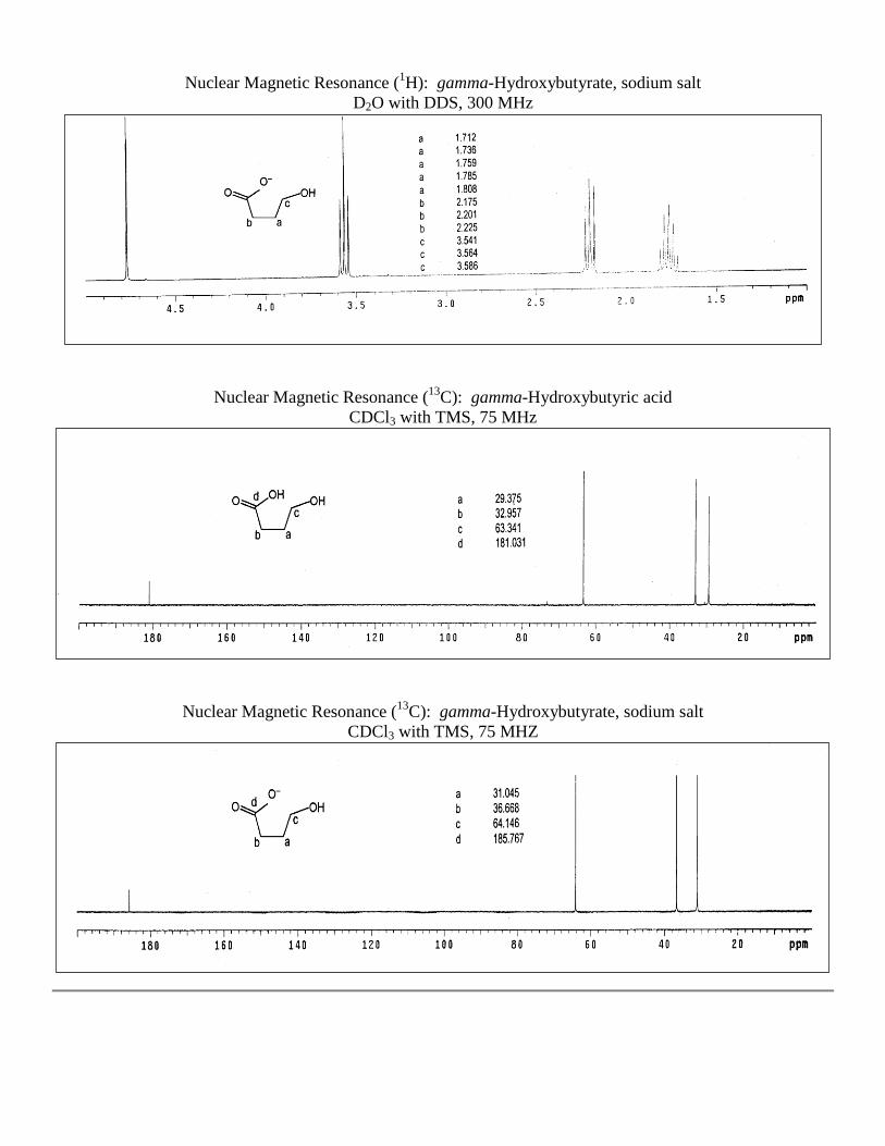
3.5. NUCLEAR MAGNETIC RESONANCE SPECTROSCOPY
GHB and GBL present proton (1H) and carbon (
13C) NMR spectra with suitably distinct peaks, whereby
mixtures of the two may be identified (see NMR spectra for GHB and GBL). Simple aqueous solutions of GHB
and GBL may be examined with minimal sample preparation that allows the relative proportions of the two
substances to be assessed directly from the composite NMR spectrum. Complex aqueous mixtures that arise
from commercial beverages require GHB and GBL to be separated prior to analysis (see Section 4, Separation
Techniques).
Method GHB-NMRS1
Sample Preparation:
Simple aqueous samples (typically 10 to 20 mg GHB /mL), may be diluted in deuterium oxide (D2O) with the
external reference standard 2,2-dimethyl-2-silapentane-5-sulfonate (DDS). GHB (or GBL) isolated by
extraction may be prepared in D2O with DDS, or in deuterated chloroform (CDCl3) with the internal reference
standard tetramethylsilane (TMS). Residual solvent peaks from the extraction solvent may be detected but do
not interfere with the identification of GHB. Filter all preparation solutions before analysis.
Instrument: Nuclear magnetic resonance spectrometer
Probe: 5-mm dual channel, room temperature
Parameters: 1H NMR:
Observation frequency: 300 MHz
Pulse angle: 30°
Acquisition time: 1.998 s
Spectral window: 4500 Hz
Filter bandwidth: 2250 Hz
Delay: 0 - 1 s
Frequency offset: 0 Hz
Number of transients: 16
13
C NMR:
Observation frequency: 75 MHz
Pulse angle: 45°
Acquisition time: 1.706 s
Spectral window: 18761.7 Hz
Filter bandwidth: 9500 Hz
Delay: 0 s
Frequency offset: 0 Hz
Number of transients: 512 (minimum)
Proton decoupler: on
Decoupler modulation frequency: 3233 Hz
4. SEPARATION TECHNIQUES
Aqueous samples containing GHB may also contain GBL due to the equilibrium between the two species (see
Section 2). The following extraction scheme can isolate the two species from aqueous solutions for subsequent
identification by IR, GC-MS or NMR.
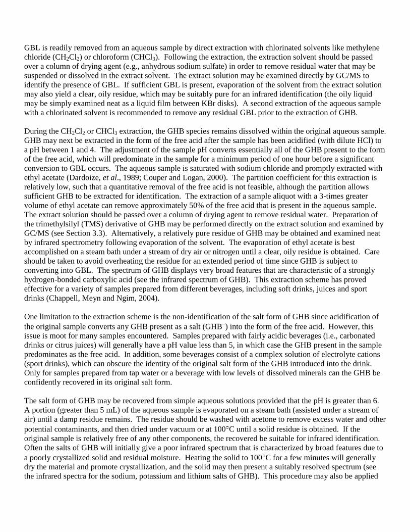
GBL is readily removed from an aqueous sample by direct extraction with chlorinated solvents like methylene
chloride (CH2Cl2) or chloroform (CHCl3). Following the extraction, the extraction solvent should be passed
over a column of drying agent (e.g., anhydrous sodium sulfate) in order to remove residual water that may be
suspended or dissolved in the extract solvent. The extract solution may be examined directly by GC/MS to
identify the presence of GBL. If sufficient GBL is present, evaporation of the solvent from the extract solution
may also yield a clear, oily residue, which may be suitably pure for an infrared identification (the oily liquid
may be simply examined neat as a liquid film between KBr disks). A second extraction of the aqueous sample
with a chlorinated solvent is recommended to remove any residual GBL prior to the extraction of GHB.
During the CH2Cl2 or CHCl3 extraction, the GHB species remains dissolved within the original aqueous sample.
GHB may next be extracted in the form of the free acid after the sample has been acidified (with dilute HCl) to
a pH between 1 and 4. The adjustment of the sample pH converts essentially all of the GHB present to the form
of the free acid, which will predominate in the sample for a minimum period of one hour before a significant
conversion to GBL occurs. The aqueous sample is saturated with sodium chloride and promptly extracted with
ethyl acetate (Dardoize, et al., 1989; Couper and Logan, 2000). The partition coefficient for this extraction is
relatively low, such that a quantitative removal of the free acid is not feasible, although the partition allows
sufficient GHB to be extracted for identification. The extraction of a sample aliquot with a 3-times greater
volume of ethyl acetate can remove approximately 50% of the free acid that is present in the aqueous sample.
The extract solution should be passed over a column of drying agent to remove residual water. Preparation of
the trimethylsilyl (TMS) derivative of GHB may be performed directly on the extract solution and examined by
GC/MS (see Section 3.3). Alternatively, a relatively pure residue of GHB may be obtained and examined neat
by infrared spectrometry following evaporation of the solvent. The evaporation of ethyl acetate is best
accomplished on a steam bath under a stream of dry air or nitrogen until a clear, oily residue is obtained. Care
should be taken to avoid overheating the residue for an extended period of time since GHB is subject to
converting into GBL. The spectrum of GHB displays very broad features that are characteristic of a strongly
hydrogen-bonded carboxylic acid (see the infrared spectrum of GHB). This extraction scheme has proved
effective for a variety of samples prepared from different beverages, including soft drinks, juices and sport
drinks (Chappell, Meyn and Ngim, 2004).
One limitation to the extraction scheme is the non-identification of the salt form of GHB since acidification of
the original sample converts any GHB present as a salt (GHB ) into the form of the free acid. However, this
issue is moot for many samples encountered. Samples prepared with fairly acidic beverages (i.e., carbonated
drinks or citrus juices) will generally have a pH value less than 5, in which case the GHB present in the sample
predominates as the free acid. In addition, some beverages consist of a complex solution of electrolyte cations
(sport drinks), which can obscure the identity of the original salt form of the GHB introduced into the drink.
Only for samples prepared from tap water or a beverage with low levels of dissolved minerals can the GHB be
confidently recovered in its original salt form.
The salt form of GHB may be recovered from simple aqueous solutions provided that the pH is greater than 6.
A portion (greater than 5 mL) of the aqueous sample is evaporated on a steam bath (assisted under a stream of
air) until a damp residue remains. The residue should be washed with acetone to remove excess water and other
potential contaminants, and then dried under vacuum or at 100 C until a solid residue is obtained. If the
original sample is relatively free of any other components, the recovered be suitable for infrared identification.
Often the salts of GHB will initially give a poor infrared spectrum that is characterized by broad features due to
a poorly crystallized solid and residual moisture. Heating the solid to 100 C for a few minutes will generally
dry the material and promote crystallization, and the solid may then present a suitably resolved spectrum (see
the infrared spectra for the sodium, potassium and lithium salts of GHB). This procedure may also be applied
to the solid that has been pressed within a KBr matrix since ion exchange between the alkali salts of GHB and
KBr is not observed to occur, even after heating the mixture of the solids for an extended period (several days).
5. QUANTITATIVE PROCEDURES
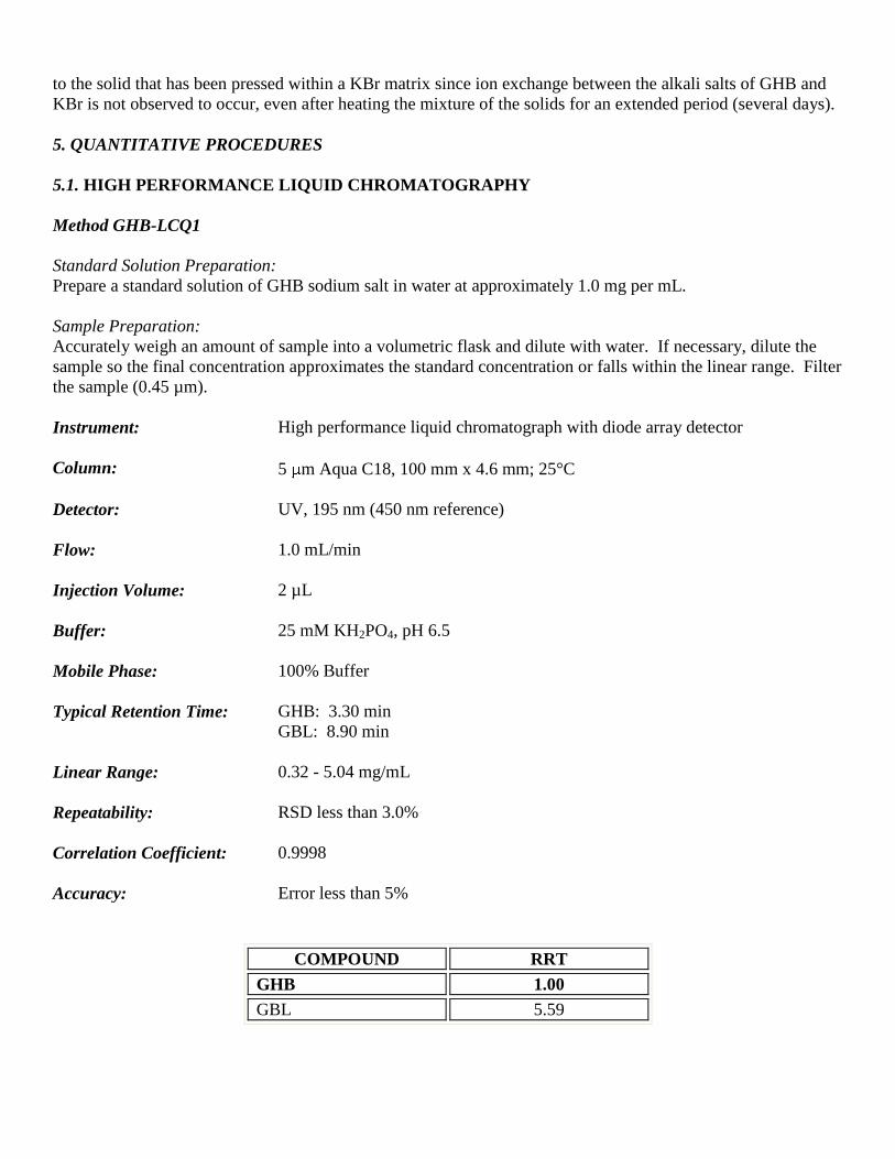
5.1. HIGH PERFORMANCE LIQUID CHROMATOGRAPHY
Method GHB-LCQ1
Standard Solution Preparation:
Prepare a standard solution of GHB sodium salt in water at approximately 1.0 mg per mL.
Sample Preparation:
Accurately weigh an amount of sample into a volumetric flask and dilute with water. If necessary, dilute the
sample so the final concentration approximates the standard concentration or falls within the linear range. Filter
the sample (0.45 µm).
Instrument: High performance liquid chromatograph with diode array detector
Column: 5 m Aqua C18, 100 mm x 4.6 mm; 25°C
Detector: UV, 195 nm (450 nm reference)
Flow: 1.0 mL/min
Injection Volume: 2 µL
Buffer: 25 mM KH2PO4, pH 6.5
Mobile Phase: 100% Buffer
Typical Retention Time: GHB: 3.30 min
GBL: 8.90 min
Linear Range: 0.32 - 5.04 mg/mL
Repeatability: RSD less than 3.0%
Correlation Coefficient: 0.9998
Accuracy: Error less than 5%
COMPOUND RRT
GHB 1.00
GBL 5.59
6. QUALITATIVE DATA
See spectra on the following pages for Infrared Spectroscopy, Mass Spectrometry, and Nuclear Magnetic
Resonance.
7. REFERENCES
Bomarito, C., “Analytical Profile of Gamma-Hydroxybutyric Acid (GHB)”. J. Clan. Invest. Chem. Assoc.,
1991, Vol. 3, No. 3, pp. 10-2.
Blackledge, R.D. and Miller, M.D. “The Identification of GHB”. Microgram, 1991, Vol. XXIV, No. 7,
pp. 172-9.
Catterton, A.J., Backstrom, E. and Bozenko, J.S. “Lithium Gamma-Hydroxybutyrate”. J. Clan. Invest. Chem.
Assoc., 2002, Vol. 12, No. 1, pp. 26-30.
Chamot, E.M. and Mason, C.W. Handbook of Chemical Microscopy, Vol. II, 2nd Ed. John Wiley & Sons: New
York, 1940.
Chappell, J.S., “The Non-Equilibrium Aqueous Solution Chemistry of Gamma-Hydroxybutyrate”. J. Clan.
Invest. Chem. Assoc., 2002, Vol. 12, No. 4, pp. 20-7.
Chappell, J.S., Meyn, A.W., and Ngim, K.K. “The Extraction and Infrared Identification of Gamma-
Hydroxybutyric Acid (GHB) from Aqueous Solutions”. J. Forensic Sci., 2004, Vol. 49, No. 1, pp. 52-9.
Chew, S.L. and Meyers, J.A. “Identification and Quantitation of Gamma-Hydroxybutyrate (NaGHB) by
Nuclear Magnetic Resonance Spectroscopy”. J. Forensic Sci., 2003, Vol. 48, pp. 292-8.
Ciolino, L.A., Mesmer, M.Z., Satzger, R.D., Machal, A.C., McCauley, H.A. and Mohrhaus, A.S. “The
Chemical Interconversion of GHB and GBL: Forensic Issues and Implications”. J. Forensic Sci., 2001,
Vol. 46, No. 6, pp. 1315-23.
Couper, F.J. and Logan, B.K. “Determination of Gamma-Hydroxybutyrate (GHB) in Biological Specimens by
Gas Chromatography - Mass Spectrometry”. J. Anal. Toxicol., 2000, Vol. 24, pp. 1-7.
CRC Handbook of Chemistry and Physics, 62nd Ed. CRC Press: Boca Raton, Florida, 1981.
Dardoize, F., Goasdoue, C., Goasdoue, N., Laborit, H.M and Topall, G. “4-Hydroxybutyric Acid (and
Analogue) Derivatives of D-Glucosamine”. Tetrahedron, 1989, Vol. 45, No. 24, pp. 7783-94.
Frost, A.A. and Pearson, R.G. Kinetics and Mechanism: A Study of Homogeneous Chemical Reactions. Wiley
and Sons: New York, 1976, pp. 327-35.
Hennessy, S.A., Moane, S.M., and McDermott, S.D. “The Reactivity of Gamma-Hydroxybutyric Acid (GHB)
and Gamma-Butyrolactone in Alcoholic Solutions”. J. Forensic Sci., 2004, Vol. 49, No. 6, pp. 1220-9.
Long, F.A. and Friedman, L. “Determination of the Mechanism of Gamma-Lactone Hydrolysis by a Mass
Spectrometric Method”. J. Am. Chem. Soc, 1950, Vol. 72, pp. 3962-5.
The Merck Index, 11th Ed. Merck & Co.: Rahway, New Jersey, 1989.
Mesmer, M.Z. and Satzger, R.D. “Determination of Gamma-Hydroxybutyrate (GHB) and Gamma-
Butyrolactone (GBL) by HPLC / UV-VIS Spectrophotometry and HPLC / Thermospray Mass Spectrometry”.
J. Forensic Sci., 1998, Vol. 43, pp. 489-92.
Morris, J.A. “Extraction of GHB for FTIR Analysis and a New Color Test for Gamma-Butyrolactone (GBL)”.
Microgram, 1999, Vol. 32, No. 8, pp. 215-221.
Morris, J.A. “Analogs of GHB; Part 2: Theoretical Perspective”. J. Clan. Invest. Chem. Assoc., 2001, Vol. 10,
pp. 14-6.
Perez-Prior, M.T., Manso, J.A., Garcia-Santos, M.D., Calle, E. and Casado, J. “Reactivity of Lactones and GHB
Formation”. J. Organic Chem., 2005, Vol. 70, pp. 420-6.
Smith, P.R. and Bozenko, J.S. “New Presumptive Tests for GHB”. Microgram, 2002, Vol. XXXV, No. 1,
pp. 9-13.
Streitwieser, A. and Heathcock, C.H. Introduction to Organic Chemistry. Macmillan: New York, 1976,
pp. 685-7.
Vose, J., Tighe, T., Schwartz, M. and Buel, E. “Detection of Gamma-Butyrolactone (GBL) as a Natural
Component of Wine”. J. Forensic Sci., 2001, Vol. 46, No. 5, pp. 1164-7.
8. ADDITIONAL RESOURCES
Forendex
Wikipedia
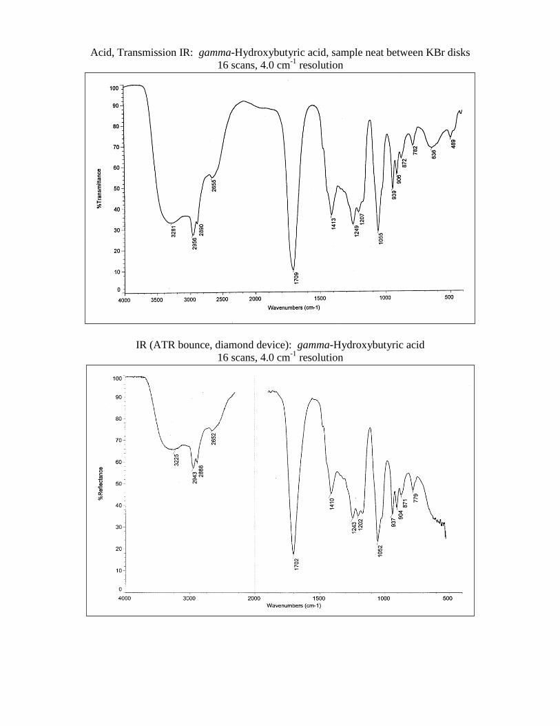
Acid, Transmission IR: gamma-Hydroxybutyric acid, sample neat between KBr disks
16 scans, 4.0 cm-1
resolution
IR (ATR bounce, diamond device): gamma-Hydroxybutyric acid
16 scans, 4.0 cm-1
resolution
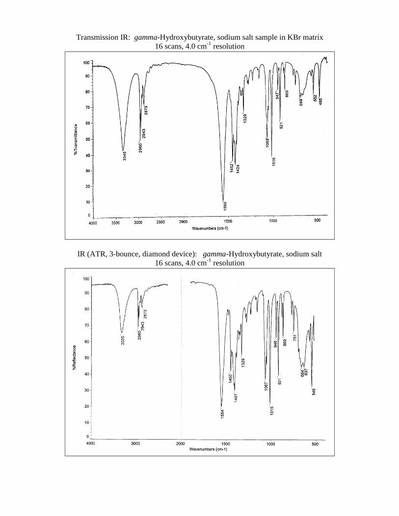
Transmission IR: gamma-Hydroxybutyrate, sodium salt sample in KBr matrix
16 scans, 4.0 cm-1
resolution
IR (ATR, 3-bounce, diamond device): gamma-Hydroxybutyrate, sodium salt
16 scans, 4.0 cm-1
resolution
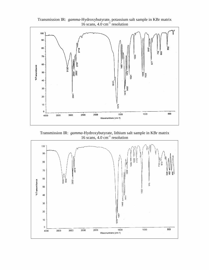
Transmission IR: gamma-Hydroxybutyrate, potassium salt sample in KBr matrix
16 scans, 4.0 cm-1
resolution
Transmission IR: gamma-Hydroxybutyrate, lithium salt sample in KBr matrix
16 scans, 4.0 cm-1
resolution
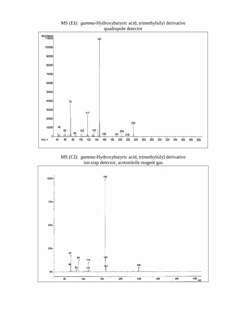
MS (EI): gamma-Hydroxybutyric acid, trimethylsilyl derivative
quadrupole detector
MS (CI): gamma-Hydroxybutyric acid, trimethylsilyl derivative
ion-trap detector, acetonitrile reagent gas
MS (Electrospray (+)): gamma-Hydroxybutyrate, sodium salt
0.02 M ammonium acetate (pH 7.5) buffer
Nuclear Magnetic Resonance (1H): gamma-Hydroxybutyric acid
D2O with DDS, 300 MHz
Nuclear Magnetic Resonance (1H): gamma-Hydroxybutyrate, sodium salt
D2O with DDS, 300 MHz
Nuclear Magnetic Resonance (13
C): gamma-Hydroxybutyric acid
CDCl3 with TMS, 75 MHz
Nuclear Magnetic Resonance (13
C): gamma-Hydroxybutyrate, sodium salt
CDCl3 with TMS, 75 MHZ


![Conventional and Inverted Photovoltaic Cells Fabricated ...koreascience.or.kr/article/JAKO201416760764766.pdf61–butyric acid methyl ester or [6,6]-phenyl-C 71-butyric acid methyl](https://img.pdfslide.us/doc/110x75/6095158a83c7e40411746c95/conventional-and-inverted-photovoltaic-cells-fabricated-61abutyric-acid-methyl.jpg)