Embed Size (px)
Citation preview
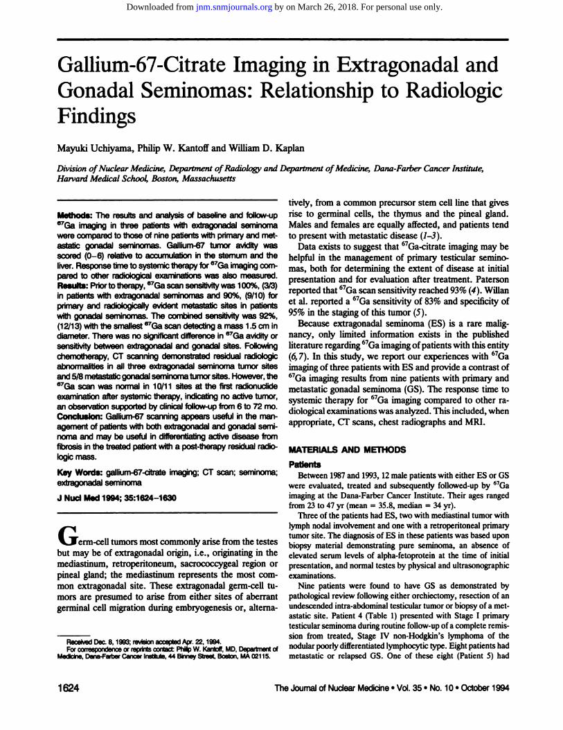
tively, from a common precursorstem cell line that givesrise to germinal cells, the thymus and the pineal gland.Males and females are equally affected, and patients tendto present with metastatic disease (1—3).
Data exists to suggest that 67Ga-citrateimagingmay behelpful in the management of primary testicular seminomas, both for determining the extent of disease at initialpresentation and for evaluation after treatment. Patersonreportedthat67Gascan sensitivity reached93%(4). Willanet al. reported a 67Gasensitivity of 83%and specificity of95% in the staging of this tumor (5).
Because extragonadal seminoma (ES) is a rare malignancy, only limited information exists in the publishedliteratureregarding67Gaimagingofpatients with this entity(6, 7). In this study, we report our experiences with 67Ga
imagingof three patientswith ES and provide a contrastof67Gaimaging results from nine patients with primaryandmetastatic gonadal seminoma (GS). The response time tosystemic therapy for 67Ga imaging compared to other radiological examinations was analyzed. This included, whenappropriate,CF scans, chest radiographsand MIII.
MATERIALSAND METHODS
PatientsBetween1987and1993,12malepatientswitheitherES orOS
were evaluated,treatedand subsequentlyfollowed-upby 67Gaimaging at the Dana-Farber Cancer Institute. Their ages rangedfrom23to 47yr (mean= 35.8,median= 34yr).
Threeof thepatientshadES, twowithmediastinaltumorwithlymphnodalinvolvementandonewitha retroperitonealprimarytumor site. The diagnosis of ES in these patients was based uponbiopsy material demonstrating pure seminoma, an absence ofelevated serum levels of alpha-fetoproteinat the time of initialpresentation,andnormaltestesby physicalandultrasonographicexaminations.
Nine patients were found to have OS as demonstrated bypathological review following either orchiectomy, resection of anundescendedintra-abdominaltesticulartumororbiopsyof a metastatic site. Patient 4 (Table 1) presented with Stage I primarytesticularseminomaduringroutinefollow-upof a complete remission from treated, Stage IV non-Hodgldn's lymphoma of thenodular poorly differentiated lymphocytic type. Eight patients hadmetastatic or relapsed OS. One of these eight (Patient 5) had
Methods:The resultsand analysisof baselineand follow-up67Ga imaging in three patients with extragonadal seminomawere compared to those of nine patients withprimaryand metastatic gonadal seminomas. Gallium-67 tumor avidity wasscored (0—6)relativeto accumulation in the sternum and theliver.Response timeto systemictherapy for @Gaimagingcornpared to other radiologicalexaminations was also measured.Results:Priortotherapy, @Gascansensitivitywas100%,(3/3)in patients with extragonadal serninornas and 90%, (9/10) forpnmaty and radiOlOgicallyevident metastatic sites in patients
@thgonadal seminomas. The com@nedsens@v@was 92%,(12/13) withthe smallest @“Gascan detecting a mass 1.5 cm indiameter. There was no significantdifferencein 67@3@avidityorsensitivitybetween extragonadal and gonadal sites. Followingchemotherapy, CT scanning demonstrated residual radiOIOgiCabnormalities in all three extragonadal serninorna tumor sitesand 5/8 metastaticgonadal serninomatumorsites. However,the67Ga scan was normal in 10/11 sites at the first radIOnUclldeexaminationafter systemic therapy, indicatingno active tumor,an observationsupported by dinicalfollow-upfrom6 to 72 mo.Conclusion: Galliurn-67scanningappearsusefulin the managernent of patients withboth extragonadal and gonadal seminorna and may be useful in differentiatingactive disease fromfibrosis in the treated patient with a post-therapy residual radiologicmass.
Key Words: gallium-67-dtrateimaging;CT scan;seminoma;extragonadal seminoma
J NucIMed1994;35:1624-1630
erm-cell tumors most commonly arise from the testesbut may be of extragonadalorigin, i.e., originatingin themediastinum, retroperitoneum, sacrococcygeal region orpineal gland; the mediastinum represents the most common extragonadal site. These extragonadal germ-cell tumors are presumed to arise from either sites of aberrantgerminal cell migration during embryogenesis or, altema
ReceivedDec.8, 1993;[email protected], 1994.Forcorrespondenceor reprintsoord@ Ph@ W. Kantoif,MD,Departmentof
Medicine,D@ia-Fwb@CancerInstitute,44 BinneyStreet,Boston,MA02115.
1624 The Journalof NuclearMedicine•Vol.35 •No. 10 •October1994
Gallium-67-Citrate Imaging in Extragonadal andGonada! Seminomas: Relationship to RadiologicFindingsMayuki Uchiyama, Philip W. Kantoff and Wffliam D. Kaplan
DivLcion ofNuclear Medicine, Depatiment ofRadiolo@ and Depamnent ofMedicine, Dana-Farber Cancer Institute,Har.'ard Medical Sch@x'4 Boston, Massachusetts
by on March 26, 2018. For personal use only. jnm.snmjournals.org Downloaded from

metastases in two differentsites, i.e., mediastinaland retroperitoneal aggregated mass, on the same occasion. A second patient,(Patient6) relapsedin two differenttumorsites, i.e., bilaterallungand mediastinum, on two separate occasions. Thus, two siteswere indicatedin this patient.Six othermetastaticor relapsedlocations included one mediastinal site, one pleura, one soft tissues of the shoulder and three retroperitonealsites, resulting inninesitesof metastaticOSatthetimeof initialpresentationoratthe timeof relapsein eightpatients. In total therewere ten sites ofprimary or metastatic OS in nine patients.
These patients had a follow-up period rangingfrom 6 to 72 mo(mean = 26.6, median = 22 mo).
Therapeutic RegimenAll three patients with ES and six patients with metastatic OS,
includingPatient6,were treatedwithcisplatin-basedcombinationchemotherapy. Patient 4, with Stage I primarytesticular seminoma,was treatedwithinfradiaphragmaticradiationtherapyfollowingorchiectomy.Patient9,withrelapseinthesofttissueoftheright shoulder, was treated with cisplatin-based combination chemotherapyandadjuvantradiationtherapy.Patient11,whohadametastaticsite in a left renal hilar node, was treated with infradiaphragmaticradiationtherapy.
GaIlIum-67ImagingAll 12patients had a baseline 670a scan; 10patients had at least
one follow-up670a study post-therapy.A total of 31 670a studieswereobtainedinthe 12patients.SPECTstudieswereperformedforthoraciclesions.All 670astudieswererereadby twonuclearmedicine physicians without knowledge of the clinical history,physical findings,originalreportor other radiologicalfindings.
Patientswere intravenouslyinjectedwith 336.7—392.2MBq(9.1—10.6mCi)of67Oa-citrate and initial imagingwas performed at72 hr, using a gamma camera fitted with a medium-energycollimator.Dependinguponthespecificcamerautilized,pulse-heightanalyzers were set on the 93- and 184-keV photopeaks or the 93-,184-and 296-keVphotopeaks.
One million count views were obtained of the anterior thorax,with500kcountviews collectedof theposteriorthorax,anteriorandposteriorabdomenandpeMs.Pairedviewsofthe lateralheadand neck and axillaewere obtainedfor 300k,with the secondimageof thepairobtainedfortime.
Gallium-67 Avidity ScoresOallium-67 tumor avidity was scored (0—6)relative to accumu
lationin the sternumon the anteriorview of the thoraxandtheliver on the anteriorview of the abdomen (Table 2); a score of 0was equivalentto backgroundaccumulation,a score of 1 wasdefinedas faint accumulationperceptiblygreaterthan background,2 was equivalentto 670a avidityless thanthatof thesternum on the anterior thorax view, 3 was equivalent to 670aavidity equal to that of the sternum on the anterior view, 4 was670aintensitygreaterthanthe sternumbutless thanthe liver,ascore of 5 was 670a uptake equal to that of the liver, and a scoreof 6 was 670auptakegreaterthanthatin the liver.Scoringwasperformedon planarimages.For thoracictumors,scoringwasperformedfollowing identificationof the correct finding,i.e., cxistence, location, relation to the organs on SPECF studies. Weexcludedthe findingof the effect of therapyor inflammatorydisease.
Radlologic Staging and Follow-upCT scanswere performedat the timeof originaldiagnosisand
at 2- to 3-mo follow-up intervals during the first year, at 6-mo
intervals for the next year and annually thereafter. Chest radiographs were performed monthly over the first 18 mo of follow-up.MIII examinations were performed in two patients.
RESULTSSenslthilty of Gallium-el ImagIng
For baseline 67Gaimaging, the sensitivity for ES was100% (3/3). Gallium-67 sensitivity for OS was 90% (9/10);the combined sensitivity was 92% (12/13). The smallestlesion detected by 67Ga imaging was a 1.5-cm lymph nodein the left renal hilum which was also noted on CF scan(Patient 11).
GaIIIum-67 AVIdItyof Extragonadal SemlnomaBaseline studies of ES revealed 67Gaavidity scores rang
ing from 3 to 6, with primarymediastinalsites showing themost intense uptake, scored as 6. Uptake of 67Gain lymphnodes at cervical, supraclavicular, pericardial and retroclavicular locations surrounding primary mediastinal tumors had avidity scores ranging from 4 to 5; 67Ga avidity ofa primaryretroperitonealtumorwas 3 (Table 3).
The mean score of 67Gaavidity in ES was 4.9. Followingchemotherapy, all sites showed scores of 0 on the firstfollow-up 67Gascan.
Galllum-67 AVIdItyof Gonadal SemlnomasNine out of ten disease sites of primary, metastatic and
relapsed OS revealed 67Gaavidity within mass lesions atthe time of initial presentation; scores ranged from 3 to 6(Table 3). Patient 4, followed for previously treated nonHodgkin's lymphoma in complete remission, showed 67Gaavidity scores of 2 and then 5 in a new primarysite withinthe right testicle on two sequential scans.
With respect to other patient scan results, two mediastinal and one soft-tissue site involving the right shoulderwere scored as 6, two retroperitonealtumorsand one pleural-based tumor showed scores of 5 and one patient withbilaterallung lesions showed scores of 4. One mediastinaltumor and one lymph node in the left renal hilum werescored as 3. The mean score of 67Gaavidity in OS lesionswas 4.8.
There was no significant difference between the 670aavidity scores for sites of ES or OS. Four out of fivemediastinal tumors of both ES and OS showed intenseuptake, scored as 6.
GaIIIum-67AvidityInFollow-upOne patient, who had a persistently positive 67Gaimage
of a tumorfocus in the soft tissues of the rightshoulderandaxillazy lymph node (Patient 9) was scored as 2; all otherswere scored as 0 on the first follow-up study, indicatingnoscintigraphicevidence of active tumor.
Sclntigraphic Versus Radlologic Assessment ofResponse
The response time to systemic therapyfor 67(3kimagingwas compared to other radiological examinations. Thisincluded CF scans, chest radiographsand MIII. The assessment compared the time to reach 67Ganegative status
Gailium-67Imagingin Seminomas •Uchryamaat al 1625
by on March 26, 2018. For personal use only. jnm.snmjournals.org Downloaded from

Patientno. TumorFrom
end of RXAX to exam (days)Imageresults
ClinicalRadiology F/U (mo)statusGascan
1 MediaStlnalseminoma ICE
TABLE 1Sdntigraphicand RadiologicAssessment of Response to Therapy
Basehne+34
BEP Baseline+24
+189+284
CEB Baseline+18
+245+357
Orch. & XRT BaselineVIP Baseline
+24+377
VIP Baseline+36+64+77+116
VIP Baseline+34
CEB Baseline+2+64
CEB Baseline+17
BEP Baseline+19*
XRT+127@
+232t+421t
+588t
BEP Baseline+21
+150XRT Baseline
+21CEB Baseline
+106+204
Pos.Neg.Pos.Neg.Nag.
Poe.Nag.
Poe.Poe.Nag.
Poe.Nag.Nag.
Poe.Nag.Poe.Nag.
Poe.Nag.Poe.Poe.Nag.
Nag.Nag.Nag.Nag.Poe.Nag.
Poe.Nag.Nag.
[email protected]@[email protected]@Nag.Nag.Poe.De@[email protected]@[email protected]@Poe.Nag.Poe.De@Dec.Dec.Nag.Nag.Nag.Nag.Nag.Poe.Dec.Nag.Poe.Nag.Poe.Da@Nag.
*F@@wjagchemotherapy.tF@@jngadjuvantradiationtherapy.AX= treatment;F/U= follow-up;Poe.= posithieresult;Nag.= nagativaresult;Dec.= decreaseinsiza;Inc.= ina@sseinsize;NEAD= no
evidenceofactivediSease;Orch. = orchioctom@XRT= rade@ontherapy;ICE= ifosfamide,carboØatin,etoposide;BEP = bleomycin,etopos@a,cisplalin; CEB = cathoplalin, etoposide, bleomycin; and ViP = vinblastina, @osfamide,cisplatin.
7 NEAD
NEAD26 Thymoma
35 NEAD6 NEAD
16 NEAD
42 NEAD
6 NEAD
10 NEAD
50 NEAD
32 NEAD
72 NEAD
17 NEAD
2 MedIaSlInaIseminoma
3 Retroperitonealseminoma
4 Primarytesticularseminoma5 MetaStaSisinmedlastinumand
retroperitoneum
6 Ralapseinlung
Relapse in madlastinum
7 Relapseinsupraclavicularandmedlastinalnodes
8 Relapseinpleura
9 RelapseInshoulder
10 MetastaSisin retroperitoneum
11 MetaStasisInleftrenalhilarnode
12 MetastaSisinretroperitoneum
TABLE 2Gallium-67 AV@ILyScoring
0 = backgroundaccumulation1 = faintaccumulation2 < sternaluptake3 = sternaluptake4 > sternaluptake,< liveruptake5 = liveruptake6 > liveruptake
to radiologic complete remission (Table 1). The results ofradiologic findings included change in the size of massfollowing systemic therapy(decrease, increase) andtime toreach complete remission.
Gallium-67 images returned to normal in 10 of the 11tumors with positive scintigraphic baseline and follow-upstudies at the first examination after treatment. This cccurredfrom 2 to 36 days following the completion of ther
1626 TheJournalof NuclearMedicine•Vol.35 •No. 10 •October1994
by on March 26, 2018. For personal use only. jnm.snmjournals.org Downloaded from

E@ seminomasGonadalseminomasPatientAVidItyPatientAvidItyno.Sitescoreno.Sitescore1
2Medlastlnum
CervIcaILNSupraclavicularLNPericardlalLNNeck and medlastinumRetrOcIavicUIerLN6
555644
5
6RIght
testicle
Medlastlnum
Relapse#1 bilaterallungRelapse#2 medlastinum5
654
33Retroperltoneum
Mean avidItyscore3 4.97
89
101112Medlastlnum
PleuraRIght shoulderRetroperltoneum*LeftrenalLNRetroperftoneumMean avidItyscore6
56530
4.8*Aggre@@*@r@
mass,notseparableIntodiscretefoci.LN= lymphnode.
TABLE 3Gallium-67Scan Avidityby Site for I2 Patients with Extragonadaland Gonadal Seminorna
low-up imagingwas 67Ganegative. Mediastinoscopy withbiopsy demonstrated thymoma.
DISCUSSION
The degree of 67Gauptake in metastatic testicularcarcinomas has been reported to vary according to cell type(4,@9). Gallium-67 scan sensitivity results ranged from93% for seminomas (4) to 74% for metastatic embryonalcell carcinomas and only 25%for teratomas (9).
In our present study, 67Gaimaging results for seminomawere similarto those reportedby Paterson et al (4) showing a combined sensitivity for both ES and OS of 92%.Indeed, we noted particularlyintense 67@j@avidity in thetwo patients with mediastinal ES and high 67Ga avidity formetastatic OS sites, a potentially importantissue if askedto scintigraphicallyidentify additionalsites of tumor.
Therewere only two instances ofminimal 67Gauptake inour series; a retrospectively identifiedearly primarytesticularseminoma, and a postchemotherapy study in relapsedseminoma. Other baseline and follow-up 67Ga imagesshowed high 67Gaavidity (score 3). Therefore it wouldappear that 670a avidity is in part, a function of the amountor volume of viable tumor cells.
Current treatment of ES or OS includes radiation therapy, chemotherapy or a combination of both. In patientswith advanced disease, significant improvement in treatment results have been achieved with the use of cisplatinbased chemotherapy regimens (10,11). Patients with ESappear to have a prognosis comparable to OS as in distinction to extragonadal nonseminomatous germ cell tumors(1). Our limited dataset showed a favorable prognosis inES.
Following therapy, residual masses are commonly notedin patients with seminoma. It is important to note, how
apy. Adjuvant therapy was not given to any of the ninepatients (ten total tumor sites) in whom 67Gaimages returned to normal.
On the other hand, nonscintigraphic radiologic examinations revealed residual masses in 3/3 ES sites and 5/8 OSsites from 24 to 377 days following completion of systemictherapy; all of these 11 sites were 67Ga negative. Twopatients with mediastinal ES demonstrated a decrease insize of mediastinal mass and resolution of lymph nodeinvolvement on the first follow-up CF scan after chemotherapy. The residual masses had been seen in the mcdiastinum 34 and 189 days following completion of chemotherapy. One patient with metastatic OS in themediastinum and retroperitoneum (Patient 5) (Fig. 1) demonstrated an improvement of mediastinal mass and a decrease in size of retroperitoneal masses on the first follow-up CF scan after chemotherapy. The residual masseswith stippled calcification remainedby 377 days followingcompletion of chemotherapy. Radiologic examinations revealed complete resolution in 3/8 OS sites at the first cxamination after treatment. This occurred from 6 to 37 daysfollowing the completion of therapy (Fig. 2).
In one case (Patient 9) with clinical relapse of OS in thesoft tissue of the right shoulder, 67Gaimaging identifiedactive residual disease both within the right shoulder andrightaxilla. Adjuvantradiationtherapyadministeredto thissite resulted in a subsequently negative 67Ga scan andfreedom from active tumorover a 50-mo follow-up period.
Follow-upTherewas no evidence of recurrenttumorin any treated
67Ganegative sites duringa follow-up period rangingfrom6 to 72 mo (mean = 26.6, median = 22 mo). One patientwith a mediastinal ES, (Patient 2) developed a new masswithin the previously treated area which on repeat fol
1627Gallium-67ImaginginSeminornas•Uchuyamaet al
by on March 26, 2018. For personal use only. jnm.snmjournals.org Downloaded from

C ,,@
@t'@ p
A B
FIGURE1. (A—D)BaselinestudiesofPatient5,a36-yr-oldmanwithgonadalseminoma.(A)Gallium-67imagingoftheanteriorthorax.Gallium-avidtumor was seen in leftsupraclavicular and medlastinal region, scored as 6. (B)GaIIium-67imagingof the anterior abdomen.Gallium-avidaggregatedtumorswere seen in para-aorticregion,scored as 5. The uptakeinthe pelvisdisappearedwithimaging144 hrpostinjection.(C)CT scan shows a soft-tissue density mass Inleftsupraclavicular and mediastinal region. (D)Huge retroper@oneaImassesextendedfromthe levelofupperpoleofrightkidneyto the levelofbifurcationwhichsurroundedand were inseparablefromthe vena cavaand surroundedthe aorta.
brosarcoma, that 670a was an indicatorof tumorviability(19). Kostakoglu concluded about residual Hodgkin's disease in the mediastinum, that 670a SPECF was a usefulnoninvasive modality in differentiatingrecurrence/residualdisease from fibrosis in the mediastinum when a CF scanwas nonconclusive in terms of disease activity (20). Frontet al. compared the negative predictive value (PV—)andpositive predictive value (PV+) of 67(3kscintigraphy andCF after treatment in 111 patients with lymphoma. Theyshowed the low PV+ of CF after treatment and the highPv+ of67Gastudiesandthatthedifferencesindisease-freesurvival between patients with negative and positive 67Gastudies were significant, but the differences were not significant for CF (21). They reported further that 67Gascintigraphy may diagnose a recurrence of lymphoma evenbefore clinical symptoms or other diagnostic tests, i.e.,clinical examination or abnormality on CF or chest radiograph (22).
In our patients, the postsystemic therapy CF examination showed residualmasses in 3/3 ES sites, and in 5/805
ever, that these masses infrequentlyrepresent sites of active tumor (12,13). Stomper et al reported their experiencewith CF scanning in 18patients treated with chemotherapyfor abdominal, pelvic and mediastinal seminomatous disease. Residual masses were demonstrated in 13 successfully treated patients (14). Similarly, Williams et al. reported on serial CF scans in 33 patients with advancedseminoma (15). Although the immediate postchemotherapy CF examination showed marked regression, some radiographic evidence of a residual mass was noted in 29patients. Complete radiographic resolution was seen inonly four patients. Continued CF follow-up at 1 and 2 yrrevealed furtherregression of the residual masses withoutadditional treatment. For this reason surgery is now considered unnecessary for the majority of such patients(12,13).
It has been also reported in other tumor types that thepresence of a residualmass aftertreatmentmay not alwaysindicate residualdisease (16—18).Iosilevsky et al. showedin a tumor model, using methyl cholanthrene-inducedfi
1628 The Journalof Nudear Medicine•Vol.35 •No. 10 •October1994
by on March 26, 2018. For personal use only. jnm.snmjournals.org Downloaded from

A
FIGURE2. GaIlium-67andCTscans24daysfollowingcompletionofchemotherapy.(A,B)ThereIsnoevidenceofresidualgalliumavidtumor.Faintbihilaruptakewas consideredresponse to treatment (C)CT scan demonstratesImprovementof leftsupraclavicularandmedlastinalmass. Therewas no evidence ofabnormaldensityarea inbihilarregion.(D)CTscan shows decreased insize of retroperltonealmasses withcalcIfIcation.These masses extendedfromthe levelofthe renalhilato approximatelythe levelofbifurcation.(E)CTscan 377days followingcompletionoftherapy.The confluentsofttissuewithstippledcalcificationremainedin para-aortlcregion.
sites. However 67Gaimaging returned to normal immediately in 10/11 indicating an absence of active tumor, anobservation supported by clinical follow-up rangingfrom6 to 72 mo. One patient who maintained a positive 67Gascan after chemotherapy, received adjuvant radiationtherapy and subsequently remained disease free. Gaffium-67 imaging may be useful in confirming the absence ofactive tumor in patients with residual radiologic massesafter treatment.
Gallium-67 imaging provides an effective means ofwhole-body screening in the management of patients withseminoma. The radionucide appears equally avid and sensitive for both ES and OS in both primaryand metastaticdisease sites. In addition, 67Ga may be capable of differenflaring active disease from fibrosis in the treated patientwith a post-therapy residual radiologic mass.
ACKNOWLEDGMENTS
MayukiUchiyamaand PhilipW. Kantoffare gratefulfor thefriendship, inspiration and leadership of our beloved colleagueWilliam D. Kaplan. His memory lives on.
REFERENCES1. GamickMB.Testicularcancerandothertrophoblasticdiseases.In:Wilson
3D, BraunwaldE, IsselbacherKY,et al., eds. HWTLSO,*'Spthi4*s of
inte,,sa!medicine,volume2.NewYork:McGraw-Hill,Inc.HealthProfessions Division;1991:1625—1629.
2. SarosdyMF.chapter21,Genitourinaiycancer.In:WeissOR,ed.Clinicaloncology. Norwalk, Connecticut: Appleton & Lange; 1993:180-193.
3. PolanskySM,BarwickKW,RavinCE.Primaiymediastinalseminoma.A.JR 1979;132:17—21.
4. PatersonAHG,PeckhaznMi, [email protected] ofthe testis. BrMedJ 1976;1:1118—1121.
5. WillanBD, PenneyH, CastorWR,McGowanDO.Theusefulnessofgailium-67citratescanningin testioiinr seminoma.ClinNUCIMed1997;12:813-815.
6. AyuloMA,DibosPE,AisnerSA,MoravecCL.Gailium.67citratescanningin priinaiy mediastinal seminoma. JNucl Med 198122:796-797.
7. McCOmbSPlC,Sin&)iV, OlsonWH. Primazymediastinalseminomaandthe gallium-67 total body scan: a case report. I Urol 1@1;126:560-562.
8. BaileyTB,PinskySM,MittemcyerBT,BoiskiAA,JohnsonM.A newad@ant in testistumorstaging: gallium-67citrate.JUrol 1973;110:307-310.
9. SauerbrunnBJL,AndrewsGA,HubnerKF.Ga-67-citratehnagingintumomof thegenito-tthnaiytract:reportof cooperativestudy.I NuclMed1978;19:470—475.
10.EmbersLII,WilliamsSD.Chemotherapyofdisseminatedseminoma.Cancer Clin Trials19803:307-313.
11. Wa@n Z, Beckicy SA, Pontes JE. changing concepts in treatment ofadvanced seminomatous tumors I Urn! 1983;129:303-306.
12.MorseM,HerrH,SoganiP,Bosi0, WhitmoreWF.Sur@ca1explorationofmetastaticseminomafollowingVABVI chemotherapy.ProcAm Soc ClinOncol 1983;2:143.
13. SimonSD, SiougiM, Goes GM.Treatmentof advancedseminoinawithvinblastine, actinounycinD, CyClOphOsphamide,bleo,nycinand CIS-platinum.hucAm Soc ClinOncol 19832132.
14.StamperPC,JochelsonMS.GarnickMB,RichicJP.Residualabdominalmassesafter chemotherapyfor nonseminomatoustesticularcancer: conelationof cr and histology.AIR 1985;145:743-746.
1629Galliurn-67Imaging in Seminomas •Uchiyama at al.
by on March 26, 2018. For personal use only. jnm.snmjournals.org Downloaded from

15.WilliamsMP,NaikG,HeronCW,HusbandJE.Computedtomographyofthe abdomenin advanced seminoma response to treatment. ClinRadial198738:629—633.
16.CanellosOP.Residualmassin lymphomamaynotbe residualdisease[Editonialj.I ClinOncol 1988;6:931-932.
17.ThomasF, CassetJM, @hese1P.CtaLThoracicCFscanningfollow-upofresidualmediastinalmassesaftertreatmentof Hodgkin'sdisease.RadiotherOflcol1988;11:119—122.
18.NorthLB,FullerLM,Sullivan-HalleyJA,HagemeisterFB.RegressionofmediastinalHodgkin's disease after thenapy evaluationof time interval.Radiology 1987;164:599—602.
19. IosilevskyG, FrontD, BettmanL, HardoffR, Ben-AriehY. Uptakeofgaffium-67-cftrateand [2-3Hjdeoxyglucosein the tumor model, followingchemotherapyand radiotherapy.JNucl Med 1985;26:278-282.
20. KostakogluL, Yeh SDJ, PortlockC, et al. Validationof gallium-67-citratesingle-plmtonemissioncomputedtomographyinbiopsy-confirmedresidualHodgkin'sdisease in the mediastinum.JNuclMed 199233:345-350.
21. FrontD,Ben-HaimS,Israel0, etat.LymphomapredictivevalueofGa-67scintigraphyafter treatment.Radiologylm;182359—363.
22. Front D, Bar-ShalomR, EpelbaumR, et al. Early detectionof Iyinphcmarecurrencewith gallium-67SCintigraphy.JNuclMed 199334:2101-2104.
1630 The Journal of Nudear Med@ne•Vol.35 •No. 10 •October 1994
by on March 26, 2018. For personal use only. jnm.snmjournals.org Downloaded from

1994;35:1624-1630.J Nucl Med. Mayuki Uchiyama, Philip W. Kantoff and William D. Kaplan Radiologic FindingsGallium-67-Citrate Imaging in Extragonadal and Gonadal Seminomas: Relationship to
http://jnm.snmjournals.org/content/35/10/1624This article and updated information are available at:
http://jnm.snmjournals.org/site/subscriptions/online.xhtml
Information about subscriptions to JNM can be found at:
http://jnm.snmjournals.org/site/misc/permission.xhtmlInformation about reproducing figures, tables, or other portions of this article can be found online at:
(Print ISSN: 0161-5505, Online ISSN: 2159-662X)1850 Samuel Morse Drive, Reston, VA 20190.SNMMI | Society of Nuclear Medicine and Molecular Imaging
is published monthly.The Journal of Nuclear Medicine
© Copyright 1994 SNMMI; all rights reserved.
by on March 26, 2018. For personal use only. jnm.snmjournals.org Downloaded from