Embed Size (px)
Citation preview
Gallbladder and Bile Duct Surgery (Cholecystectomy and Common Duct Exploration)
A simple guide to help answer your questions
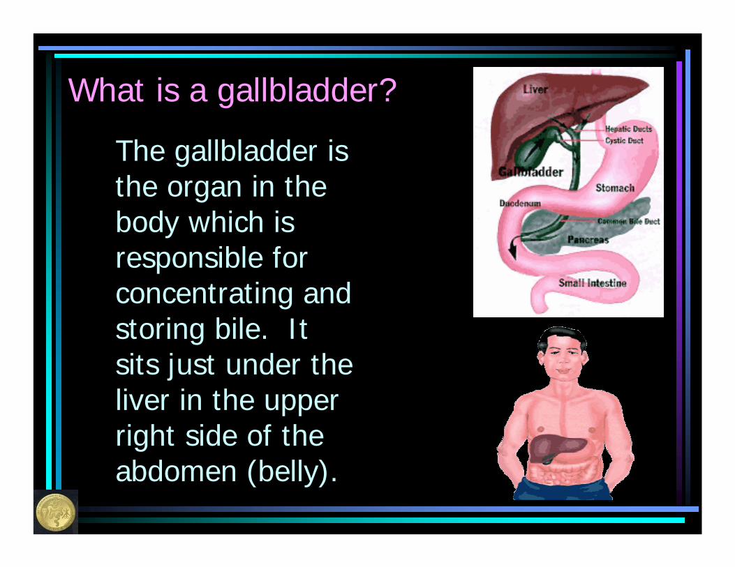
What is a gallbladder?
The gallbladder is the organ in the body which is responsible for concentrating and storing bile. It sits just under the liver in the upper right side of the abdomen (belly).

What is bile?
Bile is a digestive juice that is made by the liver. It is responsible for helping the body to digest fats and the fatty foods in our diet. Normally, bile is golden in color. Our body makes about ½ - 1 quart of bile each day.

What are the bile ducts?
Since bile is used in the digestive process, it must be taken from the liver where it is made to the intestine (bowel) where it is used. The tubes or pipes in the body which deliver the bile from the liver to the small intestines are called bile ducts. They are the size of a small straw or pencil. The bile ducts link the liver to the gallbladder as well, so that some of the bile which is made can be stored for future use.
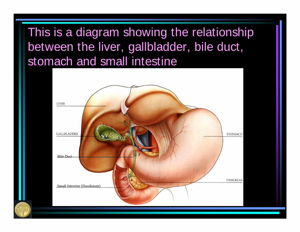
This is a diagram showing the relationship between the liver, gallbladder, bile duct, stomach and small intestine
Small Intestine (Duodenum)
Bile Duct
The area around the gallbladder and bile ducts is very complicated
The bile ducts are further categorized (named) for their relationship to the liver. The pancreas and the pancreatic duct (the tube that drains the pancreatic juices) are in very close proximity and association to the bile ducts and the small intestine. Look closely at the next slide to better understand how these relate to each other.
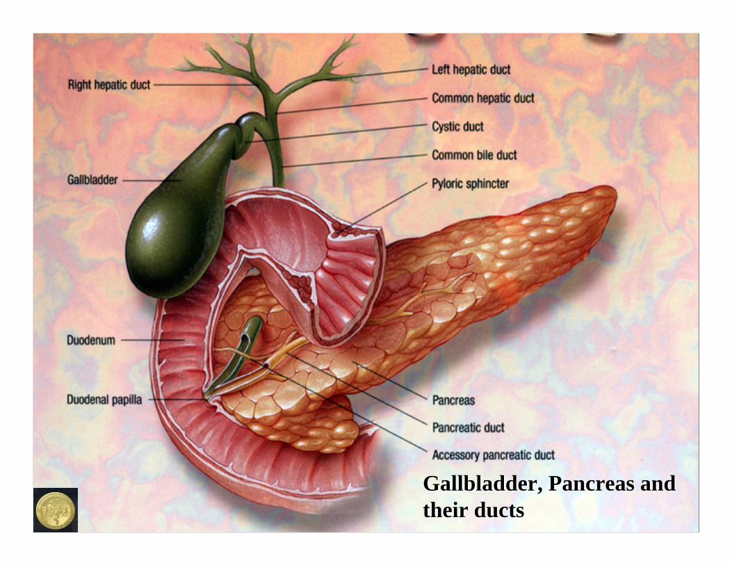
Gallbladder, Pancreas and Gallbladder, Pancreas and their ductstheir ducts
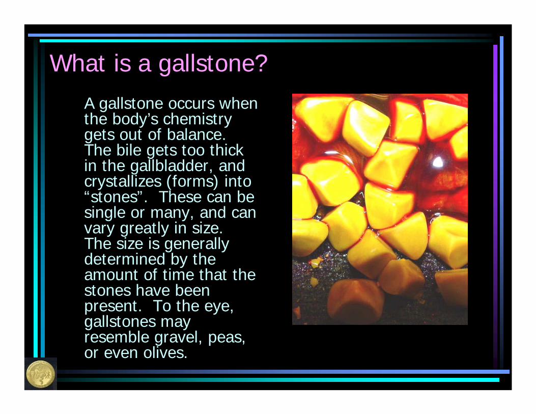
What is a gallstone?A gallstone occurs when the body’s chemistry gets out of balance. The bile gets too thick in the gallbladder, and crystallizes (forms) into “stones”. These can be single or many, and can vary greatly in size. The size is generally determined by the amount of time that the stones have been present. To the eye, gallstones may resemble gravel, peas, or even olives.
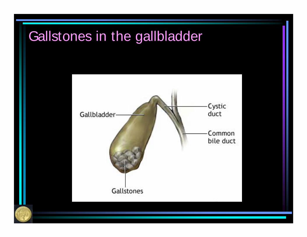
Gallstones in the gallbladder

Who gets gallstones?
Certain factors seem to be more common in people who develop gallstones
• Overweight• Women more than men – especially
those with more than one child• People over the age of forty• People who have gained or lost a large
amount of weight in a relatively short period of time

Who gets gallstones?
• Certain ethnic races/nationalities– American Indians– Eskimos– Northern Europeans
• People with certain other illnesses– Sickle cell anemia– Hemolytic anemia– Inflammatory bowel disease
What symptoms do gallstones cause?
• Many people have no symptoms (up to 50%)
• Those who do may notice the following after eating a meal which contains fat– Crampy pain in the upper right or middle
part of the belly. This pain usually radiates to the shoulder blade.
– Nausea or vomiting– Bloating or “gassiness”
What symptoms do gallstones cause?
• In more severe cases, fever, chills, a yellow tint to the whites of the eyes or dark colored urine may be present.
• Occasionally “sludge” or very thick bile can cause these symptoms even if there are no gallstones in the gallbladder.
These symptoms should prompt a visit either to your doctor or in severe cases to the emergency room.
How are gallstones detected or diagnosed?
• The most accurate (>90%) way is with an ultrasound exam (radiology test which uses sound waves).
• Other tests such as a CAT scan may detect gallstones, but are not as likely as an ultrasound to see the gallstones if they are there.
• Rarely (15%), gallstones can be seen on an ordinary X-Ray of the abdomen (belly).
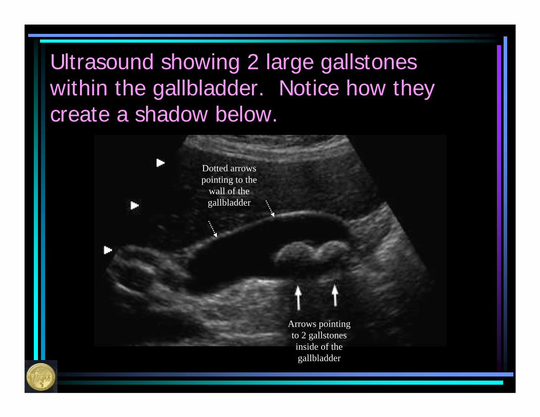
Ultrasound showing 2 large gallstones within the gallbladder. Notice how they create a shadow below.
Arrows pointing to 2 gallstones inside of the gallbladder
Dotted arrows pointing to the
wall of the gallbladder
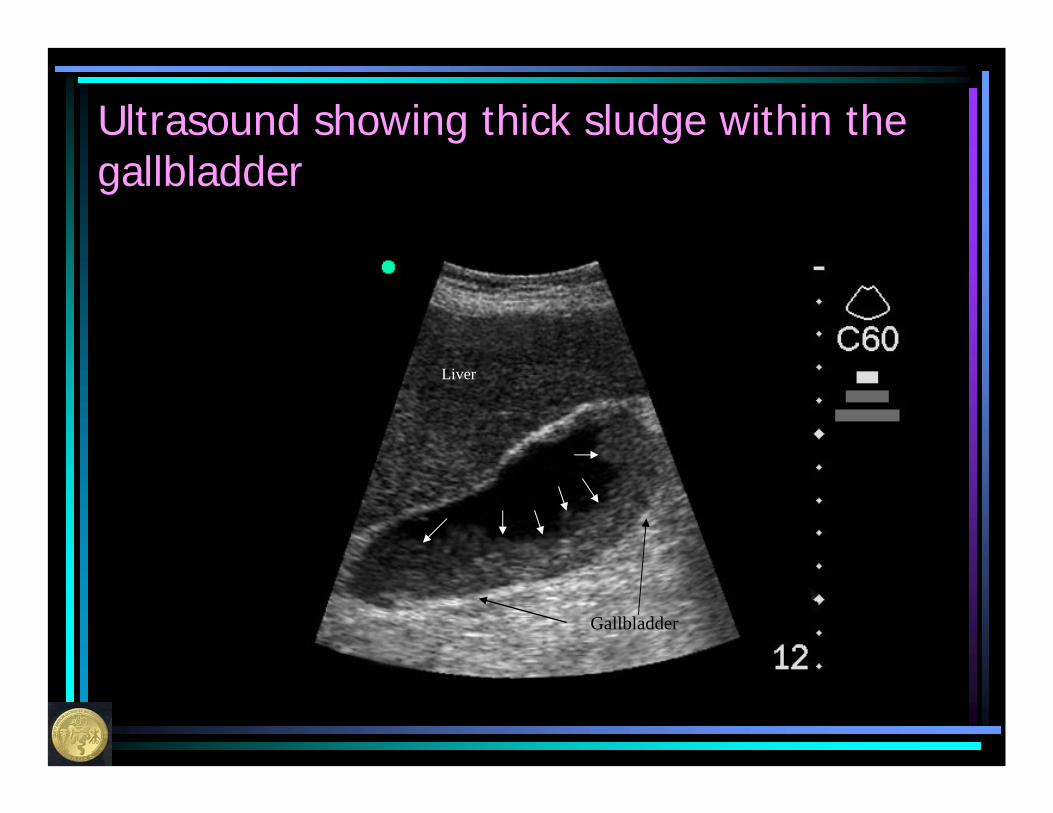
Ultrasound showing thick sludge within the gallbladder
Liver
Gallbladder
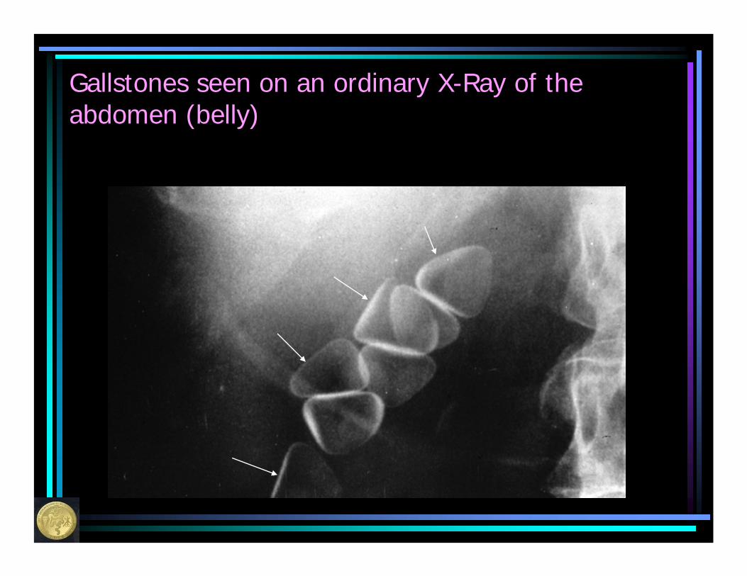
Gallstones seen on an ordinary X-Ray of the abdomen (belly)
How are gallstones treated if they are found?
• Standard treatment is to remove the gallbladder along with the stones in surgery.
• In patients who are too old or sick to undergo surgery, an attempt at dissolving them with medications or pulverizing them with ultrasound waves may be tried.
How are gallstones treated if they are found?
Sometimes, some of the gallstones get out of the gallbladder and make their way down the bile duct into the intestine. As long as these stones do not get caught in the bile duct this is not a problem. If these stones do get caught in one of the bile ducts, this can cause a very severe infection or severe inflammation of the pancreas. This will be discussed later in this presentation.
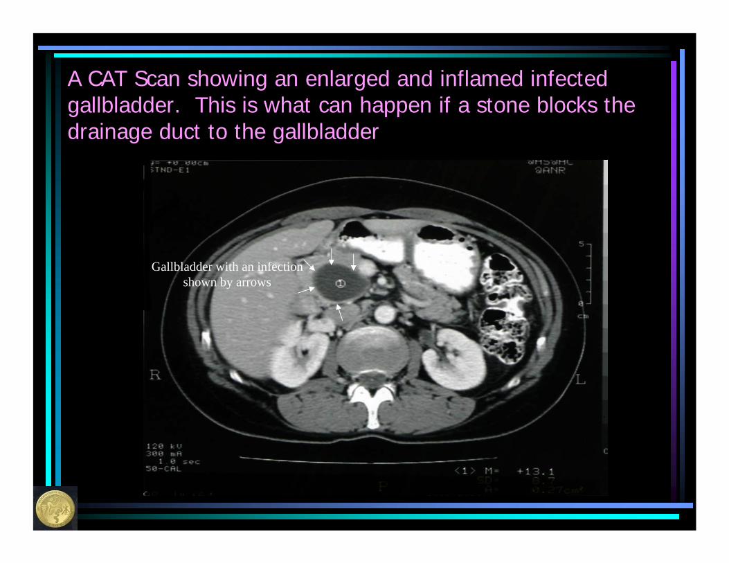
A CAT Scan showing an enlarged and inflamed infected gallbladder. This is what can happen if a stone blocks the drainage duct to the gallbladder
Gallbladder with an infectionshown by arrows
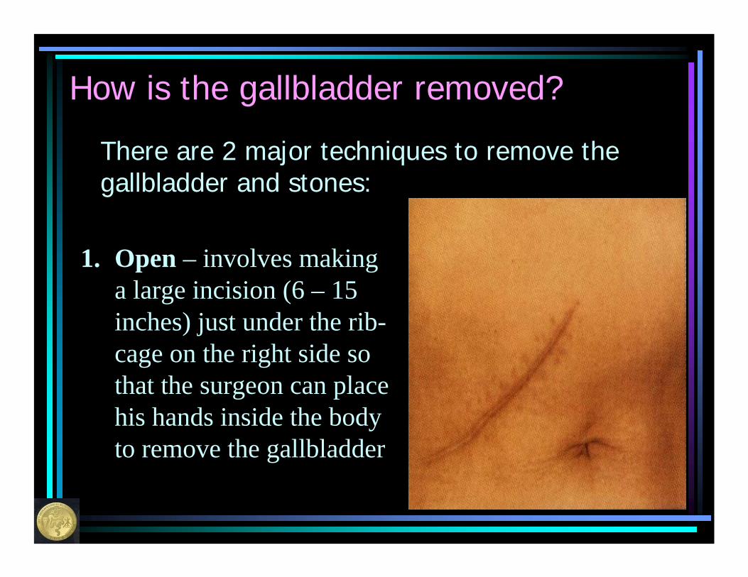
How is the gallbladder removed?
There are 2 major techniques to remove the gallbladder and stones:
1. Open – involves making a large incision (6 – 15 inches) just under the rib-cage on the right side so that the surgeon can place his hands inside the body to remove the gallbladder
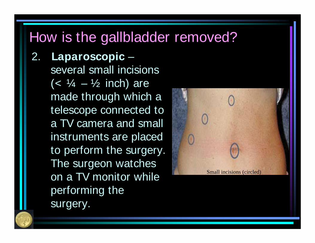
How is the gallbladder removed?2. Laparoscopic –
several small incisions (< ¼ – ½ inch) are made through which a telescope connected to a TV camera and small instruments are placed to perform the surgery. The surgeon watches on a TV monitor while performing the surgery.
Small incisions (circled)
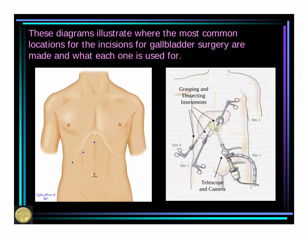
These diagrams illustrate where the most common locations for the incisions for gallbladder surgery are made and what each one is used for.
Telescope and Camera
Grasping and Dissecting Instruments
®Lap Assoc of SF
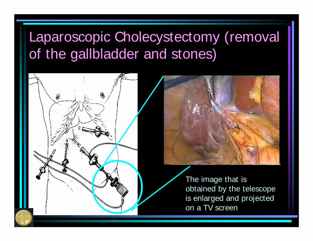
Laparoscopic Cholecystectomy (removal of the gallbladder and stones)
The image that is obtained by the telescope is enlarged and projected on a TV screen
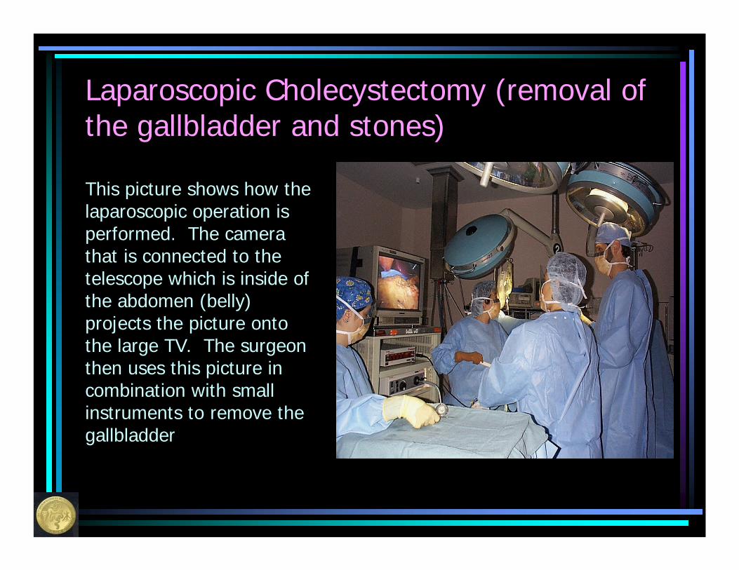
Laparoscopic Cholecystectomy (removal of the gallbladder and stones)
This picture shows how the laparoscopic operation is performed. The camera that is connected to the telescope which is inside of the abdomen (belly) projects the picture onto the large TV. The surgeon then uses this picture in combination with small instruments to remove the gallbladder

Instruments used during a laparoscopic gallbladder surgery. The size depends on your symptoms and the surgeons preference. These areplaced through the abdominal (belly) wall and enable the surgeon to operate through small incisions.
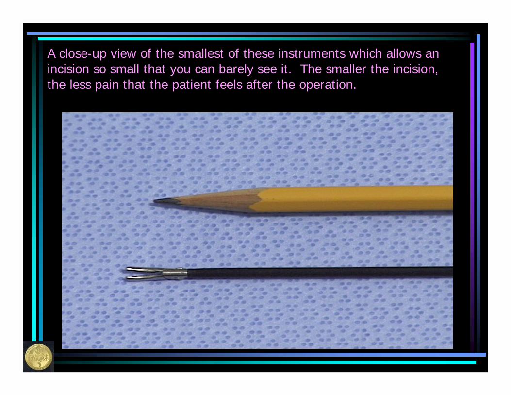
A close-up view of the smallest of these instruments which allows an incision so small that you can barely see it. The smaller the incision, the less pain that the patient feels after the operation.

When very small instruments are used for surgery, the scars left after surgery are very difficult to see and cause very little pain.
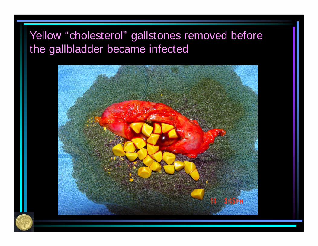
Yellow “cholesterol” gallstones removed before the gallbladder became infected
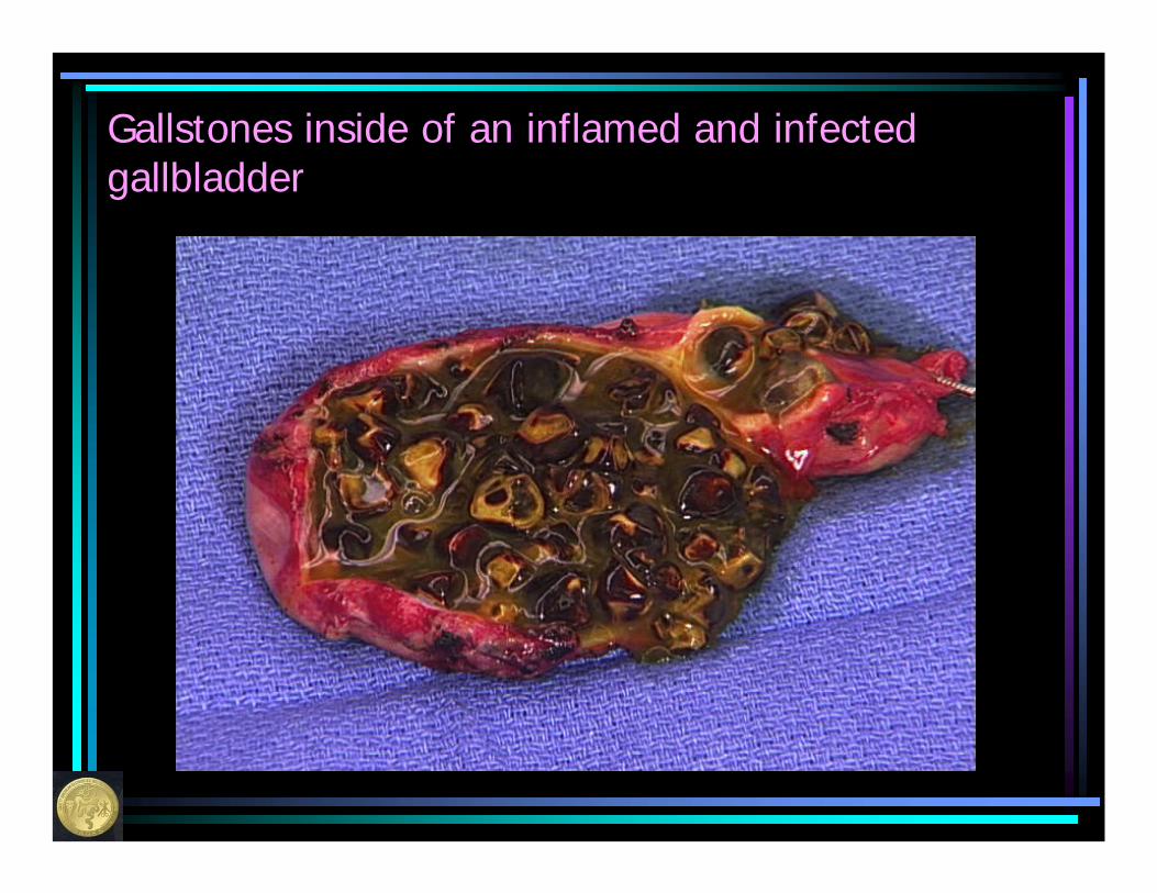
Gallstones inside of an inflamed and infectedgallbladder
Advantages of Laparoscopic Cholecystectomy
• Less scaring• Less pain • Quicker recovery • Shorter hospitalization• Faster return to work• Fewer problems with the incisions
Possible complications of gallbladder surgery
• Injury to the bile duct• Injury to the intestine of one of the
adjacent organs• Narrowing of the bile duct• Bleeding• Infection• Hernia• Leakage of bile into the abdominal
cavity
Possible complications of gallbladder surgery
• Spillage of stones into the abdominal cavity
• Missing stones in the bile duct• Bowel obstruction (blockage) from scar
tissue• Blood clot in the veins of the leg or in
the lung
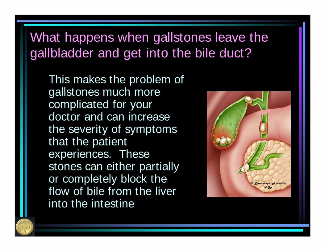
What happens when gallstones leave the gallbladder and get into the bile duct?
This makes the problem of gallstones much more complicated for your doctor and can increase the severity of symptoms that the patient experiences. These stones can either partially or completely block the flow of bile from the liver into the intestine
© Laparoscopic Associates of SF
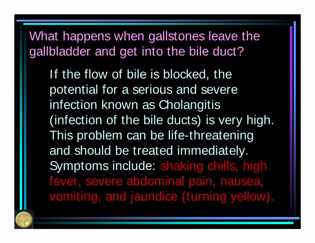
What happens when gallstones leave the gallbladder and get into the bile duct?
If the flow of bile is blocked, the potential for a serious and severe infection known as Cholangitis (infection of the bile ducts) is very high. This problem can be life-threatening and should be treated immediately. Symptoms include: shaking chills, high fever, severe abdominal pain, nausea, vomiting, and jaundice (turning yellow).

How do we know if a patient has stones in their bile ducts?
• Most of the time, patients will have symptoms that will alert the doctor to this – this may include dark colored urine, light colored stool (feces), or a yellow tint in the whites of their eyes
• Sometimes, patients will not have symptoms. Most doctors will perform lab tests to help them identify patients who have stones in their bile duct but have no symptoms
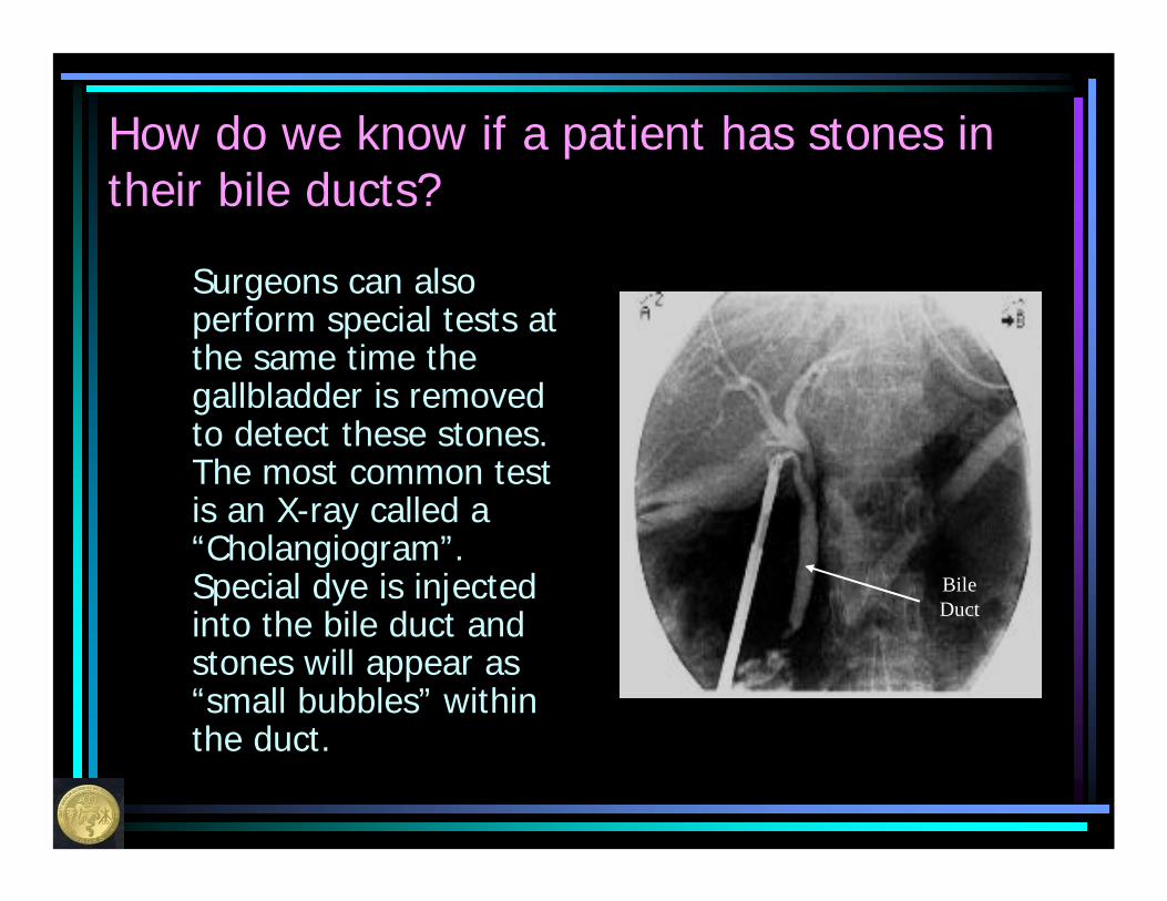
How do we know if a patient has stones in their bile ducts?
Surgeons can also perform special tests at the same time the gallbladder is removed to detect these stones. The most common test is an X-ray called a “Cholangiogram”. Special dye is injected into the bile duct and stones will appear as “small bubbles” within the duct.
Bile Duct
How do we know if a patient has stones in their bile ducts?
Another test is an ultrasound exam at the time of surgery. A small probe is placed directly on the bile duct to detect stones.
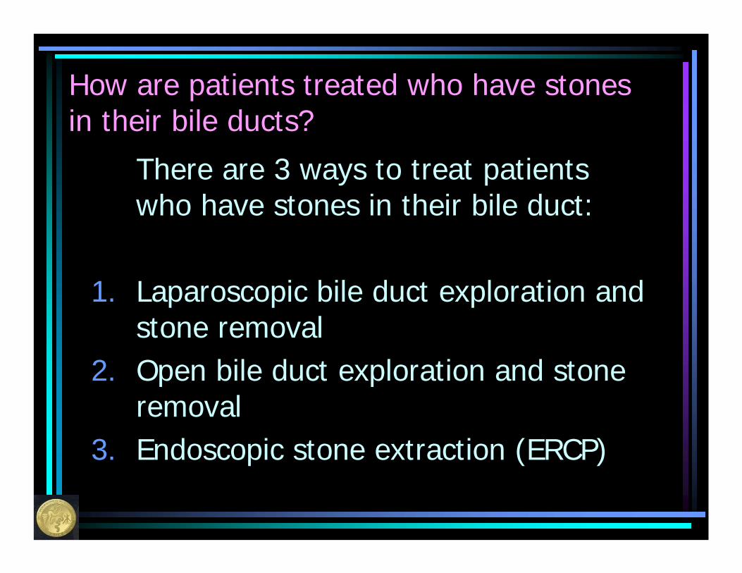
How are patients treated who have stones in their bile ducts?
There are 3 ways to treat patients who have stones in their bile duct:
1. Laparoscopic bile duct exploration and stone removal
2. Open bile duct exploration and stone removal
3. Endoscopic stone extraction (ERCP)
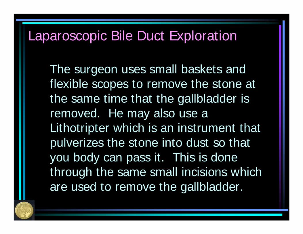
Laparoscopic Bile Duct Exploration
The surgeon uses small baskets and flexible scopes to remove the stone at the same time that the gallbladder is removed. He may also use a Lithotripter which is an instrument that pulverizes the stone into dust so that you body can pass it. This is done through the same small incisions which are used to remove the gallbladder.
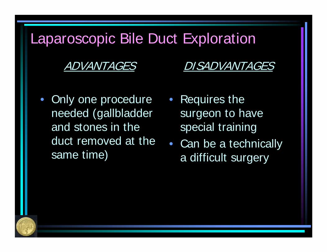
Laparoscopic Bile Duct Exploration
ADVANTAGES
• Only one procedure needed (gallbladder and stones in the duct removed at the same time)
DISADVANTAGES
• Requires the surgeon to have special training
• Can be a technically a difficult surgery
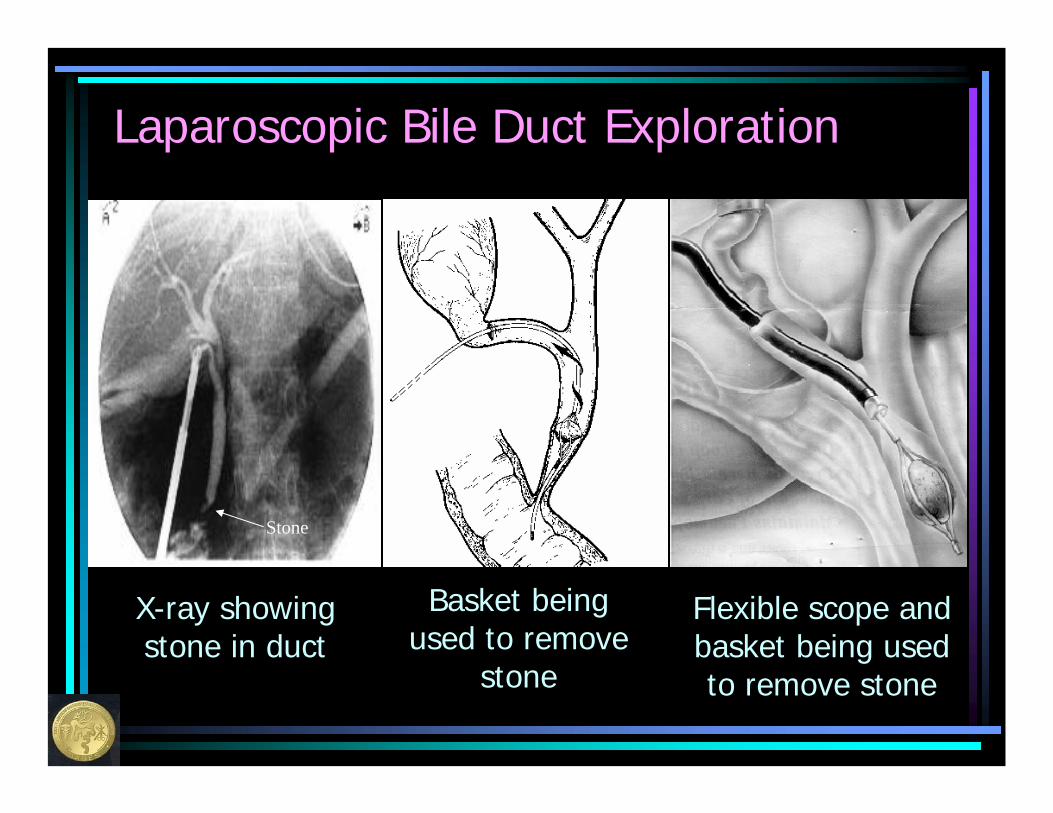
Laparoscopic Bile Duct Exploration
X-ray showing stone in duct
Stone
Basket being used to remove
stone
Flexible scope and basket being used to remove stone
Open Bile Duct Exploration
Occasionally (5-10% of the time), the stones cannot be removed using a laparoscopic approach (or your doctor has not been trained in this technique), and it is necessary to revert to an open operation. In this instance, a large incision is made (just like in the open gallbladder removal) and your surgeon uses the same techniques as described in the previous slides to remove the stones.
Open Bile Duct Exploration
ADVANTAGES
• Only one procedure needed (gallbladder and stones in the duct removed at the same time)
• Technically, less difficult than a laparoscopic procedure
DISADVANTAGES
• Large incision, and therefore more pain a longer hospital stay, and much more time to fully recover
• Much more complicated surgery than just removing the gallbladder
ERCP (endoscopic retrograde cholangiopancreatography)
A procedure performed with a flexible, lighted telescope which allows the doctor to take X-rays of the bile ducts and remove stones from the bile ducts through the small intestine.
ERCP (endoscopic retrograde cholangiopancreatography)
Since the opening of the bile duct is in the small intestine, this scope can be placed through the mouth, down through the stomach, and into the intestine where the duct is located. The duct can then be “opened” (a procedure known as sphincterotomy) so that the stones that are in the bile duct can be removed.
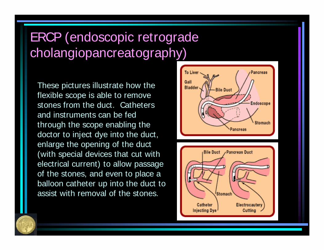
ERCP (endoscopic retrograde cholangiopancreatography)
These pictures illustrate how the flexible scope is able to remove stones from the duct. Catheters and instruments can be fed through the scope enabling the doctor to inject dye into the duct, enlarge the opening of the duct (with special devices that cut with electrical current) to allow passage of the stones, and even to place a balloon catheter up into the duct to assist with removal of the stones.
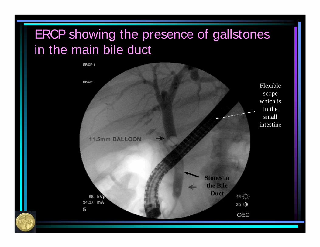
ERCP showing the presence of gallstones in the main bile duct
Stones in the Bile
Duct
Flexible scope
which is in the small
intestine
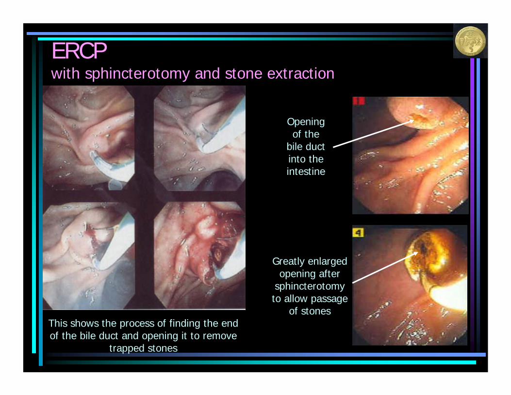
ERCPwith sphincterotomy and stone extraction
This shows the process of finding the end of the bile duct and opening it to remove
trapped stones
Opening of the
bile duct into the intestine
Greatly enlarged opening after
sphincterotomy to allow passage
of stones
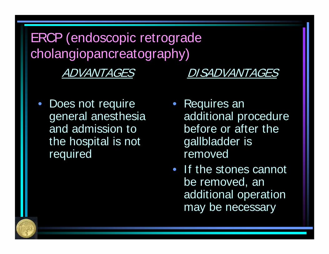
ERCP (endoscopic retrograde cholangiopancreatography)
ADVANTAGES
• Does not require general anesthesia and admission to the hospital is not required
DISADVANTAGES
• Requires an additional procedure before or after the gallbladder is removed
• If the stones cannot be removed, an additional operation may be necessary
Possible complications of ERCP
• Pancreatitis• Injury to the bile duct• Failure to remove all of the stones in
the bile duct• Bleeding• Perforation of the intestinal tract• Infection

Which is method of stone removal is best for me??
Your doctor will decide taking into consideration how severe your condition is, how healthy you are, and the individual skill and training of the doctors involved in your care.