Embed Size (px)
Citation preview
G6PD deficiency: a classic example of pharmacogenetics withon-going clinical implications
Lucio Luzzatto1 and Elisa Seneca2
1Istituto Toscano Tumori and Department of Haematology, University of Florence, Firenze, and 2Department of Haematology,
University of Naples Federico II, Napoli, Italy
Summary
That primaquine and other drugs can trigger acute haemo-
lytic anaemia in subjects who have an inherited mutation
of the glucose 6-phosphate dehydrogenase (G6PD) gene
has been known for over half a century: however, these
events still occur, because when giving the drug either the
G6PD status of a person is not known, or the risk of this
potentially life-threatening complication is under-estimated.
Here we review briefly the genetic basis of G6PD defi-
ciency, and then the pathophysiology and the clinical fea-
tures of drug-induced haemolysis; we also update the list
of potentially haemolytic drugs (which includes rasburi-
case). It is now clear that it is not good practice to give
one of these drugs before testing a person for his/her
G6PD status, especially in populations in whom G6PD
deficiency is common. We discuss therefore how G6PD
testing can be done reconciling safety with cost; this is
once again becoming of public health importance, as more
countries are moving along the pathway of malaria elimi-
nation, that might require mass administration of prima-
quine. Finally, we sketch the triangular relationship
between malaria, antimalarials such as primaquine, and
G6PD deficiency: which is to some extent protective
against malaria, but also a genetically determined hazard
when taking primaquine.
Keywords: G6PD, pharmacogenetics, Clinical Implications.
Over one century ago, Paul Ehrlich found that methylene
blue was an effective anti-malarial (Guttmann & Ehrlich,
1891); and in the 1920s, the synthesis of 8-aminoquino-
lines, specifically plasmoquine and primaquine, was a
major advance in the management of malaria (Vale et al,
2009). The first large-scale use of primaquine (PQ) took
place in the 1950s when US troops were deployed in areas
of Korea where malaria was endemic (Jones et al, 1953):
today it would be called mass drug administration (MDA).
Army doctors observed that, apart from minor gastro-
intestinal complaints, the drug was generally well tolerated;
but some of the soldiers receiving the drug became jaun-
diced and anaemic. This was relatively more common
among African Americans, and this complication, toxic
effect or side effect, was called the primaquine sensitivity
syndrome (Dern et al, 1955). A.S. Alving’s group (Chicago)
performed experiments in human volunteers in order to
detail the clinical and haematological features (see Fig 1)
of the acute haemolytic anaemia (AHA) triggered by PQ
(Tarlov et al, 1962) (the volunteers were inmates of a pen-
itentiary near Chicago who were clinically and haematolog-
ically normal at the start of the experiment, but suffered
AHA as a result of the experiment. The volunteers were
under constant medical supervision; as far as is known
they did not come to any other harm, they all recovered
without any blood transfusion and, like after an attack of
favism, there is no reason to presume that they had any
long-term ill effects. One of us has known in person some
of the physicians involved, and regards them as of high
moral standards. Nevertheless, this was human experimen-
tation without any medical benefit for the volunteers, and
would almost certainly not be allowed today). The same
group also reported that the enzymatic activity of glucose
6-phosphate dehydrogenase (G6PD) was markedly reduced
in red cells from PQ sensitive subjects (Carson et al,
1956); hence the term G6PD deficiency. Genetic analysis
demonstrated that G6PD deficiency was inherited as an
X-linked trait (Adam, 1961).
Pharmacogenetics deals with genetically determined varia-
tion in how individuals respond to drugs, in terms of both
therapeutic effects and adverse effects. This concept had
emerged when tasters and non-tasters of phenylthiocarba-
mide (PTC) had been identified, and the ability to feel that
taste was shown to be inherited (Blakeslee, 1932). As testing
for PTC tasting was easy and minimally invasive, PTC tasting
was one of the first traits widely studied at the dawn of
human population genetics. But it was with the discovery of
G6PD deficiency as the biochemical basis of PQ sensitivity
that this became a prototype study case in pharmacogenetics,
Correspondence: Professor Lucio Luzzatto, Scientific Director,
Istituto Toscano Tumori, Via Taddeo Alderotti 26N, Firenze 50139,
Italy.
E-mail: [email protected]
review
ª 2013 John Wiley & Sons Ltd, British Journal of Haematology doi:10.1111/bjh.12665
just at about the time this term was coined (Motulsky, 1957;
Vogel, 1959). Almost immediately it became clear that G6PD
deficiency was also the biochemical defect underlying
favism (Sansone & Segni, 1958), an acute syndrome associ-
ated with anaemia, haemoglobinuria and jaundice, poten-
tially fatal especially in children, intriguingly affecting only
some of the many people who eat fava beans. Favism had
been known for centuries (Fermi & Martinetti, 1905), but
it had been mis-interpreted for a long time as an allergic
reaction.
In the 1960s and 1970s, numerous drugs other than PQ
were reported as possible triggers of AHA in G6PD-defi-
cient subjects. When a new drug is introduced, ‘adverse
events’ or side effects can be expected. The medicine pack-
age sheet is not very helpful, because its compilation is
largely driven by medico-legal motives: thus, relatively triv-
ial common complaints (e.g.’headache’, ‘abdominal discom-
fort’) are listed along with very rare serious problems (e.g.
‘acute depression and suicide’). On the other hand, journal
publications required for licensing must include tables with
the frequency of each side effect as observed in formal tri-
als. The power of pharmacogenetics is that sometimes it
can assign a specific meaning to these frequency figures.
Thus, the frequency of AHA in patients receiving dapsone
may be 10%; but if we stratify the patients by G6PD sta-
tus, and we find that those 10% were all G6PD deficient,
it means that the risk of AHA is practically 0 in G6PD
normal patients, and about 100% in G6PD-deficient
patients – the original figure of 10% was simply the fre-
quency of G6PD deficiency in the patient population
where the trial was conducted. This obvious interpretation
of ‘% risk’ must be always before us. Favism, neonatal
jaundice and other manifestations of G6PD deficiency are
outside the scope of this review.
Drug-induced AHA
Clinical course
The main features of drug-induced AHA are well known
(Luzzatto & Poggi, 2009). The two drugs for which we have
extensive detailed data are PQ and dapsone. As mentioned
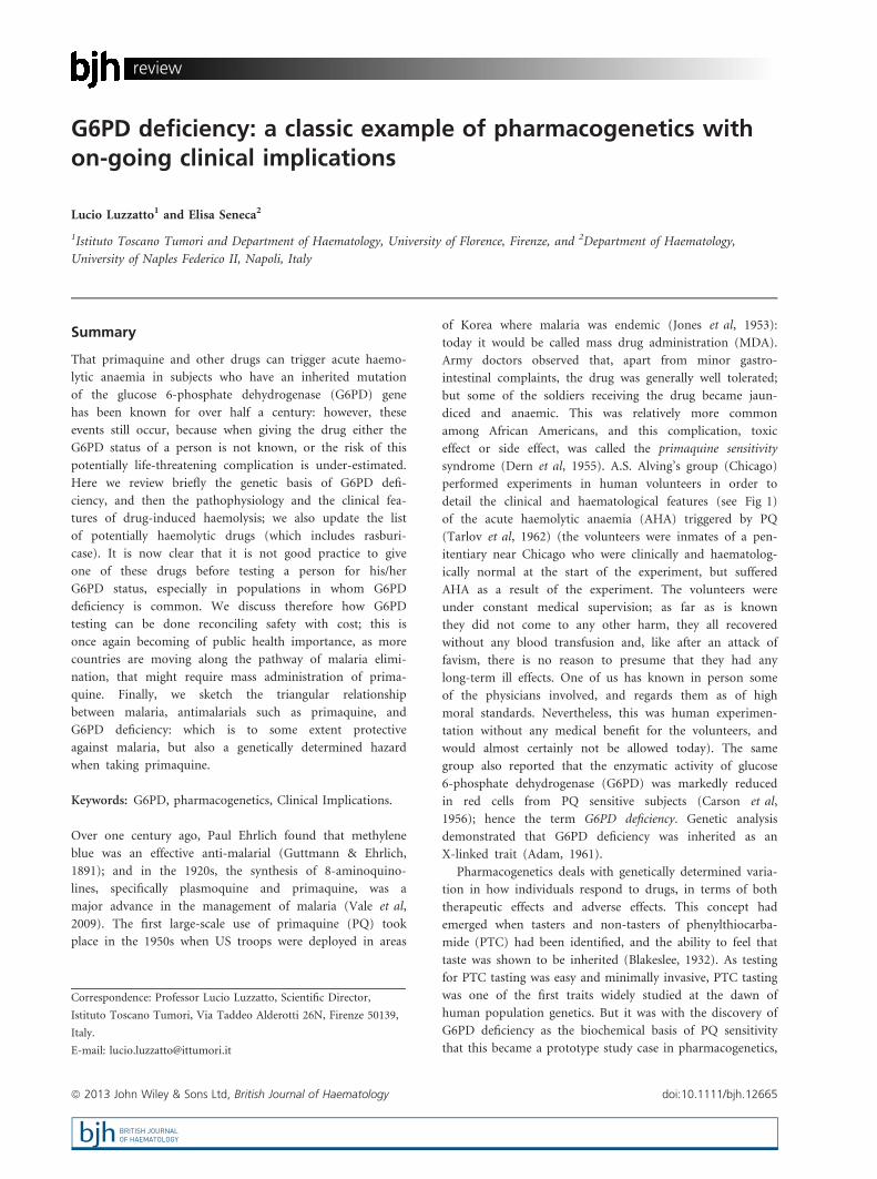
above, the clinical course of PQ-induced AHA (Fig 1) has
been studied in a number of African-American normal vol-
unteers (Tarlov et al, 1962). The clinical picture was very
similar to that of favism, which was already well known
(Luisada, 1941; Meloni et al, 1983): except that with favism
the severity is extremely variable, certainly at least in part
because the amount of fava beans consumed is highly vari-
able; with PQ 45 mg/day the pattern was instead fairly uni-
form (Tarlov et al, 1962). All subjects had a drop in Hb of
about 50 g/l, with a nadir on day 7; they all had haemoglo-
binuria for 1–3 d; they all developed characteristic changes
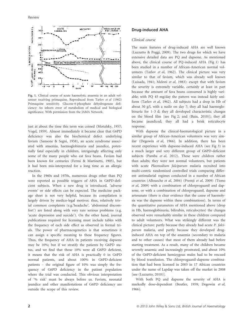
on the blood film [see Fig 2; and (Bain, 2010)]; they all
became jaundiced; they all had a brisk reticulocyte
response.
With dapsone the clinical-haematological picture in a
similar group of African-American volunteers was very sim-
ilar (Degowin et al, 1966). In addition, there has been
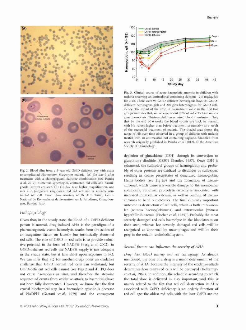
recent experience with dapsone-induced AHA (see Fig 3) in
a much larger and very different group of G6PD-deficient
subjects (Pamba et al, 2012). These were children rather
than adults; they were not normal volunteers, but patients
with acute Plasmodium falciparum malaria recruited into
multi-centric randomized controlled trials comparing differ-
ent antimalarial regimes conducted in a number of African
countries (Alloueche et al, 2004) (Premji et al, 2009) (Tiono
et al, 2009) with a combination of chlorproguanil and dap-
sone, or with a combination of chlorproguanil, dapsone and
artesunate (there is clear evidence that the cause of haemoly-
sis was the dapsone within these combinations). In terms of
the quantitative parameters of AHA mentioned above (drop
in Hb, haemoglobinuria, bilirubin, reticulocytes) the changes
observed were remarkably similar in these children compared
to adult volunteers. What was strikingly different was the
clinical picture: partly because they already had acute P. falci-
parum malaria, and partly because they developed drug-
induced AHA on top of the anaemia (secondary to malaria
and to other causes) that most of them already had before
starting treatment. As a result, many of the children became
severely anaemic and increasingly prostrated, and about 10%
of the G6PD-deficient hemizygous males had to be rescued
by blood transfusion. The chlorproguanil-dapsone combina-
tion that had been licensed in 2003 in 17 African countries
under the name of Lapdap was taken off the market in 2008
[see (Luzzatto, 2010)].
With both PQ and dapsone the severity of AHA is
markedly dose-dependent (Beutler, 1959; Degowin et al,
1966).
Fig. 1. Clinical course of acute haemolytic anaemia in an adult vol-
unteer receiving primaquine. Reproduced from Tarlov et al (1962)
Primaquine sensitivity. Glucose-6-phosphate dehydrogenase defi-
ciency: An inborn error of metabolism of medical and biological
significance. With permission from the JAMA Network.
Review
2 ª 2013 John Wiley & Sons Ltd, British Journal of Haematology
Pathophysiology
Given that, in the steady state, the blood of a G6PD-deficient
person is normal, drug-induced AHA is the paradigm of a
pharmacogenetic event: haemolysis results from the action of
an exogenous factor on latently but intrinsically abnormal
red cells. The role of G6PD in red cells is to provide reduc-
tive potential in the form of NADPH (Berg et al, 2002): in
G6PD-deficient red cells the NADPH supply is just adequate
in the steady state, but it falls short upon exposure to PQ.
We can infer that PQ (or another drug) poses an oxidative
challenge that G6PD normal red cells can withstand, but
G6PD-deficient red cells cannot (see Figs 2 and 4). PQ does
not cause haemolysis in vitro, and therefore the stepwise
sequence of events from oxidative attack to haemolysis have
not been fully documented. However, we know that the first
crucial biochemical step in a haemolytic episode is decrease
of NADPH (Gaetani et al, 1979) and the consequent
depletion of glutathione (GSH) through its conversion to
glutathione disulfide (GSSG) (Beutler, 1957). Once GSH is
exhausted, the sulfhydryl groups of haemoglobin and proba-
bly of other proteins are oxidized to disulfides or sulfoxides,
resulting in coarse precipitates of denatured haemoglobin,
Heinz bodies (see Fig 2B) and the formation of haemi-
chromes, which cause irreversible damage to the membrane:
specifically, abnormal proteolytic activity is associated with
increased intracellular calcium, as well as binding of haemi-
chromes to band 3 molecules. The final clinically important
outcome is destruction of red cells, which is both intravascu-
lar (witness haemoglobinuria) and extravascular [witness
hyperbilirubinaemia (Fischer et al, 1985)]. Probably the most
severely damaged red cells haemolyse in the bloodstream on
their own, whereas less severely damaged red cells will be
recognized as abnormal by macrophages and will be their
prey in the reticulo-endothelial system.
Several factors can influence the severity of AHA
Drug dose, G6PD activity and red cell ageing. As already
mentioned, the dose of a drug is a major determinant of the
severity of AHA, because the intensity of the oxidative attack
determines how many red cells will be destroyed (Kellermey-
er et al, 1962). In addition, the schedule according to which
the total dose is delivered is also important, and this is
mainly related to the fact that red cell destruction in AHA
associated with G6PD deficiency is an orderly function of
red cell age: the oldest red cells with the least G6PD are the
70
80
90
100
110
120
130
0 5 10 15 20 25 30 35 40 45Study day
Hae
mog
lobi
n, g
/l (+
/– S
E) Boys
BoysGirls
G6PD deficient G6PD heterozygotes G6PD normal
Fig. 3. Clinical course of acute haemolytic anaemia in children with
malaria receiving an antimalarial containing dapsone (2�5 mg/kg/day
for 3 d). There were 95 G6PD-deficient hemizygous boys, 24 G6PD-
deficient hemizygous girls and 200 girls heterozygous for G6PD defi-
ciency. The extent of the drop in haematocrit value in the first two
groups indicates that, on average, about 25% of red cells have under-
gone haemolysis. Thirteen children required blood transfusion. Note
that by the end of 6 weeks the blood counts are back to normal,
with Hb values higher than before treatment, presumably as a result
of the successful treatment of malaria. The shaded area shows the
range of Hb over time observed in a group of children with malaria
treated with an antimalarial not containing dapsone. Modified from
research originally published in Pamba et al (2012). © the American
Society of Hematology.
(A)
(B)
Fig. 2. Blood film from a 3-year-old G6PD-deficient boy with acute
uncomplicated Plasmodium falciparum malaria. (A) On day 3 after
treatment with a chlorproguanil-dapsone combination (see Pamba
et al, 2012), numerous spherocytes, contracted red cells and haemi-
ghosts (arrow) are seen. (B) On day 1, at higher magnification, one
sees a P. falciparum ring-parasitized red cell and a severely con-
tracted red cell. Blood films courtesy of Dr A B Tiono, Centre
National de Recherche et de Formation sur le Paludisme, Ouagadou-
gou, Burkina Faso.
Review
ª 2013 John Wiley & Sons Ltd, British Journal of Haematology 3
first to haemolyse, and the haemolytic process progresses
upstream toward cells with more and more G6PD activity.
Thus, with a single dose of 45 mg PQ there may be abrupt
haemolysis of, say, 45% of all red cells in a particular G6PD-
deficient person; if instead we give that same person 15 mg
PQ, only some 15% of red cells will haemolyse. As PQ is
short-lived, if we now give a further dose of 15 mg, many of
the surviving red cells will not be sensitive to the corre-
sponding blood level of PQ: therefore there will be less
haemolysis, and this will be even more true with the next
dose, because with each dose there is a selective enrichment
in red cells that, despite being genetically G6PD deficient,
have relatively higher levels of G6PD. This phenomenon can
be so marked with certain G6PD variants that patients in the
post-haemolytic state are found to be relatively resistant to
further challenge (Tarlov et al, 1962); thus, the patient may
be in a state of compensated haemolysis, also called resistance
phase.
Heterozygous versus hemizygous (or homozygous) G6PD defi-
ciency. As the G6PD gene is on the X chromosome, males
have only one allele, and if this has a mutation causing
G6PD deficiency all the red cells will be G6PD deficient.
In areas where G6PD deficiency is common, there is a non-
negligible number of homozygous females (Luzzatto & Allan,
1968), in whom both G6PD alleles are mutant: again all their
red cells will be G6PD deficient. (For brevity in this review
when discussing hemizygous males we will imply that the
same applies to homozygous females). Heterozygous females,
instead, by virtue of X chromosome inactivation in somatic
cells, have a dual population, with G6PD normal and G6PD-
deficient red cells in their blood (Beutler et al, 1962): the
average ratio is 50:50 but it is well known that the ratio is
very variable (Nance, 1964; Rinaldi et al, 1976). One can
expect therefore that, with a given dose of a given drug,
AHA in heterozygotes will be still manifest but will be, on
the average, less severe. At one extreme, in a female with a
large excess of G6PD normal red cells, haemolysis may be all
but undetectable; at the other end of the spectrum, with a
large excess of G6PD-deficient red cells, AHA may be just as
severe as in hemizygous G6PD-deficient males. These expec-
tations have been verified in a recent study of 200 heterozyg-
otes exposed to dapsone (Pamba et al, 2012).
Genetic (allelic) heterogeneity of G6PD deficiency. There are
now 187 known mutations in the G6PD gene (Minucci et al,
2012a), and at least 35 of the mutant alleles are polymorphic,
i.e. relatively common in different parts of the world. None
of the mutations in these alleles causes complete loss of
G6PD, which would be lethal (Pandolfi et al, 1995); rather,
they cause marked deficiency of G6PD in red cells by
decreasing the stability of the variant enzyme (Luzzatto et al,
2001). In other words, the normal process whereby G6PD
activity decreases as red cells age (see above) is greatly accel-
erated (Morelli et al, 1978). Thus, if old red cells have much
less G6PD activity than reticulocytes in G6PD normal blood,
the oldest red cells in G6PD-deficient blood will have nearly
zero activity. In addition, some mutations significantly affect
the binding of substrates (glucose 6-phosphate and NADP),
or catalytic efficiency of the enzyme, or both (Mason et al,
2007). Indeed, each mutant allele produces a G6PD variant
protein that, compared to normal G6PD, has a distinct pat-
tern of quantitative and qualitative changes. In view of this,
it would not be surprising if a G6PD-deficient subject with
one G6PD variant responded to a drug somewhat differently
from another G6PD-deficient subject who has a different
G6PD variant. The only available documented comparative
data are on the effect of PQ on (i) subjects with G6PD A-,
the variant common in Africa, present in the African-Ameri-
can volunteers in Chicago (see above); and (ii) subjects from
Sardinia with G6PD Mediterranean, a variant common in
that area, in the Middle East and in India. For the same dose
of PQ, by comparison to previous experience with G6PD A-,
AHA was considerably more severe with G6PD Mediterra-
nean (Pannacciulli et al, 1965); in addition, with the latter
there was little evidence of a ‘resistance phase’ upon repeated
administration (Salvidio et al, 1972). This clinical difference
is consistent with the fact that the residual G6PD activity in
red cells is about 5% of normal with G6PD Mediterranean,
whereas it is 13% of normal with G6PD A- (Luzzatto et al,
2001). In view of the fact that G6PD variants differ from
each other not only in quantity but also in quality (see
above), we cannot assume that the severity of AHA will be a
simple function of residual activity. However, because most
of the polymorphic G6PD variants for which quantitative
data are available have less residual activity than G6PD A-,
we must expect in first approximation that, other things
being equal, AHA with other variants may be more severe
than with G6PD A-.
It must be noted that when discussing genotype-pheno-
type correlations with respect to G6PD-related drug-induced
AHA there is sometimes confusion between two separate
issues. The term genotype might refer to hemizygotes and
homozygotes versus heterozygotes (item 2 above), whereby
the difference in clinical phenotype is drastic. By comparison,
when the term genotype refers to one G6PD allele versus
others (item 3 above) the differences are rather marginal,
especially as it is now clear that, even with G6PD A-, hith-
erto often regarded as a ‘mild’ variant, the clinical manifesta-
tion can be anything but mild (Pamba et al, 2012).
Current list of drugs that can cause AHA
The list of drugs that can cause AHA in G6PD-deficient sub-
jects (Table I) is similar to, but shorter than, many that have
been published since the initial World Health Organization
(WHO) report (Betke et al, 1967). Over the years some
drugs have been taken off the list simply because they are no
longer in use, and others, especially some antibacterials,
because AHA that had been attributed to them was more
Review
4 ª 2013 John Wiley & Sons Ltd, British Journal of Haematology
likely to have been triggered by the infection that had caused
them to be prescribed [for instance, AHA is common in
G6PD-deficient patients with lobar pneumonia (Tugwell,
1973)].
A recent review (Youngster et al, 2010) described a
detailed systematic analysis of the literature and concluded
that, for only seven drugs there was solid evidence that they
cause AHA in G6PD-deficient subjects. We think that the
principles of evidence-based medicine serve well the cause of
establishing the efficacy of drugs, but they are more difficult
to apply to the side-effects of drugs, where even an isolated
case report, if well documented, must be taken seriously.
Therefore our list is a bit longer, and we have preferred to
classify the effect into two groups, where predictable haemo-
lysis means that AHA can be expected in a G6PD-deficient
patient, whereas possible haemolysis means that AHA may or
may not take place, depending on dosage, other concomitant
drugs, co-morbidity and other factors. Thus, the list in
Table I is similar to that in the current British National For-
mulary (www.bnf.org).
Methylene blue (MB, also termed methylthioninium chlo-
ride) deserves special mention for two reasons. First, it is used
for the treatment of methaemoglobinemia, where it enhances
reversion of methaemoglobin back to haemoglobin via
NADPH-dependent methaemoglobin reductase. However, if a
G6PD-deficient patient has acquired methaemoglobinaemia
induced by drugs or other agents, MB will only make things
worse by causing AHA (Gauthier, 2000; Liao et al, 2002; Foltz
et al, 2006; Brewer, 2007). Second, MB has been recently
revived as an antimalarial (in combination with artemisin-
based combinations) because it has gametocytocidal action
against P. Falciparum. This is of considerable interest (see sec-
tion on primaquine below): however, MB at 15 mg/kg/day
has been associated with significant haemolysis in G6PD-defi-
cient children in Burkina Faso (Muller et al, 2013). It remains
to be seen whether at this or lower dosage the benefits may
outweigh this side effects in any individual community.
The case of rasburicase (urate oxidase)
This drug, licensed about 10 years ago for the prohpylaxis
and treatment of hyperuricaemia associated with tumour
lysis syndrome (TLS), deserves special attention for several
reasons. First, with other drugs the precise chemical mecha-
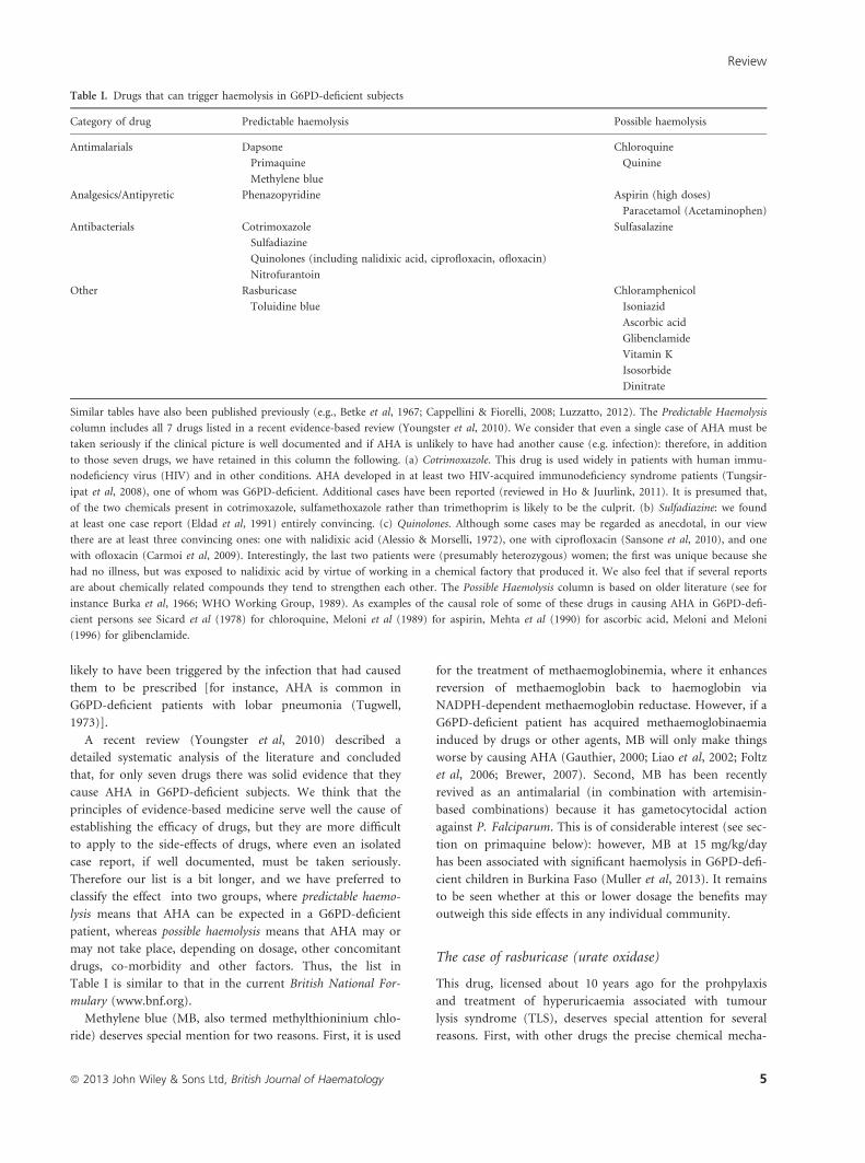
Table I. Drugs that can trigger haemolysis in G6PD-deficient subjects
Category of drug Predictable haemolysis Possible haemolysis
Antimalarials Dapsone
Primaquine
Methylene blue
Chloroquine
Quinine
Analgesics/Antipyretic Phenazopyridine Aspirin (high doses)
Paracetamol (Acetaminophen)
Antibacterials Cotrimoxazole
Sulfadiazine
Quinolones (including nalidixic acid, ciprofloxacin, ofloxacin)
Nitrofurantoin
Sulfasalazine
Other Rasburicase
Toluidine blue
Chloramphenicol
Isoniazid
Ascorbic acid
Glibenclamide
Vitamin K
Isosorbide
Dinitrate
Similar tables have also been published previously (e.g., Betke et al, 1967; Cappellini & Fiorelli, 2008; Luzzatto, 2012). The Predictable Haemolysis
column includes all 7 drugs listed in a recent evidence-based review (Youngster et al, 2010). We consider that even a single case of AHA must be
taken seriously if the clinical picture is well documented and if AHA is unlikely to have had another cause (e.g. infection): therefore, in addition
to those seven drugs, we have retained in this column the following. (a) Cotrimoxazole. This drug is used widely in patients with human immu-
nodeficiency virus (HIV) and in other conditions. AHA developed in at least two HIV-acquired immunodeficiency syndrome patients (Tungsir-
ipat et al, 2008), one of whom was G6PD-deficient. Additional cases have been reported (reviewed in Ho & Juurlink, 2011). It is presumed that,
of the two chemicals present in cotrimoxazole, sulfamethoxazole rather than trimethoprim is likely to be the culprit. (b) Sulfadiazine: we found
at least one case report (Eldad et al, 1991) entirely convincing. (c) Quinolones. Although some cases may be regarded as anecdotal, in our view
there are at least three convincing ones: one with nalidixic acid (Alessio & Morselli, 1972), one with ciprofloxacin (Sansone et al, 2010), and one
with ofloxacin (Carmoi et al, 2009). Interestingly, the last two patients were (presumably heterozygous) women; the first was unique because she
had no illness, but was exposed to nalidixic acid by virtue of working in a chemical factory that produced it. We also feel that if several reports
are about chemically related compounds they tend to strengthen each other. The Possible Haemolysis column is based on older literature (see for
instance Burka et al, 1966; WHO Working Group, 1989). As examples of the causal role of some of these drugs in causing AHA in G6PD-defi-
cient persons see Sicard et al (1978) for chloroquine, Meloni et al (1989) for aspirin, Mehta et al (1990) for ascorbic acid, Meloni and Meloni
(1996) for glibenclamide.
Review
ª 2013 John Wiley & Sons Ltd, British Journal of Haematology 5
nism whereby they cause oxidative damage is not fully
understood: for rasburicase – a genetically engineered form
of the enzyme urate oxidase – we know instead that, like all
other oxidases, one of the products of the enzyme reaction is
hydrogen peroxide, of which one molecule is produced stoi-
chiometrically for every molecule of uric acid that is catabo-
lized. In normal cells, hydrogen peroxide is promptly
degraded by either glutathione peroxidase (GSHPX) or cata-
lase, but in red cells of G6PD-deficient subjects, GSHPX
activity is impaired because GSH is in short supply (Fig 4),
and catalase may also be impaired because the intracellular
NADPH concentration is low (Gaetani et al, 1994, 1996).
Second, presumably as a result of this mechanism, in G6PD-
deficient subjects rasburicase causes both AHA and acute
methaemoglobinaemia – the latter can reach levels of up to
20%, much higher than that seen with other drugs. This lit-
erally colours the clinical picture, because the patient appears
cyanotic; and methaemoglobinaemia makes the tissue
hypoxia consequent on anaemia more severe, because meth-
aemoglobin does not carry oxygen. Third, rasburicase is used
in patients who have malignancies (such as acute leukaemia
with high blast counts, large cell lymphoma, and sometimes
other solid tumours), and who, following chemotherapy,
may develop TLS. This means that the drug is administered
to a patient who is already quite ill, may be already anaemic,
and may be already at risk of renal failure. In addition,
rasburicase has a t1/2 of 18–24 h resulting in the production
of hydrogen peroxide for several days. Fourth, the association
between rasburicase-induced AHA and G6PD deficiency is so
strong, that this is formally stated as a contra-indication. In
a recent compilation of case reports (Sonbol et al, 2013),
three patients were listed as G6PD normal, but in fact in one
of them the test was not done (Kizer et al, 2006); in two no
quantitative results were given (Kizer et al, 2006; Bauters
et al, 2011), and the test was carried out after a haemolytic
attack (when a false-normal result is possible), and after
blood transfusion: therefore the G6PD status must be
regarded as doubtful (the enzyme test ought to have been
repeated subsequently, or a molecular test should have been
done).
In an authoritative paper on the evidence-based manage-
ment of TLS, rasburicase is said to be ‘contraindicated in
patients with a history consistent with glucose-6 phosphate
dehydrogenase’ (Cairo et al, 2010). Leaving aside the typo-
graphical error whereby the word deficiency was omitted,
this suggestion is rather strange, since the majority of
patients with G6PD deficiency have never had a haemolytic
attack, and therefore they or their relatives will provide no
such history. The recommendation must be instead to do a
G6PD test before giving rasburicase, but a recurrent state-
ment in the relevant literature is that under the pressure of
impending TLS there is no time to wait for the result. In fact
a G6PD test can be done in 20 min and, were it not out-
sourced, the result should be available within 1 h. It seems
to us incongruous that in highly specialized facilities where,
for instance, arterial blood gases and flow cytometry are rou-
tine, one cannot do a simple G6PD test (even a ‘screening
test’ would be much better than nothing, see section below
on G6PD testing), costing a small fraction of a single dose of
rasburicase. No clinical unit treating acute leukaemia or lym-
phoma should be accredited if a G6PD test result cannot be
obtained promptly. The same ought to apply to neonatal
units where rasburicase is now used for ‘acute kidney injury’
(Poliseno et al, 2010): in a G6PD-deficient baby, who had
not been tested, the result was fatal (Zaramella et al, 2013).
GSSG
GSH
NADPH
NADP+
Glutathionereductase G6PD
6PGl
G6P
DrugsH2O2
other ROS
H2O
Hb MetHb
Glutathioneperoxidase
GSSG
GSH
NADPH
NADP+
Glutathionereductase G6PD
6PGl
G6P
DrugsH2O2
other ROS
H2O
Hb MetHb
IntravascularHaemolysis
Damage to RBC membrane (band 3)
Sulphaemoglobin
Haemichromes
Heinz bodies Hemighosts
ExtravascularHaemolysis
Haemoglobinuria
Bilirubin
Glutathioneperoxidase
(A)
(B)
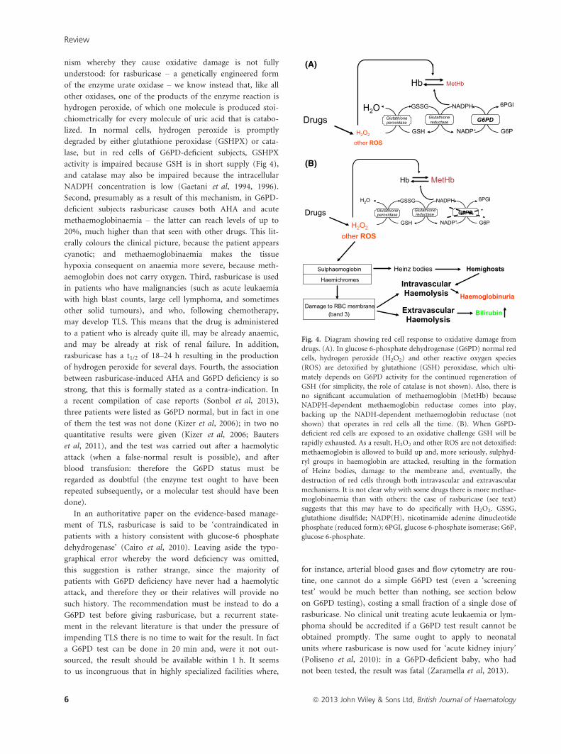
Fig. 4. Diagram showing red cell response to oxidative damage from
drugs. (A). In glucose 6-phosphate dehydrogenase (G6PD) normal red
cells, hydrogen peroxide (H2O2) and other reactive oxygen species
(ROS) are detoxified by glutathione (GSH) peroxidase, which ulti-
mately depends on G6PD activity for the continued regeneration of
GSH (for simplicity, the role of catalase is not shown). Also, there is
no significant accumulation of methaemoglobin (MetHb) because
NADPH-dependent methaemoglobin reductase comes into play,
backing up the NADH-dependent methaemoglobin reductase (not
shown) that operates in red cells all the time. (B). When G6PD-
deficient red cells are exposed to an oxidative challenge GSH will be
rapidly exhausted. As a result, H2O2 and other ROS are not detoxified:
methaemoglobin is allowed to build up and, more seriously, sulphyd-
ryl groups in haemoglobin are attacked, resulting in the formation
of Heinz bodies, damage to the membrane and, eventually, the
destruction of red cells through both intravascular and extravascular
mechanisms. It is not clear why with some drugs there is more methae-
moglobinaemia than with others: the case of rasburicase (see text)
suggests that this may have to do specifically with H2O2. GSSG,
glutathione disulfide; NADP(H), nicotinamide adenine dinucleotide
phosphate (reduced form); 6PGI, glucose 6-phosphate isomerase; G6P,
glucose 6-phosphate.
Review
6 ª 2013 John Wiley & Sons Ltd, British Journal of Haematology
Once again we must heed the principles of pharmacogenet-
ics. The full prescribing information for rasburicase states that
‘the incidence of anaphylaxis, hemolysis, and methemoglobi-
nemia was <1% of the 887 rasburicase-treated patients
entered on these clinical trials’ (see http://products.sanofi.us/
elitek/elitek.html#section-9). This is not surprising, as it
reflects the frequency of G6PD deficiency in a random group
of patients; but it is also misleading, because the effect of the
drug in any G6PD-deficient patient is serious and predictable.
A separate issue is whether in a certain clinical situation,
with the full knowledge that a patient is G6PD-deficient, one
may administer rasburicase nevertheless, having deliberated
that allopurinol is not a valid alternative, and having weighed
that the life of the patient is more at risk from TLS than from
AHA and methaemoglobinaemia, which will develop and will
be appropriately managed.
Management and Prevention of G6PD-relatedAHA
Treatment of AHA
Making a diagnosis of G6PD-related AHA has been reviewed
elsewhere (Luzzatto, 2012) and is therefore not discussed
here: probably the most important limiting factor is to think
about it. Once the diagnosis is made and the offending drug
is discontinued, all that is needed in most cases is to supervise
recovery, which will take place on its own without any spe-
cific treatment. However, there are two important exceptions.
1 If the anaemia is very severe, either because there was
pre-existing anaemia or because the fall in Hb caused by
the drug has been massive, then prompt blood transfusion
may be required to save the patient’s life. Generally speak-
ing, this situation is more dangerous in children. There
are no evidence-based guidelines, but the following guid-
ance may be helpful.
� If the haemoglobin level is below 70 g/l, proceed
with blood transfusion
� If the haemoglobin level is below 90 g/l and there is
evidence of persistent brisk haemolysis (haemoglo-
binuria), immediate blood transfusion is also indi-
cated.
� If the haemoglobin level is between 70 and 90 g/l but
there is no haemoglobinuria this means probably that
AHA is subsiding: blood transfusion can be probably
withheld provided the patient is observed closely for
at least 48 h.
2 In the context of severe AHA, acute renal failure (ARF)
may develop. This is not due to haemoglobinuria causing
damage to normal kidneys, but rather to a situation simi-
lar to hypovolaemic shock. This complication is generally
rare in children [although in one series it occurred in up
to one-third of cases: (Balaka et al, 2003)], but less rare
in adults, and is probably more likely if there was some
pre-existing renal damage. Haemodialysis may be required
in some cases, but renal function will usually recover.
G6PD testing
In general, a test for G6PD deficiency is required for one of
two purposes. (i) As a diagnostic test when G6PD-related
AHA is suspected. (ii) As a preventive measure when consid-
ering administration of a drug that is potentially haemolytic
for G6PD-deficient subjects. Item (i) is outside the scope of
this review; item (ii) is not simple. To request a G6PD test
whenever a drug listed in Table I is prescribed would be
probably asking too much; and if a person is already known
to be G6PD-deficient the test is unnecessary. On the other
hand, there are at least two circumstances when G6PD test-
ing, in our view, ought to be compulsory. (i) When a poten-
tially haemolytic drug is given in an area where G6PD
deficiency is known to be common, and more so in the con-
text of MDA: the obvious example being PQ. (ii) When a
particularly dangerous drug is given in an already compro-
mised clinical situation: the example of rasburicase has been
discussed above in some detail.
The gold standard for measuring G6PD activity is the
quantitative spectrophotometric assay. In normal red cells
from normal subjects, G6PD activity, when measured at
30°C, ranges from 7 to 10 iu/g Hb [(Dacie & Lewis, 1995):
but many laboratories give wider reference values, e.g. from
4 to 12 iu/g Hb]. In most G6PD-deficient persons in the
steady state, the red cell G6PD activity will be <20% of nor-
mal, or below 2 iu/g Hb (the activity will be higher when
there is haemolysis; see below),
Leaving aside technical issues (e.g. appropriate collection/
storage of samples), the results of G6PD testing in males is
almost always straightforward: there is a sharp demarcation
between G6PD normal and G6PD-deficient samples. This has
made it possible to adopt ‘screening tests’ much simpler than
the spectrophotometric assay. The first such methods were the
dye decolourization test (Motulsky & Campbell-Kraut, 1961)
and the methaemoglobin reduction test (Brewer et al, 1962):
although they proved quite reliable and suitable for testing
large numbers of samples, they are hardly used any more
because they have never been made into commercial products.
There are currently two commercially available rapid diagnos-
tic tests (RDT): a fluorescence spot test (Beutler et al, 1979),
and a formazan-based spot test (Tantular & Kawamoto, 2003).
To carry out the former an ultraviolet lamp is needed; for the
latter, no equipment is needed whatsoever. Therefore these
tests are eminently suitable for field work and for ‘point of
care’ use: the only limiting factors for large scale use being the
cost and proper storage of the reagent kits.
Unlike in subjects who are clinically normal at the time of
testing, in a patient with AHA the pitfall in the laboratory
diagnosis of G6PD deficiency is that, as a result of the selec-
tive destruction of the older, more deficient red cells coupled
with the entry in circulation of numerous reticulocytes, there
Review
ª 2013 John Wiley & Sons Ltd, British Journal of Haematology 7
will be a transient substantial increase in G6PD activity. The
extent of this increase will vary with different G6PD variants:
but the fact remains that the patient is being tested at
the very time when the chances of mis-classifying a G6PD-
deficient person as G6PD normal is highest, and this is true
not only for RDTs but even for the spectrophotometric
assay: therefore, a normal result ought to be re-checked
about 2 months later.
Heterozygotes for G6PD deficiency
In females, by virtue of the X chromosome inactivation phe-
nomenon, the distribution of G6PD activity values is a con-
tinuum, whereby heterozygotes have a wide range, with
values overlapping at one end with G6PD normal homozyg-
otes and at the other end with G6PD-deficient homozygotes
(Nance, 1964; Rinaldi et al, 1976). As a result, the heterozy-
gous state can be identified with confidence only in the mid-
dle range, say when the spectrophotometrically measured
G6PD activity is between 30 and 70% of normal: in all other
cases we remain uncertain between heterozygous and homo-
zygous normal or between heterozygous and homozygous
deficient. This also means that RDTs will often yield an
intermediate result, which must be regarded as ‘doubtful’. As
this problem has a biological rather than a technical basis, in
many cases the identification of heterozygotes can be rigor-
ously established only be molecular analysis (see below).
However, for the important purpose of assessing the G6PD
status before administering a potentially haemolytic drug
such as PQ, the situation is not as problematic as it might
seem. Indeed, if a heterozygote female tests as G6PD-defi-
cient, in practice her AHA is likely to be just as severe as in
a G6PD-deficient male, whereas a normal G6PD result indi-
cates that she is likely not to develop clinically significant
AHA, and a result in the middle range indicates that her
AHA is likely to be mild.
Molecular analysis
Currently, mutation analysis at the DNA level is easy: it can
be called a genotypic analysis (as opposed to the phenotypic
analysis based on enzyme activity), and G6PD testing by this
approach is attractive (Poggi et al, 1990; Minucci et al,
2012b). In many populations one or more G6PD deficiency
alleles are well known: for example, in Africa there are 3
mutations underlying the A- variant of G6PD (Beutler et al,
1989); in Sardinia the large majority of G6PD-deficient peo-
ple have the G6PD Mediterranean variant (Cappellini et al,
1996), but some have G6PD Seattle (Frigerio et al, 1994). A
DNA-based approach is fine as long as a known mutation is
found; however, if it is not found it may be that the patient
in question has an unexpected mutation (in which case it
would be unsafe to presume that the patient is G6PD nor-
mal), and therefore in such cases a phenotype-based approach
is mandatory. Of course it may not be long before complete
sequencing of all exons and intron-exon junctions may be
sufficiently cheap to become routine. For the time being the
main asset of molecular analysis is that, for instance, in stud-
ies of population genetics all heterozygotes can be detected
unambiguously, once the polymorphic variants in that popu-
lation are known. From the clinical point of view, instead, we
must remember that, as outlined in the section above, hetero-
zygotes may or may not be at risk of severe AHA: in this
respect the certain detection of every heterozygote is not nec-
essarily an advantage over knowing what proportion of an
individual heterozygote’s red cells have normal G6PD activity.
In other words, from the practical point, i.e. in terms of clini-
cal implications, the phenotypic expression is not surprisingly
more important than the genotypic classification.
Tests for drugs that may cause haemolysis
As mentioned above, PQ or dapsone do not cause haemoly-
sis of G6PD-deficient red cells in vitro: either the active mol-
ecule is a metabolite of PQ, or additional factors are
required that have not been provided in vitro, but are present
in vivo. Attempts have been made to obtain and test metabo-
lites by incubating the drug with liver microsomes (Bloom
et al, 1983); and drugs have been tested in vitro for their
ability to stimulate the pentose phosphate pathway (Gaetani
et al, 1976), since this would be expected to correlate with
haemolysis in vivo. However, neither of these approaches has
been systematically followed up. We must admit that, to
date, any new licensed drug has not been tested for its
potential to cause AHA in G6PD-deficient subjects.
Animal models
Exploring animal models has been in the best tradition of
physiology and pathophysiology for a long time. Nowadays in
many cases these can be produced by genetic engineering;
and, in a pharmacogenetic setting such as G6PD, an animal
model would be especially valuable as a test system. A targeted
‘knock-out’ of the G6PD gene in mouse was found to be an
embryonic lethal (Longo et al, 2002), thus yielding no model.
A G6PD-deficient mouse obtained by mutagenesis (Pretsch
et al, 1988) – with a level of G6PD activity in red cells that
is about 20% of that in red cells of a normal mouse AHA
(Neifer et al, 1991) – is viable. The mouse has a splicing
mutation in the G6PD gene (Sanders et al, 1997), and it has
been used extensively to investigate the possible role of G6PD
with respect to cardiovascular disorders (Matsui et al, 2006).
It appeared that this mutant mouse did not develop drug-
induced AHA [(personal communication from the late Ulrich
Bienzle of the Institute of Tropical Medicine, Berlin)]; how-
ever, very recently Zhang et al (2013) have achieved AHA in
these mice by administering PQ, although at much higher
doses (per body weight) than are used therapeutically in
humans. Thus, it appears that an outright knock-out of G6PD
is too drastic, whereas in the available mutant mice G6PD
Review
8 ª 2013 John Wiley & Sons Ltd, British Journal of Haematology
deficiency is rather mild. In future, one might take advantage
of the high sequence conservation of this gene from mouse to
human (94%), in order to try and ‘knock-in’ a human muta-
tion that gives no clinical phenotype until challenged by PQ,
i.e. any of those present in known polymorphic mutants.
Very recently, G6PD deficiency has been produced in
zebrafish embryos by the morpholino technique (Patrinostro
et al, 2013). Particularly relevant to testing new drugs,
morpholino-inhibition of G6PD synthesis has been titrated
to a level at which there was no anaemia in the absence of
challenge, but severe haemolytic anaemia developed when the
embryos were exposed to primaquine.
The current resurgence of primaquine
For some decades most of the limelight in the malaria field
has been on the treatment of acute P. falciparum malaria,
because this condition is highly lethal, especially in children.
PQ has not been prominent in this respect; in most endemic
areas chloroquine was the mainstay of treatment until the
1980s, when resistance to this agent as well as to sulfadoxine-
pyrimethamine unfortunately became widespread (Yeung
et al, 2004). Subsequently, several alternatives have been
employed, and artemisinin-containing drug combinations are
currently the recommended standard (WHO, 2010). Several
drugs are also available for the treatment of Plasmodium
vivax malaria (WHO, 2010). However, there are two specific
indications for the use of PQ. (i) With respect to P. falcipa-
rum, for the purpose of eliminating gametocytes. Standard
drug regimens are effective against the asexual parasites that
are responsible for all clinical manifestations; but becasue
they spare gametocytes, the clinically cured patient remains
infectious to mosquitoes and through them can transmit
malaria to others. (ii) In cases of P. vivax, for the purpose of
preventing relapse. Unlike with P. falciparum, where recur-
rence is always due to re-infection, with P. vivax there is
long-term persistence in the liver of a special form of the
parasite called a hypnozoite. Remarkably, PQ is still the only
effective drug in use today for these two indications. Tafe-
noquine, currently under experimentation, may be an alter-
native, but it is already known that this drug can be potently
haemolytic for G6PD-deficient patients (Shanks et al, 2001).
Because G6PD deficiency is notoriously common in areas
of endemic malaria (Howes et al, 2012), WHO recommends
testing for this before giving PQ; in practice, however, this is
rarely done. In many cases there is rather a tendency, when-
ever possible, to treat malaria with other drugs, and to with-
hold PQ in the interest of safety. However, an increasing
number of countries in Asia have set or will set themselves
the ambitious goal to eliminate P. vivax malaria: in order to
do this, PQ is needed. In addition, in areas where artemisi-
nin-resistance is beginning to occur it is especially important
to prevent transmission of P. falciparum, and again, in order
to achieve this the gametocytocidal effect of PQ is needed
(White et al, 2012).
In these situations the clash between PQ and G6PD defi-
ciency has escalated from a circumscribed pharmacogenetic
problem to a major public health issue. In principle, there
are two solutions. (i) To give PQ regardless, and let
the G6PD-deficient patients bear the consequences, with the
hope that appropriate medical supervision and intervention
will be available when necessary. (ii) To test for G6PD, and
then either exempt those who are G6PD-deficient from
receiving PQ, or give them PQ under supervision. Solution
(ii) has been regarded hitherto as impractical, but fortunately
in recent times there has been some deliberate effort to make
G6PD testing easier and more affordable: indeed, a forma-
zan-based RDT has been field-tested (Kawamoto et al, 2006).
In the meantime, WHO has very recently recommended an
important change for one of the two indications for PQ
mentioned above: for preventing transmission of P. falcipa-
rum the recommended dose has been decreased from
0�75 mg/kg (adult dose 45 mg once only) to 0�25 mg/kg
(adult dose 15 mg once only). This seems a good compro-
mise as there is evidence that the gametocytocidal action
may be sufficient, and the AHA caused in G6PD-deficient
patients will be certainly much milder (White et al, 2012).
Conclusion
Some 80 years since haemolytic complications from 8-
aminoquinolines were first observed, AHA in G6PD-deficient
persons remains a unique case in pharmacogenetics, where a
specific enzyme deficiency is the single determinant of a
severe, potentially life-threatening side effect. Unlike with
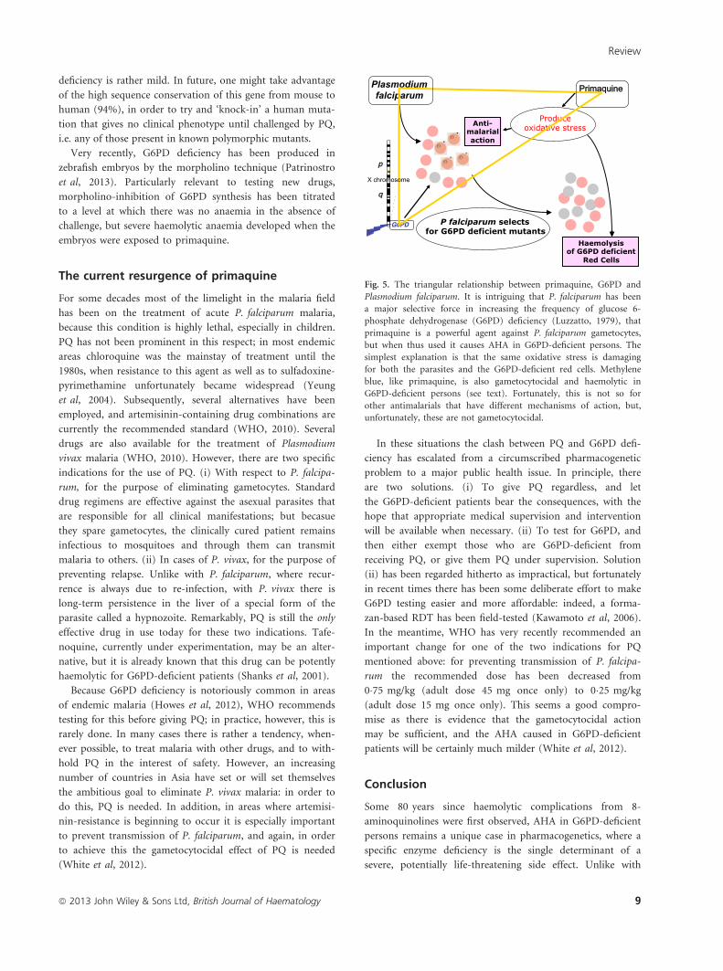
Fig. 5. The triangular relationship between primaquine, G6PD and
Plasmodium falciparum. It is intriguing that P. falciparum has been
a major selective force in increasing the frequency of glucose 6-
phosphate dehydrogenase (G6PD) deficiency (Luzzatto, 1979), that
primaquine is a powerful agent against P. falciparum gametocytes,
but when thus used it causes AHA in G6PD-deficient persons. The
simplest explanation is that the same oxidative stress is damaging
for both the parasites and the G6PD-deficient red cells. Methylene
blue, like primaquine, is also gametocytocidal and haemolytic in
G6PD-deficient persons (see text). Fortunately, this is not so for
other antimalarials that have different mechanisms of action, but,
unfortunately, these are not gametocytocidal.
Review
ª 2013 John Wiley & Sons Ltd, British Journal of Haematology 9
other enzyme-related idiosyncrasies, such as those associated
with porphyrias (Thunell et al, 2007), where the condition is
rare and there are usually alternatives to the culprit drugs,
G6PD deficiency is common in many populations, and it is
common precisely where drugs such as PQ are needed. The
intriguing triangular relationship (see Fig 5) – whereby
malaria selects for G6PD deficiency, PQ is an effective anti-
malarial, but G6PD-deficient persons are so sensitive to PQ,
has been discussed elsewhere (Luzzatto, 2010). Suffice it to
say here that, as a result, drug-induced AHA is both a clini-
cal and a public health problem.
Although they are now rather well identified and defined,
both of these problems are still challenging. On the clinical
side, G6PD deficiency must be always considered in the dif-
ferential diagnosis of haemolytic anaemia; and we should be
mindful of G6PD deficiency whenever we prescribe a poten-
tially haemolytic drug. On the public health side, it is no
longer permissible – not that we think it ever was – to sweep
the issue under the carpet when administering primaquine or
dapsone widely in areas where G6PD deficiency has a high
prevalence: until alternative drugs become available, G6PD
testing must be made available.
Acknowledgement
We are grateful to Dr Rosario Notaro, Professor Fabrizio
Pane and Dr Enzo Poggi for their support; and Ms Jessica
Nencioni for her valuable help with illustrations. We thank
Dr Alfred Tiono for making slides from G6PD-deficient chil-
dren available; and Dr Naomi Richardson for help with pro-
ducing Fig 3. In particular, we thank all G6PD-deficient
patients, those we have known and the many we have not
known in person, who have suffered drug-induced AHA,
sometimes unnecessarily.
References
Adam, A. (1961) Linkage between deficiency of
glucose 6-phosphate dehydrogenase and colour-
blindness. Nature, 189, 686–688.
Alessio, L. & Morselli, G. (1972) Occupational
exposure to nalidixic acid. British Medical Jour-
nal, 4, 110–111.
Alloueche, A., Bailey, W., Barton, S., Bwika, J.,
Chimpeni, P., Falade, C.O., Fehintola, F.A.,
Horton, J., Jaffar, S., Kanyok, T., Kremsner,
P.G., Kublin, J.G., Lang, T., Missinou, M.A.,
Mkandala, C., Oduola, A.M., Premji, Z., Rob-
ertson, L., Sowunmi, A., Ward, S.A. & Win-
stanley, P.A. (2004) Comparison of
chlorproguanil-dapsone with sulfadoxine-pyri-
methamine for the treatment of uncomplicated
falciparum malaria in young African children:
double-blind randomised controlled trial. Lan-
cet, 363, 1843–1848.
Bain, B.J. (2010) A ghostly presence-G6PD defi-
ciency. American Journal of Hematology, 85, 271.
Balaka, B., Balaka, B., Agbere, D., Bonkoungou, P.,
Gnamey, D., Kessie, K. & Assimadi, K. (2003)
Post-hemolytic renal failure in children with
glucose-6-phosphate dehydrogenase deficiency at
the University Hospital Center in Lome. Mede-
cine Tropicale: Revue Du Crops De Sant�e Colo-
nial, 63, 151–154.
Bauters, T., Bauters, T., Mondelaers, V., Robays,
H., De Wilde, H., Benoit, Y. & De Moerloose,
B. (2011) Methemoglobinemia and hemolytic
anemia after rasburicase administration in a
child with leukemia. International Journal Clini-
cal Pharmacy, 33, 58–60.
Berg, J.M., Tymoczko, J.L. & Stryer, L. (2002) Bio-
chemistry. W. H. Freeman and Co., New York.
Betke, K., Beutler, E., Brewer, G.J., Kirkman, H.N.,
Luzzatto, L., Motulsky, A.G., Ramot, B. &
Siniscalco, M. (1967) Standardization of proce-
dures for the study of glucose-6-phosphate
dehydrogenase. World Health Organization Tech-
nical Report Series, 366, 53p.
Beutler, E. (1957) The glutathione instability of
drug-sensitive red cells. A new method for the
in vitro detection of drug sensitivity. Journal of
Laboratory and Clinical Medicine, 49, 84–95.
Beutler, E. (1959) The hemolytic effect of pri-
maquine and related compounds. Blood, 14,
103–139.
Beutler, E., Yeh, M. & Fairbanks, V.F. (1962) The
normal human female as a mosaic of X-chro-
mosome activity: studies using the gene for
G6PD deficiency as a marker. Proceedings of the
National Academy of Sciences USA, 48, 9–16.
Beutler, E., Blume, K.G., Kaplan, J.C., Lohr, G.W.,
Ramot, B. & Valentine, W.N. (1979) Interna-
tional Committee for Standardization in Hae-
matology: recommended screening test for
glucose-6-phosphate dehydrogenase (G6PD)
deficiency. British Journal of Haematology, 43,
465–467.
Beutler, E., Kuhl, W., Vives-Corrons, J.L. & Prchal,
J.T. (1989) Molecular heterogeneity of glucose
6-phosphate dehydrogenase A-. Blood, 74,
2550–2555.
Blakeslee, A.F. (1932) Genetics of sensory thresh-
olds: taste for phenylthiocarbamide. Proceedings
of the National Academy Sciences USA, 18,
120–130.
Bloom, K.E., Brewer, G.J., Magon, A.M. & Wett-
erstroem, N. (1983) Microsomal incubation test
of potentially hemolytic drugs for glucose-6-
phosphate dehydrogenase deficiency. Clinical
pharmacology and therapeutics, 33, 403–409.
Brewer, G.J. (2007) Rediscovery of the susceptibil-
ity of G6PD deficient persons to methemoglobi-
nemia from oxidant drugs, and to hemolysis
from methylene blue. American Journal of
Hematology, 82, 87–88.
Brewer, G.J., Tarlov, A.R. & Alving, A.S. (1962)
The methemoglobin reduction test for primaqu-
ine-type sensitivity of erythrocytes. A simplified
procedure for detecting a specific hypersuscepti-
bility to drug hemolysis. Journal of the American
Medical Association, 180, 386–388.
Burka, E.R., Weaver, Z. III & Marks, P.A. (1966)
Clinical spectrum of hemolytic anemia associ-
ated with glucose-6-phosphate dehydrogenase
deficiency. Annals of Internal Medicine, 64,
817–825.
Cairo, M.S., Coiffier, B., Reiter, A., Younes, A. &
TLS Expert Panel (2010) Recommendations for
the evaluation of risk and prophylaxis of
tumour lysis syndrome (TLS) in adults and chil-
dren with malignant diseases: an expert TLS
panel consensus. British Journal of Haematology
149, 578–586.
Cappellini, M.D. & Fiorelli, G. (2008) Glucose-
6-phosphate dehydrogenase deficiency. Lancet,
371, 64–74.
Cappellini, M.D., Martinez di Montemuros, F., De
Bellis, G., Debernardi, S., Dotti, C. & Fiorelli, G.
(1996) Multiple G6PD mutations are associated
with a clinical and biochemical phenotype simi-
lar to that of G6PD Mediterranean. Blood, 87,
3953–3958.
Carmoi, T., Bordier, L., Bonnefoy, S., Callot, D.,
Lecoules, S. & Algayres, J.P. (2009) Ofloxacin is
contraindicated in case of G6PD deficiency: is it
evidenced based? Revue de Medecine Interne, 30,
355–357.
Carson, P.E., Alving, A.S., Flanagan, C.L. & Ickes,
C.E. (1956) Enzymatic deficiency in primaqu-
ine-sensitive erythrocytes. Science, 124, 484–485.
Dacie, J.V. & Lewis, S.M. (1995) Practical Haema-
tology. Churchill Livingstone, London.
Degowin, R.L., Eppes, R.B., Powell, R.D. & Carson,
P.E. (1966) The haemolytic effects of diaphenyl-
sulfone (DDS) in normal subjects and in those
with glucose-6-phosphate-dehydrogenase defi-
ciency. Bulletin of the World Health
Organization, 35, 165–179.
Review
10 ª 2013 John Wiley & Sons Ltd, British Journal of Haematology
Dern, R.J., Beutler, E. & Alving, A.S. (1955) The
haemolytic effect of primaquine V Primaquine
sensitivity as a manifestation of a multiple drug
sensitivity. Journal of Laboratory and Clinical
Medicine, 45, 30.
Eldad, A., Neuman, A., Weinberg, A., Benmeir, P.,
Rotem, M. & Wexler, M.R. (1991) Silver sulph-
adiazine-induced haemolytic anaemia in a glu-
cose-6-phosphate dehydrogenase-deficient burn
patient. Burns, 17, 430–432.
Fermi, C. & Martinetti, P. (1905) Studio sul favismo.
Annali di Igiene Sperimentale, 15, 76–112.
Fischer, T.M., Meloni, T., Pescarmona, G.P. &
Arese, P. (1985) Membrane cross bonding in
red cells in favic crisis: a missing link in the
mechanism of extravascular hemolysis. British
Journal of Haematology, 59, 159–169.
Foltz, L.M., Dalal, B.I., Wadsworth, L.D., Broady,
R., Chi, K., Eisenhauer, E., Kobayashi, K. &
Kollmannsburger, C. (2006) Recognition and
management of methemoglobinemia and hemo-
lysis in a G6PD-deficient patient on experimen-
tal anticancer drug Triapine. American Journal
of Hematology, 81, 210–211.
Frigerio, R., Sole, G., Lovicu, M. & Passiu, G.
(1994) Molecular and biochemical data on some
glucose-6-phosphate dehydrogenase variants
from southern Sardinia. Haematologica, 79,
319–321.
Gaetani, G.F., Mareni, C., Ravazzolo, R. & Salvid-
io, E. (1976) Haemolytic effect of two sulphona-
mides evaluated by a new method. British
Journal of Haematology, 32, 183.
Gaetani, G.F., Mareni, C., Salvidio, E., Galiano, S.,
Meloni, T. & Arese, P. (1979) Favism: eryth-
rocyte metabolism during haemolysis and retic-
ulocytosis. British Journal of Haematology, 43,
39–48.
Gaetani, G.F., Kirkman, H.N., Mangerini, R. &
Ferraris, A.M. (1994) Importance of catalase in
the disposal of hydrogen peroxide within human
erythrocytes. Blood, 84, 325–330.
Gaetani, G.F., Rolfo, M., Arena, S., Mangerini, R.,
Meloni, G.F. & Ferraris, A.M. (1996) Active
involvement of catalase during hemolytic crises
of favism. Blood, 88, 1084–1088.
Gauthier, T.W. (2000) Methylene blue-induced
hyperbilirubinemia in neonatal glucose-6-phos-
phate dehydrogenase (G6PD) deficiency. The
Journal of Maternal Fetal Medicine, 9, 252–254.
Guttmann, P. & Ehrlich, P. (1891) €Uber die Wir-
king des Methylenblau bei Malaria. Berliner
Klinische Wochenschrift, 39, 953–956.
Ho, J.M. & Juurlink, D.N. (2011) Considerations
when prescribing trimethoprim-sulfamethoxaz-
ole. CMAJ, 183, 1851–1858.
Howes, R.E., Piel, F.B., Patil, A.P., Nyangiri, O.A.,
Gething, P.W., Dewi, M., Hogg, M.M., Battle,
K.E., Padilla, C.D., Baird, J.K. & Hay, S.I.
(2012) G6PD deficiency prevalence and esti-
mates of affected populations in malaria ende-
mic countries: a geostatistical model-based map.
PLoS Medicine, 9, e1001339.
Jones, R. Jr, Jackson, L.S., Di Lorenzo, A., Marx,
R.L., Levy, B.L., Kenny, E.C., Gilbert, M.,
Johnston, M.N. & Alving, A.S. (1953) Korean
vivax malaria. III. Curative effect and toxicity of
primaquine in doses from 10 to 30 mg daily.
American Journal of Tropical Medicine and
Hygiene, 2, 977–982.
Kawamoto, F., Matsuoka, H., Kanbe, T., Tantular,
I.S., Pusarawati, S., Kerong, H.I., Damianus, W.,
Mere, D. & Dachlan, Y.P. (2006) Further inves-
tigations of glucose-6-phosphate dehydrogenase
variants in Flores Island, eastern Indonesia. Jour-
nal of Human Genetics, 51, 952–957.
Kellermeyer, R.W., Tarlov, A.R., Brewer, G.J., Car-
son, P.E. & Alving, A.S. (1962) Hemolytic effect
of therapeutic drugs. Clinical considerations of
the primaquine-type hemolysis. JAMA, 180,
394–380.
Kizer, N., Martinez, E. & Powell, M. (2006) Report of
two cases of rasburicase-induced methemoglobi-
nemia. Leukaemia & Lymphoma, 47, 2648–2650.
Liao, Y.P., Hung, D.Z. & Yang, D.Y. (2002)
Hemolytic anemia after methylene blue therapy
for aniline-induced methemoglobinemia. Veteri-
nary and Human Toxicology, 44, 19–21.
Longo, L., Vanegas, O.C., Patel, M., Rosti, V., Li,
H., Waka, J., Merghoub, T., Pandolfi, P.P., Not-
aro, R., Manova, K. & Luzzatto, L. (2002)
Maternally transmitted severe glucose 6-phos-
phate dehydrogenase deficiency is an embryonic
lethal. EMBO Journal, 21, 4229–4239.
Luisada, L. (1941) Favism: a singular disease affect-
ing chiefly red blood cells. Medicine, 20, 229–250.
Luzzatto, L. (1979) Genetics of red cells and sus-
ceptibility to malaria. Blood, 54, 961–976.
Luzzatto, L. (2010) The rise and fall of the antima-
larial Lapdap: a lesson in pharmacogenetics.
Lancet, 376, 739–741.
Luzzatto, L. (2012) Hemolytic Anemias and ane-
mias due to acute blood loss. In: Harrison’s
Principles of Internal Medcine (eds by D. Lon-
go, A.S. Fauci, D. Kasper, L. Dennis, J. Kasper,
S.L. Hauser & J. Loscalzo), Vol. 1, pp. 872–886.
McGraw-Hill, New York.
Luzzatto, L. & Allan, N.C. (1968) Relationship
between the genes for glucose 6-phosphate
dehydrogenase and haemoglobin a Nigerian
population. Nature, 219, 1041–1942.
Luzzatto, L. & Poggi, V.E. (2009) Glucose 6-
phosphate dehydrogenase deficiency. In: Hema-
tology of Infancy and Childhood (eds by S.H.
Orkin, D.G. Nathan, D. Ginsburg, T.A. Look,
D.E. Fisher & S.E. Lux), pp. 883–907. Saunders,
Philadelphia.
Luzzatto, L., Mehta, A. & Vulliamy, T.J. (2001)
Glucose-6-phosphate dehydrogenase deficiency.
In: The Metabolic & Molecular Bases of Inher-
ited Disease, Vol. 3 (eds by C. Scriver, A. Beau-
det, W. Sly & D. Valle), pp. 4517–4553.
McGraw Hill, New York.
Mason, P.J., Bautista, J.M. & Gilsanz, F. (2007)
G6PD deficiency: the genotype-phenotype asso-
ciation. Blood Reviews, 21, 267–283.
Matsui, R., Xu, S., Maitland, K.A., Mastroianni, R.,
Leopold, J.A., Handy, D.E., Loscalzo, J. & Co-
hen, R.A. (2006) Glucose-6-phosphate dehydro-
genase deficiency decreases vascular superoxide
and atherosclerotic lesions in apolipoprotein E
(-/-) mice. Arteriosclerosis, thrombosis, and vascu-
lar biology, 26, 910–916.
Mehta, J.B., Singhal, S.B. & Mehta, B.C. (1990)
Ascorbic acid-induced haemolysis in G6PD defi-
ciency. Lancet, 336, 930.
Meloni, G. & Meloni, T. (1996) Glyburide-induced
acute haemolysis in a G6PD-deficient patient
with NIDDM. British Journal of Haematology,
92, 159–160.
Meloni, T., Forteleoni, G., Dore, A. & Cutillo, S.
(1983) Favism and hemolytic anemia in glu-
cose-6-phosphate dehydrogenase-deficient sub-
jects in North Sardinia. Acta haematologica,
70, 83–90.
Meloni, T., Forteleoni, G., Ogana, A. & Franca, V.
(1989) Aspirin-induced acute hemolytic anemia
in glucose 6-phosphate dehydrogenase deficient
children with systemic arthrit1s. Acta Haemato-
logica, 81, 208–209.
Minucci, A., Moradkhani, K., Hwang, M.J., Zuppi,
C., Giardina, B. & Capoluongo, E. (2012a) Glu-
cose-6-phosphate dehydrogenase (G6PD) muta-
tions database: review of the ‘old’ and update of
the new mutations. Blood Cells, Molecules, &
Diseases, 48, 154–165.
Minucci, A., Gentile, L., Zuppi, C., Giardina, B. &
Capoluongo, E. (2012b) Rapid and simple iden-
tification of the commonest glucose-6-phosphate
dehydrogenase (G6PD) Italian mutations: from
DNA extraction to genotyping. Clinica Chimica
Acta, 413, 1018–1019.
Morelli, A., Benatti, U., Gaetani, G.F. & De Flora,
A. (1978) Biochemical mechanisms of glucose-
6-phosphate dehydrogenase deficiency. Proceed-
ings of the National Academy of Sciences of the
United States of America, 75, 1979–1983.
Motulsky, A.G. (1957) Drug reactions, enzymes
and biochemical genetics. Journal of the Ameri-
can Medical Association. Journal of the American
Medical Association, 165, 835–837.
Motulsky, A.G. & Campbell-Kraut, J.M. (1961)
Population genetics of glucose 6-phosphate
dehydrogenase deficiency of the red cell. In:
Proceedings of Conference on Genetic Polymor-
phisms and Geographic Variations in Disease
(ed. by B.S. Blumberg), pp. 159–180. Grune &
Stratton, New York.
Muller, O., Mockenhaupt, F.P., Marks, B., Meiss-
ner, P., Coulibaly, B., Kuhnert, R., Buchner, H.,
Schirmer, R.H., Walter-Sack, I., Sie, A. & Mans-
mann, U. (2013) Haemolysis risk in methylene
blue treatment of G6PD-sufficient and G6PD-
deficient West-African children with uncompli-
cated falciparum malaria: a synopsis of four
RCTs. Pharmacoepidemiology and Drug Safety,
22, 376–385.
Nance, W.E. (1964) Genetic tests with a sex-linked
marker: G-6-PD. Cold Spring Harbor Symposia
on Quantitative Biology, 29, 415–425.
Neifer, S., Jung, A. & Bienzle, U. (1991) Character-
ization of erythrocytic glucose-6-phosphate
dehydrogenase in a mouse strain with reduced
G6PD activity. Biomedica Biochimica Acta, 3,
233–238.
Review
ª 2013 John Wiley & Sons Ltd, British Journal of Haematology 11
Pamba, A., Richardson, N.D., Carter, N., Duparc,
S., Premji, Z., Tiono, A.B. & Luzzatto, L. (2012)
Clinical spectrum and severity of hemolytic ane-
mia in glucose 6-phosphate dehydrogenase-defi-
cient children receiving dapsone. Blood, 120,
4123–4133.
Pandolfi, P.P., Sonati, F., Rivi, R., Mason, P.,
Grosveld, F. & Luzzatto, L. (1995) Targeted dis-
ruption of the housekeeping gene encoding glu-
cose 6- phosphate dehydrogenase (G6PD):
G6PD is dispensable for pentose synthesis but
essential for defense against oxidative stress.
EMBO Journal, 14, 5209–5215.
Pannacciulli, I.M., Tizianello, A., Ajmar, F. & Salv-
idio, E. (1965) The course of experimentally
induced haemolytic anaemia in a primaquine
sensitive caucasian. Blood, 25, 92–95.
Patrinostro, X., Carter, M.L., Kramer, A.C. &
Lund, T.C. (2013) A model of glucose-6-phos-
phate dehydrogenase deficiency in the zebrafish.
Experimental Hematology, 41, e2.
Poggi, V., Town, M., Foulkes, N.S. & Luzzatto, L.
(1990) Identification of a single base change in a
new human mutant glucose-6-phosphate dehy-
drogenase gene by polymerase-chain-reaction
amplification of the entire coding region from
genomic DNA. The Biochemical journal, 271,
157–160.
Poliseno, L., Salmena, L., Riccardi, L., Fornari, A.,
Song, M.S., Hobbs, R.M., Sportoletti, P., Var-
meh, S., Egia, A., Fedele, G., Rameh, L., Loda,
M. & Pandolfi, P.P. (2010) Identification of the
miR-106b~25 microRNA cluster as a proto-
oncogenic PTEN-targeting intron that cooper-
ates with its host gene MCM7 in transforma-
tion. Science Signaling, 3, ra29.
Premji, Z., Umeh, R.E., Owusu-Agyei, S., Esamai,
F., Ezedinachi, E.U., Oguche, S., Borrmann, S.,
Sowunmi, A., Duparc, S., Kirby, P.L., Pamba,
A., Kellam, L., Guiguemde, R., Greenwood, B.,
Ward, S.A. & Winstanley, P.A. (2009) Chlorpro-
guanil-dapsone-artesunate versus artemether-
lumefantrine: a randomized, double-blind phase
III trial in African children and adolescents with
uncomplicated Plasmodium falciparum malaria.
PLoS ONE, 4, e6682.
Pretsch, W., Charles, D.J. & Merkle, S. (1988)
X-linked glucose-6-phosphate dehydrogenase
deficiency in mus musculus. Biochemical Genetics,
26, 89–103.
Rinaldi, A., Filippi, G. & Siniscalco, M. (1976)
Variability of red cell phenotypes between and
within individuals in an unbiased sample of 77
certain heterozygotes for G6PD deficiency in
Sardinians. American Journal of Human Genetics,
28, 496–505.
Salvidio, E., Pannacciulli, I., Ajmar, F., Gaetani, G.
& Ghio, R., Garr�e, C. (1972) Hemolytic side
effects of some antimalarial drugs. Proceeding of
the Helminthological Society of Washington, 39,
83–100.
Sanders, S., Smith, D.P., Thomas, G.A. & Williams,
E.D. (1997) A glucose-6-phosphate dehydroge-
nase (G6PD) splice site consensus sequence
mutation associated with G6PD enzyme defi-
ciency. Mutation Research, 374, 79–87.
Sansone, G. & Segni, G. (1958) Nuovi aspetti
dell’alterato biochimismo degli eritrociti dei
favici: assenza pressoche‘ completa della glucos-
6-P deidrogenasi. Bollettino della Societa‘ Italiana
di Biologia Sperimentale, 34, 327–329.
Sansone, S., Rottensteiner, J., Stocker, J., Rosanelli,
C. & Wiedermann, C.J. (2010) Ciprofloxacin-
induced acute haemolytic anaemia in a patient
with glucose-6-phosphate dehydrogenase Medi-
terranean deficiency: a case report. Annals of
Hematology, 89, 935–937.
Shanks, G.D., Oloo, A.J., Aleman, G.M., Ohrt, C.,
Klotz, F.W., Braitman, D., Horton, J. & Brueck-
ner, R. (2001) A new primaquine analogue, tafe-
noquine (WR 238605), for prophylaxis against
Plasmodium falciparum malaria. Clinical Infec-
tious Diseases, 33, 1968–1974.
Sicard, D., Kaplan, J.C. & Labie, D. (1978) Hae-
moglobinopathies and G6PD deficiency in Laos.
Lancet, 2, 571–572.
Sonbol, M.B., Yadav, H., Vaidya, R., Rana, V. &
Witzig, T.E. (2013) Methemoglobinemia and
hemolysis in a patient with G6PD deficiency
treated with rasburicase. American Journal of
Hematology, 88, 152–154.
Tantular, I.S. & Kawamoto, F. (2003) An
improved, simple screening method for detec-
tion of glucose-6-phosphate dehydrogenase defi-
ciency. Tropical Medicine & International Health,
8, 569–574.
Tarlov, A.R., Brewer, G.J., Carson, P.E. & Alving,
A.S. (1962) Primaquine sensitivity. Glucose-
6-phosphate dehydrogenase deficiency: an
inborn error of metabolism of medical and bio-
logical significance. Archives of Internal Medicine,
109, 209–234.
Thunell, S., Pomp, E. & Brun, A. (2007) Guide to
drug porphyrogenicity prediction and drug pre-
scription in the acute porphyrias. British Journal
of Clinical Pharmacology, 64, 668–679.
Tiono, A.B., Dicko, A., Ndububa, D.A., Agbenyega,
T., Pitmang, S., Awobusuyi, J., Pamba, A., Dup-
arc, S., Goh, L.E., Harrell, E., Carter, N., Ward,
S.A., Greenwood, B. & Winstanley, P. (2009)
Chlorproguanil-dapsone-artesunate versus chlor-
proguanil-dapsone: a randomized, double-blind,
phase III trial in African children, adolescents,
and adults with uncomplicated Plasmodium fal-
ciparum malaria. The American Journal of Tropi-
cal Medicine and Hygiene, 81, 969–978.
Tugwell, P. (1973) Glucose 6-phosphate dehydro-
genase deficiency in Nigerians with jaundice
associated with lobar pneumonia. Lancet, 1,
968–970.
Tungsiripat, M., Drechsler, H., Sarlone, C., Amyot,
K., Laffey, E. & Aberg, J. (2008) Prevalence and
significance of G6PD deficiency in patients of an
urban HIV clinic. Journal of International Associ-
ation of Physicians in AIDS Care (Chicago, III:
2002), 7, 88–90.
Vale, N., Moreira, R. & Gomes, P. (2009) Primaq-
uine revisited six decades after its discovery.
European Journal of Medicinal Chemistry, 44,
937–953.
Vogel, F. (1959) Moderne problem der human-
genetik.Ergebnisse der Inneren Medizin Und Kin-
derheilkunde, 12, 52–125.
White, N.J., Qiao, L.G., Qi, G. & Luzzatto, L.
(2012) Rationale for recommending a lower
dose of primaquine as a Plasmodium falciparum
gametocytocide in populations where G6PD
deficiency is common. Malaria Journal, 11, 418.
WHO (2010) Guidelines for the Treatment of
Malaria. World Health Organization, Geneva.
WHO Working Group (1989) Glucose-6-phos-
phate dehydrogenase deficiency. Bulletin of the
World Health Organization, 67, 601–611.
Yeung, S., Pongtavornpinyo, W., Hastings, I.M.,
Mills, A.J. & White, N.J. (2004) Antimalarial
drug resistance, artemisinin-based combination
therapy, and the contribution of modeling to
elucidating policy choices. American Journal of
Tropical Medicine and Hygiene, 71, 179–186.
Youngster, I., Arcavi, L., Schechmaster, R., Akay-
zen, Y., Popliski, H., Shimonov, J., Beig, S. &
Berkovitch, M. (2010) Medications and glucose-
6-phosphate dehydrogenase deficiency: an evi-
dence-based review. Drug Safety, 33, 713–726.
Zaramella, P., De Salvia, A., Zaninotto, M., Bar-
aldi, M., Capovilla, G., De Leo, D. & Chiandetti,
L. (2013) Lethal effect of a single dose of ras-
buricase in a preterm newborn infant. Pediatrics,
131, e309–e312.
Zhang, P., Gao, X., Ishida, H., Amnuaysirikul, J.,
Weina, P.J., Grogl, M., O’Neil, M.T., Li, Q.,
Caridha, D., Ohrt, C., Hickman, M., Magill, A.J.
& Ray, P. (2013) An In vivo Drug Screening
Model Using Glucose-6-Phosphate Dehydroge-
nase Deficient Mice to Predict the Hemolytic
Toxicity of 8-Aminoquinolines. American
Journal of Tropical Medicine and Hygiene, 88,
1138–1145.
Review
12 ª 2013 John Wiley & Sons Ltd, British Journal of Haematology