Embed Size (px)
Citation preview

REVIEW Open Access
G-quadruplexes: a promising target forcancer therapyNils Kosiol, Stefan Juranek, Peter Brossart, Annkristin Heine† and Katrin Paeschke*†
Abstract
DNA and RNA can fold into a variety of alternative conformations. In recent years, a particular nucleic acid structurewas discussed to play a role in malignant transformation and cancer development. This structure is called a G-quadruplex (G4). G4 structure formation can drive genome instability by creating mutations, deletions andstimulating recombination events. The importance of G4 structures in the characterization of malignant cells wascurrently demonstrated in breast cancer samples. In this analysis a correlation between G4 structure formation andan increased intratumor heterogeneity was identified. This suggests that G4 structures might allow breast cancerstratification and supports the identification of new personalized treatment options. Because of the stability of G4structures and their presence within most human oncogenic promoters and at telomeres, G4 structures arecurrently tested as a therapeutic target to downregulate transcription or to block telomere elongation in cancercells. To date, different chemical molecules (G4 ligands) have been developed that aim to target G4 structures. Inthis review we discuss and compare G4 function and relevance for therapeutic approaches and their impact oncancer development for three cancer entities, which differ significantly in their amount and type of mutations:pancreatic cancer, leukemia and malignant melanoma. G4 structures might present a promising new strategy toindividually target tumor cells and could support personalized treatment approaches in the future.
Keywords: G-quadruplex, genome instability, cancer progression, cancer therapy, malignant melanoma, pancreaticcancer, acute myeloid leukemia
IntroductionCancer is the world’s second most leading cause of death[1]. Although therapeutic strategies for many cancershave greatly advanced during the last years, still about9.6 million people died of cancer in 2018 [2]. This high-lights the need to strengthen the research on causes ofcancer development and to improve diagnostic and anti-tumor treatment options. Different promising therapeuticstrategies have been identified in the last decades. Amongthose, immunotherapy and targeted therapies have revolu-tionized anti-tumor therapy [3]. The identification of gen-etic and epigenetic abnormalities as well as tumor-growth
promoting oncogenes in tumor cells provided the ration-ale for molecular targeted therapies [4, 5]. Immune-oncology (IO) aims at boosting the patient´s own immunesystem to eliminate tumor cells. One example is the im-mune checkpoint blockade [6]. Both approaches, targetedtherapies as well as IO, significantly improved survivaloutcome in cancer patients, such as in malignant melan-oma and other cancer entities [7, 8]. A diverging newstrategy aims to modulate the 3-dimensional structure ofthe DNA with the goal to influence biological processesand genome stability.Genomic DNA canonically adopts a standard B-DNA
conformation. DNA can also fold into alternative struc-tures such as DNA hairpins, holiday junctions, cruci-forms, triplexes or G-quadruplexes (G4) [9, 10].Although the relevance of G4 structures in living cells
© The Author(s). 2021 Open Access This article is licensed under a Creative Commons Attribution 4.0 International License,which permits use, sharing, adaptation, distribution and reproduction in any medium or format, as long as you giveappropriate credit to the original author(s) and the source, provide a link to the Creative Commons licence, and indicate ifchanges were made. The images or other third party material in this article are included in the article's Creative Commonslicence, unless indicated otherwise in a credit line to the material. If material is not included in the article's Creative Commonslicence and your intended use is not permitted by statutory regulation or exceeds the permitted use, you will need to obtainpermission directly from the copyright holder. To view a copy of this licence, visit http://creativecommons.org/licenses/by/4.0/.The Creative Commons Public Domain Dedication waiver (http://creativecommons.org/publicdomain/zero/1.0/) applies to thedata made available in this article, unless otherwise stated in a credit line to the data.
* Correspondence: [email protected] Heine and Katrin Paeschke contribute equally to this work.Department of Oncology, Hematology, Rheumatology andImmune-Oncology, University Hospital Bonn, 53127 Bonn, Germany
Kosiol et al. Molecular Cancer (2021) 20:40 https://doi.org/10.1186/s12943-021-01328-4

was controversially discussed in the past, accumulatingexperimental data now supports the existence and im-portance of these structures in living cells [10–12].G4 structures can form within DNA and RNA [13]. In
a G4 structure, four guanines are held together byHoogsteen hydrogen bonds wherein each guanine canact as a donor and acceptor for two hydrogen bonds.Based on in vitro experiments it was predicted that G4structures form in regions harboring a specific G4 motif:G≥3N1-7G≥3N1-7G≥3N1-7G≥3 [13]. However, current ex-perimental data shows that G4 structures can also formwithin regions that have longer loops or less than 3 gua-nines per repeat as well as in regions that do not followthis stringent G4 motif [14, 15]. Among different factorsthe stability of the G4 structure depends on the numbersof guanines per repeat and the length of the loops [13,16].Computational as well as deep-sequencing approaches
have demonstrated that in the human genome over700.000 regions exist that could potentially fold into G4structures [14, 15, 17, 18]. Also in other organisms in-cluding viruses, yeasts and different bacterial genomesregions with a strong potential to fold into G4 weremapped genome-wide [15, 19–21]. To this date, theidentification of a G4 motif within the genome does notproof the formation of G4 structures at these regionsin vivo, but simply gives a potential to form a G4. G4formation needs to be experimentally evaluated and de-pends on different factors (e.g. protein binding, loss ofprotein binding, cell cycle phase, stress), of which not allare known, yet.G4 motifs are not randomly distributed throughout
the genome, but are enriched in certain regions (e.g.promoters, telomeres, transcription factor binding sites)[14, 22, 23]. More than 40% of human promotor regions
harbor at least one G4 motif [24]. The evolutionaryconservation, the specific location within the genome[15, 19, 25] as well as different biochemical and mo-lecular experiments underline the current model thatG4 structures form in living cells, where they sup-port/affect different biological pathways (e.g. proteinexpression, telomerase activity and genome stability)(Fig. 1) [10–12].The physiological relevance of G4 structures is further
supported by the existence of proteins that are able tobind and unfold G4 structures [11, 12, 26]. There arethree classes of G4-interacting proteins described in theliterature: G4 binding, G4 stabilizing and G4 unwindingproteins (e.g. helicases: BLM, WRN, BRIP1/FANCJ andPIF1) [27]. It has been reported that mutations and/ordeletions of these proteins (e.g. PIF1) lead to changes inthe formation of G4 structures. This in turn can resultin changes of biological pathways (transcriptionalchanges) and can also increase genome instability [28–32]. This agrees with the finding that changes withinsome G4-interacting helicases are linked to cancer pro-gression and tumorigenesis [27]. However, the link oftumorigenesis and mutations of helicases is not proofedto be related to G4 formation. Antibody stainings as wellas G4 ChIP-seq in stomach and liver tissues of immor-talized HaCaT cells showed increased levels of G4 struc-tures compared to a normal/healthy state [18, 33, 34].Similar observations were made for other cancer states. Dif-ferent lines of evidence demonstrated that changes in G4 for-mation/stability can alter telomerase activity [35, 36],transcription efficiency (inhibit or promote [11, 37, 38]), stallDNA replication and induce genome instability [39, 40].Changes can be triggered chemically (G4-ligands) or by pro-teins that modulate G4 formation. G4 ligands that modulateG4 structure formation or stabilize G4 structures were
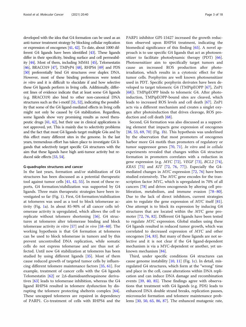
Fig. 1 Schematic overview of the effects of G4 ligands on cancer cells. Most G4 ligands cause slow growth. These growth changes are theconsequence of alteration within biological processes. Depending on the ligand and cell type G4 stabilization can lead to changes in a telomeremaintenance b gene expression of oncogenes c increased genome instability. Created with BioRender.
Kosiol et al. Molecular Cancer (2021) 20:40 Page 2 of 18
developed with the idea that G4 formation can be used as ananti-tumor treatment strategy by blocking cellular replicationor expression of oncogenes [41, 42]. To date, about 1000 dif-ferent G4 ligands have been identified [43]. These ligandsdiffer in their specificity, binding surface and cell permeabil-ity [44]. Most of them, including MM41 [45], Telomestatin[46], BRACO19 [47], TMPyP4 [48], RHPS4 [49] and PDS[50] preferentially bind G4 structures over duplex DNA.However, most of these binding preferences were testedin vitro and it is difficult to elucidate if and how selectivethese G4 ligands perform in living cells. Additionally, differ-ent lines of evidence indicate that at least some G4 ligands(e.g. BRACO19) also bind to other non-canonical DNAstructures such as the i-motif [51, 52], indicating the possibil-ity that some of the G4 ligand-mediated effects in living cellsmight not only be caused by G4 stabilization. Regardless,some ligands show very promising results as novel thera-peutic drugs [41, 42], but their use in clinical applications isnot approved, yet. This is mainly due to selectivity problemsand the fact that most G4 ligands target multiple G4s and bythis effect many different sites in the genome. In the lastyears, tremendous effort has taken place to investigate G4 li-gands that selectively target specific G4 structures with theaim that these ligands have high anti-tumor activity but re-duced side effects [53, 54].
G-quadruplex structures and cancerIn the last years, formation and/or stabilization of G4structures has been discussed as a potential therapeutictool against tumor cells [41, 42, 55]. In most of these re-ports, G4 formation/stabilization was supported by G4ligands. Three main therapeutic strategies have been in-vestigated so far (Fig. 1). First, G4 formation/stabilizationat telomeres was used as a tool to block telomerase ac-tivity (Fig. 1a). In about 85-90% of all cancer cells tel-omerase activity is upregulated, which allows the cell toreplicate without telomere shortening [56]. G4 struc-tures at telomeres alter telomerase binding and blocktelomerase activity in vitro [57] and in vivo [58–60]. Theworking hypothesis is that G4 formation at telomerescan be used to block telomerase in tumors and by thisprevent uncontrolled DNA replication, while somaticcells do not express telomerase and are thus not af-fected. Until now G4 stabilization at telomeres has beenstudied by using different ligands [35]. Most of themcause reduced growth of targeted tumor cells by influen-cing different telomere maintenance factors [35, 61]. Forexample, treatment of cancer cells with the G4 ligandsTelomestatin [62] or 2,6-diamidoanthraquinone deriva-tives [63] leads to telomerase inhibition, whereas the G4ligand RHPS4 resulted in telomere dysfunction by dis-rupting the telomere protecting shelterin complex [64].These uncapped telomeres are repaired in dependencyof PARP1. Co-treatment of cells with RHPS4 and the
PARP1 inhibitor GPI-15427 increased the growth reduc-tion observed upon RHPS4 treatment, indicating thebiomedical significance of this finding [65]. A novel ap-proach is to use specific G4 ligands that act as photosen-sitizer to facilitate photodynamic therapy (PDT) [66].Photosensitizer aim to specifically target tumors andcause an increased ROS production after photo-irradiation, which results in a cytotoxic effect for thetumor cells. Porphyrins are well known photosensitizerused in PDT. Specific porphyrin derivates have been de-veloped to target telomeric G4 (TMPipEOPP [67], ZnP1[68]). TMPipEOPP binds to telomeric G4. After photo-induction, TMPipEOPP-bound sites are cleaved, whichleads to increased ROS levels and cell death [67]. ZnP1acts via a different mechanism and creates a singlet oxy-gen after photoinduction that drives cleavage, ROS pro-duction and cell death [68].Second, G4 formation was also discussed as a support-
ing element that impacts gene expression of oncogenes[38, 53, 69, 70] (Fig. 1b). This hypothesis was underlinedby the observation that most promoters of oncogenesharbor more G4 motifs than promoters of regulatory ortumor suppressor genes [70, 71]. In vitro and in celluloexperiments revealed that changes within G4 structureformation in promoters correlates with a reduction ingene expression (e.g. MYC [72], VEGF [73], BCL2 [74],KRAS [75] and KIT [72, 76, 77]). Especially the G4-mediated changes in MYC expression [72, 76] have beenstudied extensively. The MYC gene encodes for the tran-scription factor MYC, which is upregulated in 70% of allcancers [78] and drives oncogenesis by altering cell pro-liferation, metabolism, and immune evasion [78–80].Due to the lack of direct inhibitors, current strategiesaim to regulate the gene expression of MYC itself [81].One attempt is to block its expression by inducing G4structures that are located within the MYC gene pro-moter [72, 76, 82]. Different G4 ligands have been testedto regulate MYC expression. Global studies using theseG4 ligands resulted in reduced tumor growth, which wascorrelated to decreased expression of MYC and otheroncogenes [54, 83]. But many of these ligands are not se-lective and it is not clear if the G4 ligand-dependentmechanism is via a MYC-dependent or another, yet un-known mechanism [84].Third, under specific conditions G4 structures can
cause genome instability [10, 11] (Fig. 1c). In detail, mis-regulated G4 structures, which form at the “wrong” timeand place in the cell, cause alterations within DNA repli-cation and can induce DNA damage and recombinationevents [39, 40, 85]. These findings agree with observa-tions that treatment with G4 ligands (e.g. PDS) leads toenhanced DNA double strand breaks, replication pauses,micronuclei formation and telomere maintenance prob-lems [30, 50, 65, 86, 87]. The enhanced mutagenic rate,
Kosiol et al. Molecular Cancer (2021) 20:40 Page 3 of 18

which is stimulated by G4 structures, is favored in cellsthat lack functional G4-unwinding helicases (e.g. FANCJ,BLM) [28, 88–91]. This evidence raised the idea thatgenetic alterations (e.g. point mutations, insertion, dele-tion, recombination events, telomere addition or evenepigenetic changes), which are observed in many can-cers, might be stimulated by G4 formation. If this as-sumption is correct, therapeutic strategies using G4ligands will cause increased genome instability.Increased genome instability has a dual role in cancer
research. On the one hand it stimulates tumorigenesis,but on the other hand genome instability is used as atherapeutic approach to induce apoptosis and autophagyin tumor cells (e.g. radiation therapy). A number of pub-lications demonstrated that treatment with G4 ligands iscorrelated with enhanced DNA damage, telomere dys-function and DNA damage checkpoint activation byATM [30, 50, 92–94]. Treatment of cells with the G4ligand 20A led to a G4-mediated upregulation of genesassociated with apoptosis and autophagy, which under-lines the anti-tumorigenic effect of 20A [93].Stabilization of G4 structures by PDS in cells that weredeficient in homologous recombination (HR) (e.g.BRCA1, BRCA2, Rad51 deficient cells) exhibited slowgrowth, fragile telomeres, DNA double strand breaksand checkpoint activation [94]. The authors concludedthat in the absence of a functional DNA repair machin-ery G4 stabilization by PDS exacerbates genome instabil-ity, which further drives checkpoint activation, G2/Mcell-cycle arrest and cell death [86]. This data underlinesthe hypothesis that G4 stabilization might be a promis-ing tool to target HR-deficient tumors, even for thosethat are resistant to PARP inhibition (olaparib) [94]. Atthis point the G4 ligand CX-5461 [95, 96] is in phase Iclinical trials for patients with BRCA1/2 deficient tu-mors, which is deficient for HR. CX-5461 acts as a topo-isomerase II inhibitor that impacts DNA breakproduction [97]. Also, other G4 ligands have been testedin combination with DNA damaging therapies. Co-treatment of PDS with the PRKDC inhibitor NU7441 re-sulted in enhanced growth defects. PRKDC (also knownas DNA-PK) is crucial for non-homologous end joining(NHEJ) [98].ATRX-deficient glioma cells accumulate replication
problems and DNA damage due to the lack of thehelicase ATRX [90, 99–101]. Most likely the DNAdamage is favored by G4 structures [102, 103]. Treat-ment with the G4 ligands PDS, CX-5461 or CX-3543leads to even more elevated DNA damage. In com-bination with DNA damaging agents (IR or hydroxy-urea) the cytotoxic effect becomes further accelerated[90]. The question is how and if G4 structure-mediated DNA damage might be a potential tool tomodulate DNA damage as a treatment option.
G-quadruplexes - a tool for anti-cancer therapyprospects?The current knowledge of G4 DNA suggests that theanti-tumor effects of different G4 ligands relies onchanges within telomere maintenance (Fig. 1a) (telomeredamage or telomerase regulation), changes of oncogeneexpression (Fig. 1b) and increased genome instability(Fig. 1c). While functional telomeres and active onco-gene expression is known to be important for the devel-opment of malignancies, DNA-damaging agents areusually considered cancerogenic. Genome instability andmutations are considered one of the ten hallmarks ofcancer [104]. Different cancer types vary in the amountof acquired somatic mutations [105]. In consequence,some cancers rely on alterations in a small and definedsubset of cellular pathways, while other cancers, such asmelanoma, are very versatile. This is considered an im-portant explanation for the development of resistance totargeted therapies and immunotherapies and highlightsthree important points. First, G4 stabilization drives gen-ome instability and the cytotoxic effect of G4stabilization is enhanced in cells that accumulate DNAdamage (e.g. due to IR treatment or the loss of a func-tional repair pathway – see above). This raises the ques-tion whether the mutational burden of a tumor has animpact on the outcome of G4 ligand treatment. Second,what long-term effects does the treatment with G4 li-gands have on tumor development? Third, does the G4landscape change during therapy and does this contrib-ute to genome instability that supports tumor relapse?To this date little is known regarding the risks of long-
term treatment and how G4 formation is changed overtime during treatment and how/if this contributes totumor relapse. We believe that these questions are ofhigh relevance and that they will be addressed in thenear future. Here, we aim to investigate if the current lit-erature allows us to address these questions. We chosethree different cancer entities that vary in their muta-tional burden and have been used to study G4 structure-mediated changes of cancer growth.
G-quadruplexes and melanomaMelanoma is the most aggressive skin cancer. While the5-year overall survival (OS) rate of localized melanomais about 99%, it decreases for distant metastatic melano-mas to only about 25% [106]. The development of tar-geted therapies [107, 108] and new immunotherapeuticapproaches [109] have significantly improved survivalrates, but most patients still die [110].Melanomas are generally characterized by a high mu-
tational burden [111, 112] and have frequently been usedas an example for a highly mutated cancer that blocksDNA-damage induced apoptosis by mutations in differ-ent proteins, including TP53 [113, 114], POU3F2/BRN2
Kosiol et al. Molecular Cancer (2021) 20:40 Page 4 of 18

[115], RHOJ [116] and RSK [117]. Despite this, someprevalent mutated genes such as BRAF (50% of non-chronic sun damage melanomas) [118, 119] and NRAS(20-30%) [110, 120] have been identified. NRAS is alsoknown to harbor a potential G4 motif [121].Many studies have used different melanoma cell lines to
test the relevance and effect of G4 structure stabilizationon melanoma cell growth. As shown in Table 1, a varietyof studies were conducted using different G4 ligands inmouse and human melanoma cell lines.Treatment with the G4 ligand RHPS4 leads to the in-
hibition of cancer cell growth in several different melan-oma cell lines [135]. Also, treatment with the imidazole-benzothiazole conjugate IZTZ-1 reduces the growth ofmelanoma cells [124]. Both ligands target MYC and leadto a reduction of MYC protein expression (up to 80%)after G4 stabilization [124, 131]. A systematic screen ofdifferent naphthalene diimides revealed that “G4 ligand1” targets the G4s within KIT and BCL2 and drives mel-anoma tumors into a pro-apoptotic environment. In thispaper the authors propose that G4 stabilization might berelevant in the context of resistance to targeted therapiesof BRAF mutant melanomas [126]. However, if the li-gands target other promoters and stimulate changes inother pathways (e.g. telomeres) has not been investigatedin this context, yet. A similar growth effect was observedafter treatment of B78-H1-tumor-bearing mice with theG4 ligands C14 and TMPyP4 [121]. Both ligands arephotosensitizers that caused retarded tumor growth andincreased survival time of irradiated mice. Based on theirdata, the authors propose a model in which photoclea-vage of G4 RNA at the mitogenic Ras gene was stimu-lated due to TMPyP4 and C14 binding, which led toreduced levels of RAS protein [141, 142]. Overall thesestudies measured enhanced apoptosis and necrosis[121]. It is not clear whether the observed effects wereonly because of changes within the mRNA of the Rasgenes or if additional targets such as Myc and othergenes also drove this effect.Note, treatment with either RHPS4 or BRACO19 for
3-21 days led to telomere uncapping and end-to-end fu-sion of chromosomal ends in both, prostate and melan-oma cell lines [65]. In contrast, in a UXF1138L uteruscarcinoma cell line telomere uncapping could already bedetected after 24 h of treatment with a G4 ligand [64].The authors speculate that this difference in treatmenttiming might come from the difference in telomerelength between prostate/melanoma cells (4-10 kb) anduterus carcinoma cells (2.7 kb). This raises the hypoth-esis that telomere length as well as telomerase activitymight be useful markers for the success of G4stabilization by ligands [64].In an alternative approach guanine-rich oligos (t-oli-
gos) homolog to the telomeric overhang that forms the
G4 structures were designed [143]. Within melanomacells the t-oligos led to a decreased proliferation rate, en-hanced apoptosis and reduced expression of the catalyticsubunit of the telomerase reverse transcriptase (TERT).Two major telomere-binding proteins, POT1 andTERF2, bind to these t-oligos and might cause the ob-served effects (107). Another study investigated the ef-fect of TERF2 on cancer proliferation and genomestability [136]. They observed that TERF2 inhibition re-duces tumor growth in dependency to its telomere cap-ping potential. This implies that cells with shortertelomeres are targeted more efficiently by TERF2 inhibi-tor. In subsequent experiments they revealed that G4stabilization by RHPS4 makes cells with longer telo-meres (M14 cells) sensitive to TERF2 inhibition [136].This indicates that G4 stabilization might induce telo-mere uncapping, which supports co-treatments that tar-get short or unprotected telomeres (e.g. after TERF2inhibitors) [136].
Pancreatic cancerPancreatic cancer is associated with a poor prognosis.The five-year OS rate of diagnosed patients is only 9%[144]. Once surgery is incapable of removing the tumor,treatment options are very limited. In contrast to melan-oma, pancreatic cancer is associated with a lower muta-tional burden [111] although 97% of pancreatic cancershave gene alterations [145].RAS genes (KRAS, NRAS and HRAS) represent the
most frequently mutated oncogenes in human cancer[146] including pancreatic cancer [147]. Generally, tumorcells harbor multiple genetic and epigenetic abnormalities.Nevertheless, in some cancers, tumor growth depends onone single oncogene and its continued activation. This isnamed oncogene addiction [148, 149]. For the initiation ofpancreatic ductal adenocarcinoma (PDCA), the oncogenicKRAS mutation is indispensable [150]. In pancreatic can-cer mutations in the oncogene KRAS are essential fortumorigenesis and impact directly multiple metabolicpathways of PDAC [150].As KRAS is a driver of oncogene addiction, it is tempting
to identify and characterize a therapeutic target to specific-ally down-regulate KRAS. The expression of all three RASgenes can be reduced by G4 formation [75, 151–153]. G4stabilization by G4 ligands is a promising strategy to targetthe activity of the KRAS gene promotor [70]. There arethree G4 motifs in the KRAS promoter which are located inthe nuclease hypersensitive element upstream of the tran-scription start site [75, 154–157]. It has been reported thatthe transcription factors MAZ and HNRNPA1 as well asHMGB1 can upregulate KRAS expression by binding to aG4 structure within KRAS [158, 159]. G4 structure forma-tion itself acts as a gene silencer for KRAS expression [75,160]. These biochemical studies strengthen the hypothesis
Kosiol et al. Molecular Cancer (2021) 20:40 Page 5 of 18

Table 1 Overview of studies investigating the effect of G4 ligands on different melanoma cell lines
Ligand/G4targeting
Cell line (Xh=human,Xm=mouse)
Treatmentduration(in vitro)
Growth Effect Cellular Effects Literature
Telomeremaintenance
Oncogeneregulation
Genomeinstability
Nottested
5ME B16F10m 48 h IC50: ~100 μM x [122]
Ant1,5 SKMel-5h, ROSSh, A375h,M14h
48 h IC50: 4 μM (ROSS) to 10μM (SCMel-5)
x [123]
BRACO19 B16m 24 h IC50: 28 μM x [124]
C8 B16m 72 h IC50: ~5 μM x [125]
C14Derivate fromTMPyP4; inducessinglet oxygen(1O2) production
B78-H1m n.a. In vitro:IC50 (metabolic activity):10 nMIn vivo:About 50% tumorgrowth reduction.
x [121]
c-exNDIs A375h, SK-MEL-2h 48 h IC50: ~8 nM x [126]
CORON M14h, LOX IMVIh,MALME-3Mh, SK-MEL-2h,SK-MEL-28h, SK-MEL-5h,UACC-62h
n.a. LC50: mean for all celllines ~2.5 μM
x x x [127, 128]
CX-3543(Quarfloxin)
M14h, MALME-3Mh,UACC-257h, UACC-62h
n.a. IC50: > 1 μM for all celllines
x x [129]
EMICORON M14h, LOX IMVIh, MDA-MB-435h, SK-MEL-2h, SK-MEL-28h, SK-MEL-5h,UACC-62h, UACC-257h
n.a. LC50: mean for all celllines ~3 μM
x x x [127, 128]
IZCZ-0 A375h 24 h IC50: 2.3 μM x [130]
IZCZ-3 A375h 24 h IC50: 4.2 μM x [130]
IZTC-1 B16m 24 h IC50: ~2.2 μM, ~50-65%reduced melanomagrowth in vivo. Bindspreferentially to MYCG4
x [124]
N,N'-bis(3,4-dihydroxbenzylidene)-1,2-diaminobenzene(crosslinker)
B16F1m n.a. x [131]
Naphthalenediimide derivatives(compound 2)
SKMEL-5h 48 h IC50: 1.7 μM x x [132]
PhenDC3 A375h 96 h GI20: 10 μM x [69]
Phenyl 1,2,3-triazole-thymidineligands(L1, L2, L3)
B16F10m 24 and 48h
IC50: 200 μM (L1), 125μM (L2), 50 μM (L3)
x [133]
PPL3C M14h 96 h IC50: 0.8 μM x [128, 134]
Pyridostatin A375h 96 h GI20: 1.5 μM x [69]
RHPS4 M14h, PLF2h, JR1h, JR8h,SBCL1h, SANh, LPh, LMh,JR5h, M14h
5 and 7days
IC50: 3.1 μM (5 daysM14), ~1 μM (5 daysPLF2h, JR1, JR8, SBCL1,SAN)~1 μM (7 days M14,PLF2h, JR1, JR8, SBCL1,SAN)About 50% reducedtumor growth inmelanoma xenografts
x [135–137]
Kosiol et al. Molecular Cancer (2021) 20:40 Page 6 of 18

that G4 structures within KRAS represent a promising newdrug target.G4 ligands were also used in targeted approaches in
pancreatic tumors analog to other cancer entities [72,161]. These studies revealed that application of differentG4 ligands resulted in reduced viability and growth in-hibition of pancreatic cancer cells. In detail, the G4 lig-and TMPyP4 induced tumor cell death when incubatedwith MIA Pa-Ca-2 pancreatic cancer cells while causingno effect in non-malignant cells [162]. Additional studiesdemonstrated that the G4 ligand MM41 led to reducedtumor cell growth in MIA Pa-Ca-2 xenografts [163].Also, the G4 binding porphyrins Tetrakis and Octaacetylinhibited proliferation of Ehrlich Ascites Carcinoma(EAC) solid tumors. The authors explained these growthchanges of MIA Pa-Ca-2 and EAC tumors by the re-duced expression of the KRAS and BCL2 genes in cancercells. Note, in these experiments the G4 ligands en-hanced the expression of pro-apoptotic molecules suchas BAX and TP53 and by this promoted apoptosis [164].Similarly, the G4 ligand nitidine, or TINA-modified oli-gonucleotides (insertion of (R)-1-O-(4-(1-pyrenylethy-nyl)phenylmethyl]glycerol to increase stability) thatmimic G4 structures of KRAS down-regulate KRAS pro-tein expression and inhibit pancreatic cancer cell growth[155, 160]. One possible explanation for these observa-tions is that KRAS expression is reduced due to thecompetition of G4 structure formation and MAZ bind-ing [155, 165]. An alternative, but not exclusive hypoth-esis is that the G4 structures within the promoter arecrucial for KRAS expression itself by inducing a positivefeedback loop. KRAS stimulates the expression of ILK,which regulates HNRNPA1. HNRNPA1 binds andbreaks down the G4 motif in the KRAS promotor andinduces KRAS transcription [166, 167].While most of the G4-related studies in the context of
pancreatic cancer focus on changes of KRAS expression,
one study demonstrated that G4 stabilization by BMSG-SH-3 in MIA Pa-Ca-2 xenograft tumors led to telomereshortening caused by reduced expression of TERT. Inthis study neither KRAS nor BCL2 expression changed[168].Taken together, these studies revealed promising ex-
perimental evidence that G4 structure formation altersthe expression of RAS genes and by this inhibits tumorgrowth. This can be explained by the blocking of DNApolymerases and/or altering the binding of proteins inthese regions. It is not clear which additional G4 motifsare targeted by G4 ligands and if this induces genomeinstability, alters transcription or even translation ofmultiple other sites. It would be interesting to identifyproteins that bind to G-rich non-G4 regions whosebinding behaviour is altered upon G4 structure forma-tion at oncogenes These proteins would be useful futuredrug targets to gain specificity of treatments with G4 li-gands (Table 2).
G-quadruplexes and leukemiaLeukemia is caused by the abberant proliferation ofblood cells. Different subtypes of leukemia exist accord-ing to the WHO classification depending on the affectedcell type, the cell phenotype, cytogenetic and moleculargenetic alterations. Acute myeloid leukemia (AML) isthe most common acute leukemia in adults and is char-acterized by a relatively low number of mutations [112].It is a clinically as well as genetically heterogeneous dis-ease and is also popular as a leukemia model in G4 re-search. Over the past two decades a number of differentG4 ligands (e.g. SYUIQ-5 [180], APTO-253 [181],TMPyP4 [182], telomestatin [62] and Tel03 [183]) wereused to test their effect on leukemia cell growth. Most ofthese studies demonstrated that leukemia cell growthwas inhibited after G4 structure stabilization by G4 li-gands in vitro and in vivo [62, 181, 182]. Similar to
Table 1 Overview of studies investigating the effect of G4 ligands on different melanoma cell lines (Continued)
Ligand/G4targeting
Cell line (Xh=human,Xm=mouse)
Treatmentduration(in vitro)
Growth Effect Cellular Effects Literature
Telomeremaintenance
Oncogeneregulation
Genomeinstability
Nottested
TMPyP4 B78-H1m 48 h IC50 (metabolic activity):200 nMIC50: 85 μM~65% reduced tumorgrowth in vivo (+ lighttherapy)
x [121, 122]
trans-resveratrol(tRES)
M14h, SKMEL-28h 48 and 72h
IC50: 5 μM (M14, 48 h),2.5 μM (SKMEL-28, 48 h)IC50: ~25 μM (SKMEL-28,72 h)
x x [138, 139]
Trisubstitutednaphthalimides(compounds VII,VIII and IX)
M14h 5 days IC50: ~1.5 μM (VII), ~34.7μM (IX)
x [140]
Kosiol et al. Molecular Cancer (2021) 20:40 Page 7 of 18
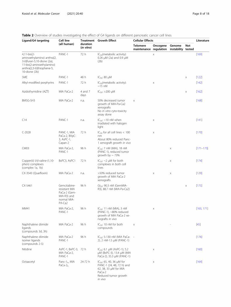
Table 2 Overview of studies investigating the effect of G4 ligands on different pancreatic cancer cell lines
Ligand/G4 targeting Cell line(all human)
Treatmentduration(in vitro)
Growth Effect Cellular Effects Literature
Telomeremaintenance
Oncogeneregulation
Genomeinstability
Nottested
4,11-bis(2-aminoethylamino) anthra[2,3-b]furan-5,10-dione (2a),11-bis(2-aminoethylamino)anthra[2,3-b]thiophene-5,10-dione (2b)
PANC-1 72 h IC50(metabolic activity):0.26 μM (2a) and 0.9 μM(2b)
x [169]
5ME PANC-1 48 h IC50: 80 μM x [122]
Alkyl-modified porphyrins PANC-1 72 h IC50(metabolic activity):~15 nM
x [142]
Azidothymidine (AZT) MIA PaCa-2 4 and 7days
IC50: >200 μM x [162]
BMSG-SH3 MIA PaCa-2 n.a. 50% decreased tumorgrowth of MIA-Pa-Ca2xenograftsNo in vitro cyto-toxicityassay done
x [168]
C14 PANC-1 n.a. IC50: ~10 nM whenirradiated with halogenlight
x [141]
C-2028 PANC-1, MIAPaCa-2, BXpC-3, AsPC-1,Capan-2
72 h IC50 for all cell lines < 100nmAbout 80% reduced Panc-1 xenograft growth in vivo
x [170]
CM03 MIA PaCa-2,PANC-1
96 h IC50: 7 nM (MIA), 18 nM(PANC-1), reduced tumorgrowth by ~ 73%
x [171–173]
Copper(ii) l/d-valine-(1,10-phen) complexes(complex 1a, 1b)
BxPC3, AsPC1 72 h IC50: ~2 μM for bothcomplexes in both celllines
x [174]
CX-3543 (Quarfloxin) MIA PaCa-2 n.a. >50% reduced tumorgrowth of MIA PaCa-2xenografts
x x [129]
CX-5461 Gemcitabine-resistant MIAPaCa-2 (Gem-MIA-R3) andnormal MIA-PA-Ca2
96 h GI50: 90.3 nM (GemMIA-R3), 88.7 nM (MIA-Pa-Ca2)
x [175]
MM41 MIA PaCa-2,PANC-1
96 h IC50: 11 nM (MIA), 3 nM(PANC-1), ~80% reducedgrowth of MIA PaCa-2 xe-nografts in vivo
x [163, 171]
Naphthalene diimideligands(compounds 3d, 3h)
MIA PaCa-2 96 h IC50: 10 nM for bothcompounds
x - [45]
Naphthalene diimideisomer ligands(compounds 2-5)
MIA PaCa-2PANC-1
96 h IC50: 5-130 nM (MIA PaCa-2), 2 nM-1.5 μM (PANC-1)
- [176]
Nitidine AsPC-1, BxPC-3,MIA PaCa-2,PANC-1
72 h IC50: 6.1 μM (AsPC-1), 5.2μM (BxPC-3), 13.4 μM (MIAPaCa-2), 35.3 μM (PANC-1)
x [160]
Octaacetyl Panc-1h, MIAPaCa-2h
24-72 h IC50: 65, 40, 36 μM forPANC-1 (24, 48, 72 h) and62, 38, 33 μM for MIAPaCa-2Reduced tumor growthin vivo
x [164]
Kosiol et al. Molecular Cancer (2021) 20:40 Page 8 of 18

previously discussed cancer entities, studies in leukemiarevealed that after G4 stabilization the expression of on-cogenes changed as well as the function of telomerase.G4 stabilization by telomestatin led to apoptosis andtelomere shortening in leukemia cells from four AMLpatients [184]. Similarily, telomere shortening and sene-sence was observed with the G4 ligand SYUIQ-5 in K-562 and HL-69 leukemia cells, because of changes in theexpression of telomerase (TERT) and TERF2 [180]. Inaddition to the relevance of G4 structure regulation attelomeres, several important leukemic oncogenes wereinhibited after using G4 ligands (including BCL2 [183],MYC [180, 181, 185], MLLT1 and AFDN [186], WT1[182], KIT [181] and KRAS [185]) in human AML-derived cell lines.As stated above, G4 structure formation might also
challenge genome stability and exhibit tumor/leukemia-promoting properties. Indeed, Tauchi and colleguesdemonstrated that telomestatin activates the ATM-dependent DNA-damage checkpoint response [62]. Insilico analysis revealed that 70% of the rearrenged genesin leukeamia contain a G4 motif [187]. This might indi-cate that G4 structures stimulate genome instability inthese tumors. This was further supported by the findingthat sites of rearrangement in TCF3, which promotesleukemia development, colocalize with regions that canform G4 structures [188]. In the future, it would be in-teresting to determine, if the G4 landscape within these
mutagenic tumors changes and to address, which G4structure newly forms and which are lost in these tu-mors. One hypothesis is that G4 structure formationcontributes to the mutagenic nature of tumors. The de-termination of the location of G4 structures within thesecancers together with deeper knowledge of the functionand relevance of these structures will be beneficial to ad-just G4 structure-driven treatments.One of the most common recurring genetic events
in AML are mutations in the C-terminal domain ofNPM1, which occur in about 30-35% of all AML pa-tients [189]. NPM1 is an abundant non-ribosomal nu-cleolar protein and essential for ribosome biogenesis.It was reported that NPM1 can bind to G4 structureslocated within the ribosomal DNA in vitro [190] andin vivo [191]. G4 binding by NPM1 is associated withits cellular localization. Experiments using the G4 lig-and TMPyP4 in OCI-AML2 cells demonstrated thatafter G4 stabilization NPM1 translocates from the nu-cleolus to the nucleoplasm. The authors explain thiseffect by the competitive effect of TMPyP4 on G4structure binding by NPM1 [191]. These findings to-gether with the observation that the most commonAML variant of NPM1 no longer binds to G4 struc-tures in ribosomal DNA [191] led to the idea that thedisruption of the G4 structure binding capability ofNPM1 is linked to nucleolar localization in AML-associated protein variants [191].
Table 2 Overview of studies investigating the effect of G4 ligands on different pancreatic cancer cell lines (Continued)
Ligand/G4 targeting Cell line(all human)
Treatmentduration(in vitro)
Growth Effect Cellular Effects Literature
Telomeremaintenance
Oncogeneregulation
Genomeinstability
Nottested
RHPS4 PAXF 736 15 dayscolonyformingassay
IC50: 0.44 μM x [177]
SOP1812 MIA PaCa-2,PANC-1,Capan-1, BXPC-3
96 h GI50: 1.3 nM (MIA PaCa-2),5.9 nM (Capan-1)Significantly reduced MIAPaCa-2 xenograft growthin vivo
x [173]
Telomestatin MIA PaCa-2 48 h IC50: 0.5 μM x [178]
Tetrakis PANC-1, MIAPaCa-2
24-72 h IC50: 60, 31, 25 μM (PANC-1) and 65, 36, 30 μM (MIAPaCa-2) for 24, 48, 72 hReduced tumor growthin vivo
x [164]
Tetrasubstitutednaphthalene diimideligands
PANC-1, MIAPaCa-2, HPAC,BxPc-3
96 h IC50: 0.1-0.2 μM (PANC-1,MIA PaCa-2, HPAC), 1.5 μM(BxPc-3)
x x [179]
TMPyP4 MIA PaCa-2,PANC-1
48 h, 4 and7 days
IC50: 21.9 μM (MIA PaCa-2, 4 days), 50 μM (PANC1, 7days), 50 μM (MIA PaCa-2,48 h), ~20 μM (PANC-1, 48h). ~30% KRAS inhibitionafter 12 and 24 h
x [122, 142,154, 162,178]
Kosiol et al. Molecular Cancer (2021) 20:40 Page 9 of 18
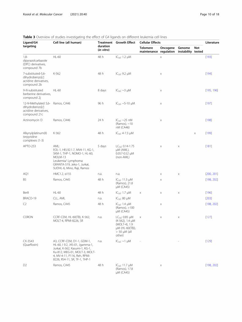
Table 3 Overview of studies investigating the effect of G4 ligands on different leukemia cell lines
Ligand/G4targeting
Cell line (all human) Treatmentduration(in vitro)
Growth Effect Cellular Effects Literature
Telomeremaintenance
Oncogeneregulation
Genomeinstability
Nottested
1,8-dipyrazolcarbazole(DPC) derivatives,compound 7b
HL-60 48 h IC50: 1.2 μM x [193]
7-substituted-5,6-dihydrobenzo[c]acridine derivatives,compound 2b
K-562 48 h IC50: 9.2 μM x [194]
9-N-substitutedberberine derivatives,compound 2j
HL-60 8 days IC50: ~3 μM x [195, 196]
12-N-Methylated 5,6-dihydrobenzo[c]acridine derivatives,compound 21c
Ramos, CA46 96 h IC50: ~5-10 μM x [197]
Actinomycin D Ramos, CA46 24 h IC50: ~25 nM(Ramos), ~10nM (CA46)
x [198]
Alkynylplatinum(II)terpyridinecomplexes (1-3)
K-562 48 h IC50: 4-7.3 μM x [199]
APTO-253 AML:EOL-1, HEL92.1.7, MV4-11, KG-1,SKM-1, THP-1, NOMO-1, HL-60,MOLM-13Leukemia/ Lymphoma:GRANTA-519, Jeko-1, Jurkat,SUDHL-6, Mino, Raji, Ramos
5 days LC50: 0.14-1.75μM (AML),0.057-0.52 μM(non-AML)
x x [181]
AQ1 HMC1.2, α155 n.a. n.a. x x [200, 201]
B5 Ramos, CA45 48 h IC50: 11.3 μM(Ramos), 21.8μM (CA45)
x [198, 202]
Ber8 HL-60 48 h IC50: 1.7 μM x x x [196]
BRACO-19 CLL, AML n.a. IC50: 80 μM [203]
C2 Ramos, CA45 48 h IC50: 1.4 μM(Ramos), >100μM (CA45)
x [198, 202]
CORON CCRF-CEM, HL-60(TB), K-562,MOLT-4, RPMI-8226, SR
n.a. LC50: 0.85 μM(K-562), 1.4 μM(MOLT-4), 1.9μM (HL-60(TB)),> 50 μM (allother)
x x x [127]
CX-3543(Quarfloxin)
A3, CCRF-CEM, D1-1, GDM-1,HL-60, I 9.2, J45-01, Jgamma-1,Jurkat, K-562, Kasumi-1, KG-1,Ku-812, MEG-01, MOLT-3, MOLT-4, MV-4-11, P116, Reh, RPMI-8226, RS4-11, SR, TF-1, THP-1
n.a. IC50: ~1 μM - - [129]
D2 Ramos, CA45 48 h IC50: 11.7 μM(Ramos), 17.8μM (CA45)
x [198, 202]
Kosiol et al. Molecular Cancer (2021) 20:40 Page 10 of 18

Table 3 Overview of studies investigating the effect of G4 ligands on different leukemia cell lines (Continued)
Ligand/G4targeting
Cell line (all human) Treatmentduration(in vitro)
Growth Effect Cellular Effects Literature
Telomeremaintenance
Oncogeneregulation
Genomeinstability
Nottested
Diquinolinyl-Pyridine Ligands(1a-c)
K-562, HL60 72 h IC50: >50 μM(1a), 3 μM (1b),>50 μM (1c, allK-562)IC50: >50 μM(1a), 18 μM (1b),>50 μM (1c, allHL60)
x [204]
Disubstitutedquindolinederivatives,compound 74a
RajiCCRF-CEMU266B2
48 h IC50: 4.7 μM(Raji), 18.1 μM(CCRF-CEM),23.0 μM(U266B2)Xenograft ofRaji cells >50%reduced tumorgrowth
x [205]
DNR K-562 48 h96 h
IC50: 0.33 μM,0.067 μM
x [206]
EMICORON CCRF-CEM, HL-60(TB), K-562,MOLT-4, RPMI-8226, SR
n.a. LC50: 3.7 μM(HL-60(TB)), >50 μM (allother)
x x x [127]
GQC-05 KG-1a, CMK, TF-1 24 h IC50: >1 μM x x [207]
LZ-11 HL-60 48 h IC50: 9.6 μM x [208]
MXR K-562 48 h96 h
IC50: 0.411 μMIC50: 0.105 μM
x [206]
Pegaharmine D HL-60 72 h IC50: 3.81 μM x [209]
Pyrazine-basedcyclometalated(C^Npz^C)Au(III)carbene complexes,compound 2 and 3
HL-60 72 h IC50: 0.31 μM(2),4.05 μM (3)
- [210]
Pyridine(2,4-dihydroxybenzaldehyde dibenzylsemicarbazone)copper(II)
MOLT-4 24 h72 h
IC50: 5 μMIC50: 1.5 μM
x [185]
QPB compound 15e HL-60 48 h IC50: 1.7 μM x [211]
Quinolino-benzo- [5,6]-dihydroisoquindoliumcompounds 3a, 3f,3g, 3j
HL-60 48 h IC50: 2 μM (3j) -12.6 μM (3g)
x [212]
TMPyP4 K-562, OCI-AML2, OCI-AML3 48 and 96h
IC50: 100-170μM (K-562, 48h), 60 μM (OCI-AML2, 96 h), 50μM (OCI-AML3,96 h)
x [182, 191,213, 214]
Telomestatin Primary blast cells from AMLpatientsOM9;22, K-562
10-30 days IC50: 5 μM (10days), 2 μMshowed growthinhibition (15/30 days)
x x [62, 184,215]
Kosiol et al. Molecular Cancer (2021) 20:40 Page 11 of 18

As a side note we would like to point out that also G4structure formation within RNAs could impact tumori-genesis and might be a useful target for cancer therapy.The translational control of oncoprotein expression isconsidered to be relevant in many solid cancers [192]and leukemias. EIF4A is an RNA helicase that promotesand sustains T-acute lymphoblastic leukemia and prefer-ably targets mRNAs with G4 motifs in their 5’UTR[192]. Silvestrol, hippuristanol and pateamine A are nat-ural compounds that target EIF4A. Inhibition of EIF4Awith silvestrol showed anti-tumor efficacy in vitro andin vivo [192]. It was proposed that after EIF4A inhibitionby silvestrol more G4 structures are formed within thesetranscripts leading to altered translation, which causedthe slow growth of tumor cells (Table 3).
DiscussionThe research of the past decades links G4 structure for-mation and unfolding to defects and changes observedin different cancer entities. Current studies revealed thatalthough most G4 ligands result in changes of cancercell growth, they act via different mechanims and mostlikely at different targets. It is not clear, if the reducedgrowth effect on cancer cells after G4 ligand treatmentis caused by multiple G4 structure-mediated changes, bya single G4 structure-driven event or by unspecific bind-ing of the ligand. The problem in the current literatureis to pinpoint the exact cause that drives growth changesand to monitor where the G4 ligand binds. Currently,there are several trials to establish G4 ligands that arespecific to only one target [83, 219] and/or to developnovel photosensitizing G4 ligands that only cause toxiceffects to G4 regions after irradiation [66]. These trialsare very promising as it is expected that these ap-proaches will reduce side effects. Additionally, the list ofG4-interacting and -regulating proteins is increasing,which will not only give insights into G4 function in
normal cells but will also provide ideas how to target/regulate specific G4 structures via the protein itself orprotein-specific inhibotors. This would be particularlyinteresting for cancers like pancreatic cancer that forsome cases are caused by a single upregulation of oneoncogene (e.g. KRAS). In these cancers a G4 structure-based block of transcription is a very promising treat-men option. The current literature demonstrates thatG4 stabilization often correlates with reduced cellulargrowth of cancers. It is not fully understood if the re-duced growth is due to changes in telomere mainten-ance, transcriptional changes or genome stability. Wewould like to emphasize that co-treatment of G4stabilization with drugs that either block DNA repair orinduce additional genome instability is currently a verypromising approach.An interesting therapeutic strategy could be to com-
bine immune checkpoint inhibitors with G4 structure-stabilizing ligands. It has been demonstrated that thesuccess of immune checkpoint inhibition correlates withthe mutational burden of a tumor [111, 112]. Hence, im-munotherapeutic approaches might benefit from a com-binational therapy with G4 ligands. It seems plausiblethat G4 ligands, which induce genome instability, mightincrease the immunogenecity of a tumor and sensitize itfor checkpoint inhibition or other immunotherapeuticapproaches.
ConclusionIn this review we presented and discussed the rele-vance of G4 structure formation and stabilization as atherapeutical approach to treat cancer cells based onthe current literature. As pointed out, different li-gands have often a negative effect on cancer cellgrowth via different mechanisms. We discussed thehypothesis, if the mutagenic burden of the tumorpositivly or negatively influences the outcome of G4
Table 3 Overview of studies investigating the effect of G4 ligands on different leukemia cell lines (Continued)
Ligand/G4targeting
Cell line (all human) Treatmentduration(in vitro)
Growth Effect Cellular Effects Literature
Telomeremaintenance
Oncogeneregulation
Genomeinstability
Nottested
Substitutedsalicylaldehydedibenzylsemicarbazonescopper(II) complexes(7, 9, 7-py and 9-py)
MOLT-4 24 h IC50: 3.1 μM (9-py), 18.1 μM (7-py), 5 μM (7),8.03 μM (9)
x [215]
Sysu12d CA46, HL-60 48 h IC50: 11.2 μM(CA46), 5 μM(HL-60)
x x [216]
SYUIQ-5 K-562, HL-60 72 h IC50: 5 μM (K-562, HL-60) or2.65 μM (K-562)
x x x [180, 217]
SYUIQ-FM05 K-562 24 h IC50: 10.83 nM x [218]
Kosiol et al. Molecular Cancer (2021) 20:40 Page 12 of 18

ligand treatment. We summarized the current re-search results that are linked to changes in G4 levelsin melanoma, pancreatic cancer and leukemia cells.The conclusions are not very clear. We oberve thatall three tumor entities show reduced cell growthupon treatment with different G4 ligands and, de-pending on the ligand, also changes in telomeremaintenance, gene expression of oncogenes and in-creased genome instability. A direct comparision isdifficult, because different studies were often doneusing different conditions. Some ligands were used inat least two entities. TMPyP4, EMICORON andCORON were tested in all three selected entities. Al-though similar effects were documented, the timing oftreatment as well as the used concentration differs inthese different studies. These differences indicate thatlower concentrations of G4 ligands (e.g. EMICORON)are required to induce G4 structure-driven toxicity incancer cells that have a high mutagenic burden (seeTable 1, 2 and 3). A similar trend was observed forBRACO-19 (28 μM in melanoma and 80 μM forleukemia). Further experimental data demonstratedthat a treatment with G4 ligands further enhances thecytotoxic effect in cells that have high levels of gen-ome instability (e.g. after radiation therapy, afterPARP inhibitor treatment or in BRCA1-deficientcells). However, in studies that used quarfloxin (CX-3543) we could not find this correlation. Future stud-ies with similar culturing conditions and additionalmolecular studies are required to proof thishypothesis.Lastly, we would like to disucss the potential impact
that G4 structure formation might have on the muta-genic burden of the tumor and what the consequencesare for tumor development and tumor relapse aftertreatment. In general, G4 structure formation, if not reg-ulated efficiently (this includes formation and unfolding),can stimulate genome instability, which includes mua-tions and deletions and complex gross chromosomal re-arrangements [28, 30, 220]. A computational studyinvestigated how the mutational burden correlates withG4 structure formation [221]. They revealed by compar-ing the location of potential G4 forming sites withcancer-associated breakpoints (using the COSMIC data-base) a significant overlap, in particular in those cancersthat harbor a mutation in TP53. This is underlined bycompuational studies in melanoma cells that link G4 re-gions with mutational hot spots [187]. A recent studyidentified a direct correlation of G4 structure formationwith mutational changes in different breast cancer en-tities [222]. This supports the notion that G4 formationindeed stimulates and influences mutation rates in dif-ferent cancers and may also contribute to subtype classi-fications [222]
Abbreviations20A: 1-ethyl-N-(phenylmethyl)-4-(tetrahydro-2H-pyran-4-ylamino)-1H-pyrazolo[3,4-b]pyridine-5-carboxamide; AFDN: Afadin, Adherens JunctionFormation Factor; AML: Acute myeloid leukemia; APTO-253: 2-(5-fluoro-2-methyl-1H-indol-3-yl)-1H-imidazo[4,5-f][1,10]phenanthroline; ATM: AtaxiaTelangiectasia Mutated; ATRX: ATRX Chromatin Remodeler; BAX: BCL2Associated X, Apoptosis Regulator; BCL2: BCL2 Apoptosis Regulator;BLM: Bloom Syndrome RecQ Like Helicase; BMSG-SH-3 : 2,7-Bis-[5-(4-methyl-piperazin-1-yl)-pentyl]-4,9-bis-[3-(4-methyl-piperazin-1-yl)-propylamino]-benzo[lmn][3,8]phenanthroline-1,3,6,8-tetraone; BRACO19: N,N′-(9-(4-(Dimethylamino)phenylamino)acridine-3,6-diyl)bis(3-(pyrrolidin-1-yl)propanamide) hydrochloride; BRAF: B-Raf Proto-Oncogene, Serine/Threo-nine Kinase; BRCA1: Breast Cancer Type 1 Susceptibility Protein;BRCA2: Breast Cancer Type 2 Susceptibility Protein; BRIP1/FANCJ: BRCA1Interacting Protein C-Terminal Helicase 1; C14: C14H28-alkyl derivative ofPMPyP4; ChIP-seq: Chromatin Immunoprecipitation and sequencing; CX-3543: Quarfloxin, 15-fluoro-N-[2-[(2S)-1-methylpyrrolidin-2-yl]ethyl]-18-oxo-14-(3-pyrazin-2-ylpyrrolidin-1-yl)-12-oxa-1-azapentacyclo[11.7.1.02,11.04,9.017,21]-henicosa-2,4,6,8,10,13(21),14,16,19-nonaene-19-carboxamide; CX-5461: 2-(4-methyl-1,4-diazepan-1-yl)-N-[(5-methylpyrazin-2-yl)methyl]-5-oxo-[1,3]benzothiazolo[3,2-a][1,8]naphthyridine-6-carboxamide; EAC: Ehrlichascites carcinoma; EIF4A: Eukaryotic translation initiation factor 4A; G4: G-quadruplex; GI20/GI50: 20%/50% growth inhibition; GPI-15427: 10-((4-Methylpiperazin-1-yl)methyl)chromeno(4,3,2-de)phthalazin-3(2H)-one;h: Hours; HaCaT: Human adult low Calcium high Temperature Keratinocytes;HMGB1: High mobility group box 1; HNRNPA1: Heterogeneous nuclearribonucleoprotein A1; HR: Homologous recombination; HRAS: HRas Proto-Oncogene, GTPase; IC50: Half maximal inhibitory concentration; ILK: IntegrinLinked Kinase; IO: Immune-oncology; IR: Ionizing radiation; IZTZ-1: Benzothiazole-2-carboxaldehyde-derivate; KIT: KIT Proto-Oncogene, Recep-tor Tyrosine Kinase; KRAS: KRAS Proto-Oncogene, GTPase; LC50: Half lethalconcentration; MAZ: MYC-associated zinc finger protein; MLLT1 : MLLT1Super Elongation Complex Subunit; MM41: 4,9-Bis((3-(4-methylpiperazin-1-yl)propyl)amino)-2,7-bis(3-morpholinopropyl)benzo[lmn][3,8]phenanthroline-1,3,6,8(2H,7H)-tetraone; MYC: MYC Proto-Oncogenes c-myc, l-myc, n-myc;n.a.: Not available; NHEJ: Non-homologous end joining;NPM1: Nucleophosmin 1; NRAS: NRAS Proto-Oncogene, GTPase; NU7441: 8-(4-Dibenzothienyl)-2-(4-morpholinyl)-4H-1-benzopyran-4-one; olaparib: 4-[[3-[4-(cyclopropanecarbonyl)piperazine-1-carbonyl]-4-fluorophenyl]methyl]-2H-phthalazin-1-one; OS: Overall survival; PARP1: Poly [ADP-ribose] polymerase 1;PDCA: Pancreatic ductal adenocarcinoma; PDS: Pyridostatin, 4-(2-Aminoethoxy)-N2,N6-bis[4-(2-aminoethoxy)-2-quinolinyl]-2,6-pyridinedicarboxamide; PDT: Photodynamic therapy; PIF1: Petite IntegrationFrequency 1; POT1: Protection of telomeres 1; Pou3FS/BRN2: Pou class 3homeobox 2; PRKDC: Protein Kinase, DNA-Activated, Catalytic Subunit;Rad51: RAD51 Recombinase; RAS: NRAS Proto-Oncogene, GTPase; RHOJ: Rashomolog family member J; RHPS4: 3,11-Difluoro-6,8,13-trimethylquino[4,3,2-kl]acridinium methylsulfate; ROS: Reactive oxygen species; RSK: P90 ribosomalS6 kinase; SYUIQ-5: N-(10H-indolo[3,2-b]quinolin-11-yl)-N',N'-dimethylpropane-1,3-diamine; TCF3: Transcription Factor 3; Tel03: N,N¢-Bis-(2-(dimethylamino)ethyl)-3,4,9,10-perylenetetracarboxylic acid diimide;Telomestatin: (1R)-4,8-Dimethyl-3,7,11,15,19,23, 27-heptaoxa-31-thia-33,34,35,36,37,38,39,40-octaazanonacyclo[28.2.1.12,5.16,9.110,13.114,17.118,21.122,25.126,29]tetraconta-2(40),4,6(39),8,10(38), 12,14(37),16,18(36),20,22(35), 24,26(34),28,30(33)-pentadecaene; TERF2: Telomericrepeat binding factor 2; TERT: Telomerase reverse transcriptase; TINA: Twistedintercalating nucleic acid; TMPipEOPP: 5,10,15,20-tetra-{4-[2-(1-methyl-1-piperidinyl)ethoxy]phenyl} porphyrin; TMPyP4: Meso-Tetra (N-methyl-4-pyridyl) porphine tetra tosylate; TP53: Tumor Protein P53; VEGF: VascularEndothelial Growth Factor A; WHO: World Health Organization; WRN: WernerSyndrome RecQ Like Helicase; WT1: WT1 Transcription Factor; ZnP1: Zn(II)5,10,15,20-tetrakis(N-carboxymethyl-4-pyridinium)porphyrin
AcknowledgementsWe thank the Paeschke and the Heine group for helpful discussion.
Authors’ contributionsNK, SJ, AH, KP literature search, NK, AH, KP initial draft of manuscript,NK, SJ, PB, AH, KP editing and final draft of manuscript, AH and KPsupervision and funding aquvisition. All authors approved the finalmanuscript.
Kosiol et al. Molecular Cancer (2021) 20:40 Page 13 of 18

FundingOpen Access funding enabled and organized by Projekt DEAL. Research inthe Paeschke laboratory is fund by an ERC Stg Grant (638988-G4DSB) and bythe Deutsche Forschungsgemeinschaft (DFG, German Research Foundation)– Project-ID 369799452 – TRR237”. Research in the Paeschke and Heine labare funded by the Deutsche Forschungsgemeinschaft (DFG, German Re-search Foundation) under Germany’s Excellence Strategy – EXC2151 –390873048.
Availability of data and materialsNot applicable
Ethics Approval and Consent to participateNot applicable
Consent for publicationNot applicable
Competing interestsThe authors declare no competing interests.
Received: 14 October 2020 Accepted: 1 February 2021
References1. Hannah Ritchie and Max Roser. Causes of Death - Our World in Data.
https://ourworldindata.org/causes-of-death. Accessed 4 Dec 2020.2. Cancer. https://www.who.int/news-room/fact-sheets/detail/cancer. Accessed
4 Dec 2020.3. Couzin-Frankel J. Breakthrough of the year. Cancer immunotherapy. Vol.
342. Science. 2013;2013:1432–3.4. Sun W, Shi Q, Zhang H, Yang K, Ke Y, Wang Y, et al. Advances in the
techniques and methodologies of cancer gene therapy. Discov Med. 2019;27(146):45–55.
5. Klinakis A, Karagiannis D, Rampias T. Targeting DNA repair in cancer: currentstate and novel approaches. 77, Cellular and Molecular Life Sciences. 2020.p. 677–703.
6. A R, JD W. Cancer immunotherapy using checkpoint blockade. Science.2018;359(6382).
7. Hochhaus A, Larson RA, Guilhot F, Radich JP, Branford S, Hughes TP, et al.Long-Term Outcomes of Imatinib Treatment for Chronic Myeloid Leukemia.N Engl J Med. 2017;376(10):917–27.
8. Hellmann MD, Paz-Ares L, Bernabe Caro R, Zurawski B, Kim S-W, CarcerenyCosta E, et al. Nivolumab plus Ipilimumab in Advanced Non–Small-CellLung Cancer. N Engl J Med. 2019;381(21):2020–31.
9. Bacolla A, Wells RD. Non-B DNA conformations, genomic rearrangements,and human disease. Journal of Biological Chemistry. 2004;279:47411–4.
10. Bochman ML, Paeschke K, Zakian VA. DNA secondary structures: Stabilityand function of G-quadruplex structures. Nature Reviews Genetics. 2012;13:770–80.
11. Varshney D, Spiegel J, Zyner K, Tannahill D, Balasubramanian S. Theregulation and functions of DNA and RNA G-quadruplexes. Vol. 21, NatureReviews Molecular Cell Biology. 2020. p. 459–74.
12. Spiegel J, Adhikari S, Balasubramanian S. The Structure and Function of DNAG-Quadruplexes. Vol. 2, Trends in Chemistry. 2020. p. 123–36.
13. Burge S, Parkinson GN, Hazel P, Todd AK, Neidle S. Quadruplex DNA:Sequence, topology and structure. Nucleic Acids Res. 2006;34(19):5402–5415.
14. Chambers VS, Marsico G, Boutell JM, Di Antonio M, Smith GP,Balasubramanian S. High-throughput sequencing of DNA G-quadruplexstructures in the human genome. Nat Biotechnol. 2015;33(8):877–81.
15. Marsico G, Chambers VS, Sahakyan AB, McCauley P, Boutell JM, Di AntonioM, et al. Whole genome experimental maps of DNA G-quadruplexes inmultiple species. Nucleic Acids Res. 2019;47(8):3862–74.
16. Lane AN, Chaires JB, Gray RD, Trent JO. Stability and kinetics of G-quadruplex structures. Vol. 36, Nucleic Acids Research. 2008. p. 5482–515.
17. Huppert JL, Balasubramanian S. Prevalence of quadruplexes in the humangenome. Nucleic Acids Res. 2005;33(9):2908–16.
18. Hänsel-Hertsch R, Beraldi D, Lensing SV, Marsico G, Zyner K, Parry A, et al. G-quadruplex structures mark human regulatory chromatin. Nat Genet. 2016;48(10):1267–72.
19. Capra JA, Paeschke K, Singh M, Zakian VA. G-quadruplex DNA sequencesare evolutionarily conserved and associated with distinct genomic featuresin Saccharomyces cerevisiae. PLoS Comput Biol. 2010;6(7):9.
20. Kaplan OI, Berber B, Hekim N, Doluca O. G-quadruplex prediction in E. coligenome reveals a conserved putative G-quadruplex-Hairpin-Duplex switch.Nucleic Acids Res. 2016;44(19):9083–95.
21. Lavezzo E, Berselli M, Frasson I, Perrone R, Palù G, Brazzale AR, et al. G-quadruplex forming sequences in the genome of all known human viruses:A comprehensive guide. PLOS Comput Biol. 2018;14(12):e1006675.
22. Eddy J, Maizels N. Conserved elements with potential to form polymorphicG-quadruplex structures in the first intron of human genes. Nucleic AcidsRes. 2008;36(4):1321–33.
23. Todd AK, Johnston M, Neidle S. Highly prevalent putative quadruplexsequence motifs in human DNA. Nucleic Acids Res. 2005;33(9):2901–7.
24. Huppert JL, Balasubramanian S. G-quadruplexes in promoters throughoutthe human genome. Nucleic Acids Res. 2007;35(2):406–13.
25. Nakken S, Rognes T, Hovig E. The disruptive positions in human G-quadruplex motifs are less polymorphic and more conserved than theirneutral counterparts. Nucleic Acids Res. 2009;37(17):5749–56.
26. Brázda V, Hároníková L, Liao JCC, Fojta M. DNA and RNA quadruplex-binding proteins. Int J Mol Sci. 2014;15:17493–517.
27. Sauer M, Paeschke K. G-quadruplex unwinding helicases and their functionin vivo. Vol. 45, Biochemical Society Transactions. 2017. p. 1173–82.
28. Paeschke K, Capra JA, Zakian VA. DNA Replication through G-QuadruplexMotifs Is Promoted by the Saccharomyces cerevisiae Pif1 DNA Helicase. Cell.2011;145(5):678–91.
29. Schiavone D, Guilbaud G, Murat P, Papadopoulou C, Sarkies P, Prioleau M,et al. Determinants of G quadruplex-induced epigenetic instability in REV 1-deficient cells. EMBO J. 2014;33(21):2507–20.
30. De Magis A, Manzo SG, Russo M, Marinello J, Morigi R, Sordet O, et al. DNAdamage and genome instability by G-quadruplex ligands are mediated by Rloops in human cancer cells. Proc Natl Acad Sci U S A. 2019;116(3):816–25.
31. Lopes J, Le Piazza A, Bermejo R, Kriegsman B, Colosio A, Teulade-Fichou MP,et al. G-quadruplex-induced instability during leading-strand replication.EMBO J. 2011;30(19):4033–46.
32. Puig Lombardi E, Holmes A, Verga D, Teulade-Fichou MP, Nicolas A,Londoño-Vallejo A. Thermodynamically stable and genetically unstable G-quadruplexes are depleted in genomes across species. Nucleic Acids Res.2019;47(12):6098–113.
33. Biffi G, Tannahill D, Miller J, Howat WJ, Balasubramanian S. Elevated levels ofG-quadruplex formation in human stomach and liver cancer tissues. PLoSOne. 2014;9(7).
34. Hänsel-Hertsch R, Spiegel J, Marsico G, Tannahill D, Balasubramanian S.Genome-wide mapping of endogenous G-quadruplex DNA structures bychromatin immunoprecipitation and high-throughput sequencing. NatProtoc. 2018;13(3):551–64.
35. Tan J, Lan L. The DNA secondary structures at telomeres and genomeinstability. Vol. 10, Cell and Bioscience. 2020.
36. Bryan TM. G-quadruplexes at telomeres: Friend or foe?. Vol. 25, Molecules.2020.
37. Kim N. The Interplay between G-quadruplex and Transcription. Curr MedChem. 2017;26(16):2898–917.
38. Tian T, Chen YQ, Wang SR, Zhou X. G-Quadruplex: A Regulator of GeneExpression and Its Chemical Targeting. Vol. 4, Chem. 2018. p. 1314–44.
39. Bryan TM. Mechanisms of DNA replication and repair: Insights from thestudy of G-quadruplexes. Molecules. 2019;24(19).
40. Lerner LK, Sale JE. Replication of G quadruplex DNA. Vol. 10, Genes. 2019.41. Ruggiero E, Richter SN. Survey and summary G-quadruplexes and G-
quadruplex ligands: Targets and tools in antiviral therapy. Vol. 46, NucleicAcids Research. 2018. p. 3270–83.
42. Carvalho J, Mergny JL, Salgado GF, Queiroz JA, Cruz C. G-quadruplex, Friendor Foe: The Role of the G-quartet in Anticancer Strategies. Vol. 26, Trends inMolecular Medicine. 2020. p. 848–61.
43. Li Q, Xiang JF, Yang QF, Sun HX, Guan AJ, Tang YL. G4LDB: A database fordiscovering and studying G-quadruplex ligands. Nucleic Acids Res. 2013;41(D1).
44. Haider SM, Neidle S, Parkinson GN. A structural analysis of G-quadruplex/ligand interactions. Vol. 93, Biochimie. 2011. p. 1239–51.
45. Micco M, Collie GW, Dale AG, Ohnmacht SA, Pazitna I, Gunaratnam M, et al.Structure-based design and evaluation of naphthalene diimide G-quadruplex ligands as telomere targeting agents in pancreatic cancer cells.J Med Chem. 2013;56(7):2959–74.
Kosiol et al. Molecular Cancer (2021) 20:40 Page 14 of 18

46. Kim MY, Vankayalapati H, Shin-Ya K, Wierzba K, Hurley LH. Telomestatin, apotent telomerase inhibitor that interacts quite specifically with the humantelomeric intramolecular G-quadruplex. J Am Chem Soc. 2002;124(10):2098–9.
47. Burger AM, Dai F, Schultes CM, Reszka AP, Moore MJ, Double JA, et al. TheG-quadruplex-interactive molecule BRACO-19 inhibits tumor growth,consistent with telomere targeting and interference with telomerasefunction. Cancer Res. 2005;65(4):1489–96.
48. Izbicka E, Wheelhouse RT, Raymond E, Davidson KK, Lawrence RA, Sun D,et al. Effects of cationic porphyrins as G-quadruplex interactive agents inhuman tumor cells. Cancer Res. 1999;59(3):639–44.
49. Gowan SM, Heald R, Stevens MFG, Kelland LR. Potent inhibition oftelomerase by small-molecule pentacyclic acridines capable of interactingwith G-quadruplexes. Mol Pharmacol. 2001;60(5):981–8.
50. Rodriguez R, Müller S, Yeoman JA, Trentesaux C, Riou JF, Balasubramanian S.A novel small molecule that alters shelterin integrity and triggers a DNA-damage response at telomeres. J Am Chem Soc. 2008;130(47):15758–9.
51. Pagano A, Iaccarino N, Abdelhamid MAS, Brancaccio D, Garzarella EU, DiPorzio A, et al. Common G-quadruplex binding agents found to interactwith i-motif-forming DNA: Unexpected multi-target-directed compounds.Front Chem. 2018;6.
52. Abdelhamid MAS, Gates AJ, Waller ZAE. Destabilization of i-Motif DNA atNeutral pH by G-Quadruplex Ligands. Biochemistry. 2019;58(4):245–9.
53. Asamitsu S, Bando T, Sugiyama H. Ligand Design to Acquire Specificity toIntended G-Quadruplex Structures. Vol. 25, Chemistry - A European Journal.2019. p. 417–30.
54. Felsenstein KM, Saunders LB, Simmons JK, Leon E, Calabrese DR, Zhang S,et al. Small Molecule Microarrays Enable the Identification of a Selective,Quadruplex-Binding Inhibitor of MYC Expression. ACS Chem Biol. 2016;11(1):138–48.
55. Cimino-Reale G, Zaffaroni N, Folini M. Emerging Role of G-quadruplex DNAas Target in Anticancer Therapy. Curr Pharm Des. 2016;22(44):6612–24.
56. Kim NW, Piatyszek MA, Prowse KR, Harley CB, West MD, Ho PLC, et al.Specific association of human telomerase activity with immortal cells andcancer. Science (80- ). 1994;266(5193):2011–5.
57. Zahler AM, Williamson JR, Cech TR, Prescott DM. Inhibition of telomerase byG-quartet DMA structures. Nature. 1991;350(6320):718–20.
58. Moye AL, Porter KC, Cohen SB, Phan T, Zyner KG, Sasaki N, et al. TelomericG-quadruplexes are a substrate and site of localization for humantelomerase. Nat Commun. 2015;6.
59. Paeschke K, Simonsson T, Postberg J, Rhodes D, Lipps HJ. Telomere end-binding proteins control the formation of G-quadruplex DNA structuresin vivo. Nat Struct Mol Biol. 2005;12(10):847–54.
60. Paudel BP, Moye AL, Assi HA, El-Khoury R, Cohen SB, Holien JK, et al. Amechanism for the extension and unfolding of parallel telomeric g-quadruplexes by human telomerase at single-molecule resolution. Elife.2020;9:1–9.
61. Neidle S. Human telomeric G-quadruplex: The current status of telomeric G-quadruplexes as therapeutic targets in human cancer. Vol. 277, FEBSJournal. 2010. p. 1118–25.
62. Tauchi T, Shin-Ya K, Sashida G, Sumi M, Nakajima A, Shimamoto T, et al.Activity of a novel G-quadruplex-interactive telomerase inhibitor, telomestatin(SOT-095), against human leukemia cells: Involvement of ATM-dependent DNAdamage response pathways. Oncogene. 2003;22(34):5338–47.
63. Wheelhouse RT, Sun D, Han H, Han FX, Hurley LH. Cationic porphyrins astelomerase inhibitors: The interaction of tetra- (N-methyl-4-pyridyl)porphinewith quadruplex DNA [1]. Vol. 120, Journal of the American ChemicalSociety. 1998. p. 3261–2.
64. Phatak P, Cookson JC, Dai F, Smith V, Gartenhaus RB, Stevens MFG, et al.Telomere uncapping by the G-quadruplex ligand RHPS4 inhibits clonogenictumour cell growth in vitro and in vivo consistent with a cancer stem celltargeting mechanism. Br J Cancer. 2007;96(8):1223–33.
65. Salvati E, Leonetti C, Rizzo A, Scarsella M, Mottolese M, Galati R, et al.Telomere damage induced by the G-quadruplex ligand RHPS4 has anantitumor effect. J Clin Invest. 2007;117(11):3236–47.
66. Kawauchi K, Urano R, Kinoshita N, Kuwamoto S, Torii T, Hashimoto Y, et al.Photosensitizers based on g-quadruplex ligand for cancer photodynamictherapy. Vol. 11, Genes. 2020. p. 1–13.
67. Zhu LN, Zhao SJ, Wu B, Li XZ, Kong DM. A new cationic porphyrinderivative (TMPipEOPP) with large side arm substituents: A highly selectiveG-quadruplex optical probe. PLoS One. 2012;7(5).
68. Beniaminov AD, Novikov RA, Mamaeva OK, Mitkevich VA, Smirnov IP,Livshits MA, et al. Light-induced oxidation of the telomeric G4 DNA incomplex with Zn(II) tetracarboxymethyl porphyrin. Nucleic Acids Res. 2016;44(21):10031–41.
69. Zyner KG, Mulhearn DS, Adhikari S, Cuesta SM, Di Antonio M, Erard N, et al.Genetic interactions of G-quadruplexes in humans. Elife. 2019;8.
70. Balasubramanian S, Hurley LH, Neidle S. Targeting G-quadruplexes in genepromoters: A novel anticancer strategy? Nat Rev Drug Discov. 2011;10(4):261–75.
71. De S, Michor F. DNA secondary structures and epigenetic determinants ofcancer genome evolution. Nat Struct Mol Biol. 2011;18(8):950–5.
72. Siddiqui-Jain A, Grand CL, Bearss DJ, Hurley LH. Direct evidence for aG-quadruplex in a promoter region and its targeting with a smallmolecule to repress c-MYC transcription. Proc Natl Acad Sci U S A.2002;99(18):11593–8.
73. Sun D, Guo K, Rusche JJ, Hurley LH. Facilitation of a structural transition inthe polypurine/polypyrimidine tract within the proximal promoter region ofthe human VEGF gene by the presence of potassium and G-quadruplex-interactive agents. Nucleic Acids Res. 2005;33(18):6070–80.
74. Dexheimer TS, Sun D, Hurley LH. Deconvoluting the structural and drug-recognition complexity of the G-quadruplex-forming region upstream ofthe bcl-2 P1 promoter. J Am Chem Soc. 2006;128(16):5404–15.
75. Cogoi S, Xodo LE. G-quadruplex formation within the promoter of the KRASproto-oncogene and its effect on transcription. Nucleic Acids Res. 2006;34(9):2536–49.
76. Yang D, Hurley L. Structure of the biologically relevant g-quadruplex in thec-MYC promoter. Nucleosides, Nucleotides and Nucleic Acids. 2006;25(8):951–68.
77. Rankin S, Reszka AP, Huppert J, Zloh M, Parkinson GN, Todd AK, et al.Putative DNA Quadruplex Formation within the Human c-kit Oncogene. JAm Chem Soc. 2005;10:10584–9.
78. Dang C V. MYC on the path to cancer. Vol. 149, Cell. 2012. p. 22–35.79. Lin CY, Lovén J, Rahl PB, Paranal RM, Burge CB, Bradner JE, et al.
Transcriptional amplification in tumor cells with elevated c-Myc. Cell. 2012;151(1):56–67.
80. Bretones G, Delgado MD, León J. Myc and cell cycle control. Vol. 1849,Biochimica et Biophysica Acta - Gene Regulatory Mechanisms. 2015. p. 506–16.
81. Whitfield JR, Beaulieu ME, Soucek L. Strategies to inhibit Myc and theirclinical applicability. Vol. 5, Frontiers in Cell and Developmental Biology.2017.
82. Nasiri HR, Bell NM, Mc Luckie KIE, Husby J, Abell C, Neidle S, et al. Targetinga c-MYC G-quadruplex DNA with a fragment library. Chem Commun. 2014;50(14):1704–7.
83. Calabrese DR, Chen X, Leon EC, Gaikwad SM, Phyo Z, Hewitt WM, et al.Chemical and structural studies provide a mechanistic basis for recognitionof the MYC G-quadruplex. Nat Commun. 2018;9(1):4229–9.
84. Boddupally PVL, Hahn S, Beman C, De B, Brooks TA, Gokhale V, et al.Anticancer activity and cellular repression of c-MYC by the G-quadruplex-stabilizing 11-piperazinylquindoline is not dependent on direct targeting ofthe G-quadruplex in the c-MYC promoter. J Med Chem. 2012;55(13):6076–86.
85. Valton AL, Prioleau MN. G-Quadruplexes in DNA Replication: A Problem or aNecessity? Vol. 32, Trends in Genetics. 2016. p. 697–706.
86. Rodriguez R, Miller KM, Forment JV, Bradshaw CR, Nikan M, Britton S, et al.Small-molecule-induced DNA damage identifies alternative DNA structuresin human genes. Nat Chem Biol. 2012;8(3):301–10.
87. Vannier JB, Pavicic-Kaltenbrunner V, Petalcorin MIR, Ding H, Boulton SJ.RTEL1 dismantles T loops and counteracts telomeric G4-DNA to maintaintelomere integrity. Cell. 2012;149(4):795–806.
88. Wu Y, Shin-ya K, Brosh RM. FANCJ Helicase Defective in Fanconia Anemiaand Breast Cancer Unwinds G-Quadruplex DNA To Defend GenomicStability. Mol Cell Biol. 2008;28(12):4116–28.
89. Wu W, Rokutanda N, Takeuchi J, Lai Y, Maruyama R, Togashi Y, et al. HERC2facilitates BLM and WRN helicase complex interaction with RPA to suppressG-quadruplex DNA. Cancer Res. 2018;78(22):6371–85.
90. Wang Y, Yang J, Wild AT, Wu WH, Shah R, Danussi C, et al. G-quadruplexDNA drives genomic instability and represents a targetable molecularabnormality in ATRX-deficient malignant glioma. Nat Commun. 2019;10(1).
91. London TBC, Barber LJ, Mosedale G, Kelly GP, Balasubramanian S, HicksonID, et al. FANCJ is a structure-specific DNA helicase associated with themaintenance of genomic G/C tracts. J Biol Chem. 2008;283(52):36132–9.
Kosiol et al. Molecular Cancer (2021) 20:40 Page 15 of 18

92. Rizzo A, Salvati E, Porru M, D’Angelo C, Stevens MF, D’Incalci M, et al. Stabilization ofquadruplex DNA perturbs telomere replication leading to the activation of an ATR-dependent ATM signaling pathway. Nucleic Acids Res. 2009;37(16):5353–64.
93. Beauvarlet J, Bensadoun P, Darbo E, Labrunie G, Rousseau B, Richard E, et al. Modulationof the ATM/autophagy pathway by a G-quadruplex ligand tips the balance betweensenescence and apoptosis in cancer cells. Nucleic Acids Res. 2019;47(6):2739–56.
94. Zimmer J, Tacconi EMC, Folio C, Badie S, Porru M, Klare K, et al. TargetingBRCA1 and BRCA2 Deficiencies with G-Quadruplex-Interacting Compounds.Mol Cell. 2016;61(3):449–60.
95. Bywater MJ, Poortinga G, Sanij E, Hein N, Peck A, Cullinane C, et al.Inhibition of RNA Polymerase I as a Therapeutic Strategy to PromoteCancer-Specific Activation of p53. Cancer Cell. 2012;22(1):51–65.
96. Xu H, Di Antonio M, McKinney S, Mathew V, Ho B, O’Neil NJ, et al. CX-5461is a DNA G-quadruplex stabilizer with selective lethality in BRCA1/2 deficienttumours. Nat Commun. 2017;8.
97. Bruno PM, Lu M, Dennis KA, Inam H, Moore CJ, Sheehe J, et al. The primarymechanism of cytotoxicity of the chemotherapeutic agent CX-5461 istopoisomerase II poisoning. Proc Natl Acad Sci U S A. 2020;117(8):4053–60.
98. McLuckie KIE, Di Antonio M, Zecchini H, Xian J, Caldas C, Krippendorff BF,et al. G-quadruplex DNA as a molecular target for induced syntheticlethality in cancer cells. J Am Chem Soc. 2013;135(26):9640–3.
99. Clynes D, Jelinska C, Xella B, Ayyub H, Scott C, Mitson M, et al. Suppressionof the alternative lengthening of telomere pathway by the chromatinremodelling factor ATRX. Nat Commun. 2015;6.
100. Heaphy CM, De Wilde RF, Jiao Y, Klein AP, Edil BH, Shi C, et al. Alteredtelomeres in tumors with ATRX and DAXX mutations. Science. 2011;333:425.
101. Lovejoy CA, Li W, Reisenweber S, Thongthip S, Bruno J, de Lange T, et al. Loss ofATRX, genome instability, and an altered DNA damage response are hallmarks ofthe alternative lengthening of Telomeres pathway. PLoS Genet. 2012;8:7.
102. Clynes D, Higgs DR, Gibbons RJ. The chromatin remodeller ATRX: A repeatoffender in human disease. Trends in Biochemical Sciences. 2013;38:461–6.
103. Law MJ, Lower KM, Voon HPJ, Hughes JR, Garrick D, Viprakasit V, et al. ATR-Xsyndrome protein targets tandem repeats and influences allele-specificexpression in a size-dependent manner. Cell. 2010;143(3):367–78.
104. Hanahan D, Weinberg RA. Hallmarks of cancer: The next generation. Cell.2011;144:646–74.
105. Martincorena I, Campbell PJ. Somatic mutation in cancer and normal cells.Vol. 349, Science. 2015. p. 1483–9.
106. Survival Rates for Melanoma Skin Cancer. https://www.cancer.org/cancer/melanoma-skin-cancer/detection-diagnosis-staging/survival-rates-for-melanoma-skin-cancer-by-stage.html. Accessed 4 Dec 2020.
107. Chapman PB, Hauschild A, Robert C, Haanen JB, Ascierto P, Larkin J, et al.Improved Survival with Vemurafenib in Melanoma with BRAF V600EMutation. N Engl J Med. 2011;364(26):2507–16.
108. Flaherty KT, Robert C, Hersey P, Nathan P, Garbe C, Milhem M, et al.Improved Survival with MEK Inhibition in BRAF-Mutated Melanoma. N Engl JMed. 2012;367(2):107–14.
109. Wolchok JD, Chiarion-Sileni V, Gonzalez R, Rutkowski P, Grob J-JJ, Cowey CL,et al. Overall Survival with Combined Nivolumab and Ipilimumab inAdvanced Melanoma. N Engl J Med. 2017;377(14):1345–56.
110. Kozar I, Margue C, Rothengatter S, Haan C, Kreis S. Many ways to resistance:How melanoma cells evade targeted therapies. Biochimica et BiophysicaActa - Reviews on Cancer. 2019;1871:313–22.
111. Arora S, Velichinskii R, Lesh RW, Ali U, Kubiak M, Bansal P, et al. Existing andEmerging Biomarkers for Immune Checkpoint Immunotherapy in SolidTumors. Vol. 36, Advances in Therapy. 2019. p. 2638–78.
112. Castle JC, Uduman M, Pabla S, Stein RB, Buell JS. Mutation-derivedneoantigens for cancer immunotherapy. Front Immunol. 2019;10(AUG).
113. Monnerat C, Chompret A, Kannengiesser C, Avril MF, Janin N, Spatz A, et al.BRCA1, BRCA2, TP53, and CDKN2A germline mutations in patients withbreast cancer and cutaneous melanoma. Fam Cancer. 2007;6(4):453–61.
114. Flørenes VA, Øyjord T, Holm R, Skrede M, Børresen AL, Nesland JM, et al.TP53 allele loss, mutations and expression in malignant melanoma. Br JCancer. 1994;69(2):253–9.
115. Herbert K, Binet R, Lambert JP, Louphrasitthiphol P, Kalkavan H, Sesma-Sanz L, et al. BRN2 suppresses apoptosis, reprograms DNA damage repair,and is associated with a high somatic mutation burden in melanoma.Genes Dev. 2019;33(5–6):310–32.
116. Ho H, Aruri J, Kapadia R, Mehr H, White MA, Ganesan AK. RhoJ regulatesmelanoma chemoresistance by suppressing pathways that sense DNAdamage. Cancer Res. 2012;72(21):5516–28.
117. Ray-David H, Romeo Y, Lavoie G, Déléris P, Tcherkezian J, Galan JA, et al.RSK promotes G2 DNA damage checkpoint silencing and participates inmelanoma chemoresistance. Oncogene. 2013;32(38):4480–9.
118. Curtin JA, Fridlyand J, Kageshita T, Patel HN, Busam KJ, Kutzner H, et al. DistinctSets of Genetic Alterations in Melanoma. N Engl J Med. 2005;353(20):2135–47.
119. Grimaldi AM, Cassidy PB, Leachmann S, Ascierto PA. Novel approaches inmelanoma prevention and therapy. Cancer Treat Res. 2014;159:443–55.
120. Kunz M, Hölzel M. The impact of melanoma genetics on treatmentresponse and resistance in clinical and experimental studies. CancerMetastasis Rev. 2017;36(1):53–75.
121. Rapozzi V, Zorzet S, Zacchigna M, Della Pietra E, Cogoi S, Xodo LE.Anticancer activity of cationic porphyrins in melanoma tumour-bearingmice and mechanistic in vitro studies. Mol Cancer. 2014;13(1):75.
122. Chilakamarthi U, Koteshwar D, Jinka S, Vamsi Krishna N, Sridharan K, NageshN, et al. Novel Amphiphilic G-Quadruplex Binding Synthetic Derivative ofTMPyP4 and Its Effect on Cancer Cell Proliferation and Apoptosis Induction.Biochemistry. 2018;57(46):6514–27.
123. Orlotti NI, Cimino-Reale G, Borghini E, Pennati M, Sissi C, Perrone F, et al.Autophagy acts as a safeguard mechanism against G-quadruplex ligand-mediated DNA damage. Autophagy. 2012;8(8):1185–96.
124. Wu TY, Huang Q, Huang ZS, Hu MH, Tan JH. A drug-like imidazole-benzothiazole conjugate inhibits malignant melanoma by stabilizing the c-MYC G-quadruplex. Bioorg Chem. 2020;99.
125. Lopes-Nunes J, Lifante J, Shen Y, Ximendes EC, Jaque D. Iglesias-de la CruzMC, et al. Biological studies of an ICG-tagged aptamer as drug deliverysystem for malignant melanoma. Eur J Pharm Biopharm. 2020;154:228–35.
126. Recagni M, Tassinari M, Doria F, Cimino-Reale G, Zaffaroni N, Freccero M, et al.The Oncogenic Signaling Pathways in BRAF-Mutant Melanoma Cells areModulated by Naphthalene Diimide-Like G-Quadruplex Ligands. Cells. 2019;8:10.
127. Franceschin M, Rizzo A, Casagrande V, Salvati E, Alvino A, Altieri A, et al.Aromatic Core Extension in the Series of N-Cyclic Bay-Substituted PeryleneG-Quadruplex Ligands: Increased Telomere Damage, Antitumor Activity, andStrong Selectivity for Neoplastic over Healthy Cells. ChemMedChem. 2012;7(12):2144–54.
128. Micheli E, Altieri A, Cianni L, Cingolani C, Iachettini S, Bianco A, et al. Peryleneand coronene derivatives binding to G-rich promoter oncogene sequencesefficiently reduce their expression in cancer cells. Biochimie. 2016;125:223–31.
129. Drygin D, Siddiqui-Jain A, O’Brien S, Schwaebe M, Lin A, Bliesath J, et al.Anticancer activity of CX-3543: A direct inhibitor of rRNA biogenesis. CancerRes. 2009;69(19):7653–61.
130. Hu MH, Wang YQ, Yu ZY, Hu LN, Ou TM, Chen S. Bin, et al. Discovery of a NewFour-Leaf Clover-Like Ligand as a Potent c-MYC Transcription Inhibitor SpecificallyTargeting the Promoter G-Quadruplex. J Med Chem. 2018;61(6):2447–59.
131. Yuan L, Tian T, Chen Y, Yan S, Xing X, Zhang Z, et al. Existence of G-quadruplex structures in promoter region of oncogenes confirmed by G-quadruplex DNA cross-linking strategy. Sci Rep. 2013;3.
132. Doria F, Nadai M, Folini M, Di Antonio M, Germani L, Percivalle C, et al.Hybrid ligand-alkylating agents targeting telomeric G-quadruplex structures.Org Biomol Chem. 2012;10(14):2798–806.
133. Mahesh Kumar J, Idris MM, Srinivas G, Vinay Kumar P, Meghah V, Kavitha M,et al. Phenyl 1,2,3-Triazole-Thymidine Ligands Stabilize G-Quadruplex DNA,Inhibit DNA Synthesis and Potentially Reduce Tumor Cell Proliferation over3′-Azido Deoxythymidine. PLoS One. 2013;8(8):e70798.
134. Casagrande V, Salvati E, Alvino A, Bianco A, Ciammaichella A, D’Angelo C,et al. N-cyclic bay-substituted perylene g-quadruplex ligands have selectiveantiproliferative effects on cancer cells and induce telomere damage. J MedChem. 2011;54(5):1140–56.
135. Leonetti C, Scarsella M, Riggio G, Rizzo A, Salvati E, D’Incalci M, et al. G-quadruplex ligand RHPS4 potentiates the antitumor activity of camptothecinsin preclinical models of solid tumors. Clin Cancer Res. 2008;14(22):7284–91.
136. Biroccio A, Rizzo A, Elli R, Koering CE, Belleville A, Benassi B, et al. TRF2inhibition triggers apoptosis and reduces tumourigenicity of humanmelanoma cells. Eur J Cancer. 2006;42(12):1881–8.
137. Leonetti C, Amodei S, D’Angelo C, Rizzo A, Benassi B, Antonelli A, et al.Biological activity of the G-quadruplex ligand RHPS4 (3, 11-difluoro-6,8,13-trimethyl-8H-quino[4,3,2-kl]acridinium methosulfate) is associated withtelomere capping alteration. Mol Pharmacol. 2004;66(5):1138–46.
138. Platella C, Raucci U, Rega N, D’Atri S, Levati L, Roviello GN, et al. Sheddinglight on the interaction of polydatin and resveratrol with G-quadruplex andduplex DNA: a biophysical, computational and biological approach. Int JBiol Macromol. 2020;151:1163–72.
Kosiol et al. Molecular Cancer (2021) 20:40 Page 16 of 18

139. Platella C, Guida S, Bonmassar L, Aquino A, Bonmassar E, Ravagnan G, et al.Antitumour activity of resveratrol on human melanoma cells: A possiblemechanism related to its interaction with malignant cell telomerase.Biochim Biophys Acta - Gen Subj. 2017;1861(11):2843–51.
140. Peduto A, Pagano B, Petronzi C, Massa A, Esposito V, Virgilio A, et al. Design,synthesis, biophysical and biological studies of trisubstituted naphthalimidesas G-quadruplex ligands. Bioorganic Med Chem. 2011;19(21):6419–29.
141. Faudale M, Cogoi S, Xodo LE. Photoactivated cationic alkyl-substitutedporphyrin binding to g4-RNA in the 5′-UTR of KRAS oncogene repressestranslation. Chem Commun. 2012;48(6):874–6.
142. Ferino A, Nicoletto G, D’Este F, Zorzet S, Lago S, Richter SN, et al. PhotodynamicTherapy for ras-Driven Cancers: Targeting G-Quadruplex RNA Structures withBifunctional Alkyl-Modified Porphyrins. J Med Chem. 2020;63(3):1245–60.
143. Chhabra G, Wojdyla L, Frakes M, Schrank Z, Leviskas B, Ivancich M, et al.Mechanism of Action of G-Quadruplex–Forming OligonucleotideHomologous to the Telomere Overhang in Melanoma. J Invest Dermatol.2018;138(4):903–10.
144. Bisht S, Brossart P, Feldmann G. Current Therapeutic Options for PancreaticDuctal Adenocarcinoma. Oncol Res Treat. 2018;41(10):590–4.
145. Cicenas J, Kvederaviciute K, Meskinyte I, Meskinyte-Kausiliene E, SkeberdyteA. KRAS, TP53, CDKN2A, SMAD4, BRCA1, and BRCA2 mutations in pancreaticcancer. Cancers. 2017;9.
146. Waters AM, Der CJ. KRAS: The critical driver and therapeutic target forpancreatic cancer. Cold Spring Harb Perspect Med. 2018;8:9.
147. Prior IA, Lewis PD, Mattos C. A comprehensive survey of ras mutations incancer. Vol. 72, Cancer Research. 2012. p. 2457–67.
148. Weinstein IB. Cancer: Addiction to oncogenes - The Achilles heal of cancer.Vol. 297, Science. 2002. p. 63–4.
149. Weinstein IB. Disorders in cell circuitry during multistage carcinogenesis:The role of homeostasis. Vol. 21, Carcinogenesis. 2000. p. 857–64.
150. Ying H, Kimmelman AC, Lyssiotis CA, Hua S, Chu GC, Fletcher-Sananikone E,et al. Oncogenic kras maintains pancreatic tumors through regulation ofanabolic glucose metabolism. Cell. 2012;149(3):656–70.
151. Kumari S, Bugaut A, Huppert JL, Balasubramanian S. An RNA G-quadruplexin the 5′ UTR of the NRAS proto-oncogene modulates translation. NatChem Biol. 2007;3(4):218–21.
152. Membrino A, Cogoi S, Pedersen EB, Xodo LE. G4-DNA formation in theHRAS promoter and rational design of decoy oligonucleotides for cancertherapy. PLoS One. 2011;6(9):24421.
153. Cogoi S, Shchekotikhin AE, Xodo LE. HRAS is silenced by two neighboringG-quadruplexes and activated by MAZ, a zinc-finger transcription factorwith DNA unfolding property. Nucleic Acids Res. 2014;42(13):8379–88.
154. Cogoi S, Paramasivam M, Spolaore B, Xodo LE. Structural polymorphismwithin a regulatory element of the human KRAS promoter: Formation ofG4-DNA recognized by nuclear proteins. Nucleic Acids Res. 2008;36(11):3765–80.
155. Cogoi S, Paramasivam M, Filichev V, Géci I, Pedersen EB, Xodo LE.Identification of a new G-quadruplex motif in the KRAS promoter anddesign of pyrene-modified G4-decoys with antiproliferative activity inpancreatic cancer cells. J Med Chem. 2009;52(2):564–8.
156. Morgan RK, Batra H, Gaerig VC, Hockings J, Brooks TA. Identification andcharacterization of a new G-quadruplex forming region within the kRASpromoter as a transcriptional regulator. Biochim Biophys Acta - Gene RegulMech. 2016;1859(2):235–45.
157. Cogoi S, Xodo LE. G4 DNA in ras genes and its potential in cancer therapy.Vol. 1859, Biochimica et Biophysica Acta - Gene Regulatory Mechanisms.2016. p. 663–74.
158. Cogoi S, Paramasivam M, Membrino A, Yokoyama KK, Xodo LE. The KRASpromoter responds to Myc-associated zinc finger and poly(ADP-ribose)polymerase 1 proteins, which recognize a critical quadruplex-forming GA-element. J Biol Chem. 2010;285(29):22003–16.
159. Amato J, Madanayake TW, Iaccarino N, Novellino E, Randazzo A, Hurley LH,et al. HMGB1 binds to the KRAS promoter G-quadruplex: A new player inoncogene transcriptional regulation? Chem Commun. 2018;54(68):9442–5.
160. Kaiser CE, Van Ert NA, Agrawal P, Chawla R, Yang D, Hurley LH. Insight intothe Complexity of the i-Motif and G-Quadruplex DNA Structures Formed inthe KRAS Promoter and Subsequent Drug-Induced Gene Repression. J AmChem Soc. 2017;139(25):8522–36.
161. Brown RV, Danford FL, Gokhale V, Hurley LH, Brooks TA. Demonstration that drug-targeted down-regulation of MYC in non-Hodgkins lymphoma is directly mediatedthrough the promoter G-quadruplex. J Biol Chem. 2011;286(47):41018–27.
162. Rha SY, Izbicka E, Lawrence R, Davidson K, Sun D, Moyer MP, et al. Effect ofTelomere and Telomerase Interactive Agents on Human Tumor and NormalCell Lines. Clin Cancer Res. 2000;6(3).
163. Ohnmacht SA, Marchetti C, Gunaratnam M, Besser RJ, Haider SM, Di Vita G,et al. A G-quadruplex-binding compound showing anti-tumour activity inan in vivo model for pancreatic cancer. Sci Rep. 2015;5.
164. Pattanayak R, Barua A, Das A, Chatterjee T, Pathak A, Choudhury P, et al.Porphyrins to restrict progression of pancreatic cancer by stabilizing KRASG-quadruplex: In silico, in vitro and in vivo validation of anticancer strategy.Eur J Pharm Sci. 2018;125:39–53.
165. Cogoi S, Zorzet S, Rapozzi V, Géci I, Pedersen EB, Xodo LE. MAZ-binding G4-decoy with locked nucleic acid and twisted intercalating nucleic acidmodifications suppresses KRAS in pancreatic cancer cells and delays tumorgrowth in mice. Nucleic Acids Res. 2013;41(7):4049–64.
166. Paramasivam M, Membrino A, Cogoi S, Fukuda H, Nakagama H, Xodo LE.Protein hnRNP A1 and its derivative Up1 unfold quadruplex DNA in thehuman KRAS promoter: Implications for transcription. Nucleic Acids Res.2009;37(9):2841–53.
167. Chu PC, Yang MC, Kulp SK, Salunke SB, Himmel LE, Fang CS, et al.Regulation of oncogenic KRAS signaling via a novel KRAS-integrin-linkedkinase-hnRNPA1 regulatory loop in human pancreatic cancer cells.Oncogene. 2016;35(30):3897–908.
168. Gunaratnam M, de la Fuente M, Hampel SM, Todd AK, Reszka AP, SchätzleinA, et al. Targeting pancreatic cancer with a G-quadruplex ligand. BioorgMed Chem. 2011;19(23):7151–7.
169. Miglietta G, Cogoi S, Marinello J, Capranico G, Tikhomirov AS, ShchekotikhinA, et al. RNA G-Quadruplexes in Kirsten Ras (KRAS) Oncogene as Targets forSmall Molecules Inhibiting Translation. J Med Chem. 2017;60(23):9448–61.
170. Paluszkiewicz E, Horowska B, Borowa-Mazgaj B, Peszyńska-Sularz G,Paradziej-Łukowicz J, Augustin E, et al. Design, synthesis and high antitumorpotential of new unsymmetrical bisacridine derivatives towards human solidtumors, specifically pancreatic cancers and their unique ability to stabilizeDNA G-quadruplexes. Eur J Med Chem. 2020;204:112599.
171. Marchetti C, Zyner KG, Ohnmacht SA, Robson M, Haider SM, Morton JP,et al. Targeting Multiple Effector Pathways in Pancreatic DuctalAdenocarcinoma with a G-Quadruplex-Binding Small Molecule. J MedChem. 2018;61(6):2500–17.
172. Ahmed AA, Neidle S. A G-Quadruplex-Binding Small Molecule and theHDAC Inhibitor SAHA (Vorinostat) Act Synergistically in Gemcitabine-Sensitive and Resistant Pancreatic Cancer Cells. Molecules. 2020;25(22):5407.
173. Ahmed AA, Angell R, Oxenford S, Worthington J, Williams N, Barton N, et al.Asymmetrically Substituted Quadruplex-Binding Naphthalene DiimideShowing Potent Activity in Pancreatic Cancer Models. ACS Med Chem Lett.2020;11(8):1634–44.
174. Arjmand F, Sharma S, Parveen S, Toupet L, Yu Z, Cowan JA. Copper(ii) l/d-valine-(1,10-phen) complexes target human telomeric G-quadruplex motifsand promote site-specific DNA cleavage and cellular cytotoxicity. Dalt Trans.2020;49(28):9888–99.
175. Ahmed AA, Marchetti C, Ohnmacht SA, Neidle S. A G-quadruplex-bindingcompound shows potent activity in human gemcitabine-resistantpancreatic cancer cells. Sci Rep. 2020;10:1.
176. Mpima S, Ohnmacht SA, Barletta M, Husby J, Pett LC, Gunaratnam M, et al.The influence of positional isomerism on G-quadruplex binding and anti-proliferative activity of tetra-substituted naphthalene diimide compounds.Bioorganic Med Chem. 2013;21(20):6162–70.
177. Cookson JC, Dai F, Smith V, Heald RA, Laughton CA, Stevens MFG, et al.Pharmacodynamics of the G-quadruplex-stabilizing telomerase inhibitor3,11-difluoro-6,8,13-trimethyl-8H-quino[4,3,2-kl]acridinium methosulfate(RHPS4) in vitro: Activity in human tumor cells correlates with telomerelength and can be enhanced, or antagonized, with cytotoxic agents. MolPharmacol. 2005;68(6):1551–8.
178. Liu W, Sun D, Hurley LH. Binding of G-quadruplex-interactive agents todistinct G-quadruplexes induces different biological effects in MiaPaCa cells.Nucleosides, Nucleotides and Nucleic Acids. 2005;24(10–12):1801–15.
179. Hampel SM, Sidibe A, Gunaratnam M, Riou J-F, Neidle S. Tetrasubstitutednaphthalene diimide ligands with selectivity for telomeric G-quadruplexesand cancer cells. Bioorg Med Chem Lett. 2010;20(22):6459–63.
180. Liu JN, Deng R, Guo JF, Zhou JM, Feng GK, Huang ZS, et al. Inhibition ofmyc promoter and telomerase activity and induction of delayed apoptosisby SYUIQ-5, a novel G-quadruplex interactive agent in leukemia cells.Leukemia. 2007;21:1300–2.
Kosiol et al. Molecular Cancer (2021) 20:40 Page 17 of 18

181. Local A, Zhang H, Benbatoul KD, Folger P, Sheng X, Tsai CY, et al. APTO-253stabilizes G-quadruplex DNA, inhibits MYC expression, and induces DNAdamage in acute myeloid leukemia cells. Mol Cancer Ther. 2018;17(6):1177–86.
182. Zidanloo SG, Hosseinzadeh Colagar A, Ayatollahi H, Raoof JB.Downregulation of the WT1 gene expression via TMPyP4 stabilization ofpromoter G-quadruplexes in leukemia cells. Tumor Biol. 2016;37(7):9967–77.
183. Chu B, Zhang Y, Liu HX, Bai YX, Zuo XG, Lu MQ, et al. Induction of apoptosisof leukemic tumor cells in mouse model with G-quadruplex ligand Tel03.Zhongguo Shi Yan Xue Ye Xue Za Zhi. 2010;18(1):57–60.
184. Nakajima A, Tauchi T, Sashida G, Sumi M, Abe K, Yamamoto K, et al.Telomerase inhibition enhances apoptosis in human acute leukemia cells:Possibility of antitelomerase therapy. Leukemia. 2003;17(3):560–7.
185. Goh YY, Yan YK, Tan NS, Goh SA, Li S, Teoh YC, et al. Downregulation ofoncogenic RAS and c-Myc expression in MOLT-4 leukaemia cells by asalicylaldehyde semicarbazone copper(II) complex. Sci Rep. 2016;6(1):1–12.
186. Thandapani P, Song J, Gandin V, Cai Y, Rouleau SG, Garant JM, et al. Avenrecognition of RNA G-quadruplexes regulates translation of the mixedlineage leukemia protooncogenes. Elife. 2015;4(AUGUST2015):1–30.
187. Katapadi VK, Nambiar M, Raghavan SC. Potential G-quadruplex formation atbreakpoint regions of chromosomal translocations in cancer may explaintheir fragility. Genomics. 2012;100(2):72–80.
188. Williams JD, Fleetwood S, Berroyer A, Kim N, Larson ED. Sites of instability inthe human TCF3 (E2A) gene adopt G-quadruplex DNA structures in vitro.Front Genet. 2015;6(MAY).
189. Falini B, Mecucci C, Tiacci E, Alcalay M, Rosati R, Pasqualucci L, et al.Cytoplasmic Nucleophosmin in Acute Myelogenous Leukemia with aNormal Karyotype. N Engl J Med. 2005;352(3):254–66.
190. Federici L, Arcovito A, Scaglione GL, Scaloni F, Lo Sterzo C, Di Matteo A,et al. Nucleophosmin C-terminal leukemia-associated domain interacts withG-rich quadruplex forming DNA. J Biol Chem. 2010;285(48):37138–49.
191. Chiarella S, De Cola A, Scaglione GL, Carletti E, Graziano V, Barcaroli D, et al.Nucleophosmin mutations alter its nucleolar localization by impairing G-quadruplex binding at ribosomal DNA. Nucleic Acids Res. 2013;41(5):3228–39.
192. Wolfe AL, Singh K, Zhong Y, Drewe P, Rajasekhar VK, Sanghvi VR, et al. RNAG-quadruplexes cause eIF4A-dependent oncogene translation in cancer.Nature. 2014;513(7516):65–70.
193. Chen WJ, Zhou CX, Yao PF, Wang XX, Tan JH, Li D, et al. Disubstituted 1,8-dipyrazolcarbazole derivatives as a new type of c-myc G-quadruplexbinding ligands. Bioorganic Med Chem. 2012;20(9):2829–36.
194. Guo QL, Su HF, Wang N, Liao SR, Lu YT, Ou TM, et al. Synthesis andevaluation of 7-substituted-5,6-dihydrobenzo[c]acridine derivatives as newc-KIT promoter G-quadruplex binding ligands. Eur J Med Chem. 2017;130:458–71.
195. Ma Y, Ou TM, Hou JQ, Lu YJ, Tan JH, Gu LQ, et al. 9-N-Substituted berberinederivatives: Stabilization of G-quadruplex DNA and down-regulation ofoncogene c-myc. Bioorganic Med Chem. 2008;16(16):7582–91.
196. Xiong YX, Su HF, Lv P, Ma Y, Wang SK, Miao H, et al. A newly identifiedberberine derivative induces cancer cell senescence by stabilizingendogenous G-quadruplexes and sparking a DNA damage response at thetelomere region. Oncotarget. 2015;6(34):35625–35.
197. Liao SR, Zhou CX, Wu W, Bin OTM, Tan JH, Li D, et al. 12-N-Methylated 5,6-dihydrobenzo[c]acridine derivatives: A new class of highly selective ligandsfor c-myc G-quadruplex DNA. Eur J Med Chem. 2012;53:52–63.
198. Kang HJ, Park HJ. Novel molecular mechanism for actinomycin D activity as anoncogenic promoter G-quadruplex binder. Biochemistry. 2009;48(31):7392–8.
199. Ou Z, Wang Y, Gao Y, Wang X, Qian Y, Li Y, et al. Targeting humantelomeric and c-myc G-quadruplexes with alkynylplatinum(II) terpyridinecomplexes under molecular crowding conditions. J Inorg Biochem. 2017;166:126–34.
200. Zorzan E, Elgendy R, Giantin M, Dacasto M, Sissi C. Whole-TranscriptomeProfiling of Canine and Human in Vitro Models Exposed to a G-QuadruplexBinding Small Molecule. Sci Rep. 2018;8:1.
201. Zorzan E, Da Ros S, Musetti C, Shahidian LZ, Coelho NFR, Bonsembiante F,et al. Screening of candidate G-quadruplex ligands for the human c-KITpromotorial region and their effects in multiple in-vitro models. Oncotarget.2016;7(16):21658–75.
202. Kang HJ, Park HJ. In silico identification of novel ligands for G-quadruplex inthe c-MYC promoter. J Comput Aided Mol Des. 2015;29(4):339–48.
203. Machireddy B, Sullivan HJ, Wu C. Binding of BRACO19 to a telomeric G-quadruplex DNA probed by all-atom molecular dynamics simulations withexplicit solvent. Molecules. 2019;24:6.
204. Das RN, Chevret E, Desplat V, Rubio S, Mergny JL, Guillon J. Design,synthesis and biological evaluation of new substituted diquinolinyl-pyridineligands as anticancer agents by targeting G-Quadruplex. Molecules. 2018;23(1).
205. Liu HY, Chen AC, Yin QK, Li Z, Huang SM, Du G, et al. New DisubstitutedQuindoline Derivatives Inhibiting Burkitt’s Lymphoma Cell Proliferation byImpeding c-MYC Transcription. J Med Chem. 2017;60(13):5438–54.
206. Ghazaey Zidanloo S, Hosseinzadeh Colagar A, Ayatollahi H, Bagheryan Z. G-quadruplex forming region within WT1 promoter is selectively targeted bydaunorubicin and mitoxantrone: A possible mechanism for anti-leukemiceffect of drugs. J Biosci. 2019;44(1):1–9.
207. Montoya JJ, Turnidge MA, Wai DH, Patel AR, Lee DW, Gokhale V, et al. Invitro activity of a G-quadruplex-stabilizing small molecule that synergizeswith Navitoclax to induce cytotoxicity in acute myeloid leukemia cells. BMCCancer. 2019;19(1):1251.
208. Li Z, Tan JH, He JH, Long Y, Ou TM, Li D, et al. Disubstituted quinazolinederivatives as a new type of highly selective ligands for telomeric G-quadruplex DNA. Eur J Med Chem. 2012;47(1):299–311.
209. Wang KB, Li DH, Hu P, Wang WJ, Lin C, Wang J, et al. A Series of β-Carboline Alkaloids from the Seeds of Peganum harmala Show G-Quadruplex Interactions. Org Lett. 2016;18(14):3398–401.
210. Bertrand B, Fernandez-Cestau J, Angulo J, Cominetti MMD, Waller ZAE,Searcey M, et al. Cytotoxicity of Pyrazine-Based Cyclometalated(C^Npz^C)Au(III) Carbene Complexes: Impact of the Nature of the AncillaryLigand on the Biological Properties. Inorg Chem. 2017;56(10):5728–40.
211. He JH, Liu HY, Li Z, Tan JH, Ou TM, Huang SL, et al. New quinazolinederivatives for telomeric G-quadruplex DNA: Effects of an added phenylgroup on quadruplex binding ability. Eur J Med Chem. 2013;63:1–13.
212. Ma Y, Ou TM, Tan JH, Hou JQ, Huang SL, Gu LQ, et al. Quinolino-benzo-[5,6]-dihydroisoquindolium compounds derived from berberine: A new classof highly selective ligands for G-quadruplex DNA in c-myc oncogene. Eur JMed Chem. 2011;46(5):1906–13.
213. Mikami-Terao Y, Akiyama M, Yuza Y, Yanagisawa T, Yamada O, Yamada H.Antitumor activity of G-quadruplex-interactive agent TMPyP4 in K562leukemic cells. Cancer Lett. 2008;261(2):226–34.
214. De Cola A, Pietrangelo L, Forlì F, Barcaroli D, Budani MC, Graziano V, et al.AML cells carrying NPM1 mutation are resistant to nucleophosmindisplacement from nucleoli caused by the G-quadruplex ligand TmPyP4.Vol. 5, Cell Death and Disease. 2014. p. e1427.
215. Munira Haidad Ali S, Yan YK, Lee PPF, Khong KZX, Alam Sk M, Lim KH, et al.Copper(II) complexes of substituted salicylaldehyde dibenzylsemicarbazones: Synthesis, cytotoxicity and interaction with quadruplexDNA. Dalt Trans. 2014;43(3):1449–59.
216. Su L, Zheng H, Li Z, Qiu J, Chen S, Liu J, et al. Mechanistic studies on theanticancer activity of 2,4-disubstituted quinazoline derivative. BiochimBiophys Acta - Gen Subj. 2014;1840(10):3123–30.
217. Zhou JM, Zhu XF, Lu YJ, Deng R, Huang ZS, Mei YP, et al. Senescence andtelomere shortening induced by novel potent G-quadruplex interactiveagents, quindoline derivatives, in human cancer cell lines. Oncogene. 2006;25(4):503–11.
218. Shen FH, Jin J, Li J, Wang Y, Zhu SH, Lu YJ, et al. The G-quadruplex ligand,SYUIQ-FM05, targets proto-oncogene c-kit transcription and inducesapoptosis in K562 cells. Pharm Biol. 2013;51(4):447–54.
219. Bhattacharjee S, Chakraborty S, Chorell E, Sengupta PK, Bhowmik S.Importance of the hydroxyl substituents in the B–ring of plant flavonols ontheir preferential binding interactions with VEGF G–quadruplex DNA: Multi-spectroscopic and molecular modeling studies. Int J Biol Macromol. 2018;118(Pt A):629–39.
220. Paeschke K, Bochman ML, Daniela Garcia P, Cejka P, Friedman KL,Kowalczykowski SC, et al. Pif1 family helicases suppress genome instabilityat G-quadruplex motifs. Nature. 2013;497(7450):458–62.
221. Bacolla A, Ye Z, Ahmed Z, Tainer JA. Cancer mutational burden is shaped byG4 DNA, replication stress and mitochondrial dysfunction. Prog Biophys MolBiol. 2019;147:47–61.
222. Hänsel-Hertsch R, Simeone A, Shea A, Hui WWI, Zyner KG, Marsico G, et al.Landscape of G-quadruplex DNA structural regions in breast cancer. NatGenet. 2020;52(9):878–83.
Publisher’s NoteSpringer Nature remains neutral with regard to jurisdictional claims inpublished maps and institutional affiliations.
Kosiol et al. Molecular Cancer (2021) 20:40 Page 18 of 18


![G-Quadruplexes: Prediction, Characterization, and ... · 1A) to form complex structural motifs known as G-quadruplexes [2] (Figure 1B). G-quadruplexes are of growing interest in chemistry](https://img.pdfslide.us/doc/110x75/5ce92d8c88c99308268cad7b/g-quadruplexes-prediction-characterization-and-1a-to-form-complex-structural.jpg)


![Precision medicine as a promising tool to empower research ...gsconlinepress.com/journals/gscbps/sites/default/files/GSCBPS-2018-0068.pdfsilico”’ target identification [1]. They](https://img.pdfslide.us/doc/110x75/603ba13622f8635772627722/precision-medicine-as-a-promising-tool-to-empower-research-silicoaa-target.jpg)