Embed Size (px)
Citation preview

Fusion Genes in Cancer
Wednesday, June 27th

Outline:
Transcription
Fusion genes
Examples of fusion genes in cancer
Summary and conclusions

Chromosomes and Genes
• In humans, every somatic cell has 23 pairs of chromosomes for a total of 46 chromosomes in its nucleus (except mature RBC)
• Each chromosome is made up of genes, and gene expression is a highly regulated process Chromatin regulation (epigenetics) Transcriptional regulation
• Different cell types have different gene expression patterns, and this results in cellular phenotype (skin cell vs. intestinal cell)
Lodish et al. (2000) Molecular Cell Biology

R
Gene ExpressionBeads = Histone proteinsString = DNA
Histone proteins are positively chargedDNA is negatively chargedAcetyl groups neutralize the positively charged histone proteins
Nature Reviews Drug Discovery 1, 287-299 (April 2002)
Histone Acetyltransferase(HAT) – adds acetyl groupto histone protein
Histone Deacetylase (HDAC) -Removes acetyl group fromhistone protein

Gene Transcription
DNA
RNA
PROTEIN
Corepressors Coactivators

Fusions genes in cancer
The Cancer Genome Project website lists at least 326 genes that have been shown to form gene translocations in cancer
http://www.sanger.ac.uk/genetics/CGP/Census/

Fusion genes
ftp://ftp.sanger.ac.uk/pub4/theses/kong/chapter4.pdf
“A fusion gene is a hybrid gene formed from two previously separate genes.”(Wikipedia)

Fusion genes result in aberrant gene expression
N’ C’
Gene 1 Promoter + Part of Gene 1 coding region
5’ 3’Gene 1 Gene 2Promoter
RegionCoding Region
Gene 1Truncation
Gene 2Truncation
Coding Region
Part of Gene 2 coding region
Part of Gene 2 coding region
5’ 3’Gene 2Promoter Region
Gene 2Gene 1Coding Region
Gene 1 Promoter
Gene 1Truncation
Gene 2Truncation
• Gene 2 expression and transcriptional regulation is now dictated by the Gene 1 promoter and all its regulatory units
• If Gene 1 has a highly active promoter region, Gene 2 will be overexpressed

Fusion genes
1212? ?
5’
3’
5’
3’
Unbalanced Translocation
121221
5’
3’
5’
3’
Balanced Translocation
21
TEL-AML1 (ALL) t(12 ; 21)
X
XY
Genes X and Y
Y
TEL
TELAML
DNA Is lost
DNA Is gained
12 12
5’
3’
Wild Type
X
Y
1212
5’
3’
Deletion
XY
DNA Is lost
WT
WT
TELAMLAML
TEL
Promoter
Promoter
DNA is neither gained nor lost
Y

Fusion gene detection: cancer diagnostics
• Fusion genes are commonly found in all 4 types of leukemia CML, AML, CLL, and ALL
CML = chronic myelogenous leukemiaCLL = chronic lymphocytic leukemiaALL = acute lymphoblastic leukemiaAML = acute myelogenous leukemia
• Leukemia cells can be collected by taking a blood sample from the patient
• Fusion genes are less commonly found in solid tumors, and these tumor cells can be collected by invasive surgery and biopsy of the tumor New and more sensitive detection methods are making fusion gene detection in solid tumors more feasible
• Fusion genes are detected in patient samples using Fluorescent In Situ Hybridization (FISH) or Polymerase Chain Reaction (PCR) New methods for future? high throughput sequencing

Fusion genes are detected in patient samples using Fluorescent In Situ Hybridization (FISH)
2121
5’
3’
TMPR
SS2-
ERG
Deletion (Intronic deletion)
2121
? ?
5’
3’
TMPR
SS2-
ERG
5’
3’
Unbalanced Translocation(Rearrangement, Insertion)
21 21
5’
3’
TMPR
SS2
ERG
Wild Type
Hofer et al. (2009) Cancer Research
Wild TypeDeletion Insertion

Fusion genes are detected in patient samples using Polymerase Chain Reaction (RT-PCR)
• Extract mRNA from patient tumor sample, use reverse transcriptase to convert mRNA into cDNA
• Use fusion gene specific primers to amplify cDNA; detect and quantify fusion gene presence in the patient tumor sample
Forward primer
Reverse primer
Fusion gene

Fusion gene detection: cancer diagnostics
• Fusion genes can serve as prognosis indicators, meaning if the patient harbors that certain gene fusion in a specific type of cancer the presence of the fusion can be used as a predictor of cancer aggressiveness
• However, certain fusion genes may indicate poor prognosis, but in some cases the presence of the fusion gene is actually a good thing for the patient because certain drugs have been developed that specifically inhibit the fusion gene
t(8;21) AML1/ETO – Favorable prognosist(15;17) PML/RAR – Favorable prognosist(9;22) BCR/ABL – Unfavorable prognosis
Hrusak et al. (2002) Leukemia

Examples of fusion genes in cancer
Bcr-Abl (CML and ALL)
PML-RARα (AML)
TMPRSS2-ERG (prostate cancer)
EML-ALK (lung cancer)
liquid cancer (leukemia)
Solid tumors

• Poster child for fusion genes in cancer due to the development of the drug Imatinib
“Philadelphia chromosome”
Fusion genes in cancer: Bcr-Abl (CML and ALL)
http://www.cancer.gov/cancertopics/pdq/treatment/adultALL/Patient/page1
t(9;22)
CML (Adults) 90% (Children) CML rare in childrenALL (Adults) 25-30% (Children) 2-10%
Prevalence of Bcr-Abl in leukemia patients

Fusion genes in cancer: Bcr-Abl (CML and ALL)
BCR – Breakpoint Cluster Region- contains important N-terminal region ofBcr-Abl fusion protein that is essential for dimerization and Bcr-Abl activation
Abl – nonreceptor tyrosine kinase- The Bcr-Abl fusion protein is a constitutivelyActive nonreceptor tryrosine kinase that is overactive in leukemia cell cytoplasm
N’ C’
Fusion protein
http://www.medscape.org/viewarticle/416483_2
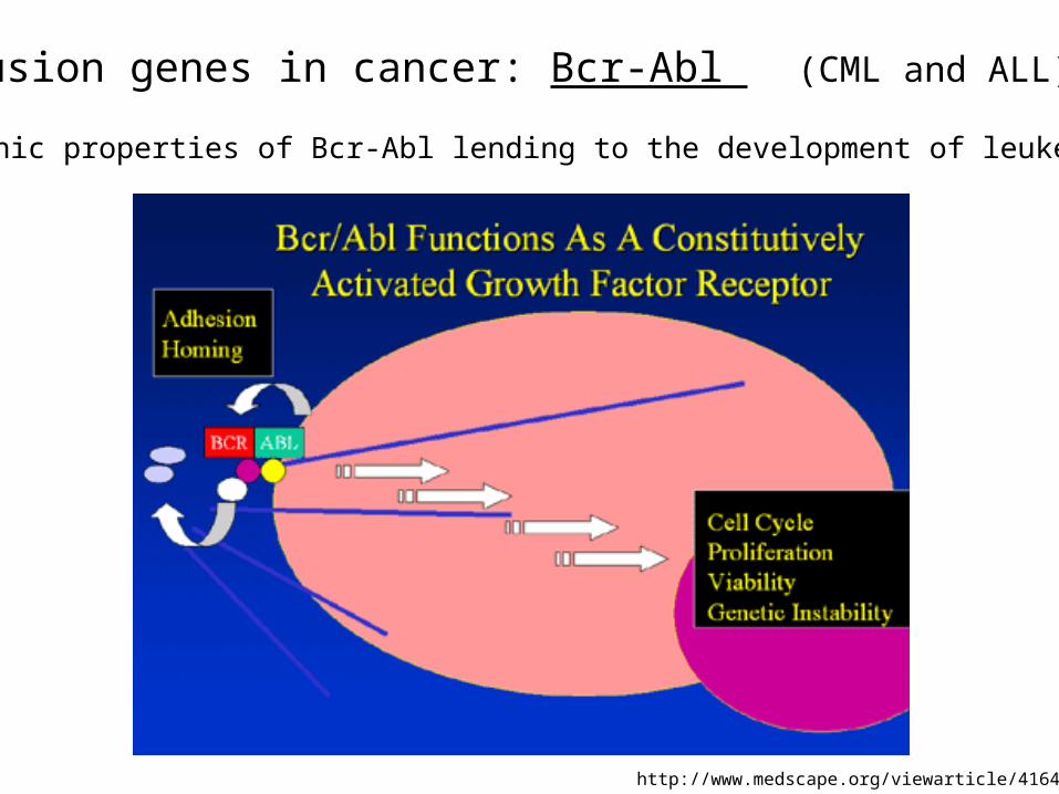
Fusion genes in cancer: Bcr-Abl (CML and ALL)
http://www.medscape.org/viewarticle/416483_2
Oncogenic properties of Bcr-Abl lending to the development of leukemia
Fusion genes in cancer: Bcr-Abl (CML and ALL)
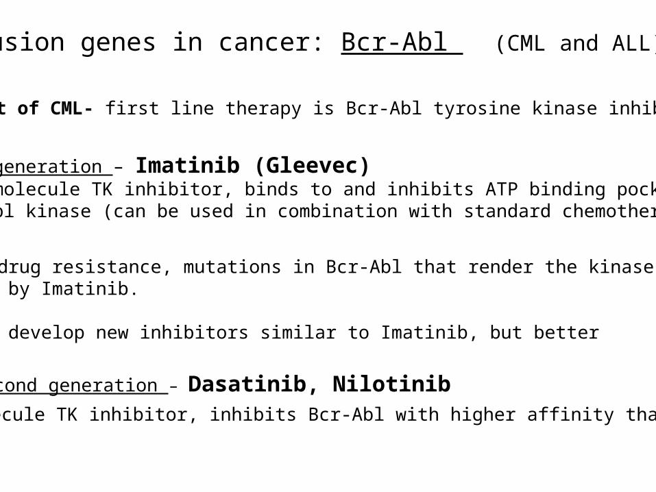
Treatment of CML- first line therapy is Bcr-Abl tyrosine kinase inhibitors
First generation – Imatinib (Gleevec)Small molecule TK inhibitor, binds to and inhibits ATP binding pocket of Bcr-Abl kinase (can be used in combination with standard chemotherapy)
Problem: drug resistance, mutations in Bcr-Abl that render the kinase no longerInhibited by Imatinib.
Solution: develop new inhibitors similar to Imatinib, but better
Second generation – Dasatinib, NilotinibSmall molecule TK inhibitor, inhibits Bcr-Abl with higher affinity than Imatinib

Fusion genes in cancer: PML-RARα (AML)t(15;17)
http://flipper.diff.org/app/items/info/482
AML (Adults) 12.5% (Children) 15%Prevalence of PML-RAR α
Bhatia et al. (2012) Mediterr J Hematol Infect Dis
Fusion genes in cancer: PML-RARα (AML)
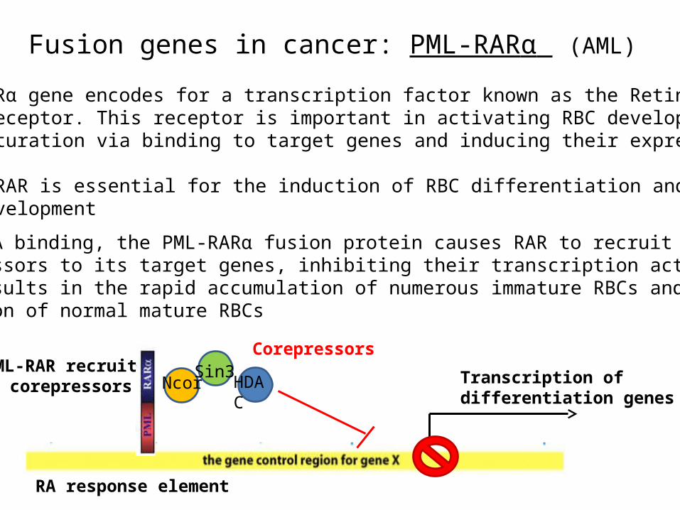
• Upon DNA binding, the PML-RARα fusion protein causes RAR to recruit corepressors to its target genes, inhibiting their transcription activation. This results in the rapid accumulation of numerous immature RBCs and the depletion of normal mature RBCs
• The RARα gene encodes for a transcription factor known as the Retinoic Acid Receptor. This receptor is important in activating RBC development and maturation via binding to target genes and inducing their expression.
Thus, RAR is essential for the induction of RBC differentiation and proper RBC development
PML-RAR recruits corepressors Transcription of
differentiation genes
RA response element
Corepressors
Ncor HDACSin3

Fusion genes in cancer: PML-RARα (AML)
“The presence of a PML-RARA fusion predicts a favorable response to differentiation therapy with all-trans retinoic acid (ATRA) and is currently the most curable subtype of acute myeloid leukemia (AML).”[1-5]
http://www.cancergeneticsitalia.com/dna-fish-probe/pmlrara/
Treatment of AML (PML-RARα)
1.) Differentiation inducing agents (e.g. all-trans retinoic acid (ATRA))
2.) Chemotherapy agents (e.g. cytarabine and anthracycline)
• Induce the immature blast cells into terminal differentiation and replenish the mature red blood cell population in patients
• Kill off the remaining immature AML blasts from patients blood stream

Fusion genes in cancer: TMPRSS2-ERG (prostate cancer)
• The TMPRSS2-ERG fusion gene is present in approximately 50% of prostate cancer patients
Tomlins et al. (2009) European Urology
Chromosome 21

Fusion genes in cancer: TMPRSS2-ERG (prostate cancer)
TMPRSS2 ERG TMPRSS2 TMPRSS2 ERGERG
ERG
ERG Target Gene ERG Target Gene
ERG
ERG Target Gene
ERG
= Androgen= Androgen Receptor
PTEN loss and/or AKT activation
Invasive carcinoma
St. John et al. (2012) - J Cancer Sci Ther - In Press
• The TMPRSS2-ERG gene fusion results in AR induced overexpression of the transcription factor ERG in prostate tumor cells
• The prognostic value of TMPRSS2-ERG fusion genes in prostate cancer remains controversial. There are currently no drugs targeting this fusion gene used in the clinic to treat prostate cancer. It was discovered at U of M in 2005

Summary and Conclusions
ALL
Better detection, new drugs, and better use of classical drugs
Some encouraging proof to not give up on the cure for cancer

Summary and Conclusions
• Selective therapies that target gene fusions in cancer have been successful in some cases and have increased overall survival rates for many patients who harbor these fusion genes, especially in leukemia
• The search for new selective therapies will most likely continue to prove beneficial in many cases, especially when treatment is combined with classical chemotherapy drugs (e.g. Methotrexate)
• New and more sensitive diagnostic techniques will be invaluable to future detection methods. This will hopefully yield more information in the detection of fusion genes in patients samples, especially in solid tumors

Questions ?
“Knowledge is power and knowing is half the battle!” (Mr. Joe, G.I.)