Embed Size (px)
Citation preview
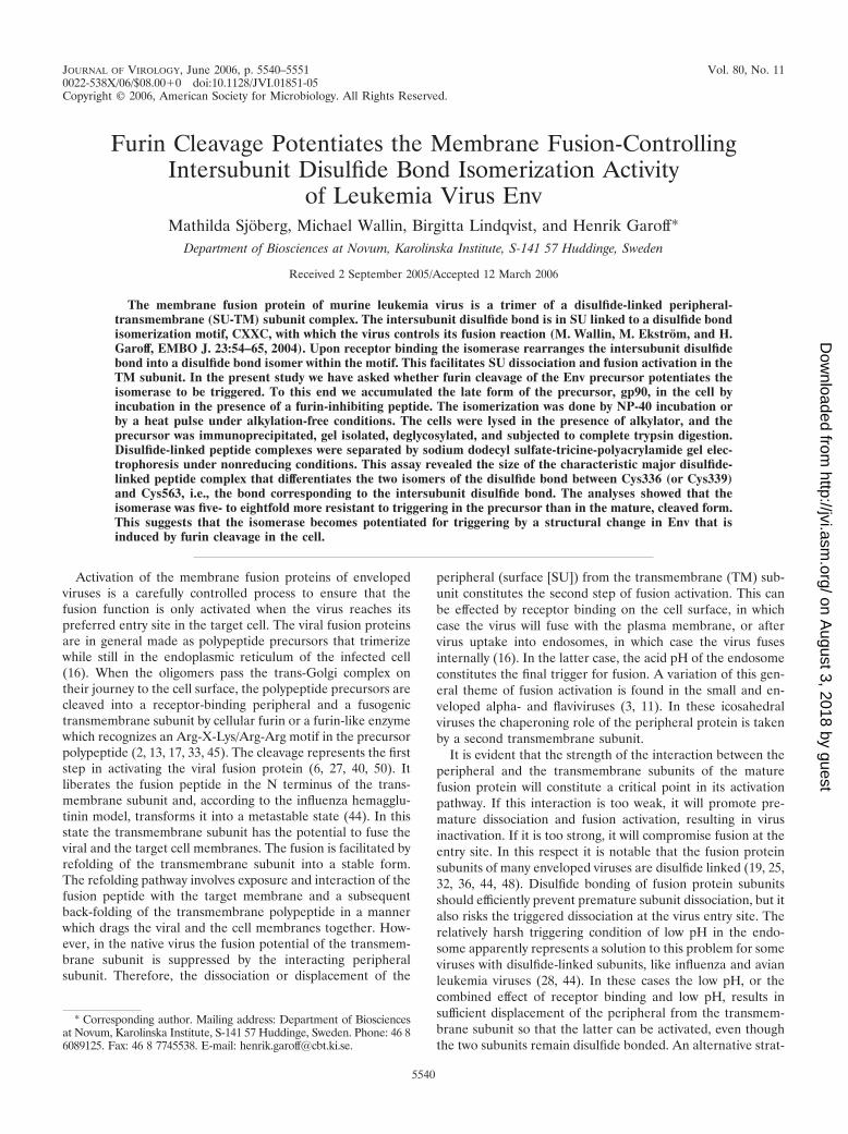
JOURNAL OF VIROLOGY, June 2006, p. 5540–5551 Vol. 80, No. 110022-538X/06/$08.00�0 doi:10.1128/JVI.01851-05Copyright © 2006, American Society for Microbiology. All Rights Reserved.
Furin Cleavage Potentiates the Membrane Fusion-ControllingIntersubunit Disulfide Bond Isomerization Activity
of Leukemia Virus EnvMathilda Sjoberg, Michael Wallin, Birgitta Lindqvist, and Henrik Garoff*
Department of Biosciences at Novum, Karolinska Institute, S-141 57 Huddinge, Sweden
Received 2 September 2005/Accepted 12 March 2006
The membrane fusion protein of murine leukemia virus is a trimer of a disulfide-linked peripheral-transmembrane (SU-TM) subunit complex. The intersubunit disulfide bond is in SU linked to a disulfide bondisomerization motif, CXXC, with which the virus controls its fusion reaction (M. Wallin, M. Ekstrom, and H.Garoff, EMBO J. 23:54–65, 2004). Upon receptor binding the isomerase rearranges the intersubunit disulfidebond into a disulfide bond isomer within the motif. This facilitates SU dissociation and fusion activation in theTM subunit. In the present study we have asked whether furin cleavage of the Env precursor potentiates theisomerase to be triggered. To this end we accumulated the late form of the precursor, gp90, in the cell byincubation in the presence of a furin-inhibiting peptide. The isomerization was done by NP-40 incubation orby a heat pulse under alkylation-free conditions. The cells were lysed in the presence of alkylator, and theprecursor was immunoprecipitated, gel isolated, deglycosylated, and subjected to complete trypsin digestion.Disulfide-linked peptide complexes were separated by sodium dodecyl sulfate-tricine-polyacrylamide gel elec-trophoresis under nonreducing conditions. This assay revealed the size of the characteristic major disulfide-linked peptide complex that differentiates the two isomers of the disulfide bond between Cys336 (or Cys339)and Cys563, i.e., the bond corresponding to the intersubunit disulfide bond. The analyses showed that theisomerase was five- to eightfold more resistant to triggering in the precursor than in the mature, cleaved form.This suggests that the isomerase becomes potentiated for triggering by a structural change in Env that isinduced by furin cleavage in the cell.
Activation of the membrane fusion proteins of envelopedviruses is a carefully controlled process to ensure that thefusion function is only activated when the virus reaches itspreferred entry site in the target cell. The viral fusion proteinsare in general made as polypeptide precursors that trimerizewhile still in the endoplasmic reticulum of the infected cell(16). When the oligomers pass the trans-Golgi complex ontheir journey to the cell surface, the polypeptide precursors arecleaved into a receptor-binding peripheral and a fusogenictransmembrane subunit by cellular furin or a furin-like enzymewhich recognizes an Arg-X-Lys/Arg-Arg motif in the precursorpolypeptide (2, 13, 17, 33, 45). The cleavage represents the firststep in activating the viral fusion protein (6, 27, 40, 50). Itliberates the fusion peptide in the N terminus of the trans-membrane subunit and, according to the influenza hemagglu-tinin model, transforms it into a metastable state (44). In thisstate the transmembrane subunit has the potential to fuse theviral and the target cell membranes. The fusion is facilitated byrefolding of the transmembrane subunit into a stable form.The refolding pathway involves exposure and interaction of thefusion peptide with the target membrane and a subsequentback-folding of the transmembrane polypeptide in a mannerwhich drags the viral and the cell membranes together. How-ever, in the native virus the fusion potential of the transmem-brane subunit is suppressed by the interacting peripheralsubunit. Therefore, the dissociation or displacement of the
peripheral (surface [SU]) from the transmembrane (TM) sub-unit constitutes the second step of fusion activation. This canbe effected by receptor binding on the cell surface, in whichcase the virus will fuse with the plasma membrane, or aftervirus uptake into endosomes, in which case the virus fusesinternally (16). In the latter case, the acid pH of the endosomeconstitutes the final trigger for fusion. A variation of this gen-eral theme of fusion activation is found in the small and en-veloped alpha- and flaviviruses (3, 11). In these icosahedralviruses the chaperoning role of the peripheral protein is takenby a second transmembrane subunit.
It is evident that the strength of the interaction between theperipheral and the transmembrane subunits of the maturefusion protein will constitute a critical point in its activationpathway. If this interaction is too weak, it will promote pre-mature dissociation and fusion activation, resulting in virusinactivation. If it is too strong, it will compromise fusion at theentry site. In this respect it is notable that the fusion proteinsubunits of many enveloped viruses are disulfide linked (19, 25,32, 36, 44, 48). Disulfide bonding of fusion protein subunitsshould efficiently prevent premature subunit dissociation, but italso risks the triggered dissociation at the virus entry site. Therelatively harsh triggering condition of low pH in the endo-some apparently represents a solution to this problem for someviruses with disulfide-linked subunits, like influenza and avianleukemia viruses (28, 44). In these cases the low pH, or thecombined effect of receptor binding and low pH, results insufficient displacement of the peripheral from the transmem-brane subunit so that the latter can be activated, even thoughthe two subunits remain disulfide bonded. An alternative strat-
* Corresponding author. Mailing address: Department of Biosciencesat Novum, Karolinska Institute, S-141 57 Huddinge, Sweden. Phone: 46 86089125. Fax: 46 8 7745538. E-mail: [email protected].
5540
on August 3, 2018 by guest
http://jvi.asm.org/
Dow
nloaded from
egy, which is apparently used by the majority of the leukemiaviruses, is to link the intersubunit disulfide bond to an isomer-ization motif, Cys-X-X-Cys (CXXC) in the peripheral subunit(48). This contains a free thiol, with which it can rearrange theintersubunit disulfide bond into an alternative disulfide isomerwithin the motif. As the isomerization of the CXXC motif istriggered by receptor binding, the virus gets rid of the inter-subunit disulfide bond at the surface of the target cell. Thisfacilitates dissociation of the peripheral subunit and fusionactivation.
The fusion activation pathway that is controlled by intersub-unit disulfide bond isomerization has been studied in somedetail using Moloney murine leukemia virus (Mo-MLV) (48).It was shown that receptor binding induced the exposure of theCXXC thiol in the peripheral subunit for modification by al-kylation. This blocked the isomerization reaction in the recep-tor-bound fraction of the viral fusion proteins (envelope [Env])and arrested fusion activation. However, the fusion could berescued by subsequent reduction of the intersubunit disulfidebond with dithiothreitol. Thus, this proved the role of theintersubunit disulfide bond isomerization in controlling thefusion activation process in Mo-MLV. It was possible to triggerthe isomerization activity not only by receptor binding but alsoby treatments of the virus with heat or urea, depleting the virusof Ca2�, and by solubilization of the viral membrane withNP-40 (46, 47). This suggested that the receptor-inducedisomerization of the intersubunit disulfide bond was mediatedby destabilization of a Ca2�-stabilized structure of the fusionprotein.
An interesting question concerns the control of the activityof the CXXC-linked disulfide bond isomerase. As the Envprecursor and Env receptor, a basic amino acid transporter,can possibly interact in the biosynthetic transport pathway,this could result in premature isomerization of the disulfidebond between Cys336 (or Cys339) and Cys563 [hereafterCys336(339)-Cys563], which corresponds to the intersubunitlinkage of the mature SU-TM complex and subsequent releaseof the fusion function directly upon furin cleavage (49). There-fore, it is possible that the isomerase activity remains sup-pressed in Env until this is cleaved by furin. Alternatively, theEnv could be synthesized with the internal CXXC isomer(Cys336-Cys339) of the disulfide bond and rearrange into theSU-TM isomer [Cys336(339)-Cys563] first upon furin cleavage.In the present study we have addressed these questions usingMo-MLV.
Earlier studies have shown that the Mo-MLV Env is made asa gp80 precursor with seven Asn (N)-linked sugar units in theSU portion of the polypeptide (8, 20, 29, 42, 43). The trans-membrane and the cytoplasmic domain of gp80 are modifiedby palmitoylation (15, 31, 52). Upon transport through theGolgi and trans-Golgi complexes, the endoglycosidase H (endoH)-sensitive high-mannose forms of the N-linked sugar units ofgp80 are processed into endo H-resistant complex units. Inaddition, the Env polypeptide is glycosylated at the hydroxylsof several Ser and Thr residues (O-glycosylation) (10, 35). Thisform of Env is recognized as gp90. In pulse-chase experiments,it is seen only transiently in very small amounts because it israpidly cleaved into the disulfide-linked SU-TM complex ofmature Env (9, 29). During maturation of virus particles bybudding at the cell surface, or shortly thereafter, the viral
protease releases a piece known as the R peptide from the endof the cytoplasmic (internal) domain or tail of TM (12, 41).
In this study we have used a disulfide-linked tryptic peptideassay to analyze when after synthesis the Cys336(339)-Cys563disulfide bond is formed in the Env precursor and to test itssensitivity to undergo CXXC-mediated isomerization. Foranalyses of the late form of the precursor, gp90, this wasenriched in cells by inhibiting its cleavage with a furin-inhibit-ing peptide (FIP). We found that the Cys336(339)-Cys563 di-sulfide bond was generated posttranslationally in gp80 and thatit was significantly more resistant to isomerization inductionboth in the early gp80 and in the late gp90 forms of theprecursor than in the mature, cleaved Env. These results sug-gest that the disulfide bond isomerase of the Mo-MLV Envprecursor is potentiated for receptor-induced activation by afurin cleavage-mediated conformational change.
MATERIALS AND METHODS
Cells and reagents. MOV-3 cells, a gift from G. Schmidt (GSF-NationalResearch Center for Environment and Health, Neuherberg, Germany) wasmaintained in Dulbecco’s modified Eagle’s medium (DMEM) containing 4.5g/liter glucose (Gibco BRL) supplemented with 10% fetal calf serum, 20 mMHEPES, and L-glutamine. HE 699, a polyclonal goat antiserum against thegp69/71 Env glycoprotein (SU) of Rauscher MLV (catalog no. VR-1519AS-Gt)was from ATCC (LGC Promochem, Borås, Sweden). Peptide N-glycosidase F(PNGase F; catalog no. 1365193) and O-glycosidase (catalog no. 11347101001)were from Roche Biochemicals (Basel, Switzerland); endo Hf (catalog no.P0703S) was from New England Biolabs (Ipswich, MA), and neuraminidase(catalog no. 480717) was from Calbiochem/Merck (Darmstadt, Germany). 14C-methylated standard proteins (catalog no. CFA 626 and CFA755) were fromAmersham Biosciences (Uppsala, Sweden).
Metabolic labeling, furin inhibition, isomerization induction, and cell lysis.Metabolic labeling of proteins in MOV-3 cells was done with [35S]Cys (Amer-sham Biosciences, Uppsala, Sweden) as described previously (32). Briefly, cells in3.5- or 6-cm tissue culture dishes were washed twice in phosphate-buffered saline(PBS), incubated in cysteine-free DMEM for 30 min (starvation), labeled for 5or 15 min (50 �Ci of [35S]Cys in 0.5 ml/3.5-cm dish) and chased for up to 2 h inDMEM containing 2 mM Cys. The cells were washed twice in PBS and solubi-lized in 1% NP-40 in lysis buffer (50 mM Tris-HCl, pH 7.5, 150 mM NaCl, 2 mMEDTA; 300 �l/3.5-cm dish) without or with 5 mM N-ethyl maleimide (NEM; inboth PBS and lysis buffer) to allow or prevent isomerization of disulfide bonds,and incubated for 2 h on ice. Finally, all samples were adjusted to 5 mM NEMand incubated for 10 min at 30°C. To inhibit furin convertase, the cells wereincubated for 1.5 h in 5 to 80 �M FIP (decanoyl-Arg-Val-Lys-Arg-chlorometh-ylketone) (catalog no. 260-022-M001; Alexis Biochemicals/Kelab Goteborg, Swe-den) prior to labeling, first in DMEM-fetal calf serum (1 h) and then during theCys starvation (0.5 h). When isomerization was induced by heat instead of NP-40,we incubated the pulse-labeled (15 min) and chased (1 h) cultures at 53°C for 4to 6 min in 50 mM Tris-HCl (pH 7.45 at 37°C), 150 mM NaCl, and 1.8 mMCaCl2. In control experiments analyzing the mature Env, parallel cultures weretreated at 53°C in the presence and absence of 20 mM NEM. To compensate fordecomposition of NEM, 10 mM fresh NEM was added every minute during theheat pulse. Lysis of the heat-treated cultures was done in the presence of NEM.In these experiments the FIP treatments were extended to include also the timeof chase.
Immunoprecipitation and deglycosylation. Mature and precursor Env proteinswere immunoprecipitated by HE699 essentially as previously described (32).Precipitations, typically 150 �l of labeled cell extract (i.e., 50% of a 3.5-cm dish),6 �l of HE699 anti-SU polyclonal antibody (pAb), and 30 �l of protein A-Sepharose slurry (1:1, vol/vol), all adjusted to 5 mM NEM, were performedovernight at 4°C. The washed immune complexes were either processed directlyfor sodium dodecyl sulfate-polyacrylamide gel electrophoresis (SDS-PAGE) oreluted for further manipulations. To elute proteins, 1 bead volume (15 �l) of 1%SDS in lysis buffer was added, and the samples were incubated for 3 min at 70°C.SDS was inactivated by the addition of 5 bead volumes (PNGase F and endo Hf)or 10 bead volumes (O-glycosidase and neuraminidase) of 1.25% NP-40 in lysisbuffer. To digest N-linked sugars, aliquots (24 �l) were supplemented with 0.5 �lof PNGase F or 4 �l of 0.5 M Na citrate, pH 5.5, and 0.5 �l of endo Hf. O-linked
VOL. 80, 2006 FUSION ACTIVATION IN RETROVIRUS 5541
on August 3, 2018 by guest
http://jvi.asm.org/
Dow
nloaded from
sugars were assessed by the addition of 1 �l of O-glycosidase, 1 �l of neur-aminidase, and 0.5 �l of PNGase F. All deglycosylations were incubated for16 h at 37°C.
Electrophoresis. Samples were adjusted to 31 g/liter SDS, 0.19 M Tris-HCl,pH 8.0, 93 g/liter sucrose, 14 mM EDTA, 0.6 mg/ml bromophenol blue, and 0.4mg/ml methionine (1� sample buffer). For reduction of disulfide bonds, 37 mMdithiothreitol was included. The samples were heated for 3 min at 70°C (6 minwhen the samples were �150 �l) and cooled to room temperature, and a finalconcentration of 9 mM iodoacetamide (or 10 mM NEM for preparative pur-poses) was added to reduced and nonreduced samples. SDS-PAGE gels (23), 8or 10.5 cm long, with 7%, 8%, or 12% total acrylamide, of which 2.6% wasbisacrylamide, and 10.5-cm SDS-tricine-PAGE gels (39) with 16.5% total acryl-amide, of which 2.6% was bisacrylamide, were run in a Mighty Small II minigelsystem (Hoefer Scientific, San Francisco, CA).
Gel purification. Metabolically labeled, immunoisolated Env proteins from720 �l of cell extract prepared under isomerizing or nonisomerizing conditionswere separated on 7% SDS-PAGE under nonreducing conditions. The bands ofinterest were detected by phosphorimaging of the wet gels (typically, a 2-hexposure) and cut out. Gel pieces from 8 to 10 equivalent lanes were crushed in2 ml of 0.05% SDS in 10 mM Tris-HCl, pH 7.5, extracted at room temperatureovernight, and removed by 0.22-�m-pore-size cellulose acetate filters (Schlei-cher-Shuell, Dassel, Germany). The extracted proteins were concentrated inMicrocon YM-30 centrifugal filter devices (Amicon/Millipore, Billerica, MA) toabout 150 �l, PNGase F was added (5 �l), and the samples were incubated at37°C overnight. The samples were adjusted to 1� sample buffer and rerun on 8%SDS-PAGE under nonreducing conditions, and the bands of interest were cutout, extracted, and concentrated to about 15 to 20 �l as above.
Tryptic digestion. Gel-purified and concentrated proteins were incubated for10 min at 56°C, cooled to room temperature, and treated with 0.15 mg/ml TPCK(N-tosyl-L-phenylalanine chloromethyl ketone)-trypsin (catalog no. 37257;Serva, Heidelberg, Germany); 2 mM CaCl2 was added, and the samples wereincubated at 37°C for 16 h.
Quantifications. The amount of radioactivity in a protein band was measuredusing phosphorimage screens (BAS-MS2025) from Fujifilm (Science ImagingScandinavia, Nacka, Sweden), a Molecular Imager FX, and the QuantityOneprogram from Bio-Rad Laboratories (Hercules, CA). To estimate the extent ofSU-TM isomerization, the amount of SU released from the SU-TM complexesduring incubation was measured. This was expressed as a percentage of total SU(i.e., the sum of free and TM-bound SU). The prevalence of the Cys336(339)-Cys563 disulfide bond in Env gp80 and gp90 precursors was determined bymeasuring the major disulfide-linked tryptic peptide complexes in digests ofprecursors that were incubated under nonisomerizing conditions. The amountsof 12-kDa complexes [molecules containing the Cys336(339)-Cys563 disulfidebond] and 9.1-kDa complexes (molecules with the internal Cys336-C339 disulfidebond) were quantified from dried SDS-tricine gels. The relative amount (R) of12-kDa complexes (R12kDa
�NEM) was calculated as a percentage of the sum of the12-kDa and 9.1-kDa complexes in the sample for each protein precursor underboth nonisomerizing (with NEM) and isomerizing (without NEM) conditions.This value was used to express the prevalence of the Cys336(339)-Cys563 disul-fide bond. The relative stability (RS) of the Cys336(339)-Cys563 disulfide bondwas calculated as relative amount of 12-kDa complex under isomerizing condi-tions as a percentage of that under nonisomerizing conditions: RS � (R12kDa
�NEM/R12kDa
�NEM) � 100. In mature Env (SU-TM) the intersubunit Cys336(339)-Cys563disulfide bond was analyzed in 12% SDS-PAGE under nonreducing conditions.The amount of SU-TM complexes (containing the intersubunit disulfide bond)and free SU subunits (without the intersubunit disulfide bond) was quantified,and the relative amount of intact complexes (RSU-TM
�NEM) present under nonisomer-izing (with NEM) and isomerizing (without NEM) conditions was calculated asa percentage of the sum of SU-TM and free SU. The relative stability of theintersubunit disulfide bond was calculated as the relative amount of SU-TMunder isomerizing conditions as a percentage of that under nonisomerizingconditions: RSSU-TM � (RSU-TM
�NEM/RSU-TM�NEM) � 100.
RESULTS
Furin inhibition results in the accumulation of gp90, a lateform of the Env precursor. We used an FIP to prevent cleavageof the Env precursor into the SU-TM complex in MOV-3 cells.To find optimal conditions for the inhibition, cell cultures wereincubated with different concentrations of FIP for 1 h, starvedfor Cys for 30 min in the presence of FIP, and then pulse
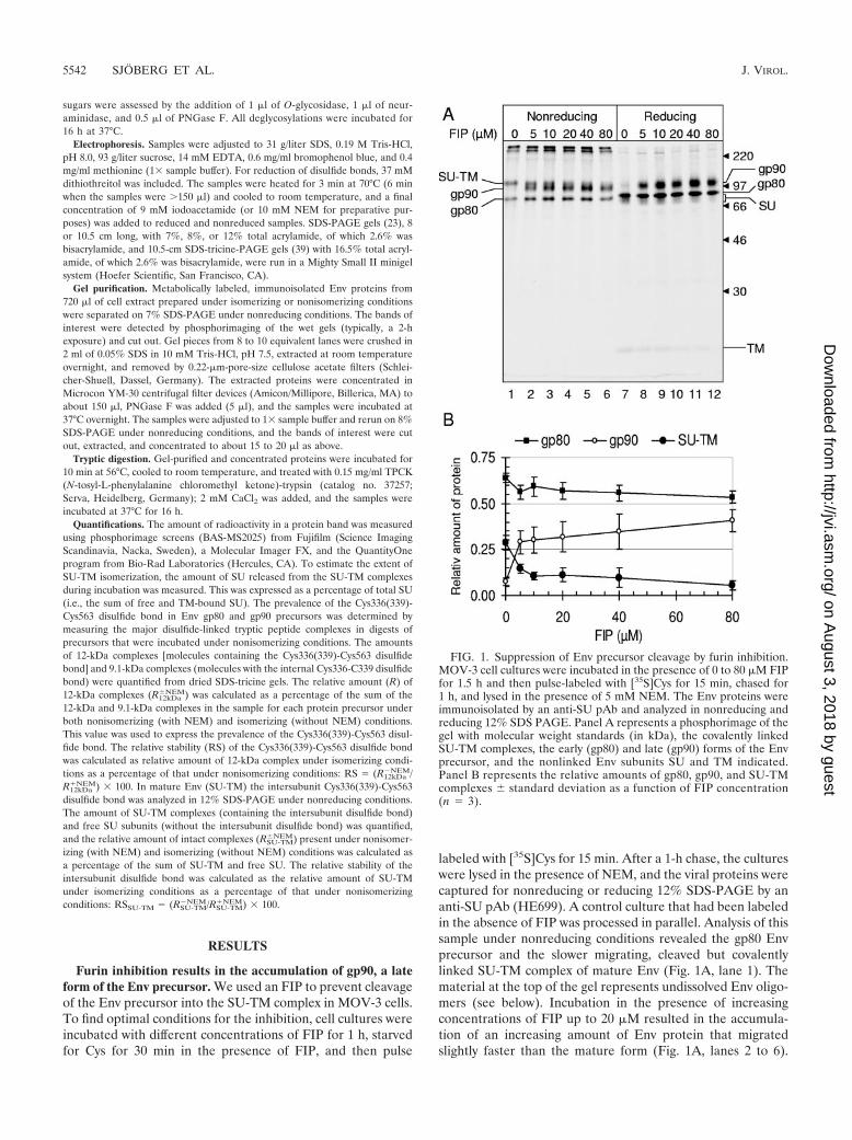
labeled with [35S]Cys for 15 min. After a 1-h chase, the cultureswere lysed in the presence of NEM, and the viral proteins werecaptured for nonreducing or reducing 12% SDS-PAGE by ananti-SU pAb (HE699). A control culture that had been labeledin the absence of FIP was processed in parallel. Analysis of thissample under nonreducing conditions revealed the gp80 Envprecursor and the slower migrating, cleaved but covalentlylinked SU-TM complex of mature Env (Fig. 1A, lane 1). Thematerial at the top of the gel represents undissolved Env oligo-mers (see below). Incubation in the presence of increasingconcentrations of FIP up to 20 �M resulted in the accumula-tion of an increasing amount of Env protein that migratedslightly faster than the mature form (Fig. 1A, lanes 2 to 6).
FIG. 1. Suppression of Env precursor cleavage by furin inhibition.MOV-3 cell cultures were incubated in the presence of 0 to 80 �M FIPfor 1.5 h and then pulse-labeled with [35S]Cys for 15 min, chased for1 h, and lysed in the presence of 5 mM NEM. The Env proteins wereimmunoisolated by an anti-SU pAb and analyzed in nonreducing andreducing 12% SDS PAGE. Panel A represents a phosphorimage of thegel with molecular weight standards (in kDa), the covalently linkedSU-TM complexes, the early (gp80) and late (gp90) forms of the Envprecursor, and the nonlinked Env subunits SU and TM indicated.Panel B represents the relative amounts of gp80, gp90, and SU-TMcomplexes � standard deviation as a function of FIP concentration(n � 3).
5542 SJOBERG ET AL. J. VIROL.
on August 3, 2018 by guest
http://jvi.asm.org/
Dow
nloaded from
Analyses by reducing SDS-PAGE showed that, whereas themature Env in the control and in the FIP-treated sample wasreleased into free SU and TM subunits, the FIP-induced fastermigrating form of Env remained as one intact chain (Fig. 1A,lanes 7 to 12). Quantification demonstrated that the FIP-in-duced protein increased with the FIP concentration at theexpense of the mature, but not gp80, Env (Fig. 1B). We con-cluded that FIP inhibited the cleavage of the Env precursor tothe SU-TM complex. The FIP-induced form of Env corre-sponded most likely to the gp90 form of gp80, which has earlierbeen observed in small amounts in pulse-chase experiments(29). With the FIP, it was possible to inhibit almost all of theEnv that was normally processed by furin. However, incuba-tions at FIP concentrations above 40 �M decreased the yield ofall forms of Env, probably as a result of the toxic effects of thedrug. Note that the high-molecular-weight material seen undernonreducing conditions was resolved into monomeric forms ofthe various Env proteins under reducing conditions.
The faster migration of gp90 compared to the SU-TM com-plex in nonreducing SDS-PAGE has not been reported before.It could be explained by a more extended conformation of theSU-TM complexes than the precursor after SDS denaturation.Alternatively, the migration difference could be due to differ-ences in sugar modifications. Though it is known that the SUsubunit of the SU-TM complex and gp90 contain both complexN-linked sugar units and also O-linked units, it is unclearwhether the modifications occur to the same extent (10, 35). Adetailed knowledge of the glycosylation status of the SU-TMcomplex and the precursor was regarded as essential for cor-rect interpretation of the mechanisms of a possible late acti-vation of the disulfide isomerase function of Env. Therefore,we compared the gel migration of gp90 and the SU-TM com-plex after enzymatic deglycosylations.
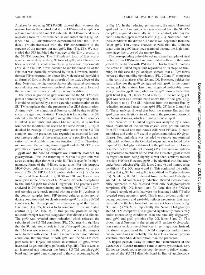
gp90 and the SU-TM complex are similarly modified byglycosylation. First, the trimming of N-linked sugar units wasassessed using digestion with endo-H. This is specific for high-mannose forms of the N-linked sugar units. For this purposeMOV-3-cell cultures were incubated in the presence or ab-sence of 20 �M FIP for 1.5 h, pulse-labeled with [35S]Cys for15 min, and then chased for 5, 60, 90, or 120 min. The cultureswere lysed in the presence of NEM and Env proteins capturedby the anti-SU pAb for endo H digestion. The products wereanalyzed in 7% nonreducing and reducing SDS-PAGE. Con-trol samples were mock treated without endo H. Analyses ofthe control samples from FIP-treated cultures under nonre-ducing conditions did not clearly resolve gp90 from the SU-TMcomplexes, but this appeared as a broadening of the matureEnv band (Fig. 2A, lanes 4 to 6) compared to the untreatedcontrol (Fig. 2A, lane 2). In this gel the proteins of highmolecular weight resolved as apparent Env dimers and trimers.The gp90 was revealed after reduction, which released thesubunits of the SU-TM complexes (Fig. 2D, lanes 4 to 6). Notethat the SU migrated closely in front of the gp80 band and thatthe TM was not resolved by the 7% gel. When the sampleswere treated with endo H and analyzed under nonreducingconditions, the migration of both gp90 and the SU-TM com-plex were left largely unaffected in contrast to gp80, whichincreased its gel mobility significantly (Fig. 2B). This is seen asan increased gap between the broad SU-TM complex/gp90band and the gp80 band compared to the corresponding bands
in Fig. 2A. In the reducing gel analyses, the endo H-treatedgp90 and the SU subunit, which was released from the SU-TMcomplex, migrated essentially as in the control, whereas theendo H-treated gp80 moved faster (Fig. 2E). Note that underthese conditions the diffuse SU band is well separated from thefaster gp80. Thus, these analyses showed that the N-linkedsugar units in gp90 have been trimmed beyond the high-man-nose stage like those of the mature Env.
The corresponding pulse-labeled and chased samples of viralproteins from FIP-treated and nontreated cells were then sub-jected to incubation with PNGase F. This treatment removesthe entire N-linked sugar unit regardless of its stage of trim-ming. In this case the gel analyses showed that all Env formsincreased their mobility significantly (Fig. 2C and F) comparedto the control analyses (Fig. 2A and D). However, neither themature Env nor the gp90 comigrated with gp80. In the nonre-ducing gel, the mature Env band migrated noticeably moreslowly than the gp80 band, whereas the gp90 closely trailed thegp80 band (Fig. 2C, lanes 2 and 4 to 6). In the reducing gel,gp90 was seen as a distinct band above that of the gp80 (Fig.2F, lanes 4 to 6). The SU, released from the mature Env byreduction, migrated faster than gp80 (Fig. 2F, lanes 2 and 4 to6). These analyses showed that both the mature Env and thegp90 carry modifications, in addition to the processed forms ofthe N-linked sugars, which are not present in gp80.
The presence of O-linked sugars was analyzed by a com-bined treatment of the 60-min chased virus protein samplefrom FIP-treated and nontreated cells with PNGase F, neur-aminidase and endo-�-N-acetyl-D-galactosaminidase (O-glyco-sidase). Neuraminidase was included to remove any terminalsialic acid residues in the O-linked sugars and was found to berequired for O-deglycosylation of both gp90 and mature Env asdescribed before (data not shown) (35). The neuraminidase–O-glycosidase treatment of the PNGase F-treated gp90 shiftedits migration from being slightly slower than similarly treatedor solely PNGase F-treated gp80 to be identical with the latterunder both reducing (Fig. 2G, lanes 2 and 4) and nonreducingconditions (Fig. 2G, lanes 6 and 8). This confirmed the earlierfinding that gp90, but not gp80, is modified by O-glycosylation(35). Similarly, the SU, released from the N- and O-deglyco-sylated SU-TM complexes by reduction, showed increased mo-bility compared to SU released from only N-deglycosylatedcomplexes (Fig. 2G, lanes 1 and 3). Note that the PNGaseF-treated sample of cells that were not incubated with FIP alsorevealed some apparent gp90. This is most evident under re-ducing conditions and probably reflects precursors that havematured into the late form but have not yet been cleaved (Fig.2G, lane 1) (29). Most importantly, the N- and O-deglycosyl-ated SU-TM complexes still migrated significantly more slowlyunder nonreducing conditions than the similarly deglycosyl-ated gp90 and gp80 proteins (Fig. 2G, lanes 5 and 7). Thisshows that differences in the extent of N- and/or O-glycosyla-tion cannot explain the differences in gel migration. Instead,the slower migration of the SU-TM complexes under nonre-ducing conditions is probably due to a less compact proteinstructure after SDS denaturation than the precursor.
A tryptic peptide assay to follow the isomerization of theCys336(339)-Cys563 disulfide bond in newly synthesized Env.Earlier we developed a tryptic peptide assay to follow isomer-ization of the SU-TM disulfide bond in Env of amphotropic
VOL. 80, 2006 FUSION ACTIVATION IN RETROVIRUS 5543
on August 3, 2018 by guest
http://jvi.asm.org/
Dow
nloaded from
and Friend MLV (48). This was based on the migration dif-ference in SDS-tricine-PAGE of the major disulfide-linkedtryptic peptide complex of Env that either had or had not beensubjected to in vitro induced isomerization of the SU-TMdisulfide bond. We used a similar assay to study the presenceof the corresponding disulfide bond, i.e., the Cys336(339)-Cys563 bond, in gp80 and gp90 of Mo-MLV. The disulfide
bond status of the mature Env of Mo-MLV has not beendetermined, but as the Mo-MLV Env has 84.2% amino acidsequence identity with Friend MLV, including all Cys residuesin the signal sequence-cleaved polypeptide chain, this is mostlikely identical with that of Friend MLV Env (34). In the latterprotein all Cys residues but the CXXC thiol and a palmitoy-lated Cys residue in the transmembrane domain of TM are
FIG. 2. Modification of Env proteins by glycosylation. MOV-3 cell cultures were treated without or with 20 �M FIP, pulse-labeled for 15 min,chased for 5 to 120 min (A to F) or 60 min (G) and lysed in the presence of NEM. Env proteins were immunoisolated and subjected to mock, endoH or PNGase F treatment (A to F) or treated with a combination of PNGase F, neuraminidase, and O-glycosidase (G) and subsequently analyzedby nonreducing (A to C and G, lanes 5 to 8) or reducing (D to F and G, lanes 1 to 4) 7% (A to F) or 8% (G) SDS-PAGE in 8-cm gels. The FIPtreatment, time of chase, and deglycosylation conditions are indicated in each panel. The molecular weight standards (arrowheads), the Envproteins (Env oligom), and in panel G the deglycosylation mode (deglyc) of the proteins are indicated.
5544 SJOBERG ET AL. J. VIROL.
on August 3, 2018 by guest
http://jvi.asm.org/
Dow
nloaded from
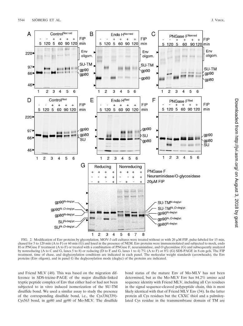
disulfide linked (15, 26, 36). After isomerization of the inter-subunit disulfide bond, the CXXC thiol forms the CXXC di-sulfide, whereas the TM Cys residue of the prior SU-TM di-sulfide exposes a free thiol (7, 48). Therefore, mature Env ofMo-MLV is predicted to yield a major deglycosylated disul-fide-linked complex of tryptic peptides of 12 kDa, which con-tains nine Cys residues (Fig. 3A and C). This includes a 4.4-kDa peptide from the SU and a 1.6-kDa peptide from the TM,
which participate in the formation of the intersubunit disulfidebond in Env, and the covalently associated 4.7-kDa SU and the0.86-kDa TM peptides. In contrast, the isomerized Env is pre-dicted to yield a major deglycosylated 9.1-kDa tryptic peptidecomplex with six Cys residues encompassing only the SU pep-tides of 4.4 and 4.7 kDa (Fig. 3B and C). In addition, both theisomerized and the nonisomerized Env sample should yieldone larger and several smaller peptide complexes. The larger,
FIG. 3. A tryptic peptide assay to follow isomerization of the Cys336(339)-Cys563 disulfide bond. Panel A shows the primary structure of thepredicted disulfide linked tryptic peptide complex, encompassing the intersubunit Cys336(339)-Cys563 disulfide bond, of the native SU-TMcomplex, and panel B shows the corresponding structures of isomerized Env. The calculated molecular weights of the complexes and includedpeptides are indicated. The N-terminal amino acid residues of the peptides are numbered. Panel C lists all predicted Cys-containing trypticpeptides and peptide complexes of native SU-TM and isomerization-released SU under nonreducing conditions. Non-iso and iso denote peptidecomplexes that are specific for the nonisomerized and isomerized states, respectively. (D) Nonreducing 12% SDS-PAGE of gel-purified,N-deglycosylated and [35S]Cys-labeled SU-TM complexes and SU subunits from cell lysates subjected to nonisomerizing and isomerizingconditions, respectively. (E) Analyses of disulfide-linked tryptic peptide complexes from the SU-TM complexes and SU subunits above. Thegel-purified, N-deglycosylated proteins were subjected to complete trypsin digestion, and the peptide products were analyzed by SDS-tricine-PAGE.
VOL. 80, 2006 FUSION ACTIVATION IN RETROVIRUS 5545
on August 3, 2018 by guest
http://jvi.asm.org/
Dow
nloaded from
an 8.9-kDa peptide complex with six Cys residues, is derivedfrom the tightly disulfide cross-linked N-terminal receptorbinding domain. However, as was earlier shown for the am-photropic MLV receptor binding domain, this should form acompact structure that migrates much faster than expectedfrom its molecular weight in nonreducing SDS-tricine-PAGE(48). The predictions were tested using the mature Env ofMo-MLV. To this end, MOV-3 cell cultures were pulse-la-beled with [35S]Cys for 15 min, chased for 60 min, and thenlysed on ice for 120 min in the presence or absence of NEM toprevent or allow NP-40-induced isomerization of the SU-TMdisulfide bond. The Env proteins were immunoisolated andsubjected to nonreducing 8% SDS-PAGE. In this analysis theSU-TM complexes from the nonisomerized sample run slightlymore slowly than any remaining gp90, whereas the free SUsubunits from the isomerized sample run closely in front ofgp80 (data not shown). The SU-TM complexes and the SUsubunits, with contaminating gp90 and gp80, respectively, werecut out, extracted from the gel, and concentrated by ultrafil-tration. The samples were then subjected to PNGase F treat-ment and a second gel purification. In their N-deglycosylatedform, the SU-TM complexes separate from contaminatinggp90 (Fig. 2C) and SU from gp80 (data not shown). Theisolated N-deglycosylated SU-TM complexes and SU subunitswere found to be essentially pure, apart from a minor amountof SU in the former preparation, a result of a limited artificialreduction of SU-TM complexes during sample preparation(Fig. 3D, lanes 1 and 2) (48). The preparations were treatedwith trypsin, and the mixtures of disulfide-linked tryptic pep-tide complexes were analyzed by nonreducing SDS-tricine-PAGE. The tryptic digest of nonisomerized Env, i.e., theSU-TM complexes, revealed a major labeled band migratingbetween the reduced 6.5- and 14-kDa markers (Fig. 3E, lane1). In contrast, the tryptic peptide complexes derived fromisomerized Env, i.e., the SU subunits, showed a major bandwith faster migration, close to the reduced 6.5-kDa marker(Fig. 3E, lane 2). This suggested that the two major bandsindeed corresponded to the predicted major peptide com-plexes, thus reflecting the isomerization status of Env. In ad-dition both analyses showed the presence of a similar set offaster migrating bands. These probably corresponded to theadditional complexes of peptides that were generated by thedigestion. Thus, the results were very similar to the corre-sponding analyses of amphotropic and Friend MLV Env, bothof which demonstrated a similar migration shift of their majordisulfide-linked tryptic peptide complexes upon isomerization(48). We conclude that we have established an assay by whichwe should be able to follow the presence or absence of theCys336(339)-Cys563 disulfide bond in Mo-MLV Env proteinsthat have not been processed by furin cleavage.
The Cys336(339)-Cys563 disulfide bond isomerization issuppressed in gp80 and gp90. In the absence of alkylator,NP-40 solubilization acts as an efficient trigger of the freeCXXC thiol in SU to attack the intersubunit disulfide bond ofthe SU-TM complex and cause its rearrangement into theCXXC-containing disulfide bond isomer (32, 48). Therefore,we used NP-40 solubilization to study the activity of theisomerase in gp80 and gp90. First we determined the thresholdcondition to induce isomerization of mature Env with NP-40.To this end, MOV-3 cell cultures were pulsed for 15 min,
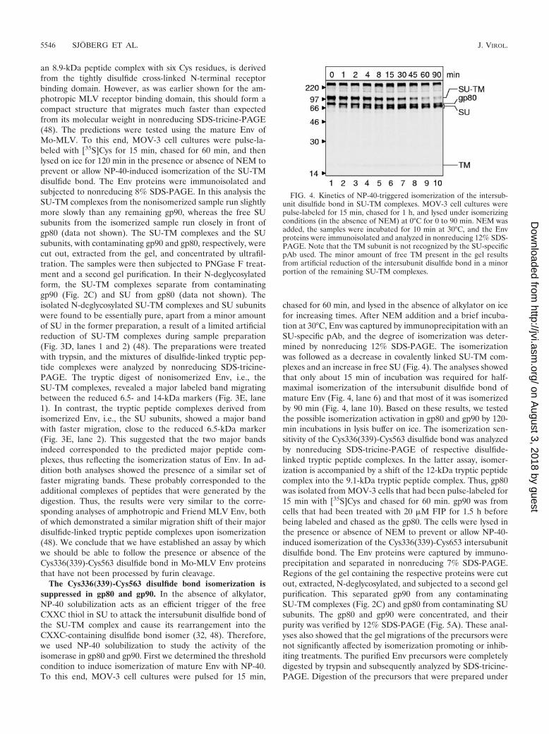
chased for 60 min, and lysed in the absence of alkylator on icefor increasing times. After NEM addition and a brief incuba-tion at 30°C, Env was captured by immunoprecipitation with anSU-specific pAb, and the degree of isomerization was deter-mined by nonreducing 12% SDS-PAGE. The isomerizationwas followed as a decrease in covalently linked SU-TM com-plexes and an increase in free SU (Fig. 4). The analyses showedthat only about 15 min of incubation was required for half-maximal isomerization of the intersubunit disulfide bond ofmature Env (Fig. 4, lane 6) and that most of it was isomerizedby 90 min (Fig. 4, lane 10). Based on these results, we testedthe possible isomerization activation in gp80 and gp90 by 120-min incubations in lysis buffer on ice. The isomerization sen-sitivity of the Cys336(339)-Cys563 disulfide bond was analyzedby nonreducing SDS-tricine-PAGE of respective disulfide-linked tryptic peptide complexes. In the latter assay, isomer-ization is accompanied by a shift of the 12-kDa tryptic peptidecomplex into the 9.1-kDa tryptic peptide complex. Thus, gp80was isolated from MOV-3 cells that had been pulse-labeled for15 min with [35S]Cys and chased for 60 min. gp90 was fromcells that had been treated with 20 �M FIP for 1.5 h beforebeing labeled and chased as the gp80. The cells were lysed inthe presence or absence of NEM to prevent or allow NP-40-induced isomerization of the Cys336(339)-Cys653 intersubunitdisulfide bond. The Env proteins were captured by immuno-precipitation and separated in nonreducing 7% SDS-PAGE.Regions of the gel containing the respective proteins were cutout, extracted, N-deglycosylated, and subjected to a second gelpurification. This separated gp90 from any contaminatingSU-TM complexes (Fig. 2C) and gp80 from contaminating SUsubunits. The gp80 and gp90 were concentrated, and theirpurity was verified by 12% SDS-PAGE (Fig. 5A). These anal-yses also showed that the gel migrations of the precursors werenot significantly affected by isomerization promoting or inhib-iting treatments. The purified Env precursors were completelydigested by trypsin and subsequently analyzed by SDS-tricine-PAGE. Digestion of the precursors that were prepared under
FIG. 4. Kinetics of NP-40-triggered isomerization of the intersub-unit disulfide bond in SU-TM complexes. MOV-3 cell cultures werepulse-labeled for 15 min, chased for 1 h, and lysed under isomerizingconditions (in the absence of NEM) at 0°C for 0 to 90 min. NEM wasadded, the samples were incubated for 10 min at 30°C, and the Envproteins were immunoisolated and analyzed in nonreducing 12% SDS-PAGE. Note that the TM subunit is not recognized by the SU-specificpAb used. The minor amount of free TM present in the gel resultsfrom artificial reduction of the intersubunit disulfide bond in a minorportion of the remaining SU-TM complexes.
5546 SJOBERG ET AL. J. VIROL.
on August 3, 2018 by guest
http://jvi.asm.org/
Dow
nloaded from
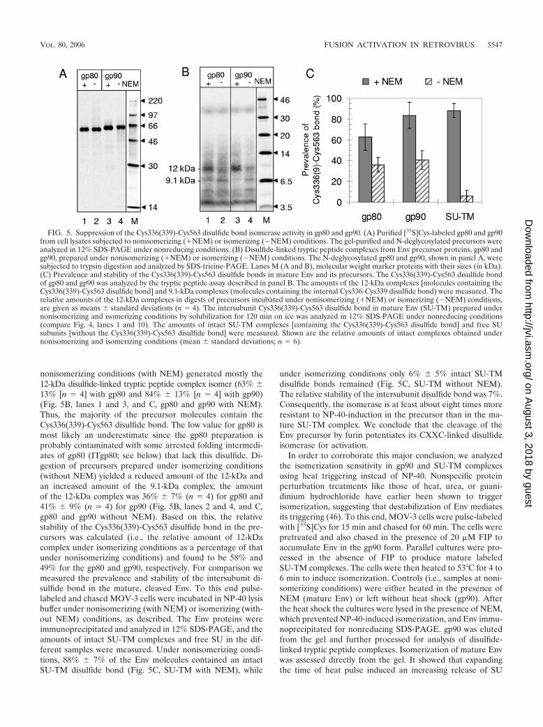
nonisomerizing conditions (with NEM) generated mostly the12-kDa disulfide-linked tryptic peptide complex isomer (63% �13% [n � 4] with gp80 and 84% � 13% [n � 4] with gp90)(Fig. 5B, lanes 1 and 3, and C, gp80 and gp90 with NEM).Thus, the majority of the precursor molecules contain theCys336(339)-Cys563 disulfide bond. The low value for gp80 ismost likely an underestimate since the gp80 preparation isprobably contaminated with some arrested folding intermedi-ates of gp80 (ITgp80; see below) that lack this disulfide. Di-gestion of precursors prepared under isomerizing conditions(without NEM) yielded a reduced amount of the 12-kDa andan increased amount of the 9.1-kDa complex; the amountof the 12-kDa complex was 36% � 7% (n � 4) for gp80 and41% � 9% (n � 4) for gp90 (Fig. 5B, lanes 2 and 4, and C,gp80 and gp90 without NEM). Based on this, the relativestability of the Cys336(339)-Cys563 disulfide bond in the pre-cursors was calculated (i.e., the relative amount of 12-kDacomplex under isomerizing conditions as a percentage of thatunder nonisomerizing conditions) and found to be 58% and49% for the gp80 and gp90, respectively. For comparison wemeasured the prevalence and stability of the intersubunit di-sulfide bond in the mature, cleaved Env. To this end pulse-labeled and chased MOV-3 cells were incubated in NP-40 lysisbuffer under nonisomerizing (with NEM) or isomerizing (with-out NEM) conditions, as described. The Env proteins wereimmunoprecipitated and analyzed in 12% SDS-PAGE, and theamounts of intact SU-TM complexes and free SU in the dif-ferent samples were measured. Under nonisomerizing condi-tions, 88% � 7% of the Env molecules contained an intactSU-TM disulfide bond (Fig. 5C, SU-TM with NEM), while
under isomerizing conditions only 6% � 5% intact SU-TMdisulfide bonds remained (Fig. 5C, SU-TM without NEM).The relative stability of the intersubunit disulfide bond was 7%.Consequently, the isomerase is at least about eight times moreresistant to NP-40-induction in the precursor than in the ma-ture SU-TM complex. We conclude that the cleavage of theEnv precursor by furin potentiates its CXXC-linked disulfideisomerase for activation.
In order to corroborate this major conclusion, we analyzedthe isomerization sensitivity in gp90 and SU-TM complexesusing heat triggering instead of NP-40. Nonspecific proteinperturbation treatments like those of heat, urea, or guani-dinium hydrochloride have earlier been shown to triggerisomerization, suggesting that destabilization of Env mediatesits triggering (46). To this end, MOV-3 cells were pulse-labeledwith [35S]Cys for 15 min and chased for 60 min. The cells werepretreated and also chased in the presence of 20 �M FIP toaccumulate Env in the gp90 form. Parallel cultures were pro-cessed in the absence of FIP to produce mature labeledSU-TM complexes. The cells were then heated to 53°C for 4 to6 min to induce isomerization. Controls (i.e., samples at noni-somerizing conditions) were either heated in the presence ofNEM (mature Env) or left without heat shock (gp90). Afterthe heat shock the cultures were lysed in the presence of NEM,which prevented NP-40-induced isomerization, and Env immu-noprecipitated for nonreducing SDS-PAGE. gp90 was elutedfrom the gel and further processed for analysis of disulfide-linked tryptic peptide complexes. Isomerization of mature Envwas assessed directly from the gel. It showed that expandingthe time of heat pulse induced an increasing release of SU
FIG. 5. Suppression of the Cys336(339)-Cys563 disulfide bond isomerase activity in gp80 and gp90. (A) Purified [35S]Cys-labeled gp80 and gp90from cell lysates subjected to nonisomerizing (�NEM) or isomerizing (�NEM) conditions. The gel-purified and N-deglycosylated precursors wereanalyzed in 12% SDS-PAGE under nonreducing conditions. (B) Disulfide-linked tryptic peptide complexes from Env precursor proteins, gp80 andgp90, prepared under nonisomerizing (�NEM) or isomerizing (�NEM) conditions. The N-deglycosylated gp80 and gp90, shown in panel A, weresubjected to trypsin digestion and analyzed by SDS-tricine-PAGE. Lanes M (A and B), molecular weight marker proteins with their sizes (in kDa).(C) Prevalence and stability of the Cys336(339)-Cys563 disulfide bonds in mature Env and its precursors. The Cys336(339)-Cys563 disulfide bondof gp80 and gp90 was analyzed by the tryptic peptide assay described in panel B. The amounts of the 12-kDa complexes [molecules containing theCys336(339)-Cys563 disulfide bond] and 9.1-kDa complexes (molecules containing the internal Cys336-Cys339 disulfide bond) were measured. Therelative amounts of the 12-kDa complexes in digests of precursors incubated under nonisomerizing (�NEM) or isomerizing (�NEM) conditions,are given as means � standard deviations (n � 4). The intersubunit Cys336(339)-Cys563 disulfide bond in mature Env (SU-TM) prepared undernonisomerizing and isomerizing conditions by solubilization for 120 min on ice was analyzed in 12% SDS-PAGE under nonreducing conditions(compare Fig. 4, lanes 1 and 10). The amounts of intact SU-TM complexes [containing the Cys336(339)-Cys563 disulfide bond] and free SUsubunits [without the Cys336(339)-Cys563 disulfide bond] were measured. Shown are the relative amounts of intact complexes obtained undernonisomerizing and isomerizing conditions (mean � standard deviations; n � 6).
VOL. 80, 2006 FUSION ACTIVATION IN RETROVIRUS 5547
on August 3, 2018 by guest
http://jvi.asm.org/
Dow
nloaded from
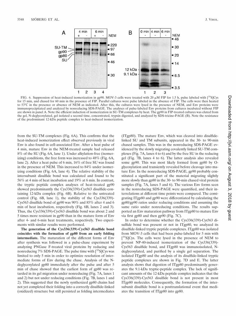
from the SU-TM complexes (Fig. 6A). This confirms that theheat-induced isomerization effect observed previously in viralEnv is also found in cell-associated Env. After a heat pulse of4 min, mature Env in the NEM-treated sample had released8% of the SU (Fig. 6A, lane 1). Under alkylation-free (isomer-izing) conditions, the free form was increased to 48% (Fig. 6A,lane 2). After a heat pulse of 6 min, 16% of free SU was foundin the presence of NEM. This increased to 84% under isomer-izing conditions (Fig. 6A, lane 4). The relative stability of theintersubunit disulfide bond was calculated and found to be56% at 4 min of heat incubation and 19% at 6 min. In contrast,the tryptic peptide complex analyses of heat-treated gp90showed predominantly the Cys336(339)-Cys563 disulfide-con-taining 12-kDa complex (Fig. 6B). Relative to the untreatedcontrol (Fig. 6B, lane 1), the stability of the Cys336(339)-Cys563 disulfide bond of gp90 was 90% and 85% after 4 and 6min of heat incubation, respectively (Fig. 6B, lanes 2 and 3).Thus, the Cys336(339)-Cys563 disulfide bond was about 2 and5 times more resistant in gp90 than in the mature form of Envafter 4- and 6-min heat treatments, respectively. Two experi-ments with similar results were performed.
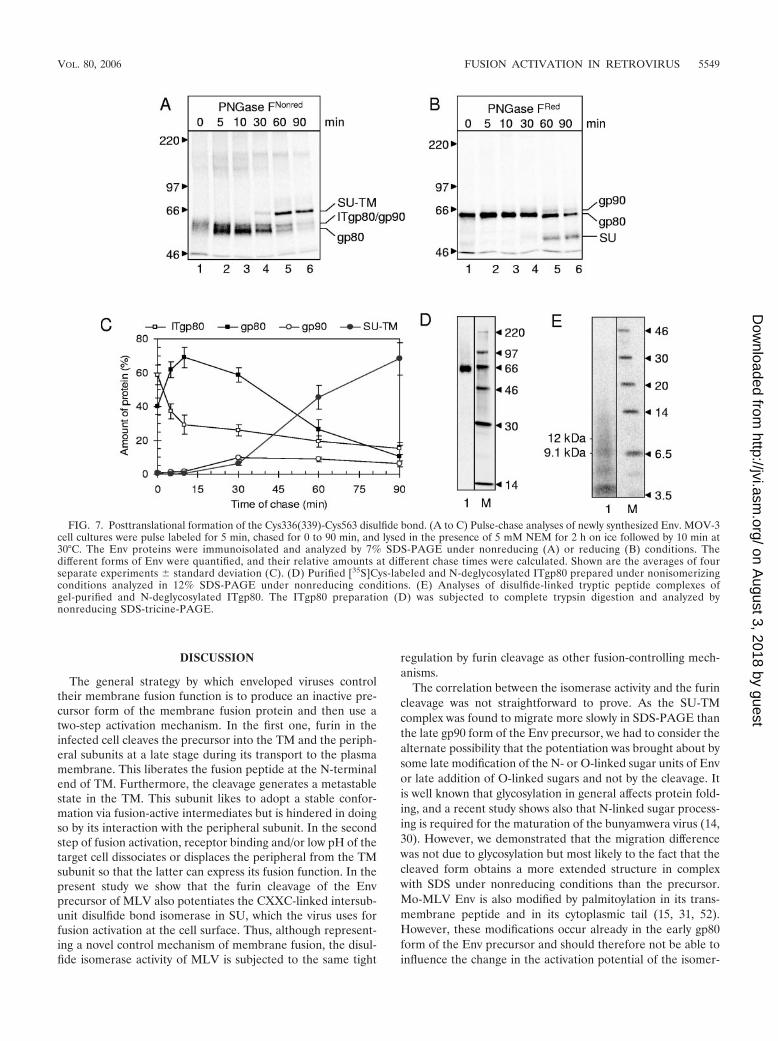
The generation of the Cys336(339)-Cys563 disulfide bondcoincides with the formation of gp80 from an early foldingintermediate. The maturation of the different forms of Envafter synthesis was followed in a pulse-chase experiment byanalyzing PNGase F-treated viral proteins by reducing andnonreducing 7% SDS-PAGE. The pulse time with [35S]Cys waslimited to only 5 min in order to optimize resolution of inter-mediate forms of Env during the chase. Analysis of the N-deglycosylated gp80 immediately after the pulse and after 5min of chase showed that the earliest form of gp80 was re-tarded in its gel migration under nonreducing (Fig. 7A, lanes 1and 2) but not under reducing conditions (Fig. 7B, lanes 1 and2). This suggested that the newly synthesized gp80 chains hadnot yet completed their folding into a correctly disulfide-linked(oxidized) structure but still persisted as folding intermediates
(ITgp80). The mature Env, which was cleaved into disulfide-linked SU and TM subunits, appeared in the 30- to 90-minchased samples. This was in the nonreducing SDS-PAGE ev-idenced by the slowly migrating covalently linked SU-TM com-plexes (Fig. 7A, lanes 4 to 6) and by the free SU in the reducinggel (Fig. 7B, lanes 4 to 6). The latter analysis also revealedsome gp90. This was most likely formed from gp80 by O-glycosylation and transiently revealed before cleavage into ma-ture Env. In the nonreducing SDS-PAGE, gp90 probably con-stituted a significant part of the material migrating slightlymore slowly than gp80 in the 30- to 90-min chased viral proteinsamples (Fig. 7A, lanes 5 and 6). The various Env forms seenin the nonreducing SDS-PAGE were quantified, and their in-terrelations at different time points were compared. The comi-grating ITgp80 and gp90 were differentiated by calculating thegp80/gp90 ratios under reducing conditions and assuming thesame ratio under nonreducing conditions. The results sup-ported an Env maturation pathway from ITgp80 to mature Envvia first gp80 and then gp90 (Fig. 7C).
In order to determine whether the Cys336(339)-Cys563 di-sulfide bond was present or not in ITgp80, we analyzed itsdisulfide-linked tryptic peptide complexes. ITgp80 was isolatedfrom MOV-3 cells that had been pulse-labeled for 5 min with[35S]Cys. The cells were lysed in the presence of NEM toprevent NP-40-induced isomerization of the Cys336(339)-Cys563 disulfide bond, and ITgp80 was immunoisolated, N-deglycosylated, and purified by a single gel separation. Theisolated ITgp80 and the analysis of its disulfide-linked trypticpeptide complexes are shown in Fig. 7D and E. The latteranalysis shows that digestion of ITgp80 predominantly gener-ates the 9.1-kDa tryptic-peptide complex. The lack of signifi-cant amounts of the 12-kDa peptide complex indicates that theCys336(339)-Cys563 disulfide bond is not present in mostITgp80 molecules. Consequently, the formation of the inter-subunit disulfide bond is a posttranslational event that medi-ates the transition of ITgp80 into gp80.
FIG. 6. Suppression of heat-induced isomerization in gp90. MOV-3 cells were treated with 20 �M FIP for 1.5 h, pulse labeled with [35S]Cysfor 15 min, and chased for 60 min in the presence of FIP. Parallel cultures were pulse labeled in the absence of FIP. The cells were then heatedto 53°C in the presence or absence of NEM as indicated. After this, the cultures were lysed in the presence of NEM, and Env proteins wereimmunoprecipitated and analyzed by nonreducing SDS-PAGE. The analyses of pulse-labeled Env proteins from cultures incubated without FIPare shown in panel A. Note the efficient induction of isomerization in SU-TM complexes by heat. The gp90 in FIP-treated cultures was eluted fromthe gel, N-deglycosylated, gel isolated a second time, concentrated, trypsin digested, and analyzed by SDS-tricine-PAGE (B). Note the resistanceof the predominant 12-kDa peptide complex to heat-induced isomerization.
5548 SJOBERG ET AL. J. VIROL.
on August 3, 2018 by guest
http://jvi.asm.org/
Dow
nloaded from
DISCUSSION
The general strategy by which enveloped viruses controltheir membrane fusion function is to produce an inactive pre-cursor form of the membrane fusion protein and then use atwo-step activation mechanism. In the first one, furin in theinfected cell cleaves the precursor into the TM and the periph-eral subunits at a late stage during its transport to the plasmamembrane. This liberates the fusion peptide at the N-terminalend of TM. Furthermore, the cleavage generates a metastablestate in the TM. This subunit likes to adopt a stable confor-mation via fusion-active intermediates but is hindered in doingso by its interaction with the peripheral subunit. In the secondstep of fusion activation, receptor binding and/or low pH of thetarget cell dissociates or displaces the peripheral from the TMsubunit so that the latter can express its fusion function. In thepresent study we show that the furin cleavage of the Envprecursor of MLV also potentiates the CXXC-linked intersub-unit disulfide bond isomerase in SU, which the virus uses forfusion activation at the cell surface. Thus, although represent-ing a novel control mechanism of membrane fusion, the disul-fide isomerase activity of MLV is subjected to the same tight
regulation by furin cleavage as other fusion-controlling mech-anisms.
The correlation between the isomerase activity and the furincleavage was not straightforward to prove. As the SU-TMcomplex was found to migrate more slowly in SDS-PAGE thanthe late gp90 form of the Env precursor, we had to consider thealternate possibility that the potentiation was brought about bysome late modification of the N- or O-linked sugar units of Envor late addition of O-linked sugars and not by the cleavage. Itis well known that glycosylation in general affects protein fold-ing, and a recent study shows also that N-linked sugar process-ing is required for the maturation of the bunyamwera virus (14,30). However, we demonstrated that the migration differencewas not due to glycosylation but most likely to the fact that thecleaved form obtains a more extended structure in complexwith SDS under nonreducing conditions than the precursor.Mo-MLV Env is also modified by palmitoylation in its trans-membrane peptide and in its cytoplasmic tail (15, 31, 52).However, these modifications occur already in the early gp80form of the Env precursor and should therefore not be able toinfluence the change in the activation potential of the isomer-
FIG. 7. Posttranslational formation of the Cys336(339)-Cys563 disulfide bond. (A to C) Pulse-chase analyses of newly synthesized Env. MOV-3cell cultures were pulse labeled for 5 min, chased for 0 to 90 min, and lysed in the presence of 5 mM NEM for 2 h on ice followed by 10 min at30°C. The Env proteins were immunoisolated and analyzed by 7% SDS-PAGE under nonreducing (A) or reducing (B) conditions. Thedifferent forms of Env were quantified, and their relative amounts at different chase times were calculated. Shown are the averages of fourseparate experiments � standard deviation (C). (D) Purified [35S]Cys-labeled and N-deglycosylated ITgp80 prepared under nonisomerizingconditions analyzed in 12% SDS-PAGE under nonreducing conditions. (E) Analyses of disulfide-linked tryptic peptide complexes ofgel-purified and N-deglycosylated ITgp80. The ITgp80 preparation (D) was subjected to complete trypsin digestion and analyzed bynonreducing SDS-tricine-PAGE.
VOL. 80, 2006 FUSION ACTIVATION IN RETROVIRUS 5549
on August 3, 2018 by guest
http://jvi.asm.org/
Dow
nloaded from
ase, which occurs later during Env maturation, in the SU-TMcomplex.
The structural basis for the furin cleavage-mediated poten-tiation of the isomerase activity is yet unknown. However, itmust involve the CXXC motif of the isomerase in the SUsubunit. In order for the CXXC thiol to attack the SU-TMdisulfide bond and cause the fusion activating disulfide bondrearrangement, it has to be deprotonated. This requires thatthere is a mechanism in Env that conditionally can lower thepKa of the CXXC thiol from its uninfluenced value of about8.3 to about neutral (24). In the case of the endoplasmicreticulum protein disulfide isomerase, structural and moleculardynamics studies have shown that an Arg side chain can tem-porarily move into the locale of the active site and therebylower the pKa of the C-terminal thiol of the CXXC motif. Thiswill facilitate the reoxidation of the reduced protein disulfideisomerase by glutathione. In this view it is possible that theisomerase of Mo-MLV Env is similarly triggered by the ap-proximation of an amino acid side chain that can influence thepKa of the CXXC thiol. According to our model, this alterationin structure is potentiated by a structural change of Env that iscaused by the precursor cleavage and finally triggered by re-ceptor binding. As the final triggering can be done by treat-ments with heat, urea, guanidinium hydrochloride, Ca2� de-pletion, and NP-40 solubilization, it is apparent that this stepmust involve the dissociation of interactions between proteindomains in the Env oligomer. This might be necessary to allowa specific restructuring of the locale of the CXXC thiol. Thepotentiation of the isomerase triggering that is brought aboutby the precursor cleavage should be represented by a specificalteration of the Env structure. The cleavage liberates theN-terminal end of TM, with the fusion peptide, and the C-terminal end of SU. This should allow for both dissociation ofprevious interactions at the cleavage site and creation of newones. In the case of influenza, hemagglutinin (HA) precursorcleavage takes place in a surface loop in the stem of themolecule. The fusion peptide at the end of the HA2 subunitthen seeks its way into a nearby groove where it will buryionizable amino acid residues (4). This structural alteration ofinfluenza HA completes the formation of the pH-sensitivemetastable HA1-HA2 complex.
A trivial explanation for the differential NP-40-inducedisomerization sensitivity of the precursor and the mature formsof Env would be the different solubilization properties withNP-40. It is reasonable to believe that the triggering of theisomerase by NP-40 must be related to structural effects (de-stabilization) caused by solubilization of the membrane-boundEnv oligomer. Therefore, if the precursor is more difficult tosolubilize, for instance due to different intracellular localiza-tion, than the mature form, this could explain the apparentisomerase suppression in the precursor. However, the lateform of the precursor, the FIP-induced gp90, was almostequally suppressed as the early form although it is expected tolargely share the same membranes, i.e., lipid environment, asthe mature form of Env. Therefore, this trivial explanationappears unlikely. Furthermore, we confirmed the relative re-sistance of gp90 to isomerization compared to the mature Envusing another isomerization induction treatment, i.e., heat(53°C).
Apart from the intersubunit disulfide isomerase, the MLV
contains one additional novel mechanism to control its fusionactivity. This is represented by the cleavage of the C-terminalend, the R peptide, of the TM cytoplasmic tail by the viralprotease during or soon after virus budding (12, 41). Previousstudies have shown that expression of the MLV Env gene inmouse 3T3 cells does not result in any significant cell-cellfusion if the region encoding the end of the cytoplasmic tail isdeleted or specifically point mutated (18, 37, 38, 51). Further-more, it has been demonstrated that MLV or MLV vectormutants with uncleaved R peptide, due to violation of theconserved amino acid sequence at the cleavage site, havegreatly reduced capacity to infect target cells (22, 38). Thus,these results suggest that the end of the cytoplasmic tail of TMsuppresses the receptor-mediated induction of the fusion func-tion in MLV.
Interestingly, the isomerase of the cell-associated SU-TMcomplex was sensitive to NP-40 and heat (53°C)-mediated trig-gering despite retention of its R peptide. However, both incu-bation at 53°C and solubilization with NP-40 represent power-ful isomerase induction treatments, which probably overridesthe control mechanism of the R peptide. Indeed, it is possiblethat the isomerase function is potentiated in two steps, first bythe furin cleavage of the late precursor form gp90 and then byviral protease cleavage of the TM cytoplasmic tail. The latterpotentiating effect might be possible to study with Env on thecell surface or in virus particles using isomerase induction viaCa2� depletion or receptor binding (47, 48). It should be notedthat it was not possible to use these induction treatments in thisstudy because of the intracellular localization of the Env pre-cursor.
The MLV membrane fusion protein appears to be a finelytuned apparatus where the action center is localized aroundthe Cys336(339)-Cys563 (or corresponding) disulfide bond. Thisis the place for the CXXC thiol catalyzed isomerization of theintersubunit disulfide bond and also the point in TM where thejackknife-like refolding might be initiated (21, 36, 48). It mightalso be close to the cleavage site as this potentiates the isomer-ase to become triggered. Therefore, the formation of this partof Env by polypeptide folding and its stabilization byCys336(339)-Cys563 disulfide bonding should be a demandingtask. Notably, it has been demonstrated that of the seven oreight N-linked sugar units in Mo-MLV or Friend MLV, onlythe conserved one just N-terminal to the CXXC motif in SU iscritical for Env maturation (8, 20). It is possible that the sugarunit transferred to this site is a target for calnexin with asso-ciated endoplasmic reticulum chaperones, including oxido-reductases. Together these might furnish the action centerready, with the Cys336(339)-Cys563 disulfide bond in place,after the complete Env chain has first been translated. Such amechanism has previously been demonstrated to be involved inthe formation of disulfide bonds of influenza HA, including theCys14-Cys466 disulfide, which corresponds to the intersubunitdisulfide of mature HA (1, 5). Consistent with this view wefound in the present study that the Cys336(339)-Cys563 disul-fide bond of Mo-MLV Env was formed posttranslationally.
ACKNOWLEDGMENTS
Swedish Science Foundation grant 2778 and Swedish Cancer Foun-dation grant 0525 to H.G. supported this work.
5550 SJOBERG ET AL. J. VIROL.
on August 3, 2018 by guest
http://jvi.asm.org/
Dow
nloaded from
REFERENCES
1. Braakman, I., H. Hoover-Litty, K. R. Wagner, and A. Helenius. 1991. Fold-ing of influenza hemagglutinin in the endoplasmic reticulum. J. Cell Biol.114:401–411.
2. Bresnahan, P. A., R. Leduc, L. Thomas, J. Thorner, H. L. Gibson, A. J.Brake, P. J. Barr, and G. Thomas. 1990. Human fur gene encodes a yeastKEX2-like endoprotease that cleaves pro-beta-NGF in vivo. J. Cell Biol.111:2851–2859.
3. Bressanelli, S., K. Stiasny, S. L. Allison, E. A. Stura, S. Duquerroy, J. Lescar,F. X. Heinz, and F. A. Rey. 2004. Structure of a flavivirus envelope glyco-protein in its low-pH-induced membrane fusion conformation. EMBO J.23:728–738.
4. Chen, J., K. H. Lee, D. A. Steinhauer, D. J. Stevens, J. J. Skehel, and D. C.Wiley. 1998. Structure of the hemagglutinin precursor cleavage site, a deter-minant of influenza pathogenicity and the origin of the labile conformation.Cell 95:409–417.
5. Daniels, R., B. Kurowski, A. E. Johnson, and D. N. Hebert. 2003. N-linkedglycans direct the cotranslational folding pathway of influenza hemaggluti-nin. Mol. Cell 11:79–90.
6. Dong, J. Y., J. W. Dubay, L. G. Perez, and E. Hunter. 1992. Mutations withinthe proteolytic cleavage site of the Rous sarcoma virus glycoprotein define arequirement for dibasic residues for intracellular cleavage. J. Virol. 66:865–874.
7. Fass, D., S. C. Harrison, and P. S. Kim. 1996. Retrovirus envelope domainat 1.7 angstrom resolution. Nat. Struct. Biol. 3:465–469.
8. Felkner, R. H., and M. J. Roth. 1992. Mutational analysis of the N-linkedglycosylation sites of the SU envelope protein of Moloney murine leukemiavirus. J. Virol. 66:4258–4264.
9. Freed, E. O., and R. Risser. 1987. The role of envelope glycoprotein pro-cessing in murine leukemia virus infection. J. Virol. 61:2852–2856.
10. Geyer, R., J. Dabrowski, U. Dabrowski, D. Linder, M. Schluter, H. H. Schott,and S. Stirm. 1990. Oligosaccharides at individual glycosylation sites in glyco-protein 71 of Friend murine leukemia virus. Eur. J. Biochem. 187:95–110.
11. Gibbons, D. L., M. C. Vaney, A. Roussel, A. Vigouroux, B. Reilly, J. Lepault, M.Kielian, and F. A. Rey. 2004. Conformational change and protein-protein inter-actions of the fusion protein of Semliki Forest virus. Nature 427:320–325.
12. Green, N., T. M. Shinnick, O. Witte, A. Ponticelli, J. G. Sutcliffe, and R. A.Lerner. 1981. Sequence-specific antibodies show that maturation of Moloneyleukemia virus envelope polyprotein involves removal of a COOH-terminalpeptide. Proc. Natl. Acad. Sci. USA 78:6023–6027.
13. Hallenberger, S., V. Bosch, H. Angliker, E. Shaw, H. D. Klenk, and W.Garten. 1992. Inhibition of furin-mediated cleavage activation of HIV-1glycoprotein gp160. Nature 360:358–361.
14. Helenius, A., and M. Aebi. 2001. Intracellular functions of N-linked glycans.Science 291:2364–2369.
15. Hensel, J., M. Hintz, M. Karas, D. Linder, B. Stahl, and R. Geyer. 1995.Localization of the palmitoylation site in the transmembrane protein p12E ofFriend murine leukaemia virus. Eur. J. Biochem. 232:373–380.
16. Hernandez, L. D., L. R. Hoffman, T. G. Wolfsberg, and J. M. White. 1996.Virus-cell and cell-cell fusion. Annu. Rev. Cell Dev. Biol. 12:627–661.
17. Hosaka, M., M. Nagahama, W. S. Kim, T. Watanabe, K. Hatsuzawa, J.Ikemizu, K. Murakami, and K. Nakayama. 1991. Arg-X-Lys/Arg-Arg motifas a signal for precursor cleavage catalyzed by furin within the constitutivesecretory pathway. J. Biol. Chem. 266:12127–12130.
18. Januszeski, M. M., P. M. Cannon, D. Chen, Y. Rozenberg, and W. F.Anderson. 1997. Functional analysis of the cytoplasmic tail of Moloneymurine leukemia virus envelope protein. J. Virol. 71:3613–3619.
19. Johnston, E. R., and K. Radke. 2000. The SU and TM envelope proteinsubunits of bovine leukemia virus are linked by disulfide bonds, both in cellsand in virions. J. Virol. 74:2930–2935.
20. Kayman, S. C., R. Kopelman, S. Projan, D. M. Kinney, and A. Pinter. 1991.Mutational analysis of N-linked glycosylation sites of Friend murine leuke-mia virus envelope protein. J. Virol. 65:5323–5332.
21. Kobe, B., R. J. Center, B. E. Kemp, and P. Poumbourios. 1999. Crystalstructure of human T cell leukemia virus type 1 gp21 ectodomain crystallizedas a maltose-binding protein chimera reveals structural evolution of retro-viral transmembrane proteins. Proc. Natl. Acad. Sci. USA 96:4319–4324.
22. Kubo, Y., and H. Amanuma. 2003. Mutational analysis of the R peptidecleavage site of Moloney murine leukaemia virus envelope protein. J Gen.Virol. 84:2253–2257.
23. Laemmli, U. K. 1970. Cleavage of structural proteins during the assembly ofthe head of bacteriophage T4. Nature 227:680–685.
24. Lappi, A. K., M. F. Lensink, H. I. Alanen, K. E. Salo, M. Lobell, A. H. Juffer,and L. W. Ruddock. 2004. A conserved arginine plays a role in the catalyticcycle of the protein disulphide isomerases. J. Mol. Biol. 335:283–295.
25. Leamnson, R. N., and M. S. Halpern. 1976. Subunit structure of the glyco-protein complex of avian tumor virus. J. Virol. 18:956–968.
26. Linder, M., D. Linder, J. Hahnen, H. H. Schott, and S. Stirm. 1992. Local-ization of the intrachain disulfide bonds of the envelope glycoprotein 71 fromFriend murine leukemia virus. Eur. J. Biochem. 203:65–73.
27. Lobigs, M., and H. Garoff. 1990. Fusion function of the Semliki Forest virusspike is activated by proteolytic cleavage of the envelope glycoprotein pre-cursor p62. J. Virol. 64:1233–1240.
28. Mothes, W., A. L. Boerger, S. Narayan, J. M. Cunningham, and J. A. Young.2000. Retroviral entry mediated by receptor priming and low pH triggeringof an envelope glycoprotein. Cell 103:679–689.
29. Ng, V. L., T. G. Wood, and R. B. Arlinghaus. 1982. Processing of the env geneproducts of Moloney murine leukaemia virus. J. Gen. Virol. 59:329–343.
30. Novoa, R. R., G. Calderita, P. Cabezas, R. M. Elliott, and C. Risco. 2005. KeyGolgi factors for structural and functional maturation of bunyamwera virus.J. Virol. 79:10852–10863.
31. Olsen, K. E., and K. B. Andersen. 1999. Palmitoylation of the intracytoplas-mic R peptide of the transmembrane envelope protein in Moloney murineleukemia virus. J. Virol. 73:8975–8981.
32. Opstelten, D. J., M. Wallin, and H. Garoff. 1998. Moloney murine leukemiavirus envelope protein subunits, gp70 and Pr15E, form a stable disulfide-linked complex. J. Virol. 72:6537–6545.
33. Ortmann, D., M. Ohuchi, H. Angliker, E. Shaw, W. Garten, and H. D. Klenk.1994. Proteolytic cleavage of wild type and mutants of the F protein ofhuman parainfluenza virus type 3 by two subtilisin-like endoproteases, furinand Kex2. J. Virol. 68:2772–2776.
34. Pearson, W. R., T. Wood, Z. Zhang, and W. Miller. 1997. Comparison ofDNA sequences with protein sequences. Genomics 46:24–36.
35. Pinter, A., and W. J. Honnen. 1988. O-Linked glycosylation of retroviralenvelope gene products. J. Virol. 62:1016–1021.
36. Pinter, A., R. Kopelman, Z. Li, S. C. Kayman, and D. A. Sanders. 1997.Localization of the labile disulfide bond between SU and TM of the murineleukemia virus envelope protein complex to a highly conserved CWLC motifin SU that resembles the active-site sequence of thiol-disulfide exchangeenzymes. J. Virol. 71:8073–8077.
37. Ragheb, J. A., and W. F. Anderson. 1994. pH-independent murine leukemiavirus ecotropic envelope-mediated cell fusion: implications for the role of theR peptide and p12E TM in viral entry. J. Virol. 68:3220–3231.
38. Rein, A., J. Mirro, J. G. Haynes, S. M. Ernst, and K. Nagashima. 1994.Function of the cytoplasmic domain of a retroviral transmembrane protein:p15E-p2E cleavage activates the membrane fusion capability of the murineleukemia virus Env protein. J. Virol. 68:1773–1781.
39. Schagger, H., and G. von Jagow. 1987. Tricine-sodium dodecyl sulfate-polyacrylamide gel electrophoresis for the separation of proteins in the rangefrom 1 to 100 kDa. Anal. Biochem. 166:368–379.
40. Scheid, A., and P. W. Choppin. 1974. Identification of biological activities ofparamyxovirus glycoproteins. Activation of cell fusion, hemolysis, and infec-tivity of proteolytic cleavage of an inactive precursor protein of Sendai virus.Virology 57:475–490.
41. Schultz, A., and A. Rein. 1985. Maturation of murine leukemia virus Envproteins in the absence of other viral proteins. Virology 145:335–339.
42. Schultz, A. M., and S. Oroszlan. 1979. Tunicamycin inhibits glycosylation ofprecursor polyprotein encoded by env gene of Rauscher murine leukemiavirus. Biochem. Biophys. Res. Commun. 86:1206–1213.
43. Shapiro, S. Z., M. Strand, and J. T. August. 1976. High molecular weightprecursor polypeptides to structural proteins of Rauscher murine leukemiavirus. J. Mol. Biol. 107:459–477.
44. Skehel, J. J., and D. C. Wiley. 2000. Receptor binding and membrane fusionin virus entry: the influenza hemagglutinin. Annu. Rev. Biochem. 69:531–569.
45. Stieneke-Grober, A., M. Vey, H. Angliker, E. Shaw, G. Thomas, C. Roberts,H. D. Klenk, and W. Garten. 1992. Influenza virus hemagglutinin withmultibasic cleavage site is activated by furin, a subtilisin-like endoprotease.EMBO J. 11:2407–2414.
46. Wallin, M., M. Ekstrom, and H. Garoff. 2005. The fusion-controlling disul-fide bond isomerase in retrovirus Env is triggered by protein destabilization.J. Virol. 79:1678–1685.
47. Wallin, M., R. Loving, M. Ekstrom, K. Li, and H. Garoff. 2005. Kineticanalyses of the surface-transmembrane disulfide bond isomerization-con-trolled fusion activation pathway in Moloney murine leukemia virus. J. Virol.79:13856–13864.
48. Wallin, M., M. Ekstrom, and H. Garoff. 2004. Isomerization of the intersub-unit disulphide-bond in Env controls retrovirus fusion. EMBO J. 23:54–65.
49. Wang, H., M. P. Kavanaugh, R. A. North, and D. Kabat. 1991. Cell-surfacereceptor for ecotropic murine retroviruses is a basic amino-acid transporter.Nature 352:729–731.
50. Ward, C. D., R. G. Paterson, and R. A. Lamb. 1995. Mutants of theparamyxovirus SV5 fusion protein: regulated and extensive syncytium for-mation. Virology 209:242–249.
51. Yang, C., and R. W. Compans. 1997. Analysis of the murine leukemia virusR peptide: delineation of the molecular determinants which are importantfor its fusion inhibition activity. J. Virol. 71:8490–8496.
52. Yang, C., and R. W. Compans. 1996. Palmitoylation of the murine leukemiavirus envelope glycoprotein transmembrane subunits. Virology 221:87–97.
VOL. 80, 2006 FUSION ACTIVATION IN RETROVIRUS 5551
on August 3, 2018 by guest
http://jvi.asm.org/
Dow
nloaded from