Embed Size (px)
Citation preview
Furanoflavonoid glycosides from Pongamia pinnata fruits§
Ghufran Ahmad, Prem P. Yadav, Rakesh Maurya*
Medicinal Chemistry Division, Central Drug Research Institute, Chattar Manzil Palace, Lucknow-226 001, India
Received 2 October 2003; accepted 28 January 2004
Abstract
Pongamia pinnata fruits afforded three new furanoflavonoid glucosides, pongamosides A-C (1–3), and a new flavonol glucoside,pongamoside D (4). The structures of these compounds were established on the basis of spectroscopic studies. This is the first timethat furanoflavone glucosides have been found as naturally occurring compounds.
# 2004 Elsevier Ltd. All rights reserved.
Keywords: Pongamia pinnata; Leguminosae; furanoflavonoid glucosides; flavone glucoside
1. Introduction
Pongamia pinnata Pierre (Leguminosae) is commonlyknown as Karanja. It is distributed throughout WesternGhats and chiefly found in tidal forests of India(Krishnamurthi, 1969). Different parts of the plant havebeen used in traditional medicines for bronchitis,whooping cough, rheumatic joints and to quench dipsiain diabetes (Kirtikar et al., 1995). Previous phytochem-ical examination of this plant indicated the presenceof furanoflavones, furanoflavonols, chromenoflavones,flavones, and furanodiketones (Talapatra et al., 1980,1982; Murty et al., 1944; Rangaswami et al., 1942;Sharma et al., 1973; Pathak et al., 1983; Toshiyuki et al.,1992). In the present communication, we describe theisolation and characterization of three new furano-flavonoid glucosides, pongamosides A-C (1–3), and anew flavonol glucoside pongamoside D (4).
2. Results and discussion
The n-BuOH soluble fraction of the EtOH extract ofPongamia pinnata fruits afforded four new compounds1–4, after repeated CC purifications.Compounds 1–4 displayed certain common structural
features; a positive Shinoda test (Grayer, 1989), Fiegletest, IR and UV suggested a flavonoid glycoside. Thesugar was identified as glucospyranose by acid hydro-lysis and co-TLC with authentic sample and NMR data(Table 1) for compounds 1–4. The anomeric protonsappeared at � 5.12 (d, J=6.6 Hz); �C 100.3, 5.09 (d,
0031-9422/$ - see front matter # 2004 Elsevier Ltd. All rights reserved.
doi:10.1016/j.phytochem.2004.01.020
Phytochemistry 65 (2004) 921–924
www.elsevier.com/locate/phytochem
§ CDRI communication no.: 6471
* Corresponding author. Tel.: +91-522-2212411-18x4440; fax:
+91-522-2223405.
E-mail address: [email protected] (R. Maurya).
J=6.9 Hz), 5.27 (d, J=6.9 Hz); �C 100.8, and 5.06 (d,J=7.2 Hz); �C 103.3 for compounds 1–4, respectively,indicating that it was linked to the phenolic OH andhave a b-configuration as suggested from coupling con-stants of anomeric proton signals.The FAB-mass spectrum of compound 1 contained a
peak at m/z 441 [M+H]+ corresponding to the mole-cular formula of C23H20O9. This conclusion was sup-ported by the elemental analysis and NMR spectra. IRabsorptions (�max 3433, 1625, 1594, 1459, 1353, 1082,758, 631 cm�1), UV spectrum (lmax 216, 261, 300 nm)and NMR data are indicative of a furanoflavone glyco-side nucleus. The 1H and 13C NMR (Table 1 and 2)showed characteristic signals for H-3 at � 7.10 (1H, s),�C 104.5, and a furan ring at � 7.58 (1H, d, 2.1 Hz, H-300), �C 107.6 and � 8.25 (1H, d, J=2.1 Hz, H-200), �C147.4. The unsubstituted C-5 and C-6 positions arerevealed by doublets for H-5 and H-6 at � 7.99(1H,J=8.7 Hz) and � 7.77 (1H, J=8.7 Hz). H-300 showedcorrelation with the H-6, in 1H–1H long range COSYspectrum, indicating that the furan ring was fused in anangular position at C-7 (oxygenated) and C-8. 1H–1HCOSY correlations observed for 1H NMR signals at �7.75 (1H, brs, H-20), � 7.29 (1H, dd, J=1.5, 7.5 Hz, H-40), � 7.52 (1H, t, J=7.5 Hz, H-50) and � 7.85 (1H, d,J=7.5 Hz, H-60) indicated ring-B as monosubstituted at
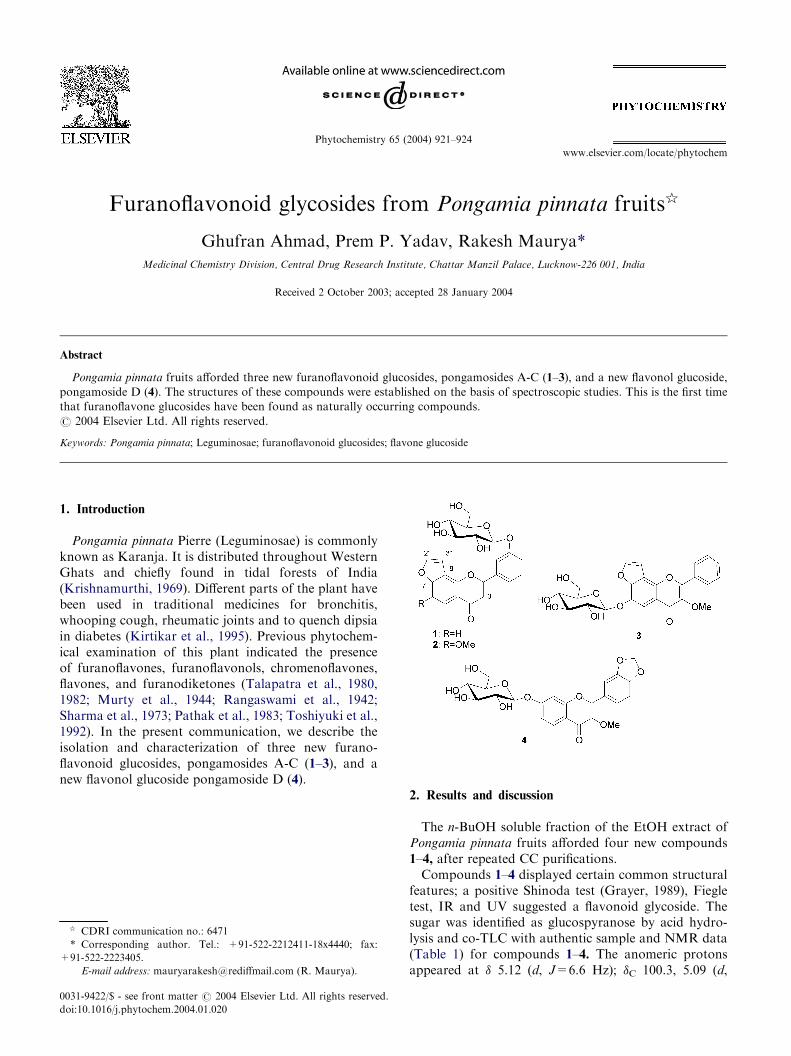
C-30. The glucosidic linkage at C-30-OH was revealed bynOe between the Glu H-1 (d 5.12) and H-20 (� 7.75), H-40 (� 7.29) signals. Furthermore, aglycone was confirmedas pongol by acid hydrolysis of compound 1 andco-TLC with authentic sample, isolated from the sameplant. Thus, based on the above information compound 1was characterized as 30-O-b-d-glucopyranosyl[200,300:7,8]furanoflavone, and we named it pongamoside-A. It isthe first time that a furanoflavone glucoside has beenfound as a naturally occurring compound.The HRFAB-mass spectrum of compound 2, shows a
[M+H]+ peak at m/z 471 consistent with the formulaC24H22O10, which was supported by the 1H NMRspectrum (Table 1) and an adduct ion at m/z 493[M+Na]+ in the FAB mass spectrum. The 1H NMRspectrum was similar to that of 1, suggesting 2 as fur-anoflavone glycoside. It could be inferred from themolecular weight and 1H signal at � 4.04 (3H, s) that 2differed from 1 by one methoxy group. The methoxygroup was placed at C-6 because H-5 appeared as sing-let at � 7.39 (1H), and the absence of long range corre-lation in the 1H–1H COSY experiment between H-300 at� 7.58 (1H, d, J=2.1 Hz) with H-6. The four aromaticprotons of ring B moiety are superimposed on those of1. The ring B protons resonated at � 7.73 (1H, brs, H-20), 7.26 (1H, dd, J=2.1, 7.8 Hz, H-40), 7.52 (1H, t,
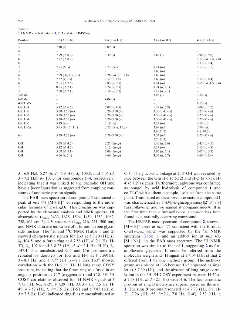
Table 11H NMR spectral data of 1, 2, 3 and 4 in DMSO-d6
Position
1 � (J in Hz) 2 � (J in Hz) 3 � (J in Hz) 4 � (J in Hz)3
7.10 (s) 7.09 (s) – –4
– – – –5
7.99 (d, 8.7) 7.39 (s) 7.62 (s) 7.98 (d, 9.0)6
7.77 (d, 8.7) – – 7.12 (dd, 2.4, 9.0)8
– – – 7.33 (d, 2.4)20
7.75 (br s) 7.73 (brs) 8.14 (m) 7.57 (d, 1.5)30
– – 7.60 (m) –40
7.29 (dd, 1.5, 7.5) 7.26 (dd, 2.1, 7.8) 7.60 (m) –50
7.52 (t, 7.5) 7.52 (t, 7.8) 7.60 (m) 7.11 (d, 8.4)60
7.85 (d, 7.5) 7.83 (d, 7.8) 8.14 (m) 7.65 (dd, 1.5, 8.4)2
8.25 (d, 2.1) 8.24 (d, 2.1) 8.24 (d, 2.1) –3
7.58 (d, 2.1) 7.58 (d, 2.1) 7.52 (d, 2.1) –3-OMe
– – 3.85 (s) 3.79 (s)6-OMe
– 4.04 (s) – –-OCH2O-
– – – 6.15 (s)Glc H-1
5.12 (d, 6.6) 5.09 (d, 6.9) 5.27 (d, 6.9) 5.06 (d, 7.2)Glc H-2
3.28–3.56 (m) 3.28–3.50 (m) 3.30–3.43 (m) 3.27–52 (m)Glc H-3
3.28–3.56 (m) 3.28–3.50 (m) 3.30–3.43 (m) 3.27–52 (m)Glc H-4
3.28–3.56 (m) 3.28–3.50 (m) 3.30–3.43 (m) 3.27–52 (m)Glc H-5
3.19 (m) 3.18 (m) 3.27 (m) 3.19 (m)Glc H-6a
3.75 (br d, 11.1) 3.72 (br d, 11.2) 3.69 (dd,3.6, 11.7)
3.70 (dd,
4.5, 10.2)
6b
3.28–3.56 (m) 3.28–3.50 (m) 3.53 (dd,5.7, 11.7)
3.27–52 (m)
OH
5.38 (d, 4.5) 5.27 (hump) 5.45 (d, 3.6) 5.43 (d, 4.5)OH
5.12 (d, 4.2) 5.12 (hump) 5.17 (brs) 5.13 (d, 4.8)OH
5.08 (d, 5.1) 5.06 (hump) 5.06 (d, 5.1) 5.07 (d, 5.1)OH
4.66 (t, 5.1) 4.66 (hump) 4.56 (d, 5.7) 4.60 (t, 5.4)922 G. Ahmad et al. / Phytochemistry 65 (2004) 921–924

J=7.8 Hz, H-50) and 7.83 (1H, d, J=7.8 Hz, H-60)indicating ring-B was monosubstituted at C-30. Furtherglycosidation of C-30 hydroxyl was confirmed by nOeexperiment; irradiation of anomeric proton at � 5.09(1H, d, J=6.9 Hz) enhances signals for H-20 (� 7.73) andH-40 (� 7.26). Thus, on the basis of the above spectraldata compound 2 was identified as 6-methoxy-30-O-b-d-glucopyranosyl[200,300:7,8] furanoflavone, a new natu-rally occurring compound, named pongamoside-B.Compound 3 was assigned the molecular formula
C24H22O10 (FAB-MS, m/z 471 [M+1]+). This conclu-sion was supported by the 1H and 13C NMR spectra.Compound 3 was recognized as a furanoflavonol glyco-side. Analysis of the 1H and 13C NMR spectra (Tables 1and 2) reveals the presence of a furan ring fused to ringA, with the characteristic proton doublets at � 7.52 and8.24 (1H each, d, J=2.1 Hz, H-300 and H-200); one aro-matic proton in ring A at d 7.62 (s, H-5), an unsub-stituted ring B, � 8.14 (2H, m, H-20, 60) and 7.60 (3H, m,H-30,40,50), and one methoxy group at � 3.85 (s); �C 59.6.The latter was located at C-3 based on nOe between themethoxy protons and the H-20 and H-60 resonances at �8.14 (2H, m). Further NMR spectra indicated a gluco-sidic moiety located at C-6 as revealed by absence ofcorrelation for H-300 in 1H-1H long range COSY experi-ment and an nOe between the anomeric proton at � 5.27and H-5 at � 7.62. Spectral similarity of the aglyconeportion of 3 to that reported in the literature for the 6-
methoxy derivative (Kamperdick et al., 1998), along withspectrochemical observations led structure of new com-pound 3 as 3-methoxy-6-O-b-d-glucopyranosyl[200,300:7,8]furanoflavone, a new naturally occurring compound,named pongamoside-C.The molecular formula C23H22O11 of compound 4
was determined by FAB-mass spectrometry (m/z 474[M+1]+) and is in accord with 1H and 13C NMR datasummarized in Tables 1 and 2. The IR, UV and NMRwere characteristics of a flavonol glycoside moiety.Ring-A exhibited an ABX spin system consisting of twodoublets at 7.98 (1H, J=9.0 Hz, H-5), � 7.33 (1H,J=2.4 Hz, H-8) and a double doublet at � 7.12 (1H,J=2.4, 9.0 Hz, H-6). The nOe correlation of theanomeric proton at d 5.06 with H-6 (d 7.12) and H-8(� 7.33) suggested the location of the glucose moiety atC-7. Absence of the characteristic flovone singlet forH-3 in the 1H NMR, the appearance of C-3 at � 139.9,and one methoxy group at � 59.4 in 13C NMR, revealedthe presence of one methoxy group at C-3. The presenceof one methylenedioxy group at C-30,40 (� 6.15, 2H, s);�C 101.7, and an ABX splitting system consisting of twodoublets at � 7.57 (1H, J=1.5 Hz, H-20), 7.11 (1H,J=8.4 Hz, H-50) and a double doublet at � 7.65 (1H,J=1.5, 8.4 Hz, H-60) indicated a disubstituted B-ring.Based on the above spectral evidences, compound 4
was characterized as 3-methoxy-30,40-methylenedioxy-7-O-b-d-glucopyranosyl flavone, and named pongamo-side D.
3. Experimental
3.1. General
Melting points were recorded on a Complab meltingpoint apparatus and are uncorrected. IR spectra (KBr)were recorded on a Perkin-Elmer RX-1 spectro-photometer. UV spectra were obtained on a Perkin-Elmer l-15 UV spectrophotometer. NMR spectra wererun on an AVANCE DPX 200 and Bruker DRX 300spectrometer, FAB MS were carried out on JEOL SX102/DA-6000 mass spectrometer. Elemental analyseswere obtained in a Carlo-Erba-1108 CHN elementalanalyzer. Column chromatography was performedusing flash silica gel (230–400 mesh). Preparative andanalytical HPLC performed on Shimadzu model LC-8Ainstrument.
3.2. Plant material
The fruits of P. pinnata were collected from Lucknowin the month of May 2000, and identified by BotanyDivision of Central Drug Research Institute. A voucherspecimen (No. 6331) is kept in the herbarium of theInstitute.
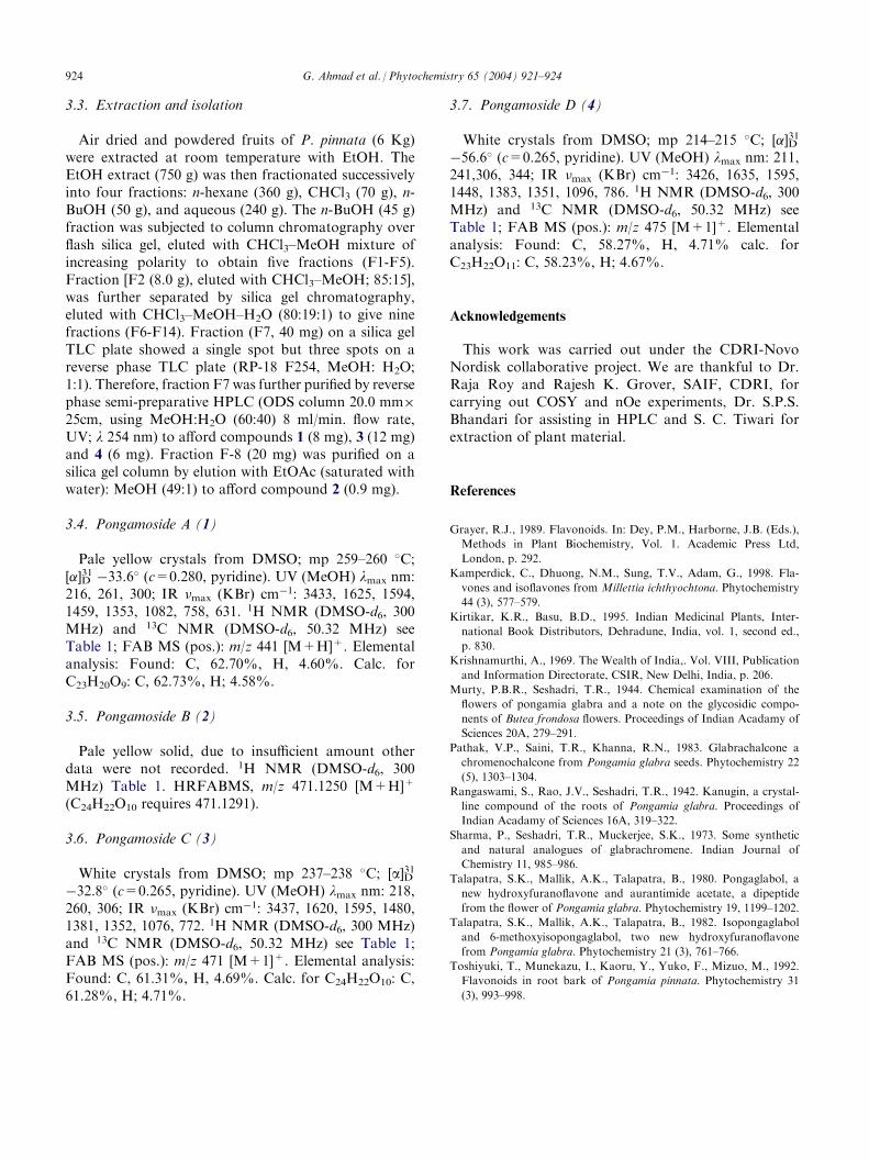
Table 213C NMR spectral data of 1, 3 and 4 in DMSO-d6
Position
1 �C 3 �C 4 �C2
150.0a 153.8 155.9a3
104.5 141.2a 139.94
176.8 173.3 172.95
120.9 103.3 126.16
110.0 140.7a 115.37
161.6a 141.2a 161.3a8
118.8 118.8 99.89
157.8a 144.5 154.2a10
117.0 119.6 118.110
132.3 130.3 123.920
119.9 128.0 108.430
157.6a 128.6 149.2a40
119.9 130.6 147.5a50
130.3 128.6 107.960
113.6 128.0 123.2200
147.4 147.4 –300
107.6 104.8 –3-OMe
– 59.6 59.4–OCH2O–
– – 101.7Glc-1
100.3 100.8 103.3Glc-2
73.2 73.2 73.0Glc-3
76.4 76.4 76.4Glc-4
69.7 69.3 69.5Glc-5
77.1 77.2 77.1Glc-6
60.7 60.4 60.5a Values may be interchanged within the column.
G. Ahmad et al. / Phytochemistry 65 (2004) 921–924 923
3.3. Extraction and isolation
Air dried and powdered fruits of P. pinnata (6 Kg)were extracted at room temperature with EtOH. TheEtOH extract (750 g) was then fractionated successivelyinto four fractions: n-hexane (360 g), CHCl3 (70 g), n-BuOH (50 g), and aqueous (240 g). The n-BuOH (45 g)fraction was subjected to column chromatography overflash silica gel, eluted with CHCl3–MeOH mixture ofincreasing polarity to obtain five fractions (F1-F5).Fraction [F2 (8.0 g), eluted with CHCl3–MeOH; 85:15],was further separated by silica gel chromatography,eluted with CHCl3–MeOH–H2O (80:19:1) to give ninefractions (F6-F14). Fraction (F7, 40 mg) on a silica gelTLC plate showed a single spot but three spots on areverse phase TLC plate (RP-18 F254, MeOH: H2O;1:1). Therefore, fraction F7 was further purified by reversephase semi-preparative HPLC (ODS column 20.0 mm�
25cm, using MeOH:H2O (60:40) 8 ml/min. flow rate,UV; l 254 nm) to afford compounds 1 (8 mg), 3 (12 mg)and 4 (6 mg). Fraction F-8 (20 mg) was purified on asilica gel column by elution with EtOAc (saturated withwater): MeOH (49:1) to afford compound 2 (0.9 mg).
3.4. Pongamoside A (1)
Pale yellow crystals from DMSO; mp 259–260 �C;[�]D31 �33.6� (c=0.280, pyridine). UV (MeOH) lmax nm:
216, 261, 300; IR �max (KBr) cm�1: 3433, 1625, 1594,
1459, 1353, 1082, 758, 631. 1H NMR (DMSO-d6, 300MHz) and 13C NMR (DMSO-d6, 50.32 MHz) seeTable 1; FAB MS (pos.): m/z 441 [M+H]+. Elementalanalysis: Found: C, 62.70%, H, 4.60%. Calc. forC23H20O9: C, 62.73%, H; 4.58%.
3.5. Pongamoside B (2)
Pale yellow solid, due to insufficient amount otherdata were not recorded. 1H NMR (DMSO-d6, 300MHz) Table 1. HRFABMS, m/z 471.1250 [M+H]+
(C24H22O10 requires 471.1291).
3.6. Pongamoside C (3)
White crystals from DMSO; mp 237–238 �C; [a]D31
�32.8� (c=0.265, pyridine). UV (MeOH) lmax nm: 218,260, 306; IR �max (KBr) cm
�1: 3437, 1620, 1595, 1480,1381, 1352, 1076, 772. 1H NMR (DMSO-d6, 300 MHz)and 13C NMR (DMSO-d6, 50.32 MHz) see Table 1;FAB MS (pos.): m/z 471 [M+1]+. Elemental analysis:Found: C, 61.31%, H, 4.69%. Calc. for C24H22O10: C,61.28%, H; 4.71%.
3.7. Pongamoside D (4)
White crystals from DMSO; mp 214–215 �C; [�]D31
�56.6� (c=0.265, pyridine). UV (MeOH) lmax nm: 211,241,306, 344; IR �max (KBr) cm
�1: 3426, 1635, 1595,1448, 1383, 1351, 1096, 786. 1H NMR (DMSO-d6, 300MHz) and 13C NMR (DMSO-d6, 50.32 MHz) seeTable 1; FAB MS (pos.): m/z 475 [M+1]+. Elementalanalysis: Found: C, 58.27%, H, 4.71% calc. forC23H22O11: C, 58.23%, H; 4.67%.
Acknowledgements
This work was carried out under the CDRI-NovoNordisk collaborative project. We are thankful to Dr.Raja Roy and Rajesh K. Grover, SAIF, CDRI, forcarrying out COSY and nOe experiments, Dr. S.P.S.Bhandari for assisting in HPLC and S. C. Tiwari forextraction of plant material.
References
Grayer, R.J., 1989. Flavonoids. In: Dey, P.M., Harborne, J.B. (Eds.),
Methods in Plant Biochemistry, Vol. 1. Academic Press Ltd,
London, p. 292.
Kamperdick, C., Dhuong, N.M., Sung, T.V., Adam, G., 1998. Fla-
vones and isoflavones from Millettia ichthyochtona. Phytochemistry
44 (3), 577–579.
Kirtikar, K.R., Basu, B.D., 1995. Indian Medicinal Plants, Inter-
national Book Distributors, Dehradune, India, vol. 1, second ed.,
p. 830.
Krishnamurthi, A., 1969. The Wealth of India,. Vol. VIII, Publication
and Information Directorate, CSIR, New Delhi, India, p. 206.
Murty, P.B.R., Seshadri, T.R., 1944. Chemical examination of the
flowers of pongamia glabra and a note on the glycosidic compo-
nents of Butea frondosa flowers. Proceedings of Indian Acadamy of
Sciences 20A, 279–291.
Pathak, V.P., Saini, T.R., Khanna, R.N., 1983. Glabrachalcone a
chromenochalcone from Pongamia glabra seeds. Phytochemistry 22
(5), 1303–1304.
Rangaswami, S., Rao, J.V., Seshadri, T.R., 1942. Kanugin, a crystal-
line compound of the roots of Pongamia glabra. Proceedings of
Indian Acadamy of Sciences 16A, 319–322.
Sharma, P., Seshadri, T.R., Muckerjee, S.K., 1973. Some synthetic
and natural analogues of glabrachromene. Indian Journal of
Chemistry 11, 985–986.
Talapatra, S.K., Mallik, A.K., Talapatra, B., 1980. Pongaglabol, a
new hydroxyfuranoflavone and aurantimide acetate, a dipeptide
from the flower of Pongamia glabra. Phytochemistry 19, 1199–1202.
Talapatra, S.K., Mallik, A.K., Talapatra, B., 1982. Isopongaglabol
and 6-methoxyisopongaglabol, two new hydroxyfuranoflavone
from Pongamia glabra. Phytochemistry 21 (3), 761–766.
Toshiyuki, T., Munekazu, I., Kaoru, Y., Yuko, F., Mizuo, M., 1992.
Flavonoids in root bark of Pongamia pinnata. Phytochemistry 31
(3), 993–998.
924 G. Ahmad et al. / Phytochemistry 65 (2004) 921–924