Embed Size (px)
Citation preview
Fundamentals of Nanotechnology:Relationship to
Food Science and Technology
Tom Huang, Winnie Chen, Tao Geng, Rafael Gomez, Rashid Bashir,
Arun Bhunia and Michael R. Ladisch
Department of Agricultural and Biological EngineeringLORRE
Purdue University

Acknowledgements
Research supported by USDA ARS Contract 1935-42000-035 National Defense University (DAB J29-03-P-0022)
Richard Linton, Director, Center of Food Safety Engineering
Randy Woodson Director and Acting Dean,
School of Agriculture
Nanoscience and Nanotechnology
Nanoscience:Fabrication, study and modeling of devices and structures where at least one dimension is 200 nm or smaller.
Nanotechnology:Enables devices that are compact, portable, energy efficient, integrate sensing, and carry out complex functions of a full-scale laboratory
Background: Microfluidics
Movement of fluids at microscopic level Micron-sized channels and featuresApplications
BiosensorsMicro-bioseparationsPathogen detection
Benefits: Microfluidic Systems
MiniaturizationConsumes less reagentsEnables higher sensitivityShorter analysis time
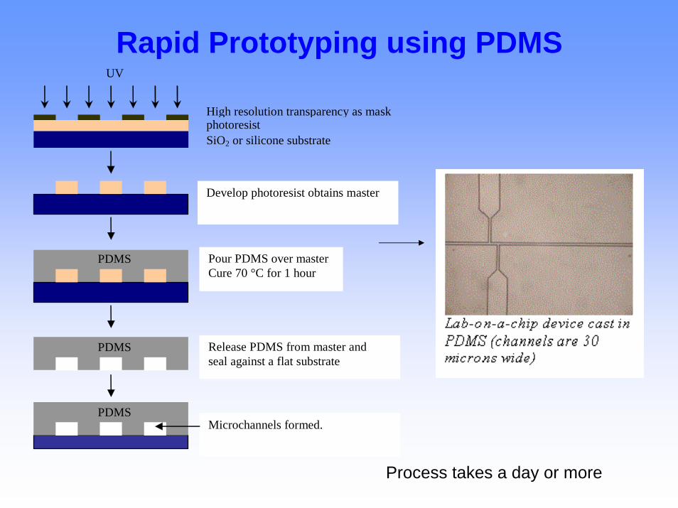
Rapid Prototyping using PDMS UV
High resolution transparency as maskphotoresistSiO2 or silicone substrate
Develop photoresist obtains master
Pour PDMS over master Cure 70 °C for 1 hour
PDMS
PDMS Release PDMS from master and seal against a flat substrate
Microchannels formed. PDMS
Process takes a day or more
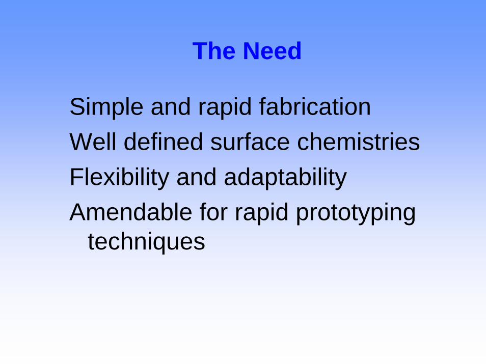
The Need
Simple and rapid fabricationWell defined surface chemistries Flexibility and adaptabilityAmendable for rapid prototyping
techniques
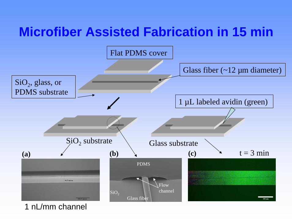
Microfiber Assisted Fabrication in 15 minFlat PDMS cover
Glass fiber (~12 µm diameter)
PDMS
SiO2
SiO2, glass, or PDMS substrate
1 µL labeled avidin (green)
Glass fiber
t = 3 minGlass substrateSiO2 substrate
(a) (b) (c)
Flow channel
1 nL/mm channel
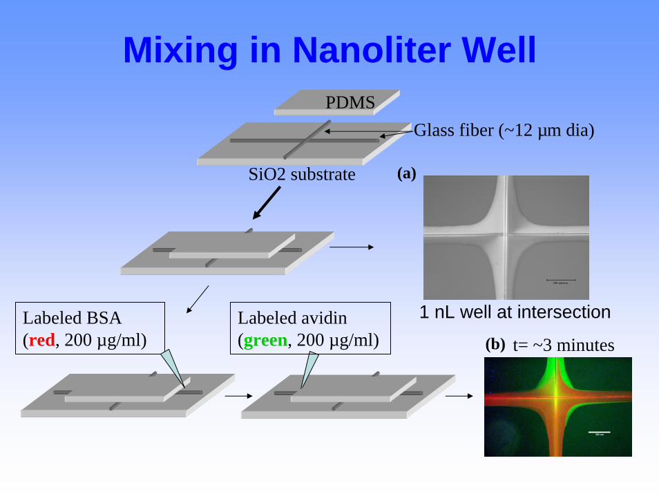
Mixing in Nanoliter Well
Glass fiber (~12 µm dia)PDMS
(a)SiO2 substrate
1 nL well at intersectionLabeled BSA (red, 200 µg/ml)
Labeled avidin (green, 200 µg/ml) t= ~3 minutes(b)
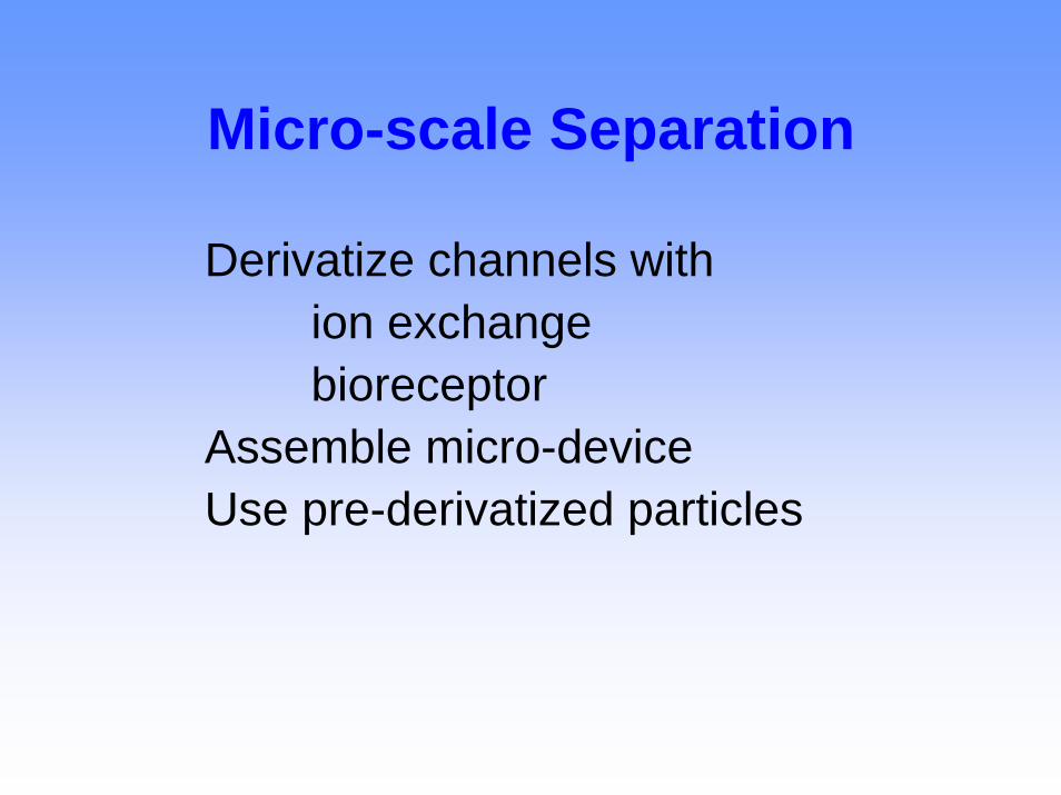
Micro-scale Separation
Derivatize channels with ion exchange bioreceptor
Assemble micro-deviceUse pre-derivatized particles
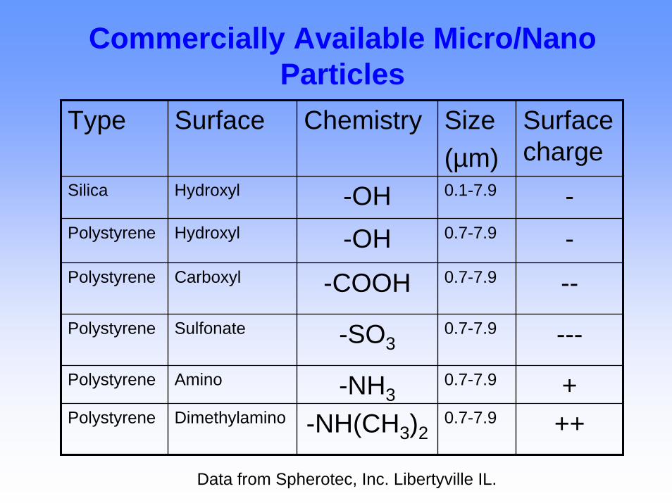
Commercially Available Micro/Nano Particles
Type Surface Chemistry Size(µm)
Surface charge
Silica Hydroxyl -OH 0.1-7.9 -Polystyrene Hydroxyl -OH 0.7-7.9 -Polystyrene Carboxyl -COOH 0.7-7.9 --Polystyrene Sulfonate -SO3
0.7-7.9 ---Polystyrene Amino -NH3
0.7-7.9 +Polystyrene Dimethylamino -NH(CH3)2
0.7-7.9 ++
Data from Spherotec, Inc. Libertyville IL.
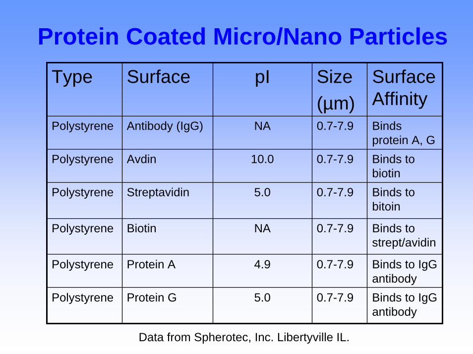
Protein Coated Micro/Nano ParticlesType Surface pI Size
(µm)Surface Affinity
Polystyrene Antibody (IgG) NA 0.7-7.9 Binds protein A, G
Polystyrene Avdin 10.0 0.7-7.9 Binds to biotin
Polystyrene Streptavidin 5.0 0.7-7.9 Binds to bitoin
Polystyrene Biotin NA 0.7-7.9 Binds to strept/avidin
Polystyrene Protein A 4.9 0.7-7.9 Binds to IgGantibody
Polystyrene Protein G 5.0 0.7-7.9 Binds to IgGantibody
Data from Spherotec, Inc. Libertyville IL.
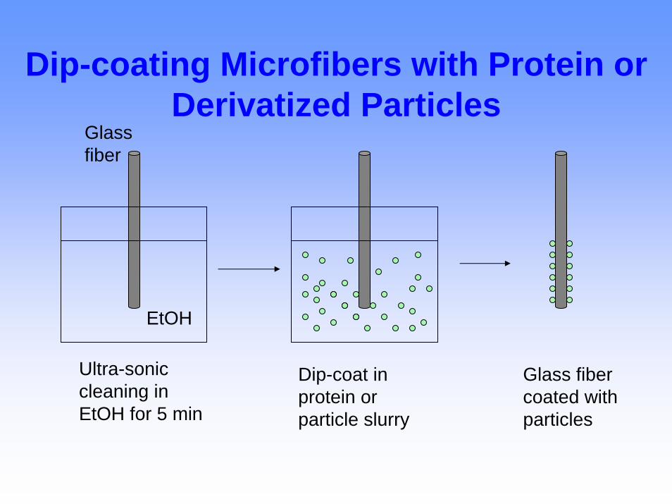
Dip-coating Microfibers with Protein or Derivatized Particles
Glass fiber
EtOH
Ultra-sonic cleaning in EtOH for 5 min
Dip-coat in protein or particle slurry
Glass fiber coated with particles
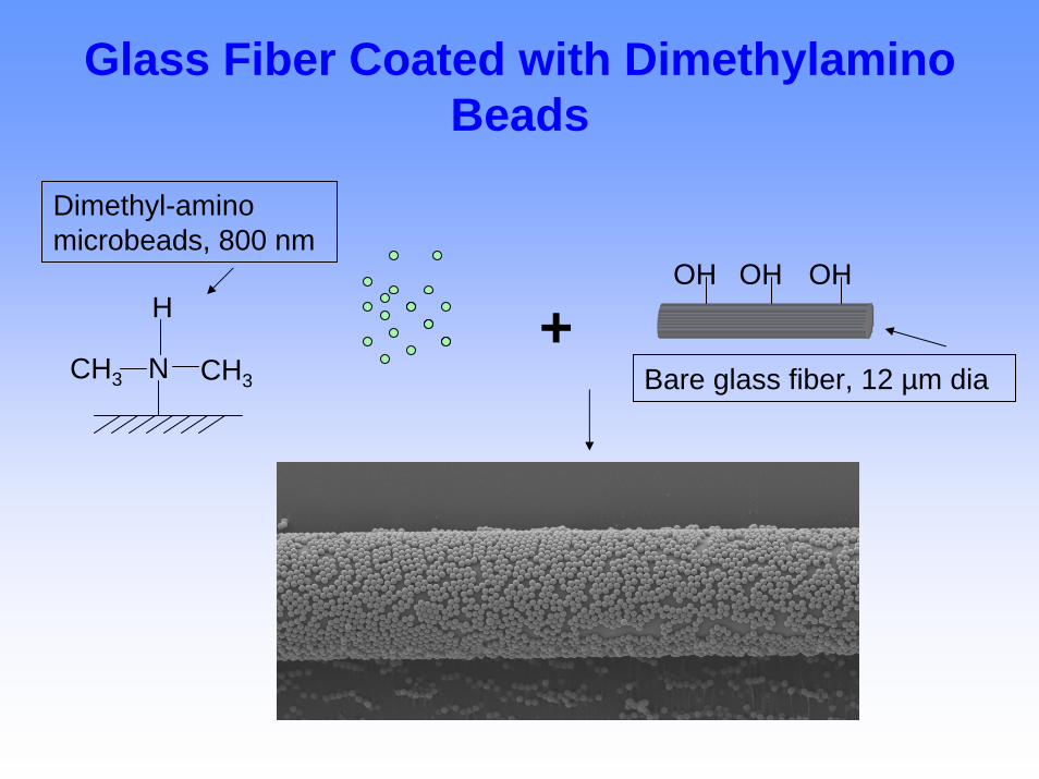
Glass Fiber Coated with DimethylaminoBeads
Dimethyl-amino microbeads, 800 nm
OH OH OH
+N CH33
H
CH Bare glass fiber, 12 µm dia
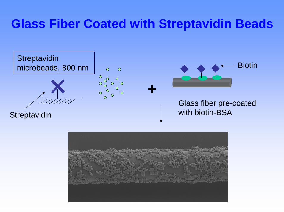
Glass Fiber Coated with Streptavidin Beads
Streptavidin microbeads, 800 nm Biotin
+Glass fiber pre-coated with biotin-BSAStreptavidin
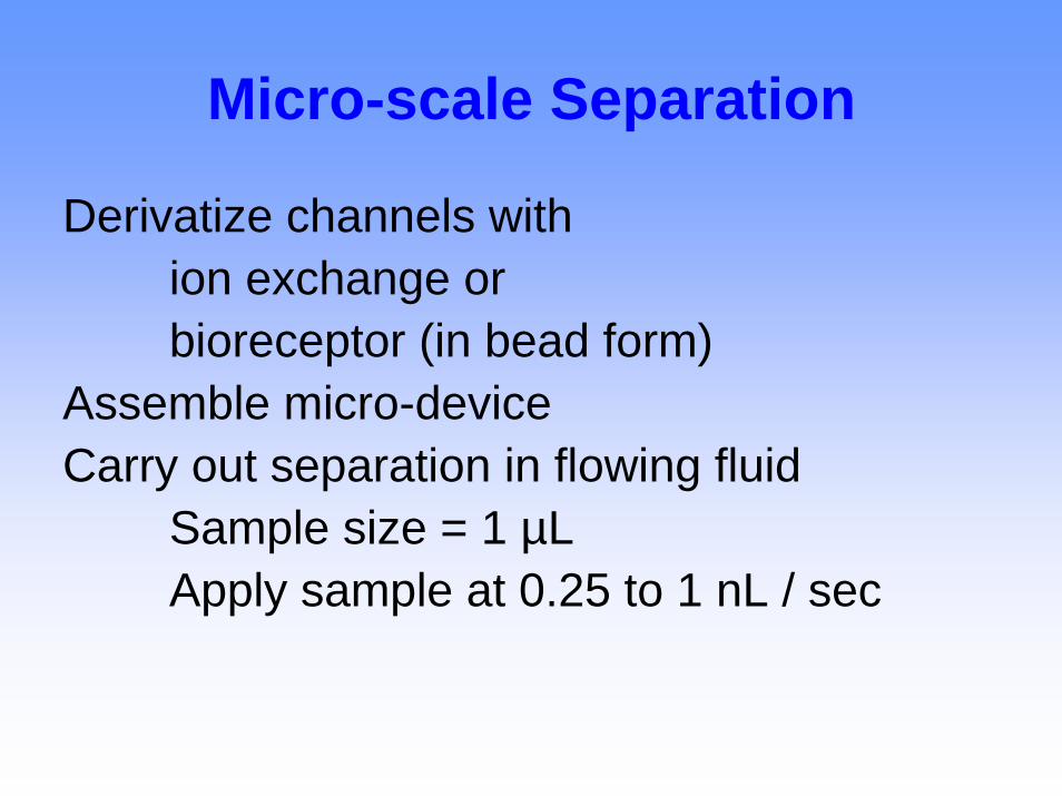
Micro-scale Separation
Derivatize channels with ion exchange orbioreceptor (in bead form)
Assemble micro-deviceCarry out separation in flowing fluid
Sample size = 1 µLApply sample at 0.25 to 1 nL / sec
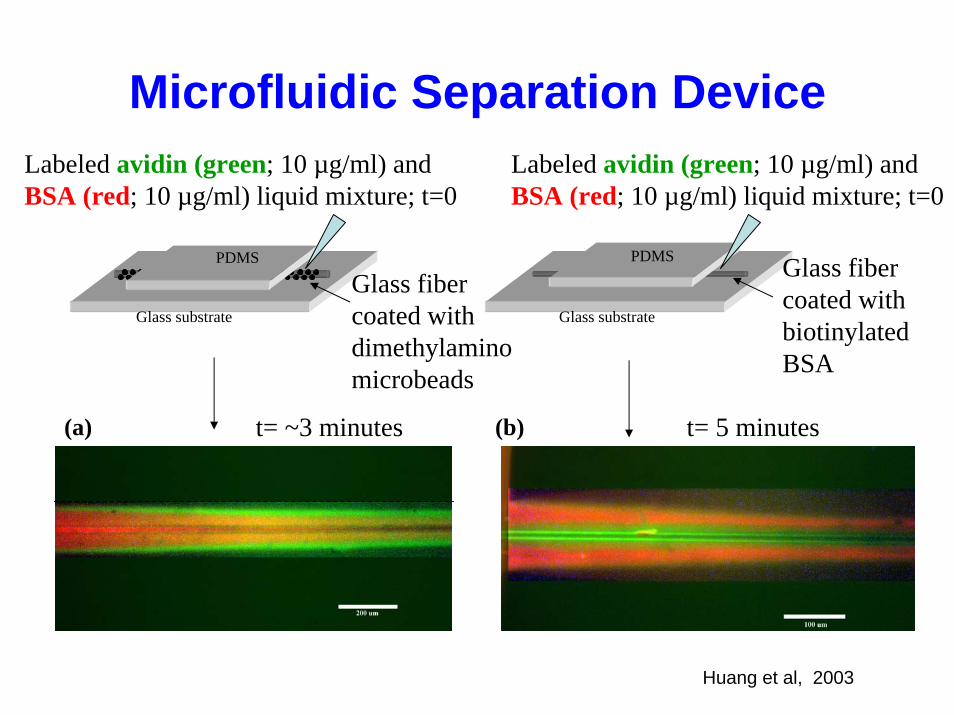
Microfluidic Separation DeviceLabeled avidin (green; 10 µg/ml) and BSA (red; 10 µg/ml) liquid mixture; t=0
Labeled avidin (green; 10 µg/ml) and BSA (red; 10 µg/ml) liquid mixture; t=0
Glass fiber coated with dimethylamino microbeads
Glass fiber coated with biotinylated BSA
Glass substrate Glass substrate
PDMSPDMS
t= ~3 minutes t= 5 minutes(a) (b)
Huang et al, 2003
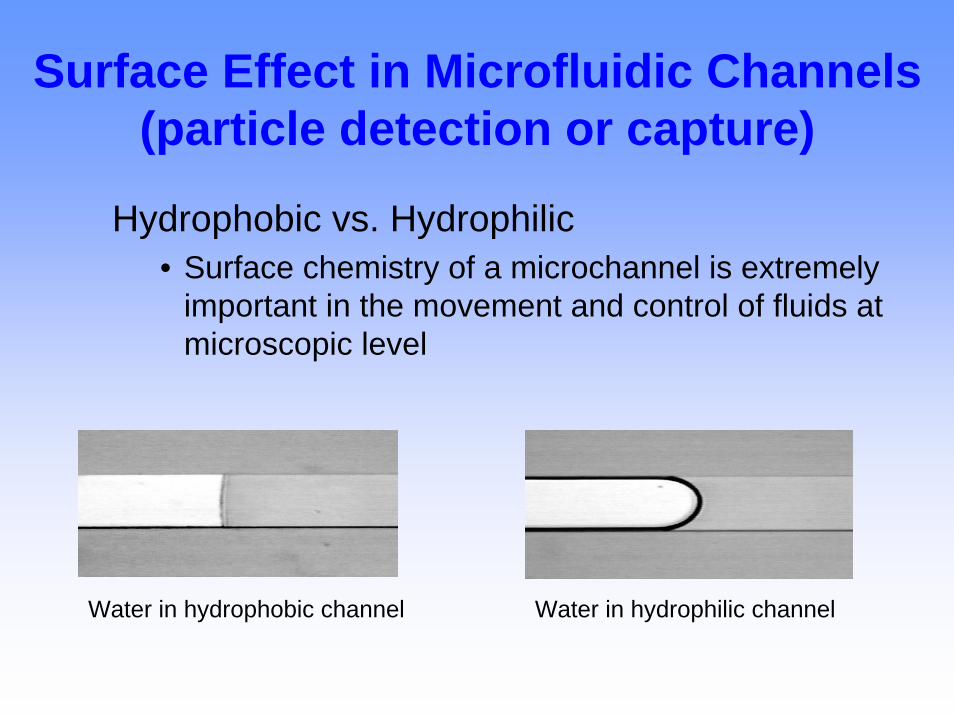
Surface Effect in Microfluidic Channels (particle detection or capture)
Hydrophobic vs. Hydrophilic• Surface chemistry of a microchannel is extremely
important in the movement and control of fluids at microscopic level
Water in hydrophobic channel Water in hydrophilic channel
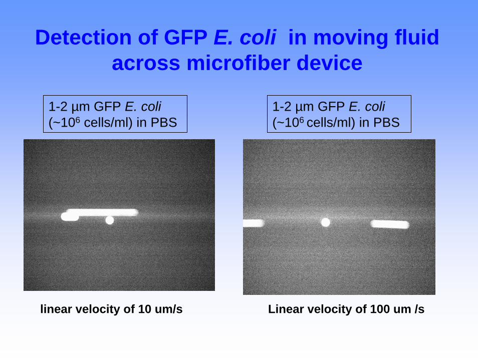
Detection of GFP E. coli in moving fluid across microfiber device
1-2 µm GFP E. coli(~106 cells/ml) in PBS
1-2 µm GFP E. coli(~106 cells/ml) in PBS
linear velocity of 10 um/s Linear velocity of 100 um /s
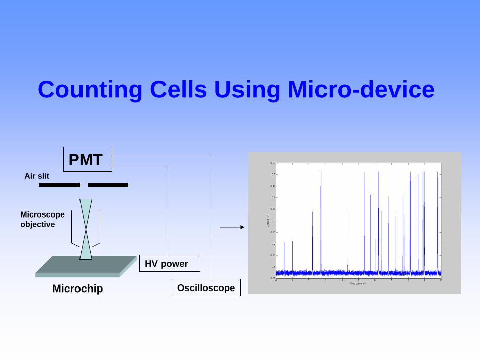
Counting Cells Using Micro-device
PMT
HV power
Air slit
Microscope objective
Microchip Oscilloscope
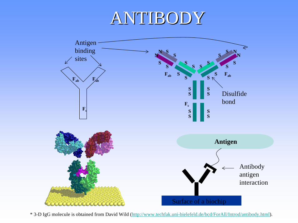
ANTIBODYANTIBODY
Fc
Fab Fab
SS
SS
SS
SS
SS
SSS
SS
S
SS
SS
SS
SS
N NNN
Fc
Fab Fab
Antigen binding sites
Disulfide bond
Antigen
Surface of a biochip
* 3-D IgG molecule is obtained from David Wild (http://www.techfak.uni-bielefeld.de/bcd/ForAll/Introd/antibody.html).
Antibody antigen interaction
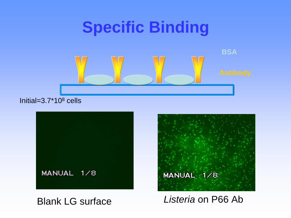
Specific Binding
Antibody
BSA
Blank LG surface Listeria on P66 Ab
Initial=3.7*108 cells
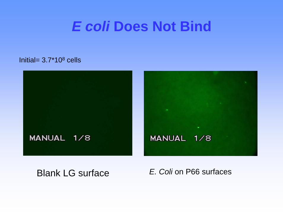
E coli Does Not Bind
Initial= 3.7*108 cells
E. Coli on P66 surfacesBlank LG surface
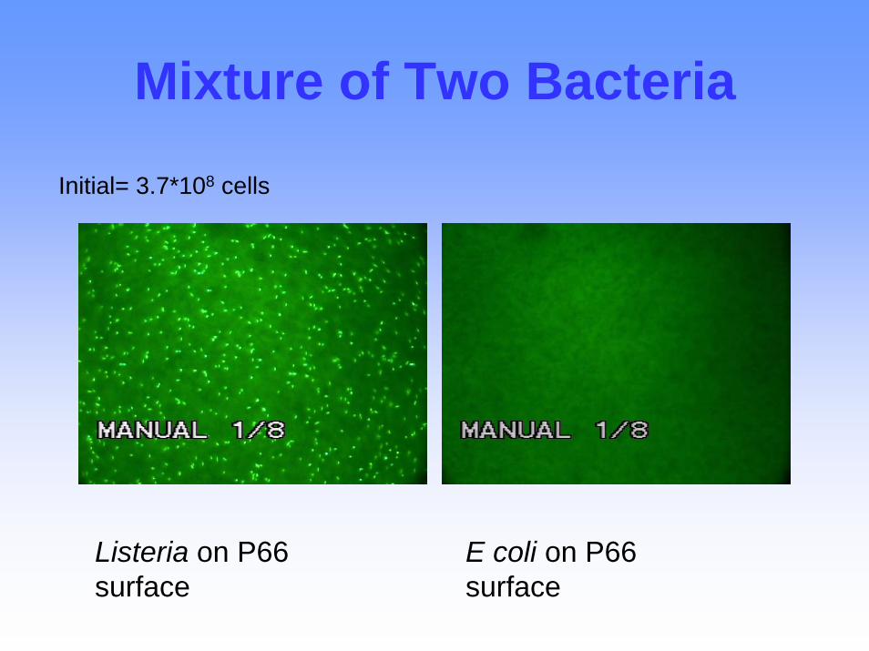
Mixture of Two Bacteria
Initial= 3.7*108 cells
Listeria on P66 surface
E coli on P66 surface
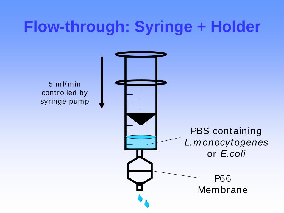
Flow-through: Syringe + Holder
5 ml/min controlled by syringe pump
PBS containing L.monocytogenes
or E.coli
P66 Membrane
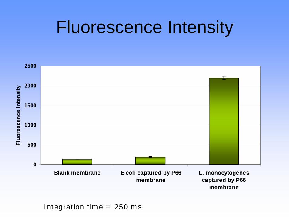
Fluorescence Intensity
0
500
1000
1500
2000
2500
Blank membrane E coli captured by P66membrane
L. monocytogenescaptured by P66
membrane
Fluo
resc
ence
Inte
nsity
Integration time = 250 ms
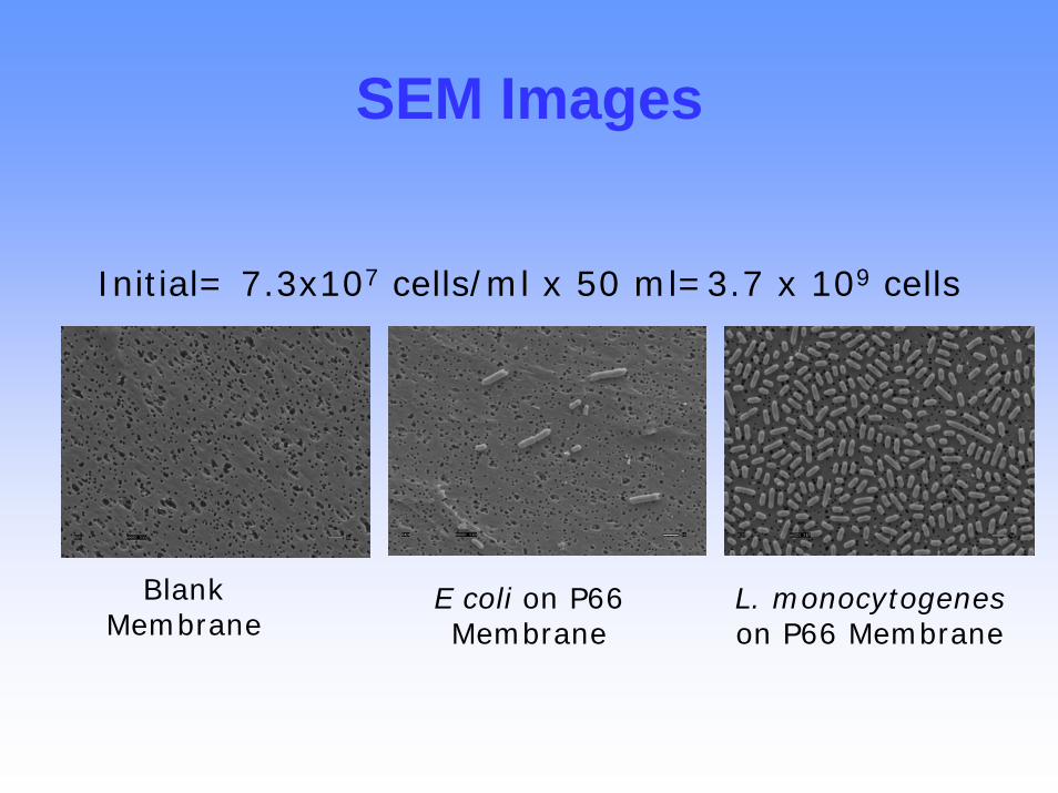
SEM Images
Blank Membrane
E coli on P66 Membrane
L. monocytogeneson P66 Membrane
Initial= 7.3x10⁷ cells/ml x 50 ml=3.7 x 10⁹ cells

Close-up of a Protein Biochip
Glass cover
In/Out ports
Channels/Wells
Epoxy adhesive
Pin
Tip of a pin
Well = 5 nanoliters30 to 50 wells/chip
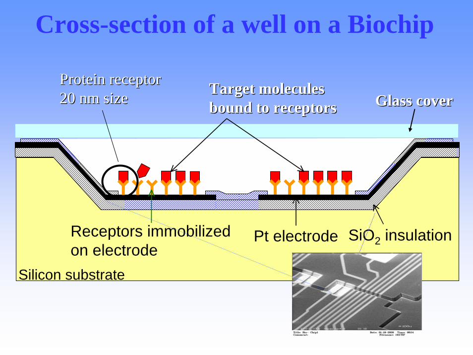
Cross-section of a well on a Biochip
Silicon substrate
Pt electrode SiO2 insulation
Glass coverGlass coverTarget molecules Target molecules bound to receptorsbound to receptors
Receptors immobilized on electrode
Protein receptorProtein receptor20 nm size20 nm size
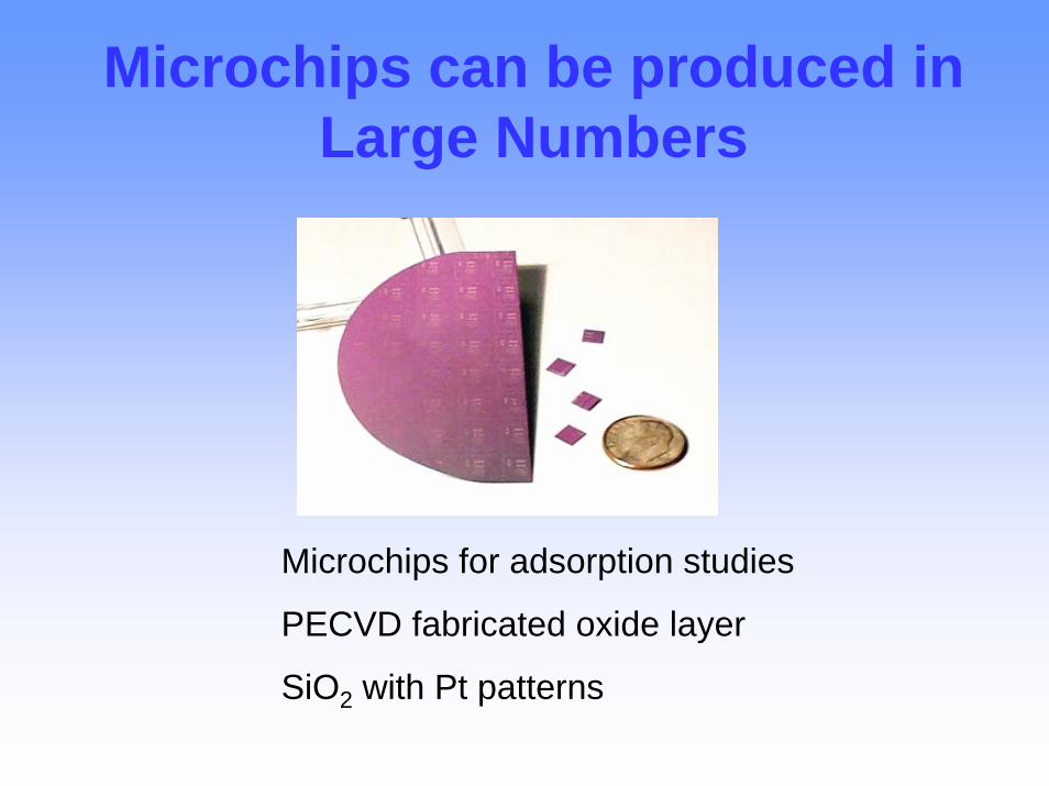
Microchips can be produced in Large Numbers
Microchips for adsorption studies
PECVD fabricated oxide layer
SiO2 with Pt patterns
To use chip1. Sample fluid2. Place fluid onto chip for
interrogation3. Electrically detect if receptor binds
something present in the fluidComplete steps 1 through 3 in three hours.
That “something” could be a pathogen that requires a rapid, preventative response.
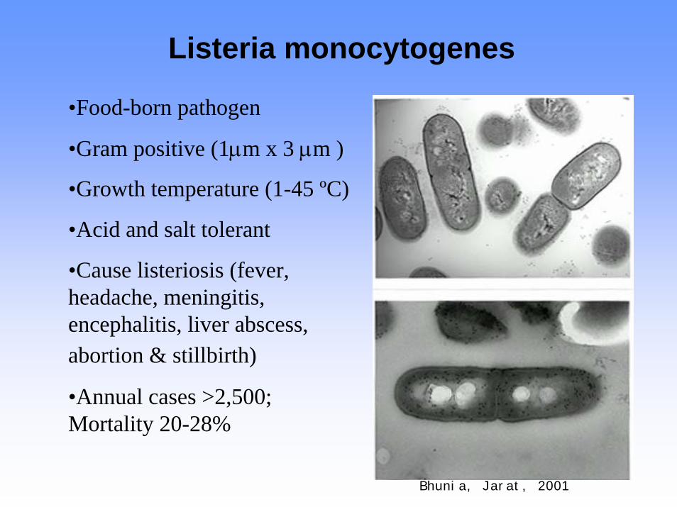
Listeria monocytogenes
•Food-born pathogen
•Gram positive (1µm x 3 µm )
•Growth temperature (1-45 ºC)
•Acid and salt tolerant
•Cause listeriosis (fever, headache, meningitis, encephalitis, liver abscess, abortion & stillbirth)
•Annual cases >2,500; Mortality 20-28%
Bhunia, Jarat, 2001
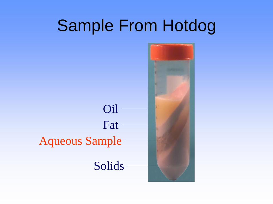
Sample From Hotdog
OilFat
Aqueous Sample
Solids
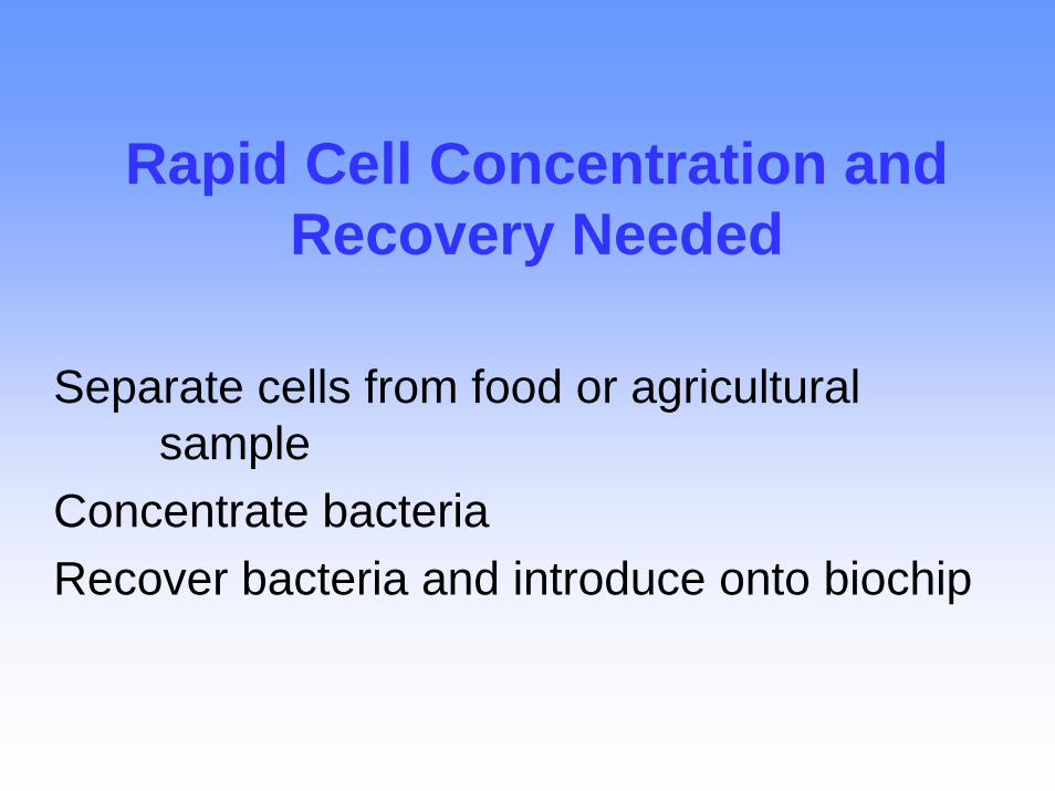
Rapid Cell Concentration and Recovery Needed
Separate cells from food or agricultural sample
Concentrate bacteria Recover bacteria and introduce onto biochip
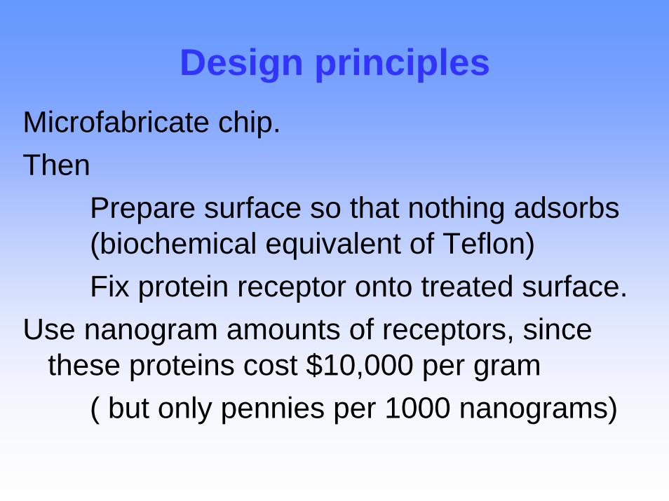
Design principlesMicrofabricate chip. Then
Prepare surface so that nothing adsorbs (biochemical equivalent of Teflon)Fix protein receptor onto treated surface.
Use nanogram amounts of receptors, since these proteins cost $10,000 per gram
( but only pennies per 1000 nanograms)
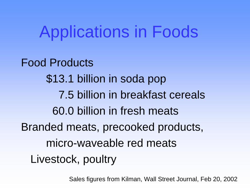
Applications in Foods
Food Products$13.1 billion in soda pop
7.5 billion in breakfast cereals60.0 billion in fresh meats
Branded meats, precooked products, micro-waveable red meats
Livestock, poultrySales figures from Sales figures from KilmanKilman, Wall Street Journal, Feb 20, 2002, Wall Street Journal, Feb 20, 2002
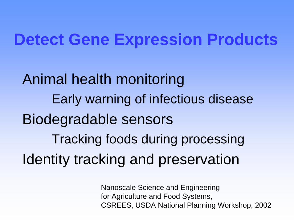
Detect Gene Expression Products
Animal health monitoringEarly warning of infectious disease
Biodegradable sensorsTracking foods during processing
Identity tracking and preservation
Nanoscale Science and Engineering for Agriculture and Food Systems, CSREES, USDA National Planning Workshop, 2002
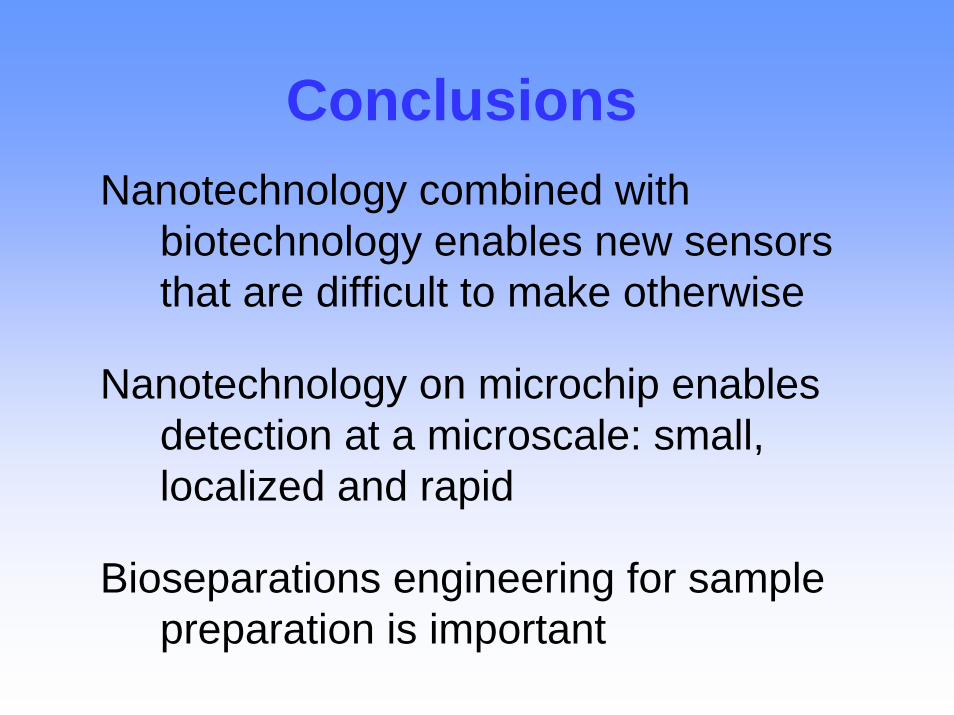
Conclusions Nanotechnology combined with
biotechnology enables new sensors that are difficult to make otherwise
Nanotechnology on microchip enables detection at a microscale: small, localized and rapid
Bioseparations engineering for sample preparation is important


![Introduction to Nanotechnology What is Nanotechnology While many definitions for nanotechnology exist, the [National Nanotechnology Initiative] NNI calls](https://img.pdfslide.us/doc/110x75/56649d9e5503460f94a88dbf/introduction-to-nanotechnology-what-is-nanotechnology-while-many-definitions.jpg)