Embed Size (px)
Citation preview
Functions of Blood
• Blood performs a number of functions dealing with:– Substance distribution– Regulation of blood levels of particular
substances– Body protection
Blood Functions: Distribution
• Blood transports:– Oxygen from the lungs and nutrients from the
digestive tract– Metabolic wastes from cells to the lungs and
kidneys for elimination– Hormones from endocrine glands to target
organs
Blood Functions: Regulation
• Blood maintains:– Appropriate body temperature by absorbing and
distributing heat to other parts of the body– Normal pH in body tissues using buffer systems– Adequate fluid volume in the circulatory
system
Blood Functions: Protection
• Blood prevents blood loss by:– Activating plasma proteins and platelets
– Initiating clot formation when a vessel is broken
• Blood prevents infection by: – Synthesizing and utilizing antibodies
– Activating complement proteins
– Activating WBCs to defend the body against foreign invaders
Physical Characteristics of Blood• Average volume of blood:
– 5–6 L for males; 4–5 L for females (Normovolemia)– Hypovolemia - low blood volume– Hypervolemia - high blood volume
• Viscosity (thickness) - 4 - 5 (where water = 1)• The pH of blood is 7.35–7.45; x = 7.4• Osmolarity = 300 mOsm or 0.3 Osm
– This value reflects the concentration of solutes in the plasma
• Salinity = 0.85%– Reflects the concentration of NaCl in the blood
• Temperature is 38C, slightly higher than “normal” body temperature
• Blood accounts for approximately 8% of body weight
Composition of Blood• Blood is the body’s only fluid tissue (a
connective tissue)
• 2 major components– Liquid = plasma (55%)– Formed elements (45%)
• Erythrocytes, or red blood cells (RBCs)
• Leukocytes, or white blood cells (WBCs)
• Platelets - fragments of megakaryocytes in marrow
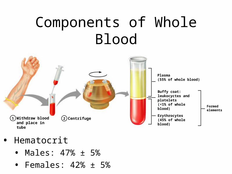
Components of Whole Blood
Withdraw blood and place in tube
1 2 Centrifuge
Plasma(55% of whole blood)
Formed elements
Buffy coat:leukocyctes and platelets(<1% of whole blood)
Erythrocytes(45% of whole blood)
• Hematocrit • Males: 47% ± 5%
• Females: 42% ± 5%
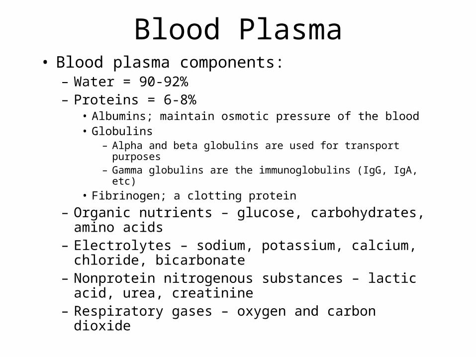
Blood Plasma• Blood plasma components:
– Water = 90-92%– Proteins = 6-8%
• Albumins; maintain osmotic pressure of the blood• Globulins
– Alpha and beta globulins are used for transport purposes– Gamma globulins are the immunoglobulins (IgG, IgA, etc)
• Fibrinogen; a clotting protein
– Organic nutrients – glucose, carbohydrates, amino acids
– Electrolytes – sodium, potassium, calcium, chloride, bicarbonate
– Nonprotein nitrogenous substances – lactic acid, urea, creatinine
– Respiratory gases – oxygen and carbon dioxide
Formed Elements• Formed elements comprise 45% of blood• Erythrocytes, leukocytes, and platelets
make up the formed elements– Only WBCs are complete cells– RBCs have no nuclei or organelles, and
platelets are just cell fragments
• Most formed elements survive in the bloodstream for only a few days
• Most blood cells do not divide but are renewed by cells in bone marrow
Erythrocytes (RBCs)• Biconcave disc
– Folding increases surface area (30% more surface area)– Plasma membrane contains spectrin
• Give erythrocytes their flexibility• Anucleate, no centrioles, no organelles
– End result - no cell division– No mitochondria means they generate ATP anaerobically
• Prevents consumption of O2 being transported
• Filled with hemoglobin (Hb) - 97% of cell contents– Hb functions in gas transport
• Hb + O2 HbO2 (oxyhemoglobin)• Most numerous of the formed elements
– Females: 4.3–5.2 million cells/cubic millimeter– Males: 5.2–5.8 million cells/cubic millimeter
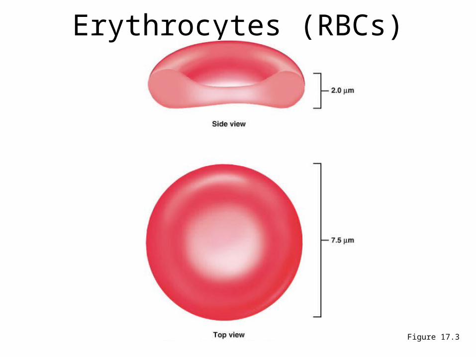
Erythrocytes (RBCs)
Figure 17.3

Erythrocyte Function• Erythrocytes are dedicated to respiratory gas
transport• Hemoglobin reversibly binds with oxygen and
most oxygen in the blood is bound to hemoglobin• Composition of hemoglobin
– A protein called globin• made up of two alpha and two beta chains
– A heme molecule• Each heme group bears an atom of iron, which can bind to one
oxygen molecule• Each hemoglobin molecule thus can transport four molecules
of oxygen
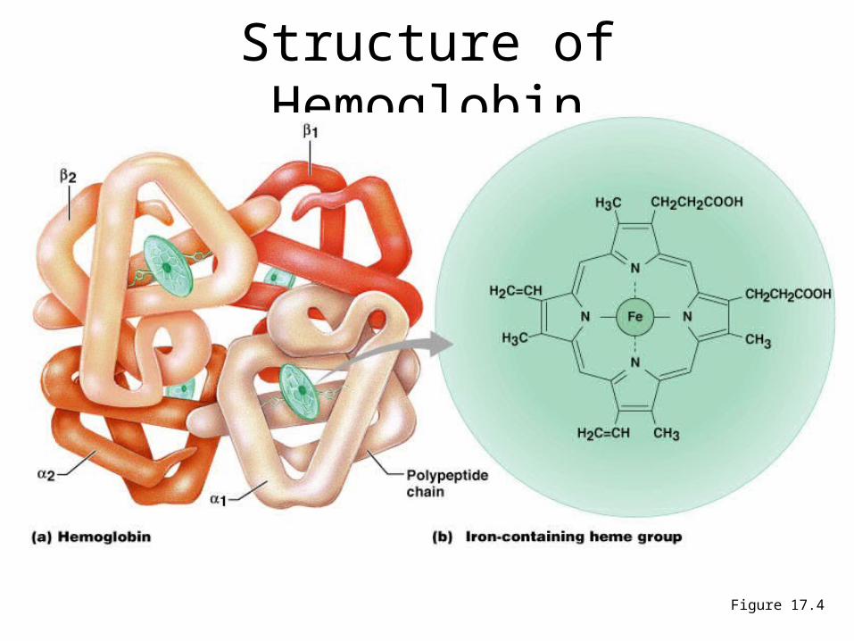
Structure of Hemoglobin
Figure 17.4
Hemoglobin
• Oxyhemoglobin – hemoglobin bound to oxygen– Oxygen loading takes place in the lungs
• Deoxyhemoglobin – hemoglobin after oxygen diffuses into tissues (reduced Hb)
• Carbaminohemoglobin – hemoglobin bound to carbon dioxide
– Carbon dioxide loading takes place in the tissues
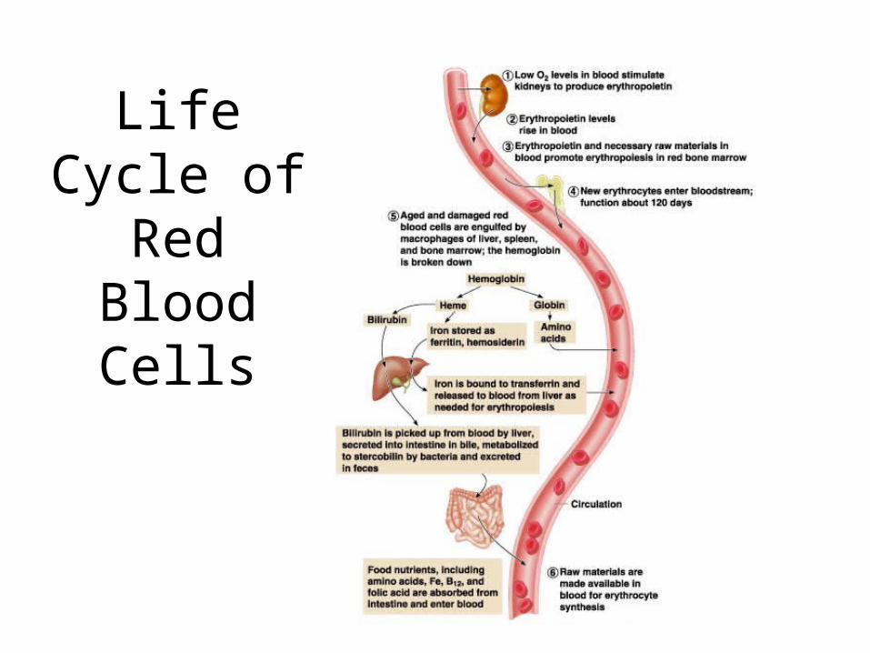
Life Cycle of Red
Blood Cells
Fate and Destruction of Erythrocytes
• The life span of an erythrocyte is 100–120 days– Travels about 750 miles in that time (LA to Albuquerque)
• Old erythrocytes become rigid and fragile, and their hemoglobin begins to degenerate
• Dying erythrocytes are engulfed by macrophages• Heme and globin are separated
– Iron is removed from the heme and salvaged for reuse• Stored as hemosiderin or ferritin in tissues
• Transported in plasma by beta-globulins as transferrin
Fate and Destruction of Erythrocytes
• Heme is degraded to a yellow pigment called bilirubin– Liver secretes bilirubin into the intestines as bile
– Intestines metabolize bilirubin into urobilinogen
– Urobilinogen leaves the body in feces, in a pigment called stercobilin
• Globin is metabolized into amino acids which are then released into the circulation
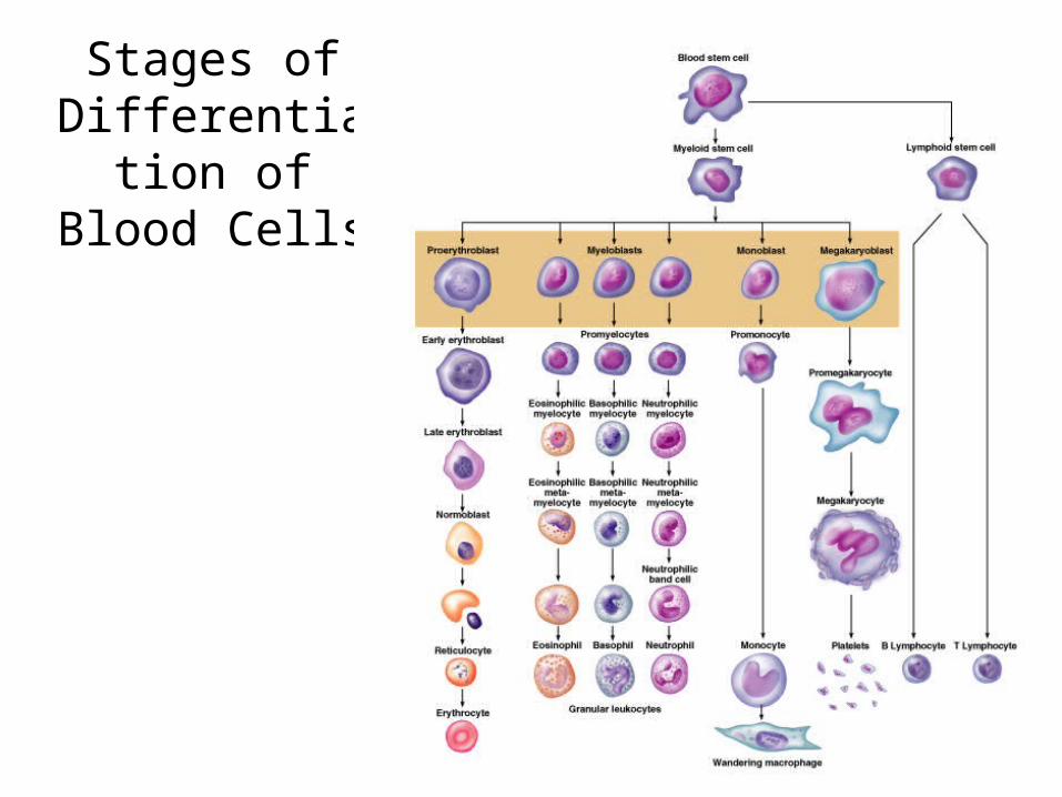
Stages of Differentiation of
Blood Cells
Figure 17.9
Production of Erythrocytes
• Hematopoiesis – blood cell formation– Occurs in the red bone marrow (myeloid tissue)
• Axial skeleton and girdles
• Epiphyses of the humerus and femur
• Marrow contains immature erythrocytes
• Composed of reticular connective tissue
• Hemocytoblasts give rise to ALL formed elements – Lymphoid stem cells - give rise to lymphocytes
– Myeloid stem cells - give rise to all other blood cells
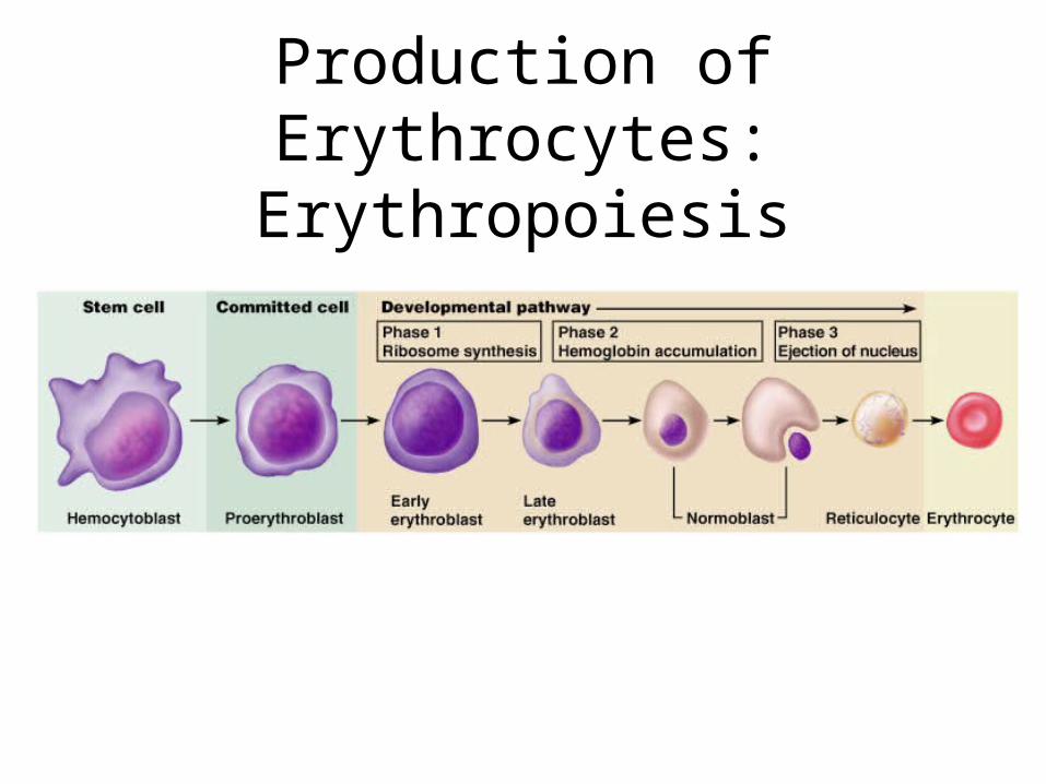
Production of Erythrocytes: Erythropoiesis
• A hemocytoblast is transformed into a committed cell called the proerythroblast
• Proerythroblasts develop into early erythroblasts• The developmental pathway consists of three phases
– Phase 1 – ribosome synthesis in early erythroblasts– Phase 2 – hemoglobin accumulation in late erythroblasts and
normoblasts– Phase 3 – ejection of the nucleus from normoblasts and formation
of reticulocytes
• Reticulocytes then become mature erythrocytes– Reticulocytes make up about 1 -2 % of all circulating erythrocytes
Production of Erythrocytes: Erythropoiesis
• Circulating erythrocytes – the number remains constant and reflects a balance between RBC production and destruction– Too few red blood cells leads to tissue hypoxia– Too many red blood cells causes undesirable blood
viscosity
• Erythropoiesis is hormonally controlled and depends on adequate supplies of iron, amino acids, and B vitamins
Regulation and Requirements for Erythropoiesis
Hormonal Control of Erythropoiesis
• Erythropoietin (EPO) release by the kidneys is triggered by:– Hypoxia due to decreased RBCs– Decreased oxygen availability– Increased tissue demand for oxygen
• Enhanced erythropoiesis increases the: – RBC count in circulating blood– Oxygen carrying ability of the blood
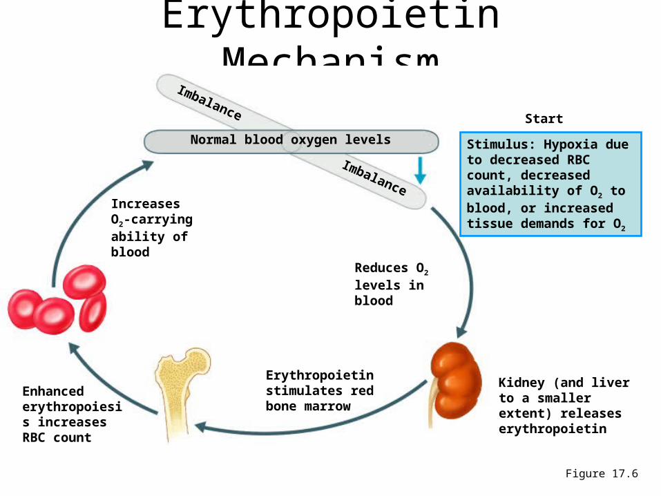
Erythropoietin Mechanism
Figure 17.6
Imbalance
Reduces O2 levels in blood
Erythropoietin stimulates red bone marrow
Enhanced erythropoiesis increases RBC count
Normal blood oxygen levels Stimulus: Hypoxia due to decreased RBC count, decreased availability of O2 to blood, or increased tissue demands for O2
Imbalance
Start
Kidney (and liver to a smaller extent) releases erythropoietin
Increases O2-carrying ability of blood
• Erythropoiesis requires:– Proteins, lipids, and carbohydrates– Iron, vitamin B12, and folic acid
• The body stores iron in Hb (65%), the liver, spleen, and bone marrow
• Intracellular iron is stored in protein-iron complexes such as ferritin and hemosiderin
• Circulating iron is loosely bound to the transport protein transferrin
Dietary Requirements of Erythropoiesis

• Polycythemia– Abnormal excess of erythrocytes
• Increases viscosity, decreases flow rate of blood
• Anemia – blood has abnormally low oxygen-carrying capacity– It is a symptom rather than a disease itself– Blood oxygen levels cannot support normal
metabolism– Signs/symptoms include fatigue, paleness,
shortness of breath, and chills
Erythrocyte Disorders
Anemia: Insufficient Erythrocytes
• Hemorrhagic anemia – result of acute or chronic loss of blood
• Hemolytic anemia – prematurely ruptured erythrocytes
• Aplastic anemia – destruction or inhibition of red bone marrow
• Iron-deficiency anemia results from:– A secondary result of hemorrhagic anemia– Inadequate intake of iron-containing foods– Impaired iron absorption
• Pernicious anemia results from:– Deficiency of vitamin B12
– Lack of intrinsic factor needed for absorption of B12
– Treatment is intramuscular injection of B12
Anemia: Decreased Hemoglobin Content
Anemia: Abnormal Hemoglobin
• Thalassemias – absent or faulty globin chain in hemoglobin – Erythrocytes are thin, delicate, and deficient in
hemoglobin
• Sickle-cell anemia – results from a defective gene– Codes for an abnormal hemoglobin called hemoglobin
S (HbS)
– This defect causes RBCs to become sickle-shaped in low oxygen situations
Polycythemia
• Polycythemia – excess RBCs that increase blood viscosity
• Three main polycythemias are:– Polycythemia vera– Secondary polycythemia– Blood doping
Leukocytes (WBCs)• Leukocytes, the only blood components that are complete cells:
– 4,800 - 10,000/cubic millimeter– Protect the body from infectious microorganisms– Can leave capillaries via diapedesis– Move through tissue spaces (amoeboid motion)– Many are phagocytic (possess numerous lysosomes)
• Two major types of leukocytes– Granulocytes: Neutrophils, Eosinophils, Basophils– Agranulocytes: Monocytes, Lymphyocytes
• Leukocytosis – WBC count over 11,000/mm3
– Normal response to bacterial or viral invasion• Leukopenia - a decrease in WBC count below 4,800/mm3 • Leukemia - a cancer of WBC
Granulocytes
• Granulocytes – neutrophils, eosinophils, and basophils– Contain cytoplasmic granules that stain
specifically (acidic, basic, or both) with Wright’s stain
– Are larger and usually shorter-lived than RBCs– Have lobed nuclei– Are all phagocytic cells
• Account for 65-75% of total WBC’s• Neutrophils have two types of granules that:
– Take up both acidic and basic dyes– Give the cytoplasm a lilac color– Contain peroxidases, hydrolytic enzymes, and
defensins (antibiotic-like proteins)
• Neutrophils are our body’s bacteria slayers• AKA “polys” or PMN’s (polymorphonuclear)
Granulocytes: Neutrophils(Polymorphonuclear leukocytes)
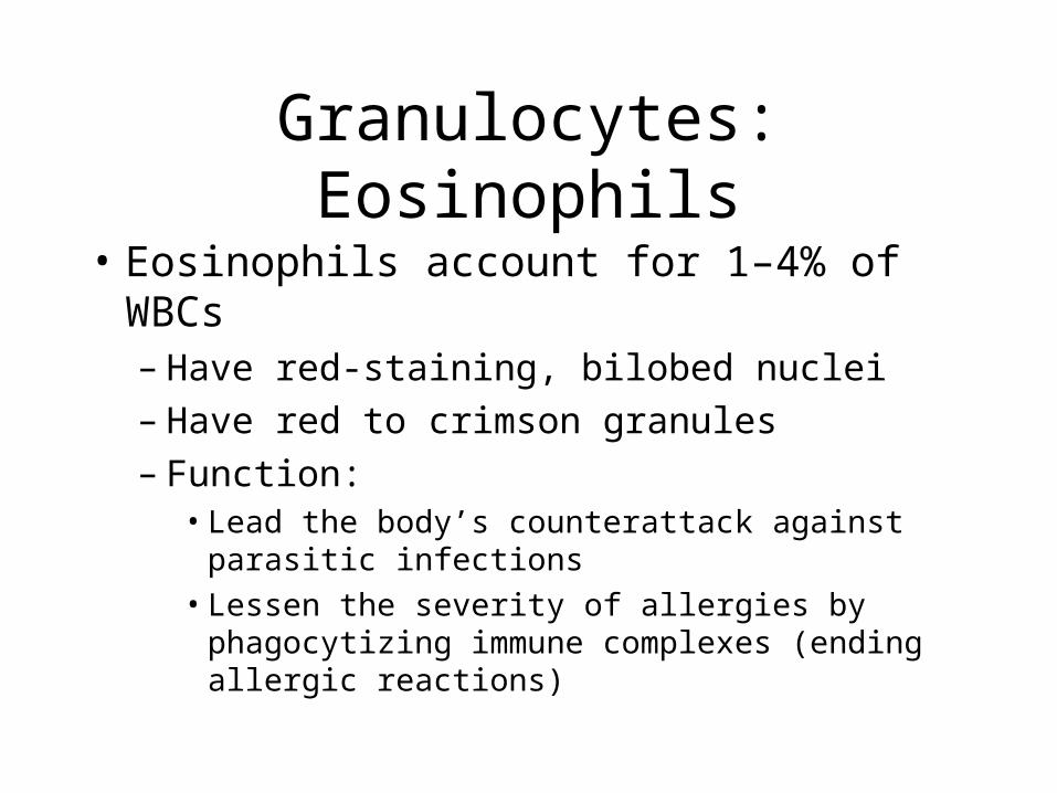
• Eosinophils account for 1–4% of WBCs – Have red-staining, bilobed nuclei– Have red to crimson granules– Function:
• Lead the body’s counterattack against parasitic infections
• Lessen the severity of allergies by phagocytizing immune complexes (ending allergic reactions)
Granulocytes: Eosinophils
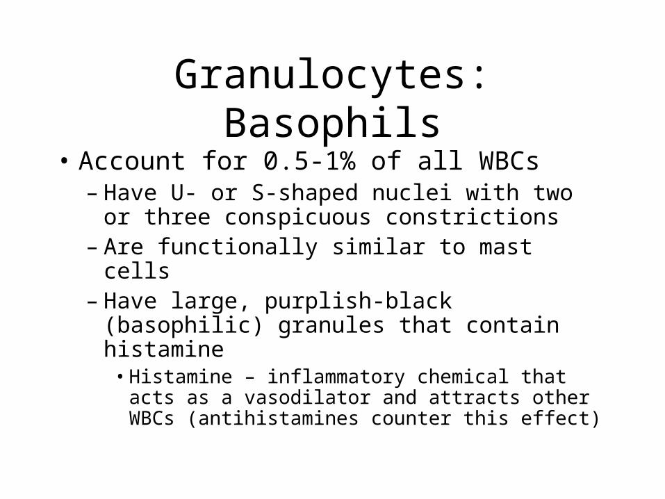
• Account for 0.5-1% of all WBCs– Have U- or S-shaped nuclei with two or three
conspicuous constrictions– Are functionally similar to mast cells– Have large, purplish-black (basophilic)
granules that contain histamine• Histamine – inflammatory chemical that acts as a
vasodilator and attracts other WBCs (antihistamines counter this effect)
Granulocytes: Basophils
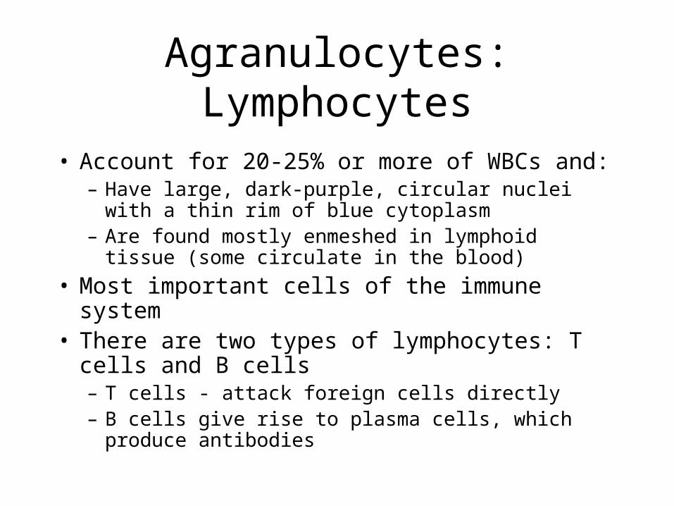
• Account for 20-25% or more of WBCs and:– Have large, dark-purple, circular nuclei with a thin rim
of blue cytoplasm– Are found mostly enmeshed in lymphoid tissue (some
circulate in the blood)
• Most important cells of the immune system• There are two types of lymphocytes: T cells and B
cells– T cells - attack foreign cells directly– B cells give rise to plasma cells, which produce
antibodies
Agranulocytes: Lymphocytes
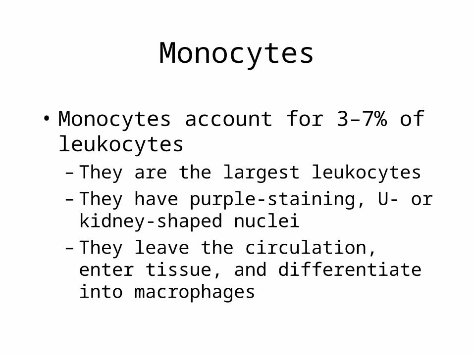
• Monocytes account for 3–7% of leukocytes – They are the largest leukocytes– They have purple-staining, U- or kidney-shaped
nuclei– They leave the circulation, enter tissue, and
differentiate into macrophages
Monocytes

• Leukopoiesis is hormonally stimulated by two families of cytokines (hematopoietic factors) – interleukins and colony-stimulating factors (CSFs)– Interleukins are numbered (e.g., IL-1, IL-2), whereas
CSFs are named for the WBCs they stimulate (e.g., granulocyte-CSF stimulates granulocytes)
• Macrophages and T cells are the most important sources of cytokines
• Many hematopoietic hormones are used clinically to stimulate bone marrow
Production of Leukocytes
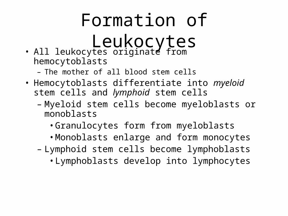
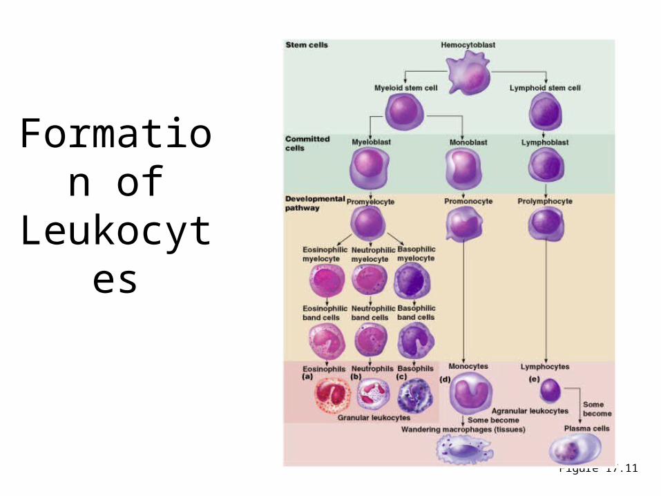
• All leukocytes originate from hemocytoblasts– The mother of all blood stem cells
• Hemocytoblasts differentiate into myeloid stem cells and lymphoid stem cells– Myeloid stem cells become myeloblasts or
monoblasts• Granulocytes form from myeloblasts• Monoblasts enlarge and form monocytes
– Lymphoid stem cells become lymphoblasts• Lymphoblasts develop into lymphocytes
Formation of Leukocytes
Formation of
Leukocytes
Figure 17.11
• Leukemia refers to cancerous conditions involving white blood cells
• Leukemias are named according to the abnormal white blood cells involved– Myelocytic leukemia – involves myeloblasts– Lymphocytic leukemia – involves lymphocytes
• Acute leukemia involves blast-type cells and primarily affects children
• Chronic leukemia is more prevalent in older people
Leukocytes Disorders: Leukemias
• Immature white blood cells are found in the bloodstream in all leukemias
• Bone marrow becomes totally occupied with cancerous leukocytes
• Severe anemia ensues due to excess production of WBC’s
• The white blood cells produced, though numerous, are not functional
• Death is caused by internal hemorrhage and overwhelming infections
• Treatments include irradiation, antileukemic drugs, and bone marrow transplants
Leukemia
• Platelets are fragments of megakaryocytes• Their granules contain serotonin, Ca2+,
enzymes, ADP, and platelet-derived growth factor (PDGF)
• Platelets function in the clotting mechanism by forming a temporary plug that helps seal breaks in blood vessels
• Platelets not involved in clotting are kept inactive by Nitric Oxide (NO) and prostaglandins
Platelets
• RBC membranes have glycoprotein antigens on their external surfaces
• These antigens are:– Unique to the individual – Recognized as foreign if transfused into another
individual– Promoters of agglutination and are referred to as
agglutinogens
• Presence or absence of these antigens is used to classify blood groups
Human Blood Groups
• Humans have 30 varieties of naturally occurring RBC antigens
• The antigens of the ABO and Rh blood groups cause vigorous transfusion reactions when they are improperly transfused
• Other blood groups (M, N, Dufy, Kell, and Lewis) are mainly used for legalities
Blood Groups
• The ABO blood groups consists of:– Two antigens (A and B) on the surface of the RBCs – Two antibodies in the plasma (anti-A and anti-B)
• An individual with ABO blood may have various types of antigens and spontaneously preformed antibodies
• Agglutinogens and their corresponding antibodies cannot be mixed without serious hemolytic reactions
ABO Blood Groups
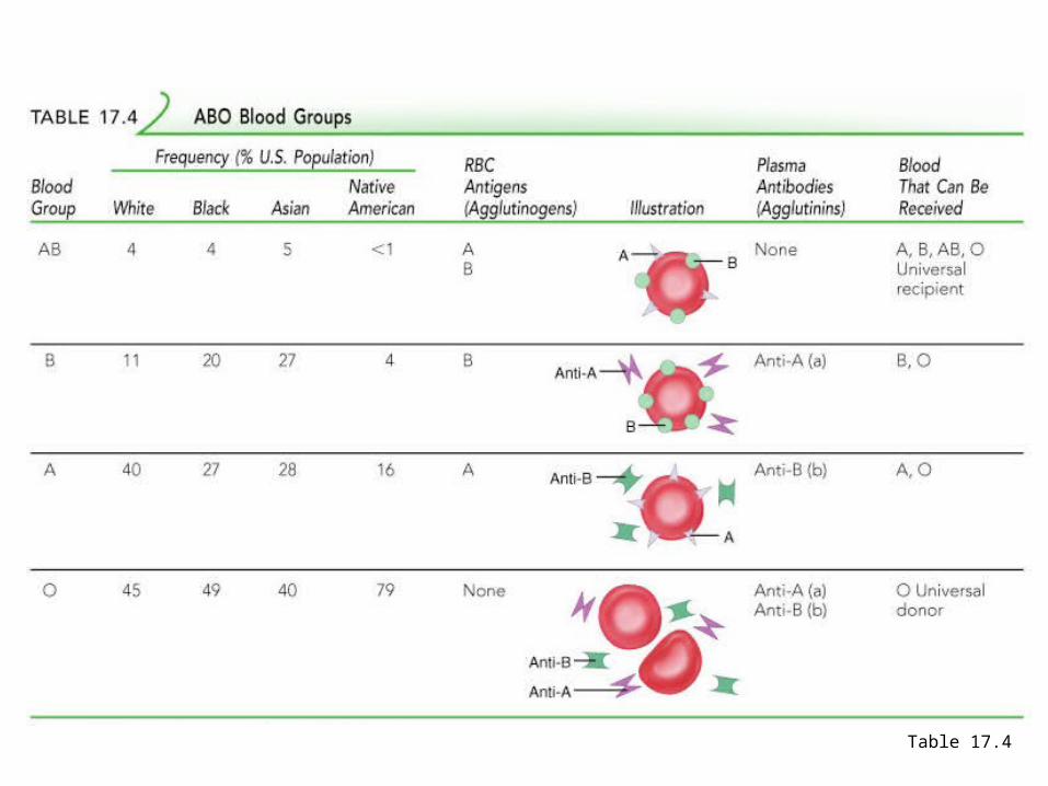
ABO Blood Groups
Table 17.4
• Presence of the Rh agglutinogens on RBCs is indicated as Rh+; 85% of population is +
• Lack of antigen indicated as Rh -; 15% of popn.• Anti-Rh antibodies are not spontaneously formed
only in Rh– individuals• However, if an Rh– individual receives Rh+ blood,
anti-Rh antibodies form• A second exposure to Rh+ blood will result in a
typical transfusion reaction
Rh Blood Groups
• May occur in an Rh- mom pregnanet with an Rh+ fetus• Hemolytic disease of the newborn – Rh+ antibodies of a
sensitized Rh– mother cross the placenta and attack and destroy the RBCs of an Rh+ baby
• Rh– mother becomes sensitized when Rh+ blood (from a previous pregnancy of an Rh+ baby or a Rh+ transfusion) causes her body to synthesis Rh+ antibodies
• The drug RhoGAM can prevent the Rh– mother from becoming sensitized
• Treatment of hemolytic disease of the newborn involves pre-birth transfusions and exchange transfusions after birth
Hemolytic Disease of the Newborn
• Transfusion reactions occur when mismatched blood is infused
• Donor’s cells are attacked by the recipient’s plasma agglutinins causing:– Diminished oxygen-carrying capacity– Clumped cells that impede blood flow– Ruptured RBCs that release free hemoglobin into the
bloodstream
• Circulating hemoglobin precipitates in the kidneys and causes renal failure
Transfusion Reactions
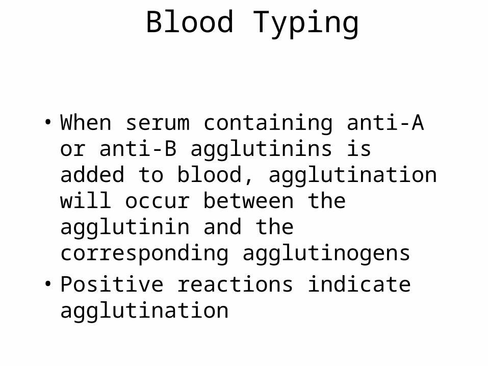
Blood Typing
• When serum containing anti-A or anti-B agglutinins is added to blood, agglutination will occur between the agglutinin and the corresponding agglutinogens
• Positive reactions indicate agglutination
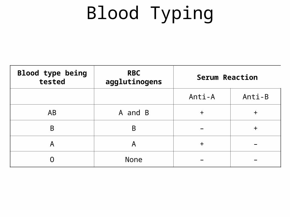
Blood Typing
Blood type being tested RBC agglutinogens Serum Reaction
Anti-A Anti-B
AB A and B + +
B B – +
A A + –
O None – –