Embed Size (px)
Citation preview
Citation: Ikeda E, Nakagawa M, Ogawa M, Takeo M and Tsuji T. Functional Tooth Regeneration as a Next-Generation Therapy. J Dent & Oral Disord. 2020; 6(6): 1146.
J Dent & Oral Disord - Volume 6 Issue 6 - 2020ISSN: 2572-7710 | www.austinpublishinggroup.com Tsuji et al. © All rights are reserved
Journal of Dentistry & Oral DisordersOpen Access
Abstract
Regenerative technology has advanced dramatically based on new findings in the fields of developmental biology, stem cell biology, and tissue engineering technology. Stem cell therapy has been attempted as a treatment for organ dysfunction due to injury, disease, and ageing. The next generation of regenerative therapy is the replacement of a dysfunctional organ. In dentistry, treatment to replace lost teeth with artificial materials, such as using bridges and implants, has been established as conventional treatment. However, tooth functions, including masticatory function and responsiveness to mechanical stress, are not fully restored by these treatments due to the difficulties in incorporation of artificial materials into live system. Tooth function is vital for survival, and functional tooth regeneration is an urgent issue for the coming super-aging society. The emergence of the organ germ method, which can regenerate organ germ utilizing fate determined organ-inductive potential embryonic epithelial and mesenchymal cells, has provided evidence for the concept of fully functional regeneration of organ in vivo. More recently, integration of implant into periodontal tissue through periodontal ligament and partial regeneration of physiological function of teeth has been archived by combination of artificial materials and biological materials, demonstrating that not all materials need to be of biological origin for regenerating functional tooth. In this review article, we will introduce the history and current trends aimed at regenerating a full functional tooth, and we will discuss the potential issue and future directions for putting recent achievements into practical use for the curing of oral and maxillofacial disease.
Keywords: Bio-hybrid implant; Organ replacement regenerative therapy; Bioengineered tooth; Organ germ method; Epithelial-mesenchymal interaction
IntroductionAdvances in regenerative research have been made in many
fields, such as embryonic development, stem cell biology and tissue engineering technology [1-4]. The first generation of regenerative therapy is stem cell transplantation using tissue-derived stem cells, embryonic stem (ES) cells or induced pluripotent stem (iPS) cells; such therapies have been attempted to repair damaged tissues due to disease or trauma [5-9]. Stem cell transplantation therapies then advanced to clinical application for diseases including leukaemia, diabetes, cardiac infarction, and liver disease [4-8,10-17]. The second generation of regenerative medicine is the scaffold method, in which a complex cell mixture and a biodegradable scaffold are employed to grow a tissue of desired form; one example is cell sheet technology [18-20]. These methods have been used for the clinical treatment of various complex tissue reconstitutions. The next generation regenerative therapy is to regenerate fully functional entire three-dimensional (3D) organs [21-26].
During embryonic development, almost all organs arise from the respective organ germs as a result of reciprocal interactions between epithelial and mesenchymal stem cells, according to each individual organ-forming field. Previously, we reported that a bioengineered tooth germ, a model of an ectodermal organ germ, regenerated by mimicking development, including epithelial-mesenchyme
Review Article
Functional Tooth Regeneration as a Next-Generation TherapyIkeda E1,2, Nakagawa M1,2, Ogawa M1,3, Takeo M1 and Tsuji T1* 1RIKEN Center for Biosystems Dynamics Research, Kobe, Hyogo, Japan2Department of Oral and Maxillofacial Surgery, Nara Medical University, Kashihara-shi, Nara, Japan3Organ Technologies Inc., Tokyo, Japan
*Corresponding author: Takashi Tsuji, RIKEN Center for Biosystems Dynamics Research (BDR), Kobe, Hyogo, Japan
Received: June 25, 2020; Accepted: July 20, 2020; Published: July 27, 2020
interaction; this tooth germ could generate a structurally correct tooth after transplantation under a subrenal capsule using in vivo and in vitro organ cultures [21].
In dentistry, regenerative therapy is a desirable method for replacing a tooth with a bioengineered one with complete oral functions. Tooth loss due to oral disease, such as dental caries, periodontal disease and traumatic injury, causes fundamental general health problems due to oral function disorders, including pronunciation, mastication, swallowing, and occlusion, which occur due to cooperation between the oral and maxillofacial regions [26,27]. Therefore, tooth regeneration therapy aims to restore oral function and successfully incorporate the tooth with the surrounding oral environment, which is a crucial issue of quality of life. Research in stem cell transplantation for tooth regeneration and for partial regeneration of dental tissues using cell sheets began in approximately 2002 [27]. However, there were many limitations to the regeneration of functional tooth. In 2007 [21], whole tooth regeneration was demonstrated with full functional recovery. Then, there was the report of the bioengineered tooth germ reconstructed using a organ germ method and a fully functioning bioengineered tooth developed that exhibited the correct tissue structure, masticatory function, responsiveness to mechanical stress and perceptive potential following transplantation into a tooth-loss region [21-23]. Since then, it has become possible to restore the physiological function of teeth using a bio-hybrid implant carrying
J Dent & Oral Disord 6(6): id1146 (2020) - Page - 02
Tsuji T Austin Publishing Group
Submit your Manuscript | www.austinpublishinggroup.com
a regenerative periodontal ligament. The bio-hybrid implant with periodontal tissue similar to natural tissue has been reported, and this strategy demonstrated the feasibility of “next-generation oral implant treatment” with physiological oral functions, such as orthodontic movement and nerve transmission [28].
In this review, we describe the recent progress in partial tooth-tissue repair and whole-tooth regeneration, and we discuss the strategy of tooth regeneration that can provide functional recovery and substitute dental treatments using artificial materials.
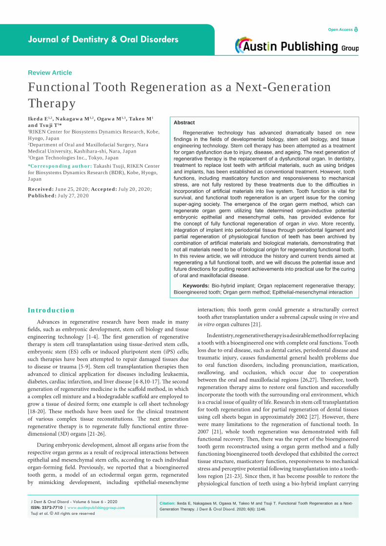
Tooth developmentIn tooth germ development, the dental lamina first thickens
(lamina stage). Then, the epithelium thickens at the future location of a tooth, and then there is subsequent epithelial budding to the underlying neural crest-derived mesenchyme (placode stage). Tooth germ formation is initiated on embryonic days (EDs) 10-11 in mice by epithelial signals, which is followed by the condensing of dental mesenchymal cells around the developing epithelial bud (bud stage). The tooth germ is developed via the interaction of the oral mucosal epithelium and mesenchyme. During these processes, the expression of homeobox genes is induced by the actions of growth factors, cytokines and adhesion molecules. Activation of these genes guides each tooth bud along a pathway to grow a tooth of a given size and shape that is predetermined by its position [29,30]. At ED13.5-14.5, the enamel knot acts as a signalling centre that is responsible for the formation and maintenance of the dental papilla. First enamel knots are formed at the tooth bud and appear at E14.5; then, during the transition from the bud to the cap stage, the first enamel knot, which acts as a signalling centre to orchestrate tooth development, is formed in the dental epithelium (cap stage). Secondary enamel knots start to appear at the late cap stage and are connected to cusp formation. At ED16-18, the epithelial and mesenchymal cells in the tooth germ terminally differentiate into ameloblasts, which later become enamel, and odontoblasts that will form dentin (bell stage). The mesenchyme also differentiates into dental pulp and periodontal tissues, which will become cementum, periodontal ligament and alveolar bone [30] (Figure 1).
Concept of tooth regenerationThe teeth and their multicellular structure play important roles
in physiological function, including mastication, swallowing and pronunciation, and they are indispensable for adequate general
health. The functions of teeth rely on their characteristic three-dimensional multicellular structure, which establishes functional cooperation with the maxillofacial region. In dental treatment, tooth loss due to periodontal disease or dental caries is restored with artificial materials such as bridges and implant treatments [31-34]. Although bridges have been widely used as conventional dental treatments, the tooth material must be shaped by forming an abutment tooth from the adjacent tooth [31,32]. On the other hand, dental implants that directly connect with the alveolar bone have been widely applied for the rehabilitation of tooth loss [33,34]. The dental implant with a screw-type artificial tooth root is embedded in the alveolar bone and obtain a certain degree of occlusal force and masticatory force. Although these artificial treatments have been effective, it is impossible to recover the physiological function of the periodontal ligament, such as the response to mechanical stress. As another option, treatment with tooth/tooth germ transplantation that may lead to whole tooth regeneration has a low success rate due to challenges associated with engraftment, and it is limited to autotransplantation due to immunological hurdles with other types of grafting. Further technological improvements based on biological findings are needed to enable full restoration of the physiological function of the tooth.
In the progress of dental research, stem cell transplantation and scaffold approaches based on stem cell biology in the dental field have been available for tooth tissue repair, although the tissue repair performed to date could only recover limited and/or partial functions. To restore dental function, a therapy based on whole tooth regeneration and the bio-hybrid implant with periodontal ligament function has been developed, and research is ongoing to refine it further. In the following discussion, we will present the history of dental regenerative medicine from its beginning through current research, as well as the details of each regenerative therapy.
Dental tissue repair and engineering The use of cells derived from patients’ own bodies is an important
method that will allow transplantation immunity to be avoided, which in turn will make dental regenerative medicine possible. In the field of dental regenerative medicine, dental tissue repair and engineering using stem cells derived from dental tissues have been reported. Previous research has gradually elucidated the fact that there are somatic stem cells in tissues associated with tooth that can
Figure 1: Development and growth of tooth. During the initiation of development, the ectoderm thickens (lamina Stage). In the bud stage, mesenchymal cells condense around the developing epithelia bud (ED12-13). During the cap stage, the epithelia bud enlarges, and mesenchymal cells gather and form dental mensenchyme, first enamel knot (EK) is formed in the epithelium (ED13.5-14.5). In the early bell stage, the tooth germ consists of all three components: the enmel organ, dental papilla and dental follicle (ED16-17). Then, the late bell stage, amelogenesis and odontogenesis occur (ED18-). Finally, tooth maturation occurs, which is followed by hard tissue development, including enamel and dentin, and tooth erupt into the the oral cavity with root formation.
J Dent & Oral Disord 6(6): id1146 (2020) - Page - 03
Tsuji T Austin Publishing Group
Submit your Manuscript | www.austinpublishinggroup.com
be utilized in the regeneration of tooth tissues.
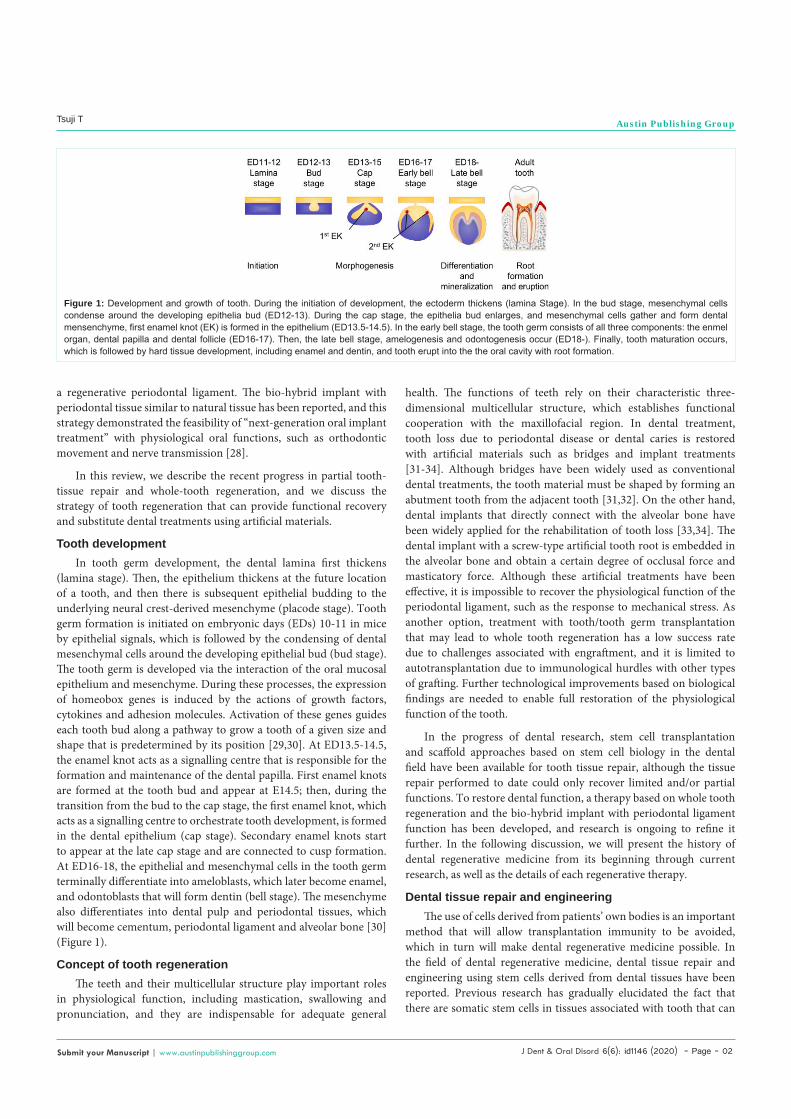
As stem cells derived from dental tissues, Dental Pulp-Derived Stem Cells (DPSCs), Stem Cells From Human Exfoliated Deciduous tooth (SHED), Periodontal Ligament Stem Cells (PDLSCs) and Stem Cells from the Apical Papilla (SCAP) in the apical region of human immature tooth have been reported [35-39]. DPECs and SHED are available for stem cell-mediated dentin-pulp complex regeneration. SCAP are also able to generate typical dentin-pulp complex structures after transplantation in vivo and are thus considered a valuable stem cell source for tissue repair and tissue engineering. Transplantation experiments have shown that PDLSCs in the human periodontal ligament can be used to regenerate periodontal tissue [38]. After transplantation, PDLSCs can generate the cementum and periodontal ligament. A root-shaped composite of hydroxyapatite containing SCAP coated with PDLSC-seeded gel foam could grow to mineralized root-like tissue with periodontal ligament in pig
extraction tooth sockets [39]. These cells are thought to be useful cell types for regeneration of dental tissue injury [40-42] (Figure 2).
Several studies have also reported tooth formation potency in cells not derived from dental tissue. One study showed that the reconstructed dental germ epithelial tissue derived from 10-day-old foetuses, ES cells or nerve stem cells were able to differentiate into odontogenic mesenchyme [43]. It has also been reported that the possibility of the reconstruction of cell strains derived from the oral mucosal epithelial cells of p53 knock-out mice and the molar mesenchymal tissue of 16.5-day-old fetuses can be utilized for the regeneration of bioengineered tooth [42]. These studies indicate the possibility that cells other than those of tooth tissues may have tooth formation potency. Although these stem cells are useful, they exhibit only partial tissue regeneration and do not generate a fully functional tooth.
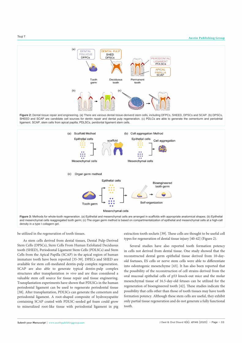
Figure 3: Methods for whole-tooth regeneration. (a) Epithelial and mesenchymal cells are arranged in scaffolds with appropriate anatomical shapes; (b) Epithelial and mesenchymal cells reaggregated tooth germ; (c) The organ germ method is based on compartmentalization of epithelial and mesenchymal cells at a high-cell density in a type I collagern gel.
Figure 2: Dental tissue repair and engineering. (a) There are various dental tissue-deriverd stem cells, including DFPCs, SHEED, DPSCs and SCAP. (b) DPSCs, SHEED and SCAP are candidate cell sources for dentin repair and dental pulp regeneration. (c) PDLCs are able to generate the cementurm and periodntal ligament. SCAP, stem cells from apical papilla; PDLSCs, peridontal ligament stem cells.
J Dent & Oral Disord 6(6): id1146 (2020) - Page - 04
Tsuji T Austin Publishing Group
Submit your Manuscript | www.austinpublishinggroup.com
Tooth/tooth germ transplantationIn the dental field, stem cell research has become increasingly
popular. Stem cell transplantation therapy has essentially focused on the use of a patient’s own stem cells as a source because immune rejection has been an important problem after allotransplantation of tissues and cells, including stem cells. Autotransplantation of a tooth or tooth germ is now available for biological dental treatment of tooth loss. Tooth/tooth germ transplantation successfully promotes partial functional recovery [44-46]. However, the limitation of this approach is that it is indicated only for an extremely limited number of patients who have a suitable healthy tooth/tooth germ for transplantation. Therefore, to ensure the safety of medical practice, it is desirable to perform transplantation of the bioengineered organ reconstructed using a patient’s own stem cells/organs to prevent an immunological response [47-50]. However, autologous transplantation of a natural tooth/tooth germ is limited by the number of teeth required. A recent study demonstrated that a tooth regenerative technology based on split tooth germ by artificial mechanical force may be utilized as a treatment [51]. In any case, these studies have demonstrated the prospects of functional recovery of tooth, thus paving the way for whole tooth regeneration.
Whole-tooth regenerationTooth regenerating method using a scaffold: As one of the
three-dimensional tissue engineering methods, scaffolds are used in complex with mixtures of cells to reorganize tissues into an intended shape. Several studies have demonstrated that scaffold constructs are fabricated in a very intricate configuration and seeded with tissue stem cells to generate new tissue. These reports showed that tissue-
specific cells could be grown in a scaffold to form a specific shape, and the technique might be used in the future for the clinical repair of damaged tissues.
The feasibility of bioengineering tooth using a cell-scaffold composite has also been reported. Previous studies using biodegradable polymer scaffolds have reported the partial generation of tooth tissue structures, including enamel, dentin and dental pulp, which was achieved through seeding epithelial and mesenchymal cells isolated from porcine tooth germ [18,51-60]. Recently, regeneration of complexes of cementum, periodontal ligament and bone using scaffolds such as 3D-printing materials and membrane materials has been reported [61,62]. It was also reported that 3D-printed scaffolds fabricated from the patient’s tooth were able to mimic the anatomical shape with limited potential for regeneration of dental tissue structure [63].
While the scaffold approach offers a strong foundation for reproducing the tooth morphology, it cannot build the complex structure of the tooth due to its inability to induce cell-cell interactions that are essential for the development of normal organs. Moreover, it has a low engraftment rate in transplantation and thus cannot support the fully functional recovery of the periodontal ligament. Fully regenerating the proper tooth structure using scaffolds requires advances in basic research (Figure 3).
Cell aggregation methodThe other approach for the reconstitution of the bioengineered
tooth germ is the cell reaggregation method. The aggregation of epithelial cells and mesenchymal cells involves multicellular assembly and self-reorganization of each cell type, and it occurs through cell
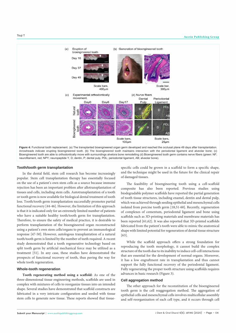
Figure 4: Functional tooth replacement. (a) The transplanted bioengineered organ germ developed and reached the occlusal plane 49 days after transplantation. Arrowheads indicate erupting bioengineered tooth. (b) The bioengineered tooth maintains interaction with the periodontal ligament and alveolar bone; (c) Bioengineered tooth are able to orthodontically move with surroundings alvelora bone remodelling (d) Bioengineered tooth germ contains nerve fibers (green: NF; neurofilament, red; NPY; neuropeptide Y, D; dentin, P; dental pulp, PDL; periodontal ligament, AB; alveolar bone).
J Dent & Oral Disord 6(6): id1146 (2020) - Page - 05
Tsuji T Austin Publishing Group
Submit your Manuscript | www.austinpublishinggroup.com
migration and selective cell adhesions until cells reach an established equilibrium [63-70]. Next, reciprocal interactions of epithelial cell layers and mesenchymal cell layers initiate organogenesis, including regulation of differentiation and morphogenesis [71-73]. In the dental field, many researchers have isolated epithelial cells and mesenchymal cells from the tooth germs of experimental animals. It has been reported that bioengineering cell aggregates reconstructing epithelial and mesenchymal tooth germ cells have the potential for tooth regeneration after transplantation [74,75]. The bioengineered cell aggregate mix of epithelial and mesenchymal stem cells isolated from tooth germ could generate the correct tooth germ structure by self-reorganisation through the cell rearrangement of epithelial and mesenchymal cells, however, the function of the regenerated tooth germ was not investigated [76]. Although these reports indicated that a cell aggregation method is useful for creating bioengineered tooth germ for dental regenerative therapy, the bioengineering method that can replicate the organogenesis of the embryo and that is adaptable to a wide variety of organ germ types is desired (Figure 3). Compared to the scaffold approach, the cell aggregation approach addresses the issue of cell-to-cell interactions to a greater extent. However, as the interactions resulting from the self-organization potentials of cells cannot be controlled, this approach is not efficient for tooth development.
Functional tooth replacementTo overcome the difficulties of inducing epithelial-mesenchymal
interactions, we developed the bioengineering method named the “organ germ method”, which is accomplished by cell compartmentalization between epithelial and mesenchymal cells at the high cell density in type I collagen gel [20-25]. Using a mouse model, we have shown that the bioengineered tooth germ can develop the correct tooth structure and successfully erupt into the oral cavity after transplantation into the tooth loss region [21]. The enamel and
dentin hardness of the bioengineered tooth components were in the normal range [21,22]. We have shown that bioengineered tooth are able to orthodontically move with surrounding alveolar bone remodelling underlying the proper localization of osteoclasts and osteoblasts in response to mechanical stress, showing penetration of blood vessels and nerve fibres. These findings demonstrate the potential for successful recovery of masticatory performance and natural tooth tissue using bioengineered tooth technology. Additionally, in the case of a transplanted bioengineered mature tooth unit comprising a mature tooth, periodontal ligament and alveolar bone, that unit can be engrafted into the tooth loss region through bone integration in the recipient. Successful engraftment of such a bioengineered tooth unit was achieved following transplantation at a position to reach the occlusal plane of the opposing tooth [22]. The technology of bioengineered tooth could contribute to the realization of whole-tooth replacement regenerative therapy as next-generation therapy (Figure 4).
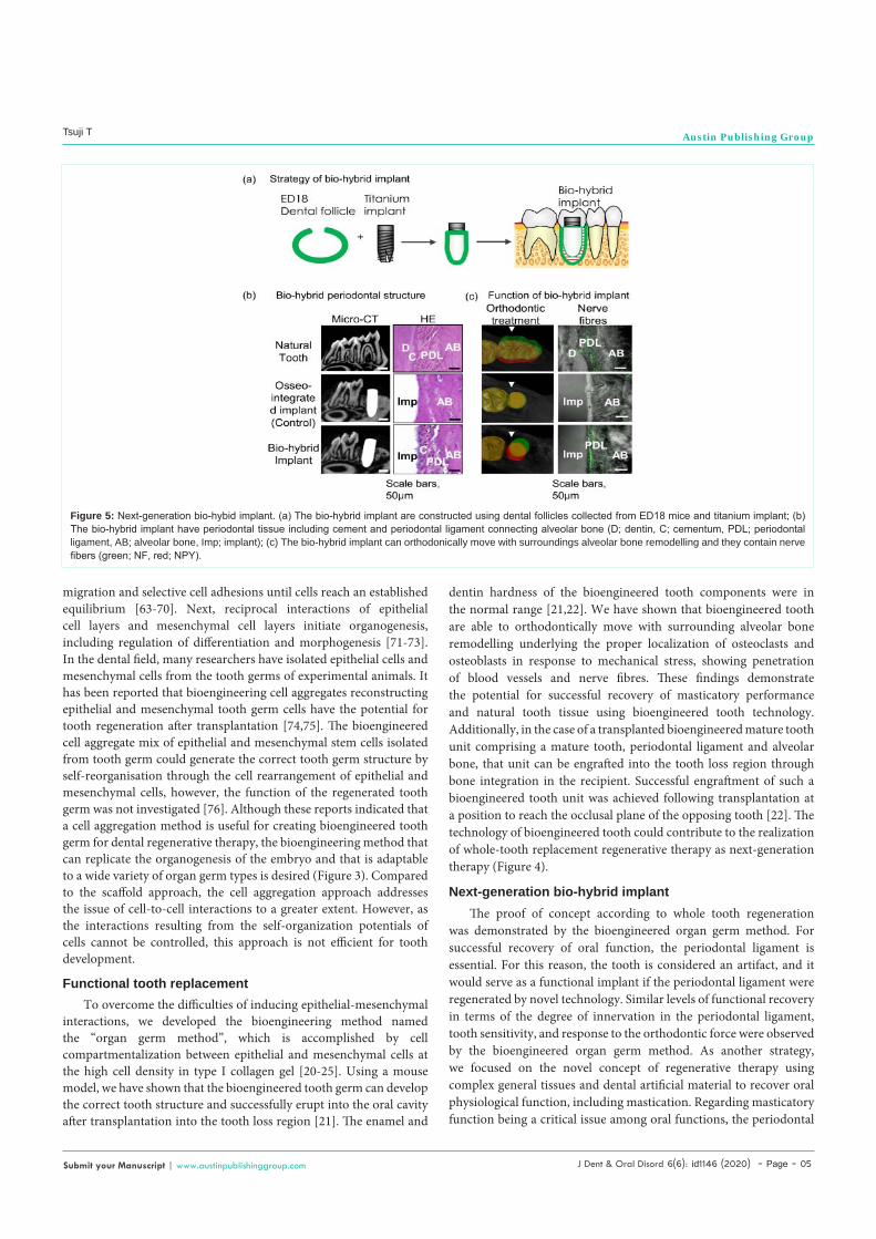
Next-generation bio-hybrid implantThe proof of concept according to whole tooth regeneration
was demonstrated by the bioengineered organ germ method. For successful recovery of oral function, the periodontal ligament is essential. For this reason, the tooth is considered an artifact, and it would serve as a functional implant if the periodontal ligament were regenerated by novel technology. Similar levels of functional recovery in terms of the degree of innervation in the periodontal ligament, tooth sensitivity, and response to the orthodontic force were observed by the bioengineered organ germ method. As another strategy, we focused on the novel concept of regenerative therapy using complex general tissues and dental artificial material to recover oral physiological function, including mastication. Regarding masticatory function being a critical issue among oral functions, the periodontal
Figure 5: Next-generation bio-hybid implant. (a) The bio-hybrid implant are constructed using dental follicles collected from ED18 mice and titanium implant; (b) The bio-hybrid implant have periodontal tissue including cement and periodontal ligament connecting alveolar bone (D; dentin, C; cementum, PDL; periodontal ligament, AB; alveolar bone, Imp; implant); (c) The bio-hybrid implant can orthodonically move with surroundings alveolar bone remodelling and they contain nerve fibers (green; NF, red; NPY).
J Dent & Oral Disord 6(6): id1146 (2020) - Page - 06
Tsuji T Austin Publishing Group
Submit your Manuscript | www.austinpublishinggroup.com
ligament is an important tissue with a proprioceptive function, and it acts as a viscoelastic cushion for occlusal force by virtue of its fibres.
Currently, dental implant treatment is widely adopted as an effective approach for restoring occlusion, which contributes to achieving a long-term, stable prognosis without damaging healthy teeth. On the other hand, an implant, unlike a natural tooth, does not have periodontal ligament, which maintains the connection between the root and the alveolar bone, since the body of an implant is in direct contact with the alveolar bone [29]. Therefore, the procedure cannot be performed in young patients as their jaw bones are still growing, and impaired occlusion due to the movement of the teeth that can occur with time would be difficult to treat. Hence, there are growing expectations that the development of a new dental treatment technique could enable the restoration of the functional occlusion system in a more biologically relevant manner.
With a view towards resolving the abovementioned issues, we validated a treatment concept for a next-generation implant that consists of periodontal tissue and was created based on the mechanism of tooth development. It has been found that dental follicular tissue, which is formed during the process of tooth development, consists of stem cells that can be differentiated into the cementum, periodontal ligaments and alveolar bone. These are all components of periodontal tissues that fuse with the jaw bones while forming periodontal tissue around the dental roots, and they function throughout the life of the person [29]. Given the above results, we conducted the following experiment: dental follicular tissue from mouse embryos, which is at which periodontal tissue can be formed, was placed around a hydroxyapatite (HA)-coated implant, and the implant was placed in adult murine tooth-loss models. After transplantation, the alveolar
bone was fused with the surface of the implant in the HA implant group, which was the study group.
The existing structure was still adopted in the next-generation implant; however, this experiment found that periodontal tissue equivalent to that of a natural tooth was formed on the surface of the implant, to which dental follicular tissue was attached; additionally, the results showed that the periodontal tissue consisted of cementum, periodontal ligaments and alveolar bone. Furthermore, it was found that this bio-hybrid implant could reproduce the physiological functions of a tooth, such as orthodontic movement and neurotransmission. The fact that an artificial material with a regenerative periodontal ligament allowed nearly complete recovery of masticatory function suggests that not all materials need to be of biological origin for regenerating tooth. Therefore, the bio-hybrid implant may be used in next-generation dental restoration treatments in combination with maxillofacial treatments (Figure 5).
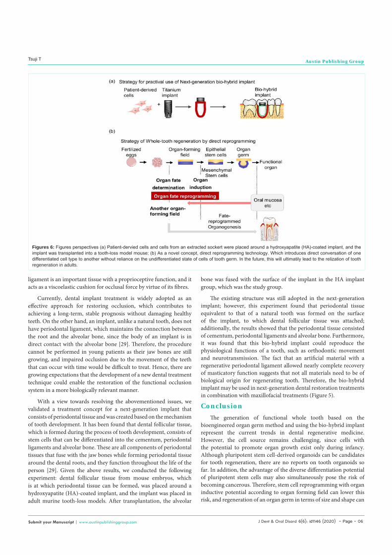
ConclusionThe generation of functional whole tooth based on the
bioengineered organ germ method and using the bio-hybrid implant represent the current trends in dental regenerative medicine. However, the cell source remains challenging, since cells with the potential to promote organ growth exist only during infancy. Although pluripotent stem cell-derived organoids can be candidates for tooth regeneration, there are no reports on tooth organoids so far. In addition, the advantage of the diverse differentiation potential of pluripotent stem cells may also simultaneously pose the risk of becoming cancerous. Therefore, stem cell reprogramming with organ inductive potential according to organ forming field can lower this risk, and regeneration of an organ germ in terms of size and shape can
Figures 6: Figures perspectives (a) Patient-dervied cells and cells from an extracted sockert were placed around a hydroxyapatite (HA)-coated implant, and the implant was transplanted into a tooth-loss model mouse; (b) As a novel concept, direct reprogramming technology. Which introduces direct conversation of one differentiated cell type to another without reliance on the undifferentiated state of cells of tooth germ. In the future, this will ultimatily lead to the relization of tooth regeneration in adults.
J Dent & Oral Disord 6(6): id1146 (2020) - Page - 07
Tsuji T Austin Publishing Group
Submit your Manuscript | www.austinpublishinggroup.com
be established by recapturing embryonic development. As a further novel concept, there is hope that direct reprogramming technology, which induces direct conversion of one differentiated cell type to another without reliance on the undifferentiated state of cells, may be utilized. If epithelial and mesenchymal stem cells, which have tooth induction potency, can be utilized via direct reprogramming, this may enable prevailing over the problems associated with dental regeneration methods, such as the organ germ method (Figure 6).
Owing to efforts towards the social acceptance of the bio-hybrid implant the clinical application of dental regeneration has become a reality. In the future, in addition to conventional organoid research, organ regeneration through direct reprogramming may help in regenerative dentistry to regenerate a three-dimensional tooth, thereby narrowing the gap between research and clinical use of whole tooth regeneration. The development of these techniques will help to establish a complete functional recovery of a lost tooth in clinical dental therapy in the foreseeable future through the use of biological methods.
AcknowledgementThis work was partially supported by a MEXT Grant-in-Aid for
Scientific Research (A) (No. 19H01180) awarded to T. Tsuji.
References 1. Brockes JP, Kumar A. Appendage regeneration in adult vertebrates and
implications for regenerative medicine Science. 2005; 310: 1919-23.
2. Langer RS, Vacanti JP. Tissue engineering: the challenges ahead Sci Am. 1999; 280: 86-9.
3. Atala A. Tissue engineering, stem cells and cloning: current concepts and changing trends. Expert Opin Biol Ther. 2005; 5: 879-92.
4. Madeira C, Santhagunam A, Salgueiro JB, Cabral JM. Advanced cell therapies for articular cartilage regeneration Trends Biotechnol. 2015; 33: 35-42.
5. Trounson A, Daley GQ, Pasque V, Plath K. A new route to human embryonic stem cells Nat Med. 2013; 19: 820-1.
6. Takebe T, Zhang RR, Koike H, Kimura M, Yoshizawa E, Enomura M, et al. Generation of a vascularized and functional human liver from an iPSC-derived organ bud transplant Nat Protoc. 2014; 9: 396-409.
7. Clevers H. Modeling Development and Disease with Organoids Cell. 2016; 165: 1586-97.
8. Addis RC, Epstein JA. Induced regeneration--the progress and promise of direct reprogramming for heart repair Nat Med. 2013; 19: 829-36.
9. Kamao H, Mandai M, Okamoto S, Sakai N, Suga A, Sugita S, et al. Characterization of human induced pluripotent stem cell-derived retinal pigment epithelium cell sheets aiming for clinical application. Stem Cell Reports. 2014; 2: 205-18.
10. Peggs KS, Hunter A, Chopra R, Parker A, Mahendra P, Milligan D, et al. Clinical evidence of a graft-versus-Hodgkin’s-lymphoma effect after reduced-intensity allogeneic transplantation Lancet. 2005; 365: 1934-41.
11. Hehlmann R. Chronic myeloid Leukaemia Lancet. 2007; 370: 342-50.
12. Lindvall O, Kokaia Z, Martinez-Serrano A. Stem cell therapy for human neurodegenerative disorders-how to make it work Nat Med. 2004;10: 42-50.
13. Sousa JE, Costa MA, Tuzcu EM, Yadav JS, Ellis S. New frontiers in interventional cardiology Circulation. 2005; 111: 671-81.
14. Prockop DJ. Marrow stromal cells as stem cells for non hematopoietic tissues Science. 1997; 276: 71-4.
15. Toma JG, Akhavan M, Fernandes KJ, Barnabe-Heider F, Sadikot A,
Kaplan DR, et al. Isolation of multipotent adult stem cells from the dermis of mammalian skin Nat Cell Biol. 2001; 3: 778-84.
16. Zuk PA, Zhu M, Ashjian P, De Ugarte DA, Huang JI, Mizuno H, et al. Human adipose tissue is a source of multipotent stem cells Mol Biol Cell. 2002; 13: 4279-95.
17. Lee JY, Qu-Petersen Z, Cao B, Kimura S, Jankowski R, Cummins J, et al. Clonal isolation of muscle-derived cells capable of enhancing muscle regeneration and bone healing J Cell Biol. 2000; 150 : 1085-100.
18. Cao Y, Vacanti JP, Paige KT, Upton J, Vacanti CA. Transplantation of chondrocytes utilizing a polymer-cell construct to produce tissue-engineered cartilage in the shape of a human ear Plast Reconstr Surg. 1997; 100: 297-302.
19. Caplan AI, Bruder SP. Mesenchymal stem cells: building blocks for molecular medicine in the 21st century Trends Mol Med. 2001; 7: 259-64.
20. Guo S, Guo W, Ding Y, Gong J, Zou Q, Xie D, et al. Comparative study of human dental follicle cell sheets and periodontal ligament cell sheets for periodontal tissue regeneration Cell Transplant. 2013; 22: 1061-73.
21. Nakao K, Morita R, Saji Y, Ishida K, Tomita Y, Ogawa M, et al. The development of a bioengineered organ germ method Nat Methods. 2007; 4: 227-30.
22. Ikeda E, Morita R, Nakao K, Ishida K, Nakamura T, Takano-Yamamoto T, et al. Fully functional bioengineered tooth replacement as an organ replacement therapy Proc Natl Acad Sci U S A. 2009; 106: 13475-80.
23. Oshima M, Mizuno M, Imamura A, Ogawa M, Yasukawa M, Yamazaki H, et al. Functional tooth regeneration using a bioengineered tooth unit as a mature organ replacement regenerative therapy PLoS One. 2011; 6: 21531.
24. Toyoshima KE, Asakawa K, Ishibashi N, Toki H, Ogawa M, Hasegawa T, et al. Fully functional hair follicle regeneration through the rearrangement of stem cells and their niches Nat Commun. 2012; 3: 784.
25. Ogawa M, Oshima M, Imamura A, Sekine Y, Ishida K, Yamashita K, et al. Functional salivary gland regeneration by transplantation of a bioengineered organ germ Nat Commun. 2013; 4: 2498.
26. Hirayama M, Tsubota K, Tsuji T. Generation of a Bioengineered Lacrimal Gland by Using the Organ Germ Method Methods Mol Biol. 2017; 1597: 153-65.
27. Young CS, Terada S, Vacanti JP, Honda M, Bartlett JD, Yelick PC. Tissue engineering of complex tooth structures on biodegradable polymer scaffolds J Dent Res. 2002; 81: 695-700.
28. Oshima M, Inoue K, Nakajima K, Tachikawa T, Yamazaki H, Isobe T, et al. Functional tooth restoration by next-generation bio-hybrid implant as a bio-hybrid artificial organ replacement therapy Sci Rep. 2014; 4: 6044.
29. Jernvall J, Thesleff I. Tooth shape formation and tooth renewal: evolving with the same signals Development. 2012; 139: 3487-97.
30. Saito M, Nishida E, Sasaki T, Yoneda T, Shimizu N. The KK-Periome database for transcripts of periodontal ligament development J Exp Zool B Mol Dev Evol. 2009; 312: 495-502.
31. Rosenstiel , Land M. Contemporary fixed prosthodontics: Mosby Press: Missouri; 2015.
32. Pokorny PH, Wiens JP, Litvak H. Occlusion for fixed prosthodontics: a historical perspective of the gnathological influence J Prosthet Dent. 2008; 99: 299-313.
33. PI B, GA Z. Tissue-integrated prostheses. In osseointegration in clinical dentistry. T A, editor: Berlin: Quintessence Pub Co Press; 1985.
34. Burns DR, Beck DA, Nelson SK. A review of selected dental literature on contemporary provisional fixed prosthodontic treatment: report of the Committee on Research in Fixed Prosthodontics of the Academy of Fixed Prosthodontics J Prosthet Dent. 2003; 90: 474-97.
35. Gronthos S, Mankani M, Brahim J, Robey PG, Shi S. Postnatal human Dental Pulp Stem Cells (DPSCs) Proc Natl Acad Sci U S A. 2000; 97: 13625-30.
36. Gronthos S, Brahim J, Li W, Fisher LW, Cherman N, Boyde A, et al. Stem
J Dent & Oral Disord 6(6): id1146 (2020) - Page - 08
Tsuji T Austin Publishing Group
Submit your Manuscript | www.austinpublishinggroup.com
cell properties of human dental pulp stem cells J Dent Res. 2002; 81: 531-5.
37. Miura M, Gronthos S, Zhao M, Lu B, Fisher LW, Robey PG, et al. SHED: stem cells from human exfoliated deciduous teeth Proc Natl Acad Sci U S A. 2003; 100: 5807-12.
38. Seo BM, Miura M, Gronthos S, Bartold PM, Batouli S, Brahim J, et al. Investigation of multipotent postnatal stem cells from human periodontal ligament. Lancet. 2004; 364: 149-55.
39. Sonoyama W, Liu Y, Fang D, Yamaza T, Seo BM, Zhang C, et al. Mesenchymal stem cell-mediated functional tooth regeneration in swine PLoS One. 2006; 1: 79.
40. Gancheva MR, Kremer KL, Gronthos S, Koblar SA. Using Dental Pulp Stem Cells for Stroke Therapy. Front Neurol. 2019; 10: 422.
41. Gancheva MR, Kremer KL, Gronthos S, Koblar SA. Using Dental Pulp Stem Cells for Stroke Therapy. Frontiers in neurology. 2019; 10: 422.
42. Hu L, Liu Y, Wang S. Stem cell-based tooth and periodontal regeneration Oral Diseases. 2018; 24: 696-705.
43. Ohazama A, Modino SA, Miletich I, Sharpe PT. Stem-cell-based tissue engineering of murine teeth. J Dent Res. 2004; 83: 518-22.
44. Bauss O, Engelke W, Fenske C, Schilke R, Schwestka-Polly R. Autotransplantation of immature third molars into edentulous and atrophied jaw sections Int J Oral Maxillofac Surg. 2004; 33: 558-63.
45. Lai FS. Autotransplantation of an unerupted wisdom tooth germ without its follicle immediately after removal of an impacted mandibular second molar: a case report J Can Dent Assoc. 2009; 75: 205-8.
46. Gerard E, Membre H, Gaudy JF, Mahler P, Bravetti P. Functional fixation of autotransplanted tooth germs by using bioresorbable membranes Oral Surg Oral Med Oral Pathol Oral Radiol Endod. 2002; 94: 667-72.
47. Tan J, Wu W, Xu X, Liao L, Zheng F, Messinger S, et al. Induction therapy with autologous mesenchymal stem cells in living-related kidney transplants: a randomized controlled trial Jama. 2012; 307: 1169-77.
48. Quarto R, Mastrogiacomo M, Cancedda R, Kutepov SM, Mukhachev V, Lavroukov A, et al. Repair of large bone defects with the use of autologous bone marrow stromal cells N Engl J Med. 2001; 344: 385-6.
49. Morizane A, Doi D, Kikuchi T, Okita K, Hotta A, Kawasaki T, et al. Direct comparison of autologous and allogeneic transplantation of iPSC-derived neural cells in the brain of a non-human primate Stem Cell Reports. 2013; 1: 283-92.
50. Ono M, Oshima M, Ogawa M, Sonoyama W, Hara ES, Oida Y, et al. Practical whole-tooth restoration utilizing autologous bioengineered tooth germ transplantation in a postnatal canine model Sci Rep. 2017; 7: 44522.
51. Yamamoto N, Oshima M, Tanaka C, Ogawa M, Nakajima K, Ishida K, et al. Functional tooth restoration utilising split germs through re-regionalisation of the tooth-forming field Sci Rep. 2015; 5: 18393.
52. Honda M, Morikawa N, Hata K, Yada T, Morita S, Ueda M, et al. Rat costochondral cell characteristics on poly (L-lactide-co-epsilon-caprolactone) scaffolds Biomaterials. 2003; 24: 3511-9.
53. Iwatsuki S, Honda MJ, Harada H, Ueda M. Cell proliferation in teeth reconstructed from dispersed cells of embryonic tooth germs in a three-dimensional scaffold Eur J Oral Sci. 2006; 114: 310-7.
54. Duailibi MT, Duailibi SE, Young CS, Bartlett JD, Vacanti JP, Yelick PC. Bioengineered teeth from cultured rat tooth bud cells J Dent Res. 2004; 83: 523-8.
55. Yelick PC, Vacanti JP. Bioengineered teeth from tooth bud cells Dent Clin North Am. 2006; 50: 191-203.
56. Sumita Y, Honda MJ, Ohara T, Tsuchiya S, Sagara H, Kagami H, et al. Performance of collagen sponge as a 3-D scaffold for tooth-tissue engineering Biomaterials. 2006; 27: 3238-48.
57. Honda MJ, Tsuchiya S, Sumita Y, Sagara H, Ueda M. The sequential seeding of epithelial and mesenchymal cells for tissue-engineered tooth regeneration Biomaterials. 2007; 28: 680-9.
58. Ohara T, Itaya T, Usami K, Ando Y, Sakurai H, Honda MJ, et al. Evaluation of scaffold materials for tooth tissue engineering. J Biomed Mater Res A. 2010; 94: 800-5.
59. Steinberg MS. Adhesion in development: an historical overview. Dev Biol. 1996; 180: 377-88.
60. Zhang W, Vazquez B, Oreadi D, Yelick PC. Decellularized Tooth Bud Scaffolds for Tooth Regeneration. Journal of dental research. 2017; 96: 516-23.
61. Park CH, Rios HF, Jin Q, Bland ME, Flanagan CL, Hollister SJ, et al. Biomimetic hybrid scaffolds for engineering human tooth-ligament interfaces Biomaterials. 2010; 31: 5945-52.
62. Wu M, Wang J, Zhang Y, Liu H, Dong F. Mineralization Induction of Gingival Fibroblasts and Construction of a Sandwich Tissue-Engineered Complex for Repairing Periodontal Defects. Med Sci Monit. 2018; 24: 1112-23.
63. Kim K, Lee CH, Kim BK, Mao JJ. Anatomically shaped tooth and periodontal regeneration by cell homing. J Dent Res. 2010; 89: 842-7.
64. Steinberg M. The nature and origin of “ECM”, a putative mediator of mutual cell adhesions Am Zool 1962; 2: 561-2.
65. Steinberg MS. On the mechanism of tissue reconstruction by dissociated cells. I. Population kinetics, differential adhesiveness. and the absence of directed migration. Proc Natl Acad Sci U S A. 1962; 48: 1577-82.
66. Steinberg MS. Mechanism of tissue reconstruction by dissociated cells. II. Time-course of events. Science. 1962; 137: 762-3.
67. Steinberg MS. On the mechanism of tissue reconstruction by dissociated cells, I. Free energy relations and the reoganization of fused, heteronomic tissue fragments Proc Natl Acad Sci U S A. 1962; 48: 1769-76.
68. Layer PG, Robitzki A, Rothermel A, Willbold E. Of layers and spheres: the reaggregate approach in tissue engineering Trends Neurosci. 2002; 25: 131-4.
69. Layer PG, Rothermel A, Willbold E. From stem cells towards neural layers: a lesson from re-aggregated embryonic retinal cells Neuroreport. 2001; 12: 39-46.
70. Sharpe PT, Young CS. Test-tube teeth Sci Am. 2005; 293: 34-41.
71. Thesleff I. Epithelial-mesenchymal signalling regulating tooth morphogenesis J Cell Sci. 2003; 116: 1647-8.
72. Tucker A, Sharpe P. The cutting-edge of mammalian development; how the embryo makes teeth. Nat Rev Genet. 2004; 5: 499-508.
73. Thesleff I. From understanding tooth development to bioengineering of teeth. Eur J Oral Sci. 2018; 126: 67-71.
74. Hu B, Nadiri A, Kuchler-Bopp S, Perrin-Schmitt F, Peters H, Lesot H. Tissue engineering of tooth crown, root, and periodontium. Tissue Eng. 2006; 12: 2069-75.
75. Yamamoto H, Kim EJ, Cho SW, Jung HS. Analysis of tooth formation by reaggregated dental mesenchyme from mouse embryo J Electron Microsc (Tokyo). 2003; 52: 559-66.
76. Song Y, Zhang Z, Yu X, Yan M, Zhang X, Gu S, et al. Application of lentivirus-mediated RNAi in studying gene function in mammalian tooth development Dev Dyn. 2006; 235: 1334-44.


![089 ' # '6& *#0 & 7Chapter 5 Tooth Tissue-engineered odontogenesis One important goal of dental research is the efficient regeneration of lost teeth [1, 2]. Tooth formation, or odontogenesis,](https://img.pdfslide.us/doc/110x75/60327f1bc15cec2a855c31cf/089-6-0-7-chapter-5-tooth-tissue-engineered-odontogenesis-one.jpg)