Embed Size (px)
Citation preview
Functional neuroimaging correlates of finger-tappingtask variations: An ALE meta-analysis
Suzanne T. Witt,a,! Angela R. Laird,b and M. Elizabeth Meyeranda,!
aDepartment of Medical Physics, University of Wisconsin, Madison, Wisconsin, USAbResearch Imaging Center, University of Texas Health Science Center, San Antonio, Texas, USA
Received 14 January 2008; revised 24 March 2008; accepted 1 April 2008Available online 16 April 2008
Finger-tapping tasks are one of themost commonparadigmsused to studythe human motor system in functional neuroimaging studies. These taskscan vary both in the presence or absence of a pacing stimulus as well as inthe complexity of the tapping task. Avoxel-wise, coordinate-based meta-analysis was performed on 685 sets of activation foci in Talairach spacegathered from 38 published studies employing finger-tapping tasks.Clusters of concordance were identified within the primary sensorimotorcortices, supplementary motor area, premotor cortex, inferior parietalcortices, basal ganglia, and anterior cerebellum. Subsequent analysesperformed on subsets of the primary set of foci demonstrated that theuse of a pacing stimulus resulted in a larger, more diverse network ofconcordance clusters, in comparison to varying the complexity of thetapping task. The majority of the additional concordance clustersoccurred in regions involved in the temporal aspects of the tapping task,rather than its execution. Tapping tasks employing a visual pacingstimulus recruited a set of nodes distinct from the results observed in thosetasks employing either an auditory or no pacing stimulus, suggestingdiffering cognitive networks when integrating visual or auditory pacingstimuli into simple motor tasks. The relatively uniform network ofconcordance clusters observed across the more complex finger-tappingtasks suggests that further complexity, beyond the use of multi-fingersequences or bimanual tasks, may be required to fully reveal those brainregions necessary to execute truly complex movements.© 2008 Elsevier Inc. All rights reserved.
Keywords: Meta-analysis; Finger tapping; Motor; Activation likelihoodestimation; ALE; Paced finger tapping; Auditory stimulus; Visual stimulus;Self-paced movement; Movement complexity; Sequential finger movements;Bimanual
Introduction
Finger-tapping tasks are commonly used to study the humanmotor system in functional neuroimaging studies. Tapping taskshave the advantage of being simple enough to use in the study ofboth normal control subjects as well as those with neuropathol-ogies affecting the motor system, while being flexible enough toaccommodate numerous modifications. These tasks can varyacross studies both by the use or lack of a pacing stimulus andin the relative complexity of the tapping task.
Pacing stimuli are used to ensure that all subjects uniformlyperform a given finger-tapping task at a predetermined rate. Thestimuli are usually in the form of a regularly paced, repetitive auditoryor visual cue, such as that produced by a metronome (e.g. Catalanet al., 1998; Colebatch et al., 1991; Sadato et al., 1996a) or blinkinglight (e.g. Indovina and Sanes, 2001; Jäncke et al., 2000b),respectively. Such finger-tapping tasks performed in the presenceof a pacing stimulus are referred to as externally guided or externallygenerated. In contrast, the task can be performed in the absence of anypacing stimulus (i.e. self-paced). Such self-paced tapping tasks arereferred to as internally guided or internally generated. The resultsfrom studies investigating the effects of auditory and visual pacingstimuli have reported different networks of active brain regions,however, these results are not consistent across different studies.
Pacing stimuli are also often used in conjunction with morecomplex finger-tapping tasks such as multi-finger sequential orbimanual tapping tasks. For the purposes of the ensuing analyses,multi-finger sequential tapping tasks were taken to be complex interms of the increased number of fingers involved in the task; factorssuch as the rate of movement and the length of the sequence were notspecifically considered. Bimanual tasks were taken to be any taskinvolving the tapping of fingers on both hands, regardless of thesymmetry. These types of complex finger-tapping tasks are oftenemployed to elicit neural activation that is more representative ofwhat would be observed in typical, everyday manual movementsthat may not be practical to complete within the confines of aMRI orPET scanner. The use of complex finger-tapping tasks also allowsfor the further study of secondary and tertiary neural motor regions
www.elsevier.com/locate/ynimgNeuroImage 42 (2008) 343–356
! Corresponding authors. Department of Medical Physics, University ofWisconsin, 1530 Medical Sciences Center, 1300 University Avenue,Madison, WI 53706, USA. Fax: +1 608 265 9840.
E-mail addresses: [email protected] (S.T. Witt), [email protected](M.E. Meyerand).
Available online on ScienceDirect (www.sciencedirect.com).
1053-8119/$ - see front matter © 2008 Elsevier Inc. All rights reserved.doi:10.1016/j.neuroimage.2008.04.025
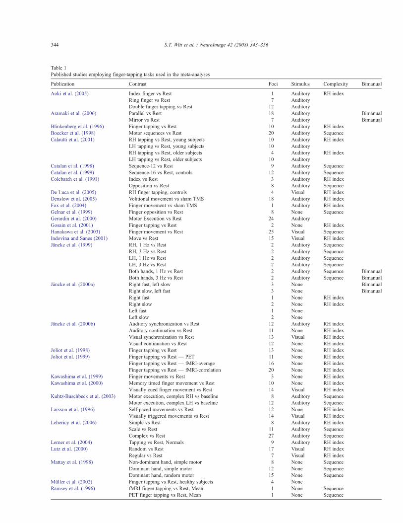
Table 1Published studies employing finger-tapping tasks used in the meta-analyses
Publication Contrast Foci Stimulus Complexity Bimanual
Aoki et al. (2005) Index finger vs Rest 1 Auditory RH indexRing finger vs Rest 7 AuditoryDouble finger tapping vs Rest 12 Auditory
Aramaki et al. (2006) Parallel vs Rest 18 Auditory BimanualMirror vs Rest 7 Auditory Bimanual
Blinkenberg et al. (1996) Finger tapping vs Rest 10 Auditory RH indexBoecker et al. (1998) Motor sequences vs Rest 20 Auditory SequenceCalautti et al. (2001) RH tapping vs Rest, young subjects 10 Auditory RH index
LH tapping vs Rest, young subjects 10 AuditoryRH tapping vs Rest, older subjects 4 Auditory RH indexLH tapping vs Rest, older subjects 10 Auditory
Catalan et al. (1998) Sequence-12 vs Rest 9 Auditory SequenceCatalan et al. (1999) Sequence-16 vs Rest, controls 12 Auditory SequenceColebatch et al. (1991) Index vs Rest 3 Auditory RH index
Opposition vs Rest 8 Auditory SequenceDe Luca et al. (2005) RH finger tapping, controls 4 Visual RH indexDenslow et al. (2005) Volitional movement vs sham TMS 18 Auditory RH indexFox et al. (2004) Finger movement vs sham TMS 1 Auditory RH indexGelnar et al. (1999) Finger opposition vs Rest 8 None SequenceGerardin et al. (2000) Motor Execution vs Rest 24 AuditoryGosain et al. (2001) Finger tapping vs Rest 2 None RH indexHanakawa et al. (2003) Finger movement vs Rest 25 Visual SequenceIndovina and Sanes (2001) Move vs Rest 15 Visual RH indexJäncke et al. (1999) RH, 1 Hz vs Rest 2 Auditory Sequence
RH, 3 Hz vs Rest 2 Auditory SequenceLH, 1 Hz vs Rest 2 Auditory SequenceLH, 3 Hz vs Rest 2 Auditory SequenceBoth hands, 1 Hz vs Rest 2 Auditory Sequence BimanualBoth hands, 3 Hz vs Rest 2 Auditory Sequence Bimanual
Jäncke et al. (2000a) Right fast, left slow 3 None BimanualRight slow, left fast 3 None BimanualRight fast 1 None RH indexRight slow 2 None RH indexLeft fast 1 NoneLeft slow 2 None
Jäncke et al. (2000b) Auditory synchronization vs Rest 12 Auditory RH indexAuditory continuation vs Rest 11 None RH indexVisual synchronization vs Rest 13 Visual RH indexVisual continuation vs Rest 12 None RH index
Joliot et al. (1998) Finger tapping vs Rest 13 None RH indexJoliot et al. (1999) Finger tapping vs Rest — PET 11 None RH index
Finger tapping vs Rest — fMRI-average 16 None RH indexFinger tapping vs Rest — fMRI-correlation 20 None RH index
Kawashima et al. (1999) Finger movements vs Rest 3 None RH indexKawashima et al. (2000) Memory timed finger movement vs Rest 10 None RH index
Visually cued finger movement vs Rest 14 Visual RH indexKuhtz-Buschbeck et al. (2003) Motor execution, complex RH vs baseline 8 Auditory Sequence
Motor execution, complex LH vs baseline 12 Auditory SequenceLarsson et al. (1996) Self-paced movements vs Rest 12 None RH index
Visually triggered movements vs Rest 14 Visual RH indexLehericy et al. (2006) Simple vs Rest 8 Auditory RH index
Scale vs Rest 11 Auditory SequenceComplex vs Rest 27 Auditory Sequence
Lerner et al. (2004) Tapping vs Rest, Normals 9 Auditory RH indexLutz et al. (2000) Random vs Rest 17 Visual RH index
Regular vs Rest 7 Visual RH indexMattay et al. (1998) Non-dominant hand, simple motor 8 None Sequence
Dominant hand, simple motor 12 None SequenceDominant hand, random motor 15 None Sequence
Müller et al. (2002) Finger tapping vs Rest, healthy subjects 4 NoneRamsey et al. (1996) fMRI finger tapping vs Rest, Mean 1 None Sequence
PET finger tapping vs Rest, Mean 1 None Sequence
344 S.T. Witt et al. / NeuroImage 42 (2008) 343–356
that may not be active during a simple, unimanual index finger-tapping task.
Results from studies employing finger-tapping tasks can bedivergent due to variations in the experimental paradigms used,making them difficult to interpret across studies. Additionally, studiescan choose to focus on a few specific neural regions (e.g. Jänckeet al., 2000a; Colebatch et al., 1991; De Luca et al., 2005), resulting inpartial descriptions of the underlying neural network involved in agiven tapping task. A quantitative meta-analysis technique, such asthat proposed independently by Turkeltaub et al. (2002) and Cheinet al. (2002) provides a method to assess the degree of concordanceacross multiple studies. The results of such an analysis can be usefulin determining a more complete network of neural regions involvedin a given task or paradigm as well as in forming new hypotheses andinterpreting results from subjects with neurological impairments.
This present studywas not the first to use quantitativemeta-analysistechniques to assess concordance across studies examining the humanmotor system. Chouinard and Paus (2006) employed a similartechnique to further elucidate the roles of the primary motor andpremotor cortices in various motor tasks. Four motor-related tasks –movement response selection, movement response to a stimulus,execution of object-related hand movements, and observation ofobject-related hand movements – were chosen to map out the roles ofthe dorsal and ventral premotor cortices in these tasks. Chouinard andPaus were successful in utilizing meta-analysis to identify severaldistinct nonprimary motor areas within the motor cortex.
In the present meta-analysis, our aim was to isolate the corpusof published literature for simple hand movements (i.e., fingertapping), and identify the entire network of brain regionsassociated with this type of motor task. Our intent was to examineagreement across studies not only in the motor cortex, but alsothroughout all cortical, subcortical, and cerebellar regions. Inaddition, meta-analysis was used to differentiate the brain regionsthat are active during the most common variations of finger-tapping tasks: auditorially-paced, visually-paced, self-paced, singleindex finger, unimanual, dominant hand (RH) multi-fingersequence, and bimanual, as well as to compare these networksamong the tapping task variations. We hypothesized that the choiceof finger-tapping task variation would have a strong influence onthe observed network of active brain regions.
Methods
Several literature searches were performed in Medline to find thepublished corpus of literature prior to July 2006 involving finger-tapping tasks in unimpaired, right-handed subjects. References fromall relevant papers were also examined. From these search results,only those papers which reported activations as coordinates instereotactic space (x,y,z) were considered. Papers directly addressingmotor learning or using over-trained subjects such as professionalmusicians were excluded. Results from 38 papers (22 fMRI and 16PET; Table 1) were selected (685 foci), and three analyses wereperformed. The first pooled the results from all of the included studies.For the second, studies were divided into three groups based on thetype or lack of pacing stimulus employed: auditory stimulus (22papers; 403 foci), visual stimulus (7 papers; 109 foci), and no stimulus(13 papers; 173 foci). The final analysis divided the studies into threegroups determined by the complexity of the tapping task used: righthand index finger (23 papers; 311 foci), RHmulti-finger sequence (15papers; 242 foci), and bimanual (5 papers; 90 foci). All MNIcoordinates were transformed to Talairach space using the icbm2taltransform (Lancaster et al., 2007), which has shown to provideimproved fit over the mni2tal transform (Brett et al., 2001, 2002).When applying the Lancaster transform, software-specific versionswere used for FSL (icbm_fsl2tal) and SPM (icbm_spm2tal)coordinates, to correct for varying normalization methods withineach software package. Activation likelihood estimate (ALE) mapswere created for each grouping by modeling each focus as a three-dimensional Gaussian function with a FWHM of 12 mm (Turkeltaubet al., 2002). Statistical significance was assessed using a permutationtest with 5000 permutations, corrected formultiple comparisons usingthe false discovery rate (FDR) (Laird et al., 2005). The resultant mapswere thresholded at Pb0.05.
Results
The ALE map for the main effects of all finger-tapping taskvariations included in this study is shown in Fig. 1. Common, robustconcordance was seen in bilateral sensorimotor cortices (L: !38,!26,50; R: 36,!22,54), supplementary motor area (SMA) (!4,!8,52), left ventral premotor cortex (!54,!2,32), bilateral inferior
Table 1 (continued)
Publication Contrast Foci Stimulus Complexity Bimanual
Riecker et al. (2006) Main effects during index finger movement, young subjects 6 Auditory RH indexMain effects during index finger movement, older subjects 8 Auditory RH index
Rounis et al. (2005) Main effects of movement 17 Auditory RH indexSadato et al. (1996a) Various length sequences 6 Auditory SequenceSadato et al. (1997) Mirror vs Rest 13 Auditory Sequence Bimanual
Parallel vs Rest 15 Auditory Sequence BimanualRight unimanual vs Rest 3 Auditory RH indexLeft unimanual vs Rest 6 AuditoryBimanual mirror vs Rest 12 Auditory BimanualBimanual parallel vs Rest 13 Auditory Bimanual
Seitz et al. (2000) Irregularly paced right finger movement 4 Auditory RH indexWilson et al. (2004) Moving fingers 2 None Sequence BimanualYoo et al. (2005) Group-level finger-tapping activation 17 Auditory Sequence
A total of 38 studies with 74 contrasts and 685 foci was included in the finger-tapping meta-analyses (listed in alphabetical order by first author). The type or lackof pacing stimulus and the level of complexity as well as whether the task was bimanual are indicated for each contrast.
345S.T. Witt et al. / NeuroImage 42 (2008) 343–356
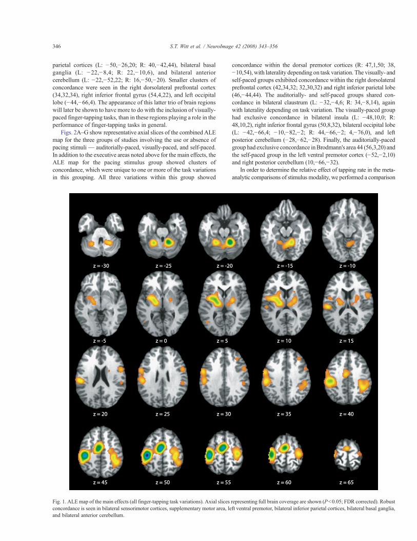
parietal cortices (L: !50,!26,20; R: 40,!42,44), bilateral basalganglia (L: !22,!8,4; R: 22,!10,6), and bilateral anteriorcerebellum (L: !22,!52,22; R: 16,!50,!20). Smaller clusters ofconcordance were seen in the right dorsolateral prefrontal cortex(34,32,34), right inferior frontal gyrus (54,4,22), and left occipitallobe (!44,!66,4). The appearance of this latter trio of brain regionswill later be shown to have more to do with the inclusion of visually-paced finger-tapping tasks, than in these regions playing a role in theperformance of finger-tapping tasks in general.
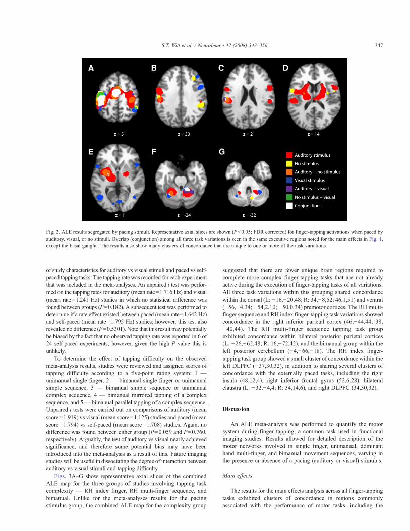
Figs. 2A–G show representative axial slices of the combined ALEmap for the three groups of studies involving the use or absence ofpacing stimuli — auditorially-paced, visually-paced, and self-paced.In addition to the executive areas noted above for the main effects, theALE map for the pacing stimulus group showed clusters ofconcordance, which were unique to one or more of the task variationsin this grouping. All three variations within this group showed
concordance within the dorsal premotor cortices (R: 47,1,50; 38,!10,54), with laterality depending on task variation. The visually- andself-paced groups exhibited concordance within the right dorsolateralprefrontal cortex (42,34,32; 32,30,32) and right inferior parietal lobe(46,!44,44). The auditorially- and self-paced groups shared con-cordance in bilateral claustrum (L: !32,!4,6; R: 34,!8,14), againwith laterality depending on task variation. The visually-paced grouphad exclusive concordance in bilateral insula (L: !48,10,0; R:48,10,2), right inferior frontal gyrus (50,8,32), bilateral occipital lobe(L: !42,!66,4; !10,!82,!2; R: 44,!66,!2; 4,!76,0), and leftposterior cerebellum (!28,!62,!28). Finally, the auditorially-pacedgroup had exclusive concordance inBrodmann's area 44 (56,3,20) andthe self-paced group in the left ventral premotor cortex (!52,!2,10)and right posterior cerebellum (10,!66,!32).
In order to determine the relative effect of tapping rate in the meta-analytic comparisons of stimulus modality, we performed a comparison
Fig. 1. ALE map of the main effects (all finger-tapping task variations). Axial slices representing full brain coverage are shown (Pb0.05; FDR corrected). Robustconcordance is seen in bilateral sensorimotor cortices, supplementary motor area, left ventral premotor, bilateral inferior parietal cortices, bilateral basal ganglia,and bilateral anterior cerebellum.
346 S.T. Witt et al. / NeuroImage 42 (2008) 343–356
of study characteristics for auditory vs visual stimuli and paced vs self-paced tapping tasks. The tapping rate was recorded for each experimentthat was included in the meta-analyses. An unpaired t test was perfor-med on the tapping rates for auditory (mean rate=1.716 Hz) and visual(mean rate=1.241 Hz) studies in which no statistical difference wasfound between groups (P=0.182). A subsequent test was performed todetermine if a rate effect existed between paced (mean rate=1.642 Hz)and self-paced (mean rate=1.795 Hz) studies; however, this test alsorevealed no difference (P=0.5301). Note that this result may potentiallybe biased by the fact that no observed tapping rate was reported in 6 of24 self-paced experiments; however, given the high P value this isunlikely.
To determine the effect of tapping difficulty on the observedmeta-analysis results, studies were reviewed and assigned scores oftapping difficulty according to a five-point rating system: 1 —
unimanual single finger, 2 — bimanual single finger or unimanualsimple sequence, 3 — bimanual simple sequence or unimanualcomplex sequence, 4 — bimanual mirrored tapping of a complexsequence, and 5— bimanual parallel tapping of a complex sequence.Unpaired t tests were carried out on comparisons of auditory (meanscore=1.919) vs visual (mean score=1.125) studies and paced (meanscore=1.794) vs self-paced (mean score=1.708) studies. Again, nodifference was found between either group (P=0.059 and P=0.760,respectively). Arguably, the test of auditory vs visual nearly achievedsignificance, and therefore some potential bias may have beenintroduced into the meta-analysis as a result of this. Future imagingstudies will be useful in dissociating the degree of interaction betweenauditory vs visual stimuli and tapping difficulty.
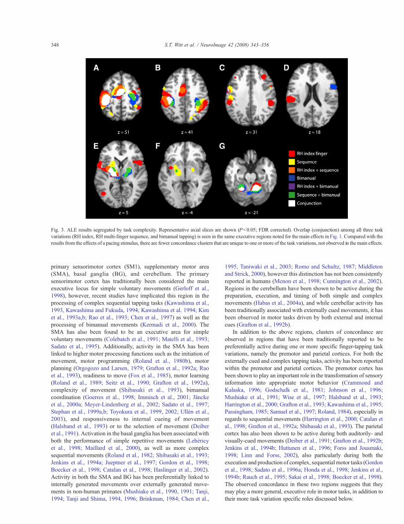
Figs. 3A–G show representative axial slices of the combinedALE map for the three groups of studies involving tapping taskcomplexity — RH index finger, RH multi-finger sequence, andbimanual. Unlike for the meta-analyses results for the pacingstimulus group, the combined ALE map for the complexity group
suggested that there are fewer unique brain regions required tocomplete more complex finger-tapping tasks that are not alreadyactive during the execution of finger-tapping tasks of all variations.All three task variations within this grouping shared concordancewithin the dorsal (L: !16,!20,48; R: 34,!8,52; 46,1,51) and ventral(!56,!4,34; !54,2,10; !50,0,34) premotor cortices. The RH multi-finger sequence and RH index finger-tapping task variations showedconcordance in the right inferior parietal cortex (46,!44,44; 38,!40,44). The RH multi-finger sequence tapping task groupexhibited concordance within bilateral posterior parietal cortices(L: !26,!62,48; R: 16,!72,42), and the bimanual group within theleft posterior cerebellum (!4,!66,!18). The RH index finger-tapping task group showed a small cluster of concordance within theleft DLPFC (!37,30,32), in addition to sharing several clusters ofconcordance with the externally paced tasks, including the rightinsula (48,12,4), right inferior frontal gyrus (52,6,28), bilateralclaustra (L: !32,!4,4; R: 34,14,6), and right DLPFC (34,30,32).
Discussion
An ALE meta-analysis was performed to quantify the motorsystem during finger tapping, a common task used in functionalimaging studies. Results allowed for detailed description of themotor networks involved in single finger, unimanual, dominanthand multi-finger, and bimanual movement sequences, varying inthe presence or absence of a pacing (auditory or visual) stimulus.
Main effects
The results for the main effects analysis across all finger-tappingtasks exhibited clusters of concordance in regions commonlyassociated with the performance of motor tasks, including the
Fig. 2. ALE results segregated by pacing stimuli. Representative axial slices are shown (Pb0.05; FDR corrected) for finger-tapping activations when paced byauditory, visual, or no stimuli. Overlap (conjunction) among all three task variations is seen in the same executive regions noted for the main effects in Fig. 1,except the basal ganglia. The results also show many clusters of concordance that are unique to one or more of the task variations.
347S.T. Witt et al. / NeuroImage 42 (2008) 343–356
primary sensorimotor cortex (SM1), supplementary motor area(SMA), basal ganglia (BG), and cerebellum. The primarysensorimotor cortex has traditionally been considered the mainexecutive locus for simple voluntary movements (Gerloff et al.,1998), however, recent studies have implicated this region in theprocessing of complex sequential tapping tasks (Kawashima et al.,1993, Kawashima and Fukuda, 1994; Kawashima et al. 1994; Kimet al., 1993a,b; Rao et al., 1993; Chen et al., 1997) as well as theprocessing of bimanual movements (Kermadi et al., 2000). TheSMA has also been found to be an executive area for simplevoluntary movements (Colebatch et al., 1991; Matelli et al., 1993;Sadato et al., 1995). Additionally, activity in the SMA has beenlinked to higher motor processing functions such as the initiation ofmovement, motor programming (Roland et al., 1980b), motorplanning (Orgogozo and Larsen, 1979; Grafton et al., 1992a; Raoet al., 1993), readiness to move (Fox et al., 1985), motor learning(Roland et al., 1989; Seitz et al., 1990; Grafton et al., 1992a),complexity of movement (Shibasaki et al., 1993), bimanualcoordination (Goerres et al., 1998; Immisch et al., 2001; Jänckeet al., 2000a; Meyer-Lindenberg et al., 2002; Sadato et al., 1997;Stephan et al., 1999a,b; Toyokura et al., 1999, 2002; Ullén et al.,2003), and responsiveness to internal cueing of movement(Halsband et al., 1993) or to the selection of movement (Deiberet al., 1991). Activation in the basal ganglia has been associated withboth the performance of simple repetitive movements (Lehéricyet al., 1998; Maillard et al., 2000), as well as more complexsequential movements (Roland et al., 1982; Shibasaki et al., 1993;Jenkins et al., 1994a; Jueptner et al., 1997; Gordon et al., 1998;Boecker et al., 1998; Catalan et al., 1998; Haslinger et al., 2002).Activity in both the SMA and BG has been preferentially linked tointernally generated movements over externally generated move-ments in non-human primates (Mushiake et al., 1990, 1991; Tanji,1994; Tanji and Shima, 1994, 1996; Brinkman, 1984; Chen et al.,
1995, Taniwaki et al., 2003; Romo and Schultz, 1987; Middletonand Strick, 2000), however this distinction has not been consistentlyreported in humans (Menon et al., 1998; Cunnington et al., 2002).Regions in the cerebellum have been shown to be active during thepreparation, execution, and timing of both simple and complexmovements (Habas et al., 2004a), and while cerebellar activity hasbeen traditionally associated with externally cued movements, it hasbeen observed in motor tasks driven by both external and internalcues (Grafton et al., 1992b).
In addition to the above regions, clusters of concordance areobserved in regions that have been traditionally reported to bepreferentially active during one or more specific finger-tapping taskvariations, namely the premotor and parietal cortices. For both theexternally cued and complex tapping tasks, activity has been reportedwithin the premotor and parietal cortices. The premotor cortex hasbeen shown to play an important role in the transformation of sensoryinformation into appropriate motor behavior (Crammond andKalaska, 1996; Godschalk et al., 1981; Johnson et al., 1996;Mushiake et al., 1991; Wise et al., 1997; Halsband et al., 1993;Harrington et al., 2000; Grafton et al., 1993; Kawashima et al., 1995;Passingham, 1985; Samuel et al., 1997; Roland, 1984), especially inregards to sequential movements (Harrington et al., 2000; Catalan etal., 1998; Grafton et al., 1992a; Shibasaki et al., 1993). The parietalcortex has also been shown to be active during both auditorily- andvisually-cued movements (Deiber et al., 1991; Grafton et al., 1992b;Jenkins et al., 1994b; Huttunen et al., 1996; Forss and Jousmaki,1998; Linn and Forss, 2002), also particularly during both theexecution and production of complex, sequentialmotor tasks (Gordonet al., 1998; Sadato et al., 1996a; Honda et al., 1998; Jenkins et al.,1994b; Rauch et al., 1995; Sakai et al., 1998; Boecker et al., 1998).The observed concordance in these two regions suggests that theymay play a more general, executive role in motor tasks, in addition totheir more task variation specific roles discussed below.
Fig. 3. ALE results segregated by task complexity. Representative axial slices are shown (Pb0.05; FDR corrected). Overlap (conjunction) among all three taskvariations (RH index, RHmulti-finger sequence, and bimanual tapping) is seen in the same executive regions noted for the main effects in Fig. 1. Compared with theresults from the effects of a pacing stimulus, there are fewer concordance clusters that are unique to one or more of the task variations, not observed in the main effects.
348 S.T. Witt et al. / NeuroImage 42 (2008) 343–356
Pacing stimulus effects
In line with the ALE map for the main effects of all finger-tapping task variations (Fig. 1), conjunction analysis revealed thatthe three groups (auditorially-, visually-, and self-paced) sharedcommon, robust concordance in the primary sensorimotor cortex,supplementary motor area, and anterior cerebellum (Fig. 2). Thisshared concordance is in agreement with the body of literature onexternally and internally generated motor tasks, suggesting that theseareas are involved in the execution motor tasks under both conditions(Weeks et al., 2001; Grafton et al., 1992b). Within the basal ganglia,there was no robust concordance among all three groups. Theauditorially- and self-paced groups demonstrated concordancewithinthe putamen and thalamus. The use of visually-paced tapping tasksonly revealed concordance within the left globus pallidus external(GPe). These results suggest that the basal ganglia may be activeduring both externally and internally generated motor tasks but onlyfor those externally generated tasks driven by an auditory cue.
The concordance within the dorsal premotor cortex (PMd; BA 6)was lateralized based on task variation, with the visually-paced groupshowing bilateral concordance, the auditorially-paced group in the rightPMd, and the self-paced group in the left PMd.With the exception of thevisually-paced group's clusters in the right PMd, none showed distinctclusters of concordance in this region. Rather, the clusters comprisingthe primary sensorimotor cortices extended anterior enough toencompass the cortical region usually defined as the dorsal premotorcortex, and the location of this extension in the left hemisphere was inagreement with the region of the left PMd identified through a previousmeta-analysis study (Chouinard and Paus, 2006) as being involved inthe execution of simple movements in response to an external stimulus.The premotor cortex, in general, has traditionally been associated withthe execution of movements under sensory guidance (Goldberg, 1985).Removal of the PMd in humans, in particular, has been shown to disruptthe ability to use arbitrary cues to withhold or perform a particularmovement (Petrides, 1982, 1985; Halsband and Passingham, 1982,1985). More recently, Schubotz and von Cramon (2003) posited ageneral trend for lateral premotor cortex dominance over externallyguided movements and medial premotor cortex dominance overinternally guided movements, implying that the premotor cortex isinvolved in movement execution regardless of the presence or absenceof an external cue, which is confirmed by concordance within thisregion for both the externally and internally paced tasks.
From Fig. 2B, one can observe a definite somatotopy to theconcordance cluster within the right dorsolateral prefrontal cortex(DLPFC; BA 9), with the visually-paced group exhibiting concordancein the lateral aspect and the self-paced group in the more medial aspect.The DLPFC has been consistently linked to self-paced movements.Jahanshahi et al. (1995) found greater activation within the DLPFCduring self-initiated movements. This study also noted, in line withprevious results (Deiber et al., 1991; Frith et al., 1991; Playford et al.,1992) that the DLPFC was the area that significantly distinguishedinternally generated movements from externally triggered movements,especially in regards to internal temporal processing (Rao et al., 2001;Thaut, 2003), so its observed concordance for visually-paced move-ments is not in line with previous results. However, this area is alsoassociated with sustained attention (Pardo et al., 1991). It may be thatexternally generated finger-tapping tasks that use a visual pacingstimulus may not be as automatic as those the make use of an auditorypacing stimulus.
The inferior parietal lobe (LPi; BA 40) has been linked to theencoding of sequence-specific information (Honda et al., 1998; Jenkins
et al., 1994b; Rauch et al., 1995; Sakai et al., 1998) as well assensorimotor integration processes (Huttunen et al., 1996; Forss andJousmaki, 1998; Linn and Forss, 2002). Vaillancourt et al. (2006)showed increased activation in rightLPi, in conjunction with right PMdand right PMv, in the presence of frequent visual pacing stimulus duringa visuomotor task. Its increased activity observed during internallygenerated movements has been hypothesized to be a consequence ofincreased attentional demands directed towards somatosensory inputfrom the limbs (Debaere et al., 2003).
Aswith the dorsal premotor cortices, the concordance in the bilateralclaustra was also lateralized based on task variation, with the cluster inthe left hemisphere corresponding to the auditorially-paced group andthat in the right hemisphere to the self-paced group. Activity in theclaustrum has been linked to sensorimotor integration (Edelstein andDenaro, 2004). Neuroanatomical studies have shown that the non-human primate claustrum shares reciprocal connections with structuresin the frontal lobe, including themotor, premotor, and cingulate cortices;the visual cortices in the occiplital lobe; the temporal cortex; the parieto-occipital and posterior parietal cortices; and somatosensory areas (CrickandKoch, 2005). A prior PETstudy in humans revealed involvement ofthe claustrum, along with the insula, in cross-model matching tasks thatrequire the simultaneous evaluation of information frommore than onesensory domain (Hadjikhani and Roland, 1998). The results from thisstudy, in particular, lend support to the hypothesis that the claustrumserves to combine and bind different attributes of objects, both withinand across modalities. Crick and Koch (2005) further suggest that theclaustrummay contain specialized mechanisms that permit informationwithin its own extent to synchronize different perceptual, cognitive, andmotor modalities. That these two different pacing modalities activatedthe left and right claustra separately suggest a potential segregation ofsensory processing within the claustrum.
The concordance seen in bilateral insula, right inferior frontal gyrus(IFG), Brodmann's areas 18 and 37, and left posterior cerebellum,specifically Crus I (Schmahmann et al., 1999) can all be related to theprocessing of the visual pacing stimuli. Areas 18 and 37 are commonlyassociated with the processing of visual information, and activity inCrus I has been linked to visuomotor processing in the presence offrequent but not infrequent visual stimuli (Vaillancourt et al., 2006).Anatomic studies have shown that the insular cortex (BA 13) hasnumerous afferent and efferent connectionswith a diverse array of brainstructures including the motor and somatosensory cortices (Augustine,1996). With regard to its motor-related connections, researchers havesuggested that the insula plays an important role as a motor associationarea involved in the movement of the upper limbs, including the hands(Chollet et al., 1991;Weiller et al., 1992) and in saccadic movements ofthe eyes (Petit et al., 1993). This region has also been shown to beinvolved in a variety of timing tasks including interval sequenceencoding (Schubotz et al., 2000) and sensorimotor synchronization(Rubia et al., 2000). Cerasa et al. (2006) proposed that this region, alongwith the right IFG (BA 9), may guide the timing of sequentialmovements through both the internal subvocalization of the intervalduration and multi-modal integration. This hypothesis is supported byobserved insular and right IFG activity during both acoustically (Raoet al., 1997; Riecker et al., 2002) and visually (Cerasa et al., 2005;Penhune et al., 1998) stimulated timing tasks.
The self-paced group exhibited small clusters of concordancewithin the left inferior ventral premotor cortex (BA 6) and the rightcerebellar pyramis (VIII–IX; Schmahmann et al., 1999). The roleof the ventral premotor cortex is discussed more fully below inrelation to its observed concordance for the complex finger-tappingtasks, but as it is most often associated with the execution of
349S.T. Witt et al. / NeuroImage 42 (2008) 343–356
visually guided movements (Kurata, 1993, 1994a; Rizzolatti et al.,1996; Debaere et al., 2003), its apparent concordance for the self-paced tapping task group represents a deviation from the acceptedrole of the PMv. This suggests a role of the PMv in movementbeyond sensorimotor integration. There is little published about theright cerebellar pyramis, however, Rijntjes et al. (1999) providedevidence that it may be the location of a third homunculus. If this istrue, it is uncertain why activation was not seen in this area for allfinger-tapping task variations.
Brodmann's area 44 (frontal opercular cortex; inferior frontalgyrus) has been connected to finger movements (Binkofski et al.,1999; Harrington et al., 2000; Schlaug et al., 1994), motorimagination (Decety et al., 1994; Grafton et al., 1996; Stephanet al., 1995), motor learning (Seitz, 1992), and motor observation(Haslinger et al., 2002; Chouinard and Paus, 2006). Rao et al. (1997)found this region to be active during auditory continuation tasks,suggesting that it may play a role in the internal timing ofmovements.Studies have further found the right inferior fronal gyrus along withthe right superior temporal gyrus form a network associated with theretrieval and rehearsal of auditory information, particularly in theabsence of any external stimulus (Zatorre et al., 1996), making thisnetwork key in the subvocal rehearsal systems (Paulesu et al., 1993).Additional studies have proposed that this region, along with thelateral aspect of BA 6, forms the human ventral premotor cortex(Tomaiuolo et al., 1999). Activity in the frontal opercular and ventralpremotor cortices has been linked to the learning of implicit andexplicit motor sequences (Hazeltine et al., 1997; Rauch et al., 1995;Seitz, 1992) and novel visuomotor associations (Toni et al., 2001;Toni et al., 2002). In non-human primates, studies have shown thatthe ventral premotor cortex plays an important role during visuallyguided movements (Kurata and Hoffman, 1994), so its concordancefor the auditorially-paced tasks – many of which are sequentialtapping tasks – may have more to do with the performance of thesequential task as opposed to the modality of the pacing sequence.
The use or lack of a pacing stimulus has an effect on the network ofbrain regions observed to be active during finger-tapping tasks. The useof a visual stimulus requires the recruitment of a number of brainregions distinct from either the use of an auditory stimulus or nostimulus. This network includes the bilateral insula, right inferiorfrontal gyrus (IFG), Brodmann's areas 18 and 37, and left posteriorcerebellum. The self-paced tasks appear to bemore demanding than theauditorily-paced tasks, necessitating more complex cognitive control,as evidenced by observed concordance in several frontal and prefrontalregions including more lateral aspects of the premotor and parietalcortices, as well as the dorsolateral pretrontal cortex. The auditorily-paced tasks did not appear to recruit many additional regions beyondthose observed in the main effects (Fig. 1). However, the observedconcordance in Brodmann's Area 44 is of interest, as while in thisregion has previously been associated with the internal timing system,its observed concordance during the auditorily-paced tasks, suggests arole in both the internal timing system as well as the sensorimotorintegration of external, auditory pacing stimuli. Taken together, theseresults suggest that there is a stronger distinction between the visually-paced tasks and either the auditorily- or self-paced tasks than betweenthe auditorily- and self-paced tasks, as evidenced by greater overlap ofthe ALE results for the auditorily- and self-paced tasks.
Task complexity effects
Comparing the studies based on the complexity of the tappingtask employed – RH index finger, RH multi-finger sequence, and
bimanual – conjunction analysis yielded common concordance inthe primary sensorimotor cortex, SMA, basal ganglia, and anteriorcerebellum. The concordance was contralateral to the dominanthand for the RH index finger-tapping task group in all areas exceptthe anterior cerebellum, in which case it was bilateral. Theconcordance within these executive regions was bilateral for thebimanual tapping group, which was expected. It was also bilateralfor the sequence tapping group, even though the majority of thestudies included in this group used unimanual sequence tasks.
Concordance within the dorsal premotor cortices was againlateralized based on task variation, with the RH index finger groupexhibiting bilateral clusters of concordance, the sequence groupright hemispherical, and the bimanual group left hemispherical. ThePMd, in addition to its afore-described role in integrating sensoryinformation into movements, has also been shown to be involved inmodulating movement frequency and complexity (Nakai et al.,2003; Debaere et al., 2004; Ullén et al., 2003). Kermadi et al. (2000)demonstrated that this region contains bimanual specific neurons,with the left PMd playing a fundamental role in the control ofbimanual movements in right-handed subjects (Hlustík et al., 2002).The right PMd has been shown to play a role in the execution ofmotor sequences, even those performed with the dominant hand inright-handed subjects (Sadato et al., 1996a). The cluster ofconcordance centered on the right primary sensorimotor cortex forthe sequence tapping task group extended anterior into the corticalarea traditionally considered to be the PMd, supporting thisconclusion. However, the only definite concordance within theright PMd was seen in the results for the RH index finger-tappinggroup. Since the two clusters of concordance were in similarlocations to those seen for the auditorily- and visually-paced groups(and likewise with the anterior extension of the left sensorimotorcluster), their appearance in the RH index finger results may havemore to do with that group's inclusion of externally paced tappingtasks, as opposed to the region's role in simple, dominant hand indexfinger tapping. Particularly, the location of the anterior extension ofthe left sensorimotor cortex into the left PMd – as discussed abovefor in reference to the use of pacing stimuli – is in agreement with thelocus for simple movements in response to an external stimulus(Chouinard and Paus, 2006).
The clusters of concordance in the ventral aspect of the premotorcortex were located in the left hemisphere (BA 6) for the RH indexfinger and sequence groups; the bimanual group also exhibited asmall cluster in the left hemisphere (BA 6), in addition to a largercluster in the right hemisphere (BA 44), where the role of BA 44 inmotor tasks and the PMv has been previously addressed in referenceto its observed concordance for the auditorily-paced tapping taskgroup. As stated above, the ventral premotor cortex has consistentlybeen shown to be active during visually guided movements (Kurata,1993, 1994a; Rizzolatti et al., 1996; Debaere et al., 2003), especiallyduring reaching and grasping tasks (Kurata, 1994b, Kurata andHoffman, 1994; Kurata and Hoshi, 1999; Rizzolatti et al., 1987,1990). Fogassi et al. (1992) posited that the PMv represents a body-centered frame of reference which is involved in manipulatingmovement direction towards a target location. Since all of the studiesused in these meta-analyses involved finger-tapping tasks, it is morelikely that the observed concordance within this region supports theresults of Stephan et al. (1995) who found activation within the leftPMv, in particular, during the execution of upper limb movements.This suggests that PMv activity may constitute part of the normalphysiologic processes during fingermovement. In addition to its rolein sensorimotor integration, the left PMv in particular has been
350 S.T. Witt et al. / NeuroImage 42 (2008) 343–356
implicated speech and language production, as its extent has beenshown to encompass part of Broca's region (Binkofski and Buccino,2004), and as one of the key structures in the human mirror neuronsystem, demonstrating activation during observation, execution, andimitation of action (Iacoboni et al., 1999; Iacoboni and Dapretto,2006).
As mentioned above, the inferior parietal lobe has beenimplicated in the performance of sequential finger movements(Jenkins et al., 1994a), confirmed by the concordance observed inthe right hemisphere for the RH multi-finger sequence group. Alarge cluster of concordance was also observed in the right LPi forthe RH index finger-tapping group, suggesting that, in addition tothis region's role in sensorimotor integration and sequenceperformance, it may play a role in the execution of movements ingeneral. There was also an apparent somatotopywithin the right LPi,with the region controlling sequence production and performancelocated more anteriorly to that controlling sensorimotor integrationand general movement production. Such a functional dichotomywithin this region has not yet, to the best of the authors' knowledge,been reported.
The roles of the insular cortex, right inferior frontal gyrus,claustrum, and right dorsolateral prefrontal cortex have beenpreviously discussed in terms of their roles in externally pacedfinger-tapping tasks. The observed concordance within theseregions for the RH index finger group is more likely due to theinclusion of these studies using external pacing cues in the RH indexfinger-tapping task group, as opposed any specific role these regionshave in simple, right hand index finger tapping. However in regardsto the insular cortex, its role in movement timing would seem tosuggest that this region should exhibit concordance for more thanjust the visually-paced studies and RH index finger studies,especially since this region is also implicated in such timing tasksas interval sequence encoding (Schubotz et al., 2000) andsensorimotor synchronization (Rubia et al., 2000). The concordancecluster within the left DLPFC, observed for the RH index fingergroup, cannot be fully explained, as this region has been implicatedinmotor preparation for imitative tasks as well as in the selection andcombination of individual motor elements into new motor tasks(Buccino et al., 2004), and the RH index finger-tapping tasksemployed by the studies were neither imitative nor novel.
Only the ALE maps for the RH multi-finger sequence tappingtask group showed concordance within bilateral posterior parietalcortex (BA7, precuneus). Neurons in the posterior parietal cortex(PPC) have been shown to be responsive to hand manipulation andmovements in extra-personal space (Mountcastle et al., 1975), andthe region itself is thought of as an integrative system involved in theprocessing of spatial aspects of movements (Boecker et al., 1998;Sadato et al., 1996a; Roland et al., 1980a). Since finger-tapping tasksdo not necessitate extensive movements in extra-personal space, theobserved concordance within the PPC may suggest that this area isalso involved in the temporal aspects of the task, the integration ofsensory information into the movement sequence, as well as theproduction of movement sequences in general (Gordon et al., 1998).Studies have shown that this region, particularly the right PPC, isactive during complex sequential motor tasks (Boecker et al., 1998;Jenkins et al., 1994a; Wenderoth et al., 2005; Grafton et al., 1992b).Further studies have demonstrated the role of the PPC in theintegration of both auditory and visual cues into movement selectionand execution (Deiber et al., 1991; Grafton et al., 1992b; Jenkinset al., 1994a). In addition to serving as a higher-order motorstructure, activity within the PPC has been linked to the executive
processing of working memory, in particular, updating, order, andmanipulation tasks (Wager and Smith, 2003). Further studies havealso demonstrated this region to be active during memory retrieval(Buckner et al., 1996; Smith and Jonides, 1997; Krause et al., 1999;Schmidt et al., 2002; Shannon and Buckner, 2004), suggesting thatactivity in the posterior parietal cortex may be related to the retrievalof a memorized sequence and its translation into a plan of execution(Sakai et al., 1998) in addition to its above described role in thetemporal aspects and sensory integration of movement sequences.
Concordance for the bimanual tapping task group is observed inthe posterior cerebellum, vermal lobe VI/declive (Schmahmannet al., 1999). Activity in the posterior cerebellum has beenmost oftenlinked to the temporal aspects of motor and visuospatial workingmemory tasks (Geier et al., 2007; Jantzen et al., 2007; Simmondset al., 2007). Additionally, Habas et al. (2004b) demonstrated,specifically, that vermal lobe VI may play a role in both simple andcomplex bimanual movements. These results suggest that bimanualmovements may require additional temporal processing comparedwith unimanual tasks.
Effects of tapping rate and proficiency
Although we included coordinates from studies specificallyemploying tapping tasks of varying rates (Aramaki et al., 2006;Blinkenberg et al., 1996; Jäncke et al., 1999; Jäncke et al., 2000a;Kawashima et al., 1999; Lehéricy et al., 2006; Riecker et al., 2006;Rounis et al., 2005), and previous studies have shown that the socalled “rate effect” can have an impact on the degree of bothcortical (Deiber et al., 1999; Jäncke et al., 1998; Lutz et al., 2005)and subcortical (Lutz et al., 2005) activations, it would be difficultfor us to draw any conclusions regarding the effect of varying thefrequency of a tapping task would have on the observed clusters ofconcordance. For all studies reporting a movement frequency, thefrequencies ranged from 0.25 to 4 Hz (average=1.73 Hz;mode=2 Hz). There, as yet, does not appear to be a consensusas to at what frequency or frequencies of movement the rate effectbecomes important (Deiber et al., 1999). Sadato et al. (1996b)found peak SMA activation at 0.5 Hz, with decreasing activation athigher frequencies. Blinkenberg et al. (1996), in contrast, foundpeak SMA activation at 1 Hz, with decreasing activation at higherfrequencies less than 2 Hz; a second peak was reported at 4 Hz.Finally, in regards to self-paced, repetitive simple movements inyoung, healthy subjects, Diciotti et al. (2007) demonstrated that therate effect did was not a relevant source of variability in fMRIsignal for task frequencies ranging from 0.2 to 2 Hz. Since theaverage and mode frequencies of movement reported were bothapproximately 2 Hz, it is unlikely that the rate effect had anysignificant impact on the results of our meta-analyses.
Enhanced training on motor tasks has also been shown to have aneffect on the level of activation observed in cortical motor structures,particularly the primary sensorimotor cortex (Jäncke et al., 2000c;Koeneke et al., 2006a,b). Prior functional neuroimaging studies havedemonstrated an initial decrease in primary sensorimotor corticalactivation contralateral to the moving hand during motor skillacquisition, followed by an enlargement in activation in this sameregion during the course of motor training, which has been shown tobe sustained for up to 4 weeks post-training (Jäncke et al., 2000c).Motor skill acquisition has been suggested to occur in two discretestages: the first being a fast learning, initial, within-sessionimprovement phase and the second being a slow learning phase,consisting of delayed, incremental gains in performance during
351S.T. Witt et al. / NeuroImage 42 (2008) 343–356
continued practice (Koeneke et al., 2006a). Only one study included(Boecker et al., 1998) reported using an extended training period of2weeks, in an effort to allow the complex tapping sequences employedto become more automatic. Of the remaining studies who reportedtraining (21 studies), all used training sessions just prior to scanning.Although the majority of these studies merely indicated that traininghad occurred, several studies did report the duration of these trainingsessions, which lasted from a few minutes to up to an hour. As there isno conclusive neurophysiological data demonstrating a clear effect ofthe first stage ofmotor skill learning (Koeneke et al., 2006a), the lack ofconsistency in reporting details on pre-scan training makes it againdifficult to assess what effect, if any this may have had on our results.
Conclusions
From the results of the meta-analyses performed, it appears thatthe choice or lack of a pacing stimulus has a greater effect on thenetwork of brain regions consistently reported to be active than thechoice of a more complex tapping task. For all of the taskvariations considered, though, the additional regions reported to beactive, beyond those involved in the general motor execution, seemto be involved preferentially in the temporal aspects of the tappingtask. The use of a visual pacing stimulus seems to require anetwork of brain regions that is distinct from either that observedduring the use of an auditory or no pacing stimulus. This suggeststhat the network of brain regions necessary to integrate a visualpacing stimulus into a simple motor task is separate from that foran auditory pacing stimulus, however, further study examining theeffects of pacing stimuli on the performance of simple motor tasksis warranted. The lack of diversity in brain networks consistentlyreported during the two complex finger-tapping task variationsexamined here suggests that further complexity, such as movementin extra-personal space, may be required to fully elucidate thenetwork of brain regions necessary to execute truly complex motortasks.
Acknowledgments
ARL was supported by the Human Brain Project of the NIMH(R01-MH074457-01A1; PI: Peter T. Fox). STW was supported bythe Vilas (William F) Trust Estate: Vilas Life Cycle Professorshipand NIH (R01-CA118365-01; PI: M. Elizabeth Meyerand).
References
Aoki, T., Tsuda, H., Takasawa, M., Osaki, Y., Oku, N., Hatazawa, J.,Kinoshita, H., 2005. The effect of tapping finger and mode differenceson cortical and subcortical activities: a PET study. Exp. Brain Res. 160,375–383.
Aramaki, Y., Honda, M., Okada, T., Sadato, N., 2006. Neural correlates ofthe spontaneous phase transition during bimanual coordination. Cereb.Cortex 16, 1338–1348.
Augustine, J.R., 1996. Circuitry and functional aspects of the insular lobe inprimates including humans. Brain Res. Brain Res. Rev. 22, 229–244.
Binkofski, F., Buccino, G., 2004. Motor functions of the Broca's region.Brain Lang. 89, 362–369.
Binkofski, F., Buccino, G., Posse, S., Seitz, R.J., Rizzolattie, G., Freund, H.,1999. A fronto-parietal circuit for object manipulation in man: evidencefrom an fMRI-study. Eur. J. Neurosci. 11, 3276–3286.
Blinkenberg,M., Bonde, C., Holm, S., Svarer, C., Andersen, J., Paulson, O.B.,Law, I., 1996. Rate dependence of regional cerebral activation during
performance of a repetitive motor task: a PETstudy. J. Cereb. Blood FlowMetab. 16, 794–803.
Boecker, H., Dagher, A., Ceballos-Baumann, A.O., Passingham, R.E.,Samuel, M., Friston, K.J., Poline, J., Dettmers, C., Conrad, B., Brooks,D.J., 1998. Role of the human rostral supplementary motor area and thebasal ganglia in motor sequence control: investigations with H2 15OPET. J. Neurophysiol. 79, 1070–1080.
Brett, M., Christoff, K., Cusack, R., Lancaster, J., 2001. Using the Talairachatlas with the MNI template. NeuroImage 13, S85.
Brett, M., Johnsrude, I.S., Owen, A.M., 2002. The problem of functionallocalization in the human brain. Nat. Rev., Neurosci. 3, 243–249.
Brinkman, C., 1984. Supplementary motor area of the monkey's cerebralcortex: short- and long-term deficits after unilateral ablation and theeffects of subsequent callosal section. J. Neurosci. 4, 918–929.
Buccino, G., Vogt, S., Ritzl, A., Fink, G.R., Zilles, K., Freund, H.J.,Rizzolatti, G., 2004. Neural circuits underlying imitation learning ofhand actions: an event-related fMRI study. Neuron 42, 323–334.
Buckner, R.L., Raichle, M.E., Miezin, F.M., Petersen, S.E., 1996. Functionalanatomic studies of memory retrieval for auditory words and visualpictures. J. Neurosci. 16, 6219–6235.
Calautti, C., Serrati, C., Baron, J.C., 2001. Effects of age on brain activationduring auditory-cued thumb-to-index opposition: A positron emissiontomography study. Stroke 32, 139–146.
Catalan, M.J., Honda, M., Weeks, R.A., Cohen, L.G., Hallett, M., 1998. Thefunctional neuroanatomy of simple and complex sequential fingermovements: a PET study. Brain 121, 253–264.
Catalan, M.J., Ishii, K., Honda, M., Samii, A., Hallett, M., 1999. A PETstudy of sequential finger movements of varying length in patients withParkinson's disease. Brain 122, 483–495.
Cerasa, A., Hagberg, G.E., Bianciardi, M., Sabatini, U., 2005. Visually cuedmotor synchronization: modulation of fMRI activation patterns bybaseline condition. Neurosci. Lett. 373, 32–37.
Cerasa, A., Hagberg, G.E., Peppe, A., Bianciardi, M., Gioia, M.C., Costa,A., Castriota-Scanderbeg, A., Caltagirone, C., Sabatini, U., 2006.Functional changes in the activity of cerebellum and frontostriatalregions during externally and internally timed movement in Parkinson'sdisease. Brain Res. Bull. 71, 259–269.
Chein, J.M., Fissell, K., Jacobs, S., Fiez, J.A., 2002. Functionalheterogeneity within Broca's area during verbal working memory.Physiol. Behav. 77, 635–639.
Chen, Y.C., Thaler, D., Nixon, P.D., Stern, C.E., Passingham, R.E., 1995.The functions of the medial premotor cortex. II. The timing and selectionof learned movements. Exp. Brain Res. 102, 461–473.
Chen, R., Gerloff, C., Hallett, M., Cohen, L.G., 1997. Involvement of theipsilateral motor cortex in finger movements of different complexities.Ann. Neurol. 41, 247–254.
Chollet, F., DiPiero, V.,Wise,R.J., Brooks, D.J., Dolan, R.J., Frackowiak, R.S.,1991. The functional anatomy of motor recovery after stroke in humans: astudy with positron emission tomography. Ann. Neurol. 29, 63–71.
Chouinard, P.A., Paus, T., 2006. The primary motor and premotor areas ofthe human cerebral cortex. Neurosci. 12, 143–152.
Colebatch, J.G., Deiber, M.P., Passingham, R.E., Friston, K.J., Frackowiak,R.S., 1991. Region cerebral blood flow during voluntary arm and handmovements in human subjects. J. Neurophysiol. 65, 1392–1401.
Crammond, D.J., Kalaska, J.F., 1996. Differential relation of discharge inprimary motor cortex and premotor cortex to movements versusactively maintained postures during a reaching task. Exp. Brain Res.108, 45–61.
Crick, F.C., Koch, C., 2005. What is the function of the claustrum? Philos.Trans. R. Soc. Lond., B Bio. Sci. 360, 1271–1279.
Cunnington, R., Windischberger, C., Deecke, L., Moser, E., 2002. Thepreparation and execution of self-initiated and externally-triggeredmovement: a study of event-related fMRI. NeuroImage 15, 373–385.
De Luca, M., Smith, S., De Stefano, N., Federico, A., Matthews, P.M., 2005.Blood oxygenation level dependent contrast resting state networks arerelevant to functional activity in the neocortical sensorimotor system.Exp. Brain Res. 167, 587–594.
352 S.T. Witt et al. / NeuroImage 42 (2008) 343–356
Debaere, F., Wenderoth, N., Sunaert, S., Van Hecke, P., Swinnen, S.P., 2003.Internal vs. external generation of movements: differential neuralpathways involved in bimanual coordination performed in the presenceor absence of augmented visual feedback. NeuroImage 19, 764–776.
Debaere, F., Wenderoth, N., Sunaert, S., Van Hecke, P., Swinnen, S.P., 2004.Cerebellar and premotor function in bimanual coordination: parametricneural response to spatiotemporal complexity and cycling frequency.NeuroImage 21, 1416–1427.
Decety, J., Perani, D., Jeannerod, M., Bettinardi, V., Tadary, B., Woods, R.,Mazziotta, J.C., Fazio, F., 1994. Mapping motor representations withpositron emission tomography. Nature 371, 600–602.
Deiber, M.P., Passingham, R.E., Colebatch, J.G., Friston, K.J., Nixon, P.D.,Frackowiak, R.S., 1991. Cortical areas and the selection of movement: astudy with positron emission tomography. Exp. Brain Res. 84, 393–402.
Deiber, M.P., Honda, M., Ibañez, V., Sadato, N., Hallet, M., 1999. Mesialmotor areas in self-initiated versus externally triggered movementsexamined with fMRI: effect of movement type and rate. J. Neurophysiol.81, 3065–3077.
Denslow, S., Lomarev, M., George, M.S., Bohning, D.E., 2005. Cortical andsubcortical brain effects of transcranial magnetic stimulation (TMS)-induced movement: an interleaved TMS/functional magnetic resonanceimaging study. Biol. Pyschiatry 57, 752–760.
Diciotti, S., Gavazzi, C., Della Nave, R., Boni, E., Ginestroni, A., Paoli, L.,Cecchi, P., De Stefano, N., Mascalchi, M., 2007. Self-paced frequency ofa simple motor task and brain activation: an fMRI study in healthysubjects using an on-line monitor device. NeuroImage 38, 402–412.
Edelstein, L.R., Denaro, F.J., 2004. The clastrum: a historical review of itsanatomy, physiology, cytochemistry and functional significance. Cell.Mol. Biol. (Noisy-le-grand) 50, 675–702.
Fogassi, L., Gallese, V., di Pellegrino, G., Fadiga, L., Gentilucci, J.,Luppino, G., Matelli, M., Pedotti, A., Rizzolatti, G., 1992. Space codingby premotor cortex. Exp. Brain Res. 89, 686–690.
Forss, N., Jousmaki, V., 1998. Sensorimotor integration in human primaryand secondary somatosensory cortices. Brain Res. 781, 259–267.
Fox, P.T., Raichle, M.E., Thach, W.T., 1985. Functional mapping of thehuman cerebellum with positron emission tomography. Proc. Natl. Acad.Sci. U. S. A. 82, 7462–7466.
Fox, P.T., Narayana, S., Tandon, N., Sandoval, H., Fox, S.P., Kochunov, P.,Lancaster, J.L., 2004. Column-based model of electric field excitation ofcerebral cortex. Hum. Brain Mapp. 22, 1–14.
Frith, C.D., Friston, K., Liddle, P.F., Frackowiak, R.S., 1991.Willed action andprefrontal cortex in man: a study with PET. Proc. Biol. Sci. 244, 241–246.
Geier, C.F., Garver, K.E., Luna, B., 2007. Circuitry underlying temporallyextended spatial working memory. NeuroImage 35, 904–915.
Gelnar, P.A., Krauss, B.R., Sheehe, P.R., Szeverenyi, N.M., Apkarian, A.V.,1999. A comparative fMRI study of cortical representations for thermalpainful, vibrotactial, and motor performance tasks. NeuroImage 10,460–482.
Gerardin, E., Sirigu, A., Lehéricy, S., Poline, J.B., Gaymard, B., Marsault,C., Agid, Y., Le Bihan, D., 2000. Partially overlapping neural networksfor real and imagined hand movements. Cereb. Cortex 10, 1093–1104.
Gerloff, C., Corwell, B., Chen, R., Hallett, M., Cohen, L.G., 1998. The roleof the human motor cortex in the control of complex and simple fingermovement sequences. Brain 121, 1695–1709.
Godschalk, M., Lemon, R.N., Nijs, H.G., Kuypers, H.G., 1981. Behaviourof neurons in monkey peri-arcuate and precentral cortex before andduring visually guided arm and hand movements. Exp. Brain Res. 44,113–116.
Goerres, G.W., Samuel, M., Jenkins, I.H., Brooks, D.J., 1998. Cerebralcontrol of unimanual and bimanual movements: an H2(15)O PET study.Neuroreport 9, 3631–3638.
Goldberg, G., 1985. Supplementary motor area structure and function:review and hypotheses. Behav. Brain Sci. 8, 567–616.
Gordon, A.M., Lee, J.H., Flament, D., Ugurbil, K., Ebner, T.J., 1998.Functional magnetic resonance imaging of motor, sensory, and posteriorparietal cortical areas during performance of sequential typing move-ments. Exp. Brain Res. 121, 153–166.
Gosain, A.K., Birn, R.M., Hyde, J.S., 2001. Localization of the corticalresponse to smiling using new imaging paradigms with functionalmagnetic resonance imaging. Plast. Reconstr. Surg. 108, 1136–1144.
Grafton, S.T., Mazziotta, J.C., Woods, R.P., Phelps, M.E., 1992a. Humanfunctional anatomy of visually guided finger movements. Brain 115,565–587.
Grafton, S.T., Mazziotta, J.C., Presky, S., Friston, K.J., Frackowiak, R.S.,Phelps, M.E., 1992b. Functional anatomy of human procedural learningdetermined with regional cerebral blood flow and PET. J. Neurosci. 12,2542–2548.
Grafton, S.T., Woods, R.P., Tyszka, M., 1993. Functional imaging ofprocedural motor learning: Relating cerebral blood flow with individualsubject performance. Hum. Brain Mapp. 1, 221–234.
Grafton, S.T., Fagg, A.H., Woods, R.P., Arbib, M.A., 1996. Functionalanatomy of pointing and grasping in humans. Cereb. Cortex 6, 226–237.
Habas, C., Axelrad, H., Cabanis, E.A., 2004a. The cerebellar secondhomunculus remains silent during passinve bimanual movements.Neuroreport 15, 1571–1574.
Habas, C., Axelrad, H., Nguyen, T.H., Cabanis, E.A., 2004b. Specificneocerebellar activation during out-of-phase bimanual movements.Neuroreport 15, 595–599.
Hadjikhani, N., Roland, P.E., 1998. Cross-modal transfer of informationbetween the tactile and the visual representations in the human brain: apositron emission tomographic study. J. Neurosci. 18, 1072–1084.
Halsband, U., Passingham, R.E., 1982. The role of premotor and parietalcortex in the direction of action. Brain Res. 240, 368–372.
Halsband, U., Passingham, R.E., 1985. Premotor cortex and the conditionsfor movement in monkeys (Macaca fascicularis). Behav. Brain Res. 18,269–277.
Halsband, U., Ito, N., Tanji, J., Freund, H.J., 1993. The role of premotorcortex and the supplementary motor area in the temporal control ofmovement in man. Brain 116, 243–266.
Hanakawa, T., Immisch, I., Toma, K., Dimyan, M.A., Van Gelderen, P.,Hallett, M., 2003. Functional properties of brain areas associated withmotor execution and imagery. J. Neurophysiol. 89, 989–1002.
Harrington, D.L., Rao, S.M., Haaland, K.Y., Bobholz, J.A., Mayer, A.R.,Binderx, J.R., Cox, R.W., 2000. Specialized neural systems underlyingrepresentations of sequential movements. J. Cogn. Neurosci. 12, 56–77.
Haslinger, B., Erhard, P.,Weilke, F., Ceballos-Baumnann, A.O., Bartenstein, P.,von Einsiedel, H.G., Schwaiger, M., Conrad, B., Boecker, H., 2002. Therole of lateral premotor-cerebellar-parietal circuits in motor sequencecontrol: a parametric fMRI study. Brain Res. Cogn. Brain Res. 13,159–168.
Hazeltine, E., Grafton, S.T., Ivry, R., 1997. Attention and stimuluscharacteristics determine the locus of motor-sequence encoding. APET study. Brain 120, 123–140.
Hlustík, P., Solodkin, A., Gullapalli, R.P., Noll, D.C., Small, S.L., 2002.Functional lateralization of the human premotor cortex during sequentialmovements. Brain Cogn. 49, 54–62.
Honda, M., Deiber, M.P., Ibáñez, V., Pascual-Leone, A., Zhuang, P., Hallett,M., 1998. Dynamic cortical involvement in implicit and explicit motorsequence learning. A PET study. Brain 121, 2159–2173.
Huttunen, J., Wikström, H., Korvenoja, A., Seppäläinen, A.M., Aronen, H.,Ilmoniemi, R.J., 1996. Significance of the second somatosensory cortexin sensorimotor integration: enhancement of sensory responses duringfinger movements. NeuroReport 7, 1009–1012.
Iacoboni, M., Dapretto, M., 2006. The mirror neuron system and theconsequence of its dysfunction. Nat. Rev., Neurosci. 7, 942–951.
Iacoboni, M., Woods, R.P., Brass, M., Bekkering, H., Mazziotta, J.C.,Rizzolatti, G., 1999. Cortical mechanisms of human imitation. Science286, 2526–2528.
Immisch, I., Walvogel, D., van Gelderen, P., Hallett, M., 2001. The role ofthe medial wall and its anatomical variations for bimanual antiphase andin-phase movements. NeuroImage 14, 674–684.
Indovina, I., Sanes, J.N., 2001. Combined visual attention and fingermovement effects on human brain representations. Exp. Brain Res. 140,265–279.
353S.T. Witt et al. / NeuroImage 42 (2008) 343–356
Jahanshahi,M., Jenkins, I.H., Brown, R.G.,Marsden, C.D., Passingham, R.E.,Brooks, D.J., 1995. Self-initiated versus externally triggered movements.I. An investigation usingmeasurement of regional cerebral blood flowwithPET and movement-related potentials in normal and Parkinson's diseasesubjects. Brain 118, 913–933.
Jantzen, K.J., Oullier, O., Marshall, M., Steinberg, F.L., Kelso, J.A.S., 2007.A parametric fMRI investigation of context effects in sensorimotortiming and coordination. Neuropsychologia 45, 673–684.
Jäncke, L., Specht, K., Mirzazade, S., Loose, R., Himmelbach, M., Lutz, K.,Shah, N.J., 1998. A parametric analysis of the ‘rate effect' in thesensorimotor cortex: a functional magnetic resonance imaging analysisin human subjects. Neurosci. Lett. 252, 37–40.
Jäncke, L., Specht, K., Mirzazade, S., Peters, M., 1999. The effect of finger-movement speede of the dominant and the subdominant hand on cerebellaractivation: a functional magnetic resonance imaging study. NeuroImage 9,497–507.
Jäncke, L., Peters, M., Himmelbach, M., Nösselt, T., Shah, J., Steimetz, H.,2000a. fMRI study of bimanual coordination. Neuropsychologia 38,164–174.
Jäncke, L., Loose, R., Lutz, K., Specht, K., Shah, N.J., 2000b. Corticalactivations during paced finger-tapping applying visual and auditorypacing stimuli. Brain Res. Cogn. Brain Res. 10, 51–66.
Jäncke, L., Shah, N.J., Peters, M., 2000c. Cortical activations in primary andsecondary motor areas for complex bimanual movements in professionalpianists. Cogn. Brain Res. 10, 177–183.
Jenkins, I.H., Brooks, D.J., Nixon, P.D., Frackowiak, R.S., Passingham, R.E.,1994a. Motor sequence learning: a study with positron emissiontomography. J. Neurosci. 14, 3775–3790.
Jenkins, I.H., Jahanshahi,M., Brown, R., Frackowiak, R.S.J.,Marsden, C.D.,Passingham, R.E., Brooks, D.J., 1994b. Impaired activation of mesialfrontal-cortex during self-paced movements in Parkinsons-disease.Neurology 44, A353.
Johnson, P.B., Ferraina, S., Bianchi, L., Caminti, R., 1996. Cortical networksfor visual reaching: physiological and anatomical organization of frontaland parietal lobe arm regions. Cereb. Cortex 6, 102–119.
Joliot, M., Crivello, F., Badier, J.M., Diallo, B., Tzourio, N., Mazoyer, B.,1998. Anatomical congruence of metabolic and electromagnetic acti-vation signals during a self-paced motor task: a combined PET-MEGstudy. NeuroImage 7, 337–351.
Joliot, M., Papathanassiou, D., Mellet, E., Quinton, O., Mazoyer, N.,Courtheoux, P., Mazoyer, B., 1999. FMRI and PET of self-paced fingermovement: comparison of intersubject stereotaxic averaged data.NeuroImage 10, 430–447.
Jueptner, M., Frith, C.D., Brooks, D.J., Frackowiak, R.S., Passingham, R.E.,1997. Anatomy of motor learning. II. Subcortical structures and learningby trial and error. J. Neurophysiol. 77, 1325–1337.
Kawashima, R., Fukuda, H., 1994. Functional organization of the humanprimary motor area: an update on current concepts. Rev. Neurosci. 5,347–354.
Kawashima, R., Roland, P.E., O'Sullivan, B.T., 1994. Activity in the humanprimary motor cortex related to ipsilateral hand movements. Brain Res.663, 251–256.
Kawashima, R., Yamada, K., Kinomura, S., Yamaguchi, T., Matsui, H.,Yoshioka, S., Fukuda, H., 1993. Regional cerebral blood flow changesof cortical motor areas and prefrontal areas in humans related toipsilateral and contralateral hand movement. Brain Res. 623, 33–40.
Kawashima, R., Roland, P.E., O'Sullivan, B.T., 1995. Functional anatomyof reaching and visuomotor learning: a positron emission tomographystudy. Cereb. Cortex 5, 111–122.
Kawashima, R., Inoue, K., Sugiura, M., Okada, K., Ogawa, A., Fukuda, H.,1999. A positron emission tomography study of self-paced fingermovements at difference frequencies. Neuroscience 92, 107–112.
Kawashima, R., Okuda, J., Umetsu, A., Sugiura, M., Inoue, K., Suzuki, K.,Tabuchi, M., Tsukiura, T., Narayan, S.L., Nagasaka, T., Yanagawa, I.,Fujii, T., Takahashi, S., Fukuda, H., Yamadori, A., 2000. Humancerebellum plays an important role in memory-timed finger movement:an fMRI study. J. Neurophysiol. 83, 1079–1087.
Kermadi, I., Liu, Y., Rouiller, E.M., 2000. Do bimanual motor actionsinvolve the dorsal premotor (PMd), cingulated (CMA) and posteriorparietal (PPC) cortices? Comparison with primary and supplementarymotor cortical areas. Somatos. Mot. Res. 17, 255–271.
Kim, S.G., Ashe, J., Georgopoulos, A.P.,Merkle, H., Ellermann, J.M.,Menon,R.S., Ogawa, S., Ugurbil, K., 1993a. Functional imaging of human motorcortex at high magnetic field. J. Neurophsyiol. 69, 297–302.
Kim, S.G., Ashe, J., Hendrich, K., Ellermann, J.M., Merkle, H., Ugurbil, K.,Georgopoulos, A.P., 1993b. Functional magnetic resonance imaging ofmotor cortex: hemispheric asymmetry and handedness. Science 261,615–617.
Koeneke, S., Lutz, K., Herwig, U., Ziemann, U., Jäncke, L., 2006a.Extensive training of elementary finger tapping movements changes thepattern of motor cortex excitability. Exp. Brain Res. 174, 199–209.
Koeneke, S., Lutz, K., Esslen, M., Jäncke, L., 2006b. How finger tappingpractice enhances efficiency of motor control. NeuroReport 17,1565–1569.
Krause, B.J., Schmidt, D., Mottaghy, F.M., Taylor, J., Halsband, U., Herzog,H., Tellmann, L., Müller-Gärtner, H.W., 1999. Episodic retrievalactivates the precuneus irrespective of the imagery content of wordpair associates. A PET study. Brain 122, 255–263.
Kuhtz-Buschbeck, J.P., Mahnkopf, C., Holzknecht, C., Siebner, H., Ulmer,S., Jansen, O., 2003. Effector-independent representations of simple andcomplex imagined finger movements: a combined fMRI and TMS study.Eur. J. Neurosci. 18, 3375–3387.
Kurata, K., 1993. Premotor cortex of monkeys: set- and movement-related activity reflecting amplitude and direction of wrist movements.J. Neurophysiol. 69, 187–200.
Kurata, K., 1994a. Information processing for motor control in primatepremotor cortex. Behav. Brain Res. 61, 135–142.
Kurata, K., 1994b. Site of origin of projections from the thalamus to dorsalversus ventral aspects of the premotor cortex of monkeys. Neurosci. Res.21, 71–76.
Kurata, K., Hoffman, D.S., 1994. Differential effects of muscimolmicroinjection into dorsal and ventral aspects of the premotor cortexof monkeys. J. Neurophysiol. 71, 1151–1164.
Kurata, K., Hoshi, E., 1999. Reacquisition deficits in prism adaptation aftermuscimol microinjection into the ventral premotor cortex of monkeys.J. Neurophysiol. 81, 1927–1938.
Laird, A.R., Fox, P.M., Price, C.J., Glahn, D.C., Uecker, A.M., Lancaster, J.L.,Turkeltaub, P.E., Kochunov, P., Fox, P.T., 2005. ALE meta-analysis:controlling the false discovery rate and performing statistical contrasts.Hum. Brain Mapp. 25, 155–164.
Lancaster, J.L., Tordesillas-Gutiérrez, D., Martinez, M., Salinas, F., Evans,A., Zilles, K., Mazziotta, J.C., Fox, P.T., 2007. Bias between MNI andTalairach coordinates analyzed using the ICBM-152 brain template.Hum. Brain Mapp. 28, 1194–1205.
Larsson, J., Gulyás, B., Roland, P.E., 1996. Cortical representation of self-paced finger movement. Neuroreport 7, 463–468.
Lehéricy, S., van de Moortele, P.F., Lobel, E., Paradis, A.L., Vidailhet, M.,Frouin, V., Neveu, P., Agid, Y., Marsault, C., Le Bihan, D., 1998.Somatotopical organization of striatal activation during finger and toemovement: a 3-T functional magnetic resonance imaging study. Ann.Neurol. 44, 398–404.
Lehéricy, S., Bardinet, E., Tremblay, L., van de Moortele, P.F., Pochon, J.B.,Dormont, D., Kim, D.S., Yelnik, J., Ugurbil, K., 2006. Motor control inbasal ganglia circuits using fMRI and brain atlas approaches. Cereb.Cortex 16, 149–161.
Lerner, A., Shill, H., Hanakawa, T., Bushara, K., Goldfine, A., Hallett, M.,2004. Regional cerebral blood flow correlates of the severity of writer'scramp symptoms. NeuroImage 21, 904–913.
Lin, Y.Y., Forss, N., 2002. Functional characterization of human secondsomatosensory cortex by magnetoencephalography. Behav. Brain Res.135, 141–145.
Lutz, K., Specht, K., Shah, N.J., Jäncke, L., 2000. Tapping movementsaccording to regular and irregular visual timing signals investigated withfMRI. NeuroReport 11, 1301–1306.
354 S.T. Witt et al. / NeuroImage 42 (2008) 343–356
Lutz, K., Koeneke, S., Wüstenberg, T., Jäncke, L., 2005. Asymmetry ofcortical activation during maximum and convenient tapping speed.Neurosci. Lett. 373, 61–66.
Maillard, L., Ishii, K., Bushara, K., Waldvogel, D., Schulman, A.E., Hallett,M., 2000. Mapping the basal ganglia: fMRI evidence for somatotopicrepresentation of face, hand, and foot. Neurology 55, 377–383.
Matelli, M., Rizzolatti, G., Bettinardi, V., Gilardi, M.C., Perani, D., Rizzo,G., Fazio, F., 1993. Activation of precentral and mesial motor areasduring the execution of elementary proximal and distal arm movements:a PET study. Neuroreport 4, 1295–1298.
Mattay, V.S., Callicott, J.H., Bertolino, A., Santha, A.K., Van Horn, J.D.,Tallent, K.A., Frank, J.A., Weinberger, D.R., 1998. Hemispheric controlof motor function: a whole brain echo planar fMRI study. PsychiatryRes. 83, 7–22.
Menon, V., Glover, G.H., Pfefferbaum, A., 1998. Differential activation ofdorsal basal ganglia during externally and self paced sequences of armmovements. NeuroReport 9, 1567–1573.
Meyer-Lindenberg, A., Ziemann, U., Hajak, G., Cohen, L., Berman, K.F.,2002. Transitions between dynamical states of differing stability in thehuman brain. Proc. Natl. Acad. Sci. U. S. A. 99, 10948–10953.
Middleton, F.A., Strick, P.L., 2000. Basal ganglia and cerebellar loops:motor and cognitive circuits. Brain Res. Brain Res. Rev. 31, 236–250.
Mountcastle, V.B., Lynch, J.C., Georgopoulos, A., Sakata, H., Acuna, C.,1975. Posterior parietal association cortex of the monkey: commandfunctions for operations within extrapersonal space. J. Neurophsyiol. 38,871–908.
Mushiake, H., Inase, M., Tanji, J., 1990. Selective coding of motor sequencein the supplementary motor area of the monkey cerebral cortex. Exp.Brain Res. 82, 208–210.
Mushiake, H., Inase, M., Tanji, J., 1991. Neuronal activity in the primatepremotor, supplementary, and precentral motor cortex during visuallyguided and internally determined sequential movements. J. Neurophy-siol. 66, 705–718.
Müller, J.L., Röder, C.H., Schuierer, G., Klein, H., 2002. Motor-inducedbrain activation in cortical, subcortical and cerebellar regions inschizophrenic inpatients. A whole brain fMRI fingertapping study.Prog. Neuro-psychopharmocol. Biol. Psychiatry 26, 421–426.
Nakai, T., Kato, C., Glover, G.H., Toma, K., Moriya, T., Matsuo, K., 2003.A functional magnetic resonance imaging study of internal modulationof an external visual cue for motor execution. Brain Res. 968, 238–247.
Orgogozo, J.M., Larsen, B., 1979. Activation of the supplementary motorarea during voluntary movement in man suggests it works as asupramotor area. Science 206, 847–850.
Pardo, J.V., Fox, P.T., Raichle,M.E., 1991. Localization of a human system forsustained attention by positron emission tomography. Nature 349, 61–64.
Passingham, R.E., 1985. Premotor cortex: sensory cues and movement.Behav. Brain Res. 18, 175–185.
Paulesu, E., Frith, C.D., Frackowiak, R.S., 1993. The neural correlates of theverbal component of working memory. Nature 362, 342–345.
Penhune, V.B., Zattore, R.J., Evans, A.C., 1998. Cerebellar contributions tomotor timing: a PET study of auditory and visual rhythm reproduction.J. Cogn. Neurosci. 10, 752–765.
Petit, L., Orssaud, C., Tzourio, N., Salamon, G., Mazoyer, B., Berthoz, A.,1993. PET study of voluntary saccadic eye movements in humans: basalganglia-thalamocortical system and cingulated cortex involvement.J. Neurophsyiol. 69, 1009–1017.
Petrides, M., 1982. Motor conditional associative-learning after selectiveprefrontal lesions in the monkey. Behav. Brain Res. 5, 407–413.
Petrides, M., 1985. Deficits in non-spatial conditional associated learningafter periarcuate lesions in the monkey. Behav. Brain Res. 1985, 95–101.
Playford, E.D., Jenkins, I.H., Passingham, R.E., Nutt, J., Frackowiak, R.S.,Brooks, D.J., 1992. Impaired mesial frontal and putamen activation inParkinson's disease: a positron emission tomography study. Ann.Neurol. 32, 151–161.
Ramsey, N.F., Kirkby, B.S., van Gelderen, P., Berman, K.F., Duyn, J.H.,Frank, J.A., Mattay, V.S., Van Horn, J.D., Esposito, G., Moonen, C.T.,Weinberger, D.R., 1996. Functional mapping of human sensorimotor
cortex with 3D BOLD fMRI correlates highly with H2(15)O PET rCBF.J. Cereb. Blood Flow Metab. 16, 755–764.
Rao, S.M., Binder, J.R., Bandettini, P.A., Hammeke, T.A., Yetkin, F.Z.,Jesmanowicz, A., Lisk, L.M., Morris, G.L., Mueller, W.M., Estkowski,L.D., 1993. Functional magnetic resonance imaging of complex humanmovements. Neurology 43, 2311–2318.
Rao, S.M., Harrington, D.L., Haaland, K.Y., Bobholz, J.A., Cox, R.W.,Binder, J.R., 1997. Distributed neural systems underlying the timing ofmovements. J. Neurosci. 17, 5528–5535.
Rao, S.M., Mayer, A.R., Harrington, D.L., 2001. The evolution of brainactivation during temporal processing. Nat. Neurosci. 4, 317–323.
Rauch, S.L., Savage, C.R., Brown, H.D., Curran, T., Alpert, N.M.,Kendrick, A., Fischman, A.J., Kosslyn, S.M., 1995. A PET investigationof implicit and explicit sequence learning. Hum. Brain Mapp. 3,271–286.
Riecker, A., Wildgruber, D., Dogil, G., Grodd, W., Ackermann, H., 2002.Hemispheric lateralization effects of rhythm implementation duringsyllable repetitions: an fMRI study. NeuroImage 16, 169–176.
Riecker, A., Gröschel, K., Ackermann, H., Steinbrink, C., Witte, O.,Kastrup, A., 2006. Functional significance of age-related differences inmotor activation patterns. NeuroImage 32, 1345–1354.
Rijntjes, M., Dettmers, C., Büchel, C., Kiebel, S., Frackowiak, R.S., Weiller,C., 1999. A blueprint for movement: functional and anatomicalrepresentations in the human motor system. J. Neurosci. 19, 8043–8048.
Rizzolatti, G., Gentilucci, M., Fogassi, L., Luppino, G., Matelli, M.,Ponzoni-Maggi, S., 1987. Neurons related to goal-directed motor acts ininferior act 6 of the macaque monkey. Exp. Brain Res. 67, 220–224.
Rizzolatti, G., Gentilucci, M., Camarda, R.M., Gallese, V., Luppino, G.,Metlli, M., Fogassi, L., 1990. Neurons related to reaching-grasping armmovements in the rostral part of 6 (area 6a beta). Exp. Brain Res. 82,337–350.
Rizzolatti, G., Fadiga, L., Gallese, V., Fogassi, L., 1996. Premotor cortexand the recognition of motor actions. Brain Res. Cogn. Brain Res. 3,1131–1141.
Roland, P.E., 1984. Organization of motor control by the normal humanbrain. Hum. Neurobiol. 2, 205–216.
Roland, P.E., Skinhøj, E., Lassen, N.A., Larsen, B., 1980a. Different corticalareas in man in organization of voluntary movements in extrapersonalspace. J. Neurophysiol. 43, 137–150.
Roland, P.E., Larsen, B., Lassen, N.A., Skinhøj, E., 1980b. Supplementarymotor area and other cortical areas in organization of voluntarymovements in man. J. Neurophysiol. 43, 118–136.
Roland, P.E., Meyer, E., Shibasaki, T., Yamamoto, Y.L., Thompson, C.J.,1982. Region cerebral blood flow changes in cortex and basal gangliaduring voluntary movements in normal human volunteers. J. Neuro-physiol. 48, 467–480.
Roland, P.E., Eriksson, L., Widén, L., Stone-Elander, S., 1989. Changes inregional cerebral oxidative metabolism induced by tactile learning andrecognition in man. Eur. J. Neurosci. 1, 3–18.
Romo, R., Schultz, W., 1987. Neuronal activity preceding self-initiated orexternally timed arm movements in area 6 of monkey cortex. Exp. BrainRes. 67, 656–662.
Rounis, E., Lee, L., Siebner, H.R., Rowe, J.B., Friston, K.J., Rothwell, J.C.,Frackowiak, R.S.J., 2005. NeuroImage 26, 164–176.
Rubia, K., Overmeyer, S., Taylor, E., Brammer, M., Williams, S.C.,Simmons, A., Andrew, C., Bullmore, E.T., 2000. Functional frontalisa-tion with age: mapping neurodevelopmental trajectories with fMRI.Neurosci. Biobehav. Rev. 24, 13–19.
Sadato, N., Zerffior, T.A., Campbell, G., Konishi, J., Shibasaki, H., Hallett,M., 1995. Regional cerebral blood flow changes in motor cortical areasafter transient anesthesia of the forearm. Ann. Neurol. 37, 74–81.
Sadato, N., Campbell, G., Ibáñez, V., Deiber, M., Hallett, M., 1996a.Complexity affects regional cerebral blood flow change duringsequential finger movements. J. Neurosci. 16, 2691–2700.
Sadato, N., Ibáñez, V., Deiber, M.P., Campbell, G., Leonardo, M., Hallett,M., 1996b. Frequency-dependent changes in regional cerebral bloodflow during finger movements. J. Cereb. Blood Flow Metab. 16, 23–33.
355S.T. Witt et al. / NeuroImage 42 (2008) 343–356
Sadato, N., Yonekura, Y., Waki, A., Yamada, H., Ishii, Y., 1997. Role of thesupplementary motor area and the right premotor cortex in thecoordination of bimanual finger movements. J. Neurosci. 17,9667–9674.
Sakai, K., Hikosaka, O., Miyauchi, S., Takino, R., Sasaki, Y., Pütz, B., 1998.Transition of brain activation from frontal to parietal areas in visuomotorsequence learning. J. Neurosci. 18, 1827–1840.
Samuel, M., Ceballos-Baumann, A.O., Blin, J., Uema, T., Boecker, H.,Passingham, R.E., Brooks, D.J., 1997. Evidence for lateral premotor andparietal overactivity in Parkinson's disease during sequential andbimanual movements. A PET study. Brain 120, 963–976.
Schlaug, G., Knorr, U., Seitz, R., 1994. Inter-subject variability of cerebralactivations in acquiring a motor skill: a study with positron emissiontomography. Exp. Brain Res. 98, 523–534.
Schmahmann, J.D., Doyon, J., McDonald, D., Holmes, C., Lavoie, K.,Hurwitz, A.S., Kabani, N., Toga, A., Evans, A., Petrides, M., 1999.Three-dimensional MRI atlas of the human cerebellum in proportionalstereotaxic space. NeuroImage 10, 233–260.
Schmidt, D., Krause, B.J., Mottaghy, F.M., Halsband, U., Herzog, H.,Tellmann, L., Müller-Gärtner, H.W., 2002. Brain systems engaged inencoding and retrieval of word-pair associates independent of theirimagery content or presentation modalities. Neuropsychologia 40,457–470.
Schubotz, R.I., von Cramon, D.Y., 2003. Frunctional-anatomical concepts ofhuman premotor cortex: evidence from fMRI and PET studies.NeuroImage 20, S5120–S5131.
Schubotz, R.I., Friederici, A.D., von Cramon, D.Y., 2000. Time perceptionand motor timing: a common cortical and subcortical basis revealed byfMRI. NeuroImage 11, 1–12.
Seitz, R.J., 1992. Learning of sequential finger movements in man: acombined kinematic and positron emission tomography (PET) study.Eur. J. Neurosci. 4, 154–165.
Seitz, R.J., Roland, E., Bohm, C., Greitz, T., Stone-Elander, S., 1990. Motorlearning in man: a positron emission tomographic study. Neuroreport 1,57–60.
Seitz, R.J., Stephan, K.M., Binkofski, F., 2000. Control of action asmediated by the human frontal lobe. Exp. Brain Res. 133, 71–80.
Shannon, B.J., Buckner, R.L., 2004. Functional-anatomic correlates ofmemory retrieval that suggest nontraditional processing roles formultiple distinct regions within posterior parietal cortex. J. Neurosci.24, 10084–10092.
Shibasaki, H., Sadato, N., Lyshkow, H., Yonekura, Y., Honda, M.,Nagamine, T., Suwazono, S., Magata, Y., Ikeda, A., Miyazaki, M.,1993. Both primary motor cortex and supplementary area play animportant role in complex finger movement. Brain 116, 1387–1398.
Simmonds, D.J., Fotedar, S.G., Suskauer, S.J., Pekar, J.J., Denckla, M.B.,Mostofsky, S.H., 2007. Functional brain correlates of response timevariability in children. Neuropsychologia 45, 2147–2157.
Smith, E.E., Jonides, J., 1997. Working memory: a view from neuroimaging.Cogn. Psychol. 33, 5–42.
Stephan, K.M., Fink, G.R., Passingham, R.E., Silbersweig, D., Ceballos-Baumann, A.O., Frith, C.D., Frackowiak, R.S., 1995. Functionalanatomy of the mental representation of upper extremity movementsin healthy subjects. J. Neurophysiol. 73, 373–386.
Stephan, K.M., Binkofski, F., Halsband, U., Dohle, C., Wunderlich, G.,Schnitzler, A., Tass, P., Posse, S., Herzog, H., Surm, V., Zilles, K., Seitz,R.J., Freund, H.J., 1999a. The role of ventral medial wall motor areas inbimanual coordination. A combined lesion and activation study. Brain122, 351–368.
Stephan, K.M., Binkofski, F., Posse, S., Seitz, R.J., Freund, H.J., 1999b.Cerebral midline structures in bimanual coordination. Exp. Brain Res.128, 243–249.
Taniwaki, T., Okayama, A., Yoshiura, T., Nakmura, Y., Goto, Y., Kira, J.,Tobimatsu, S., 2003. Reappraisal of the motor role of basal ganglia: afunctional magnetic resonance image study. J. Neurosci. 23, 3432–3438.
Tanji, J., 1994. The supplementary motor area in the cerebral cortex.Neurosci. Res. 19, 251–268.
Tanji, J., Shima, K., 1994. Role for supplementary motor area cells inplanning several movements ahead. Nature 371, 413–416.
Tanji, J., Shima, K., 1996. Contrast of neuronal activity between thesupplementary motor area and other cortical motor areas. In: Lüders, H.O.(Ed.), Advances in Neurology. Supplementary Sensorimotor Area. Lippin-cott-Raven, Philadelphia, pp. 95–103.
Thaut, M.H., 2003. Neural basis of rhythmic timing networks in the humanbrain. Ann. N.Y. Acad. Sci. 999, 364–373.
Tomaiuolo, F., MacDonald, D., Caramanos, Z., Posner, G., Chiavaras, M.,Evans, A.C., Petrids, M., 1999. Morphology, morphometry andprobability mapping of the pars opercularis of the inferior frontalgyrus: an in vivo MRI analysis. 1999. Eur. J. Neurosci. 11, 3033–3046.
Toni, I., Ramnani, H., Josephs, O., Ashburner, J., Passingham, R.E., 2001.Learning arbitrary visuomotor association: temporal dynamic of brainactivity. NeuroImage 14, 1048–1057.
Toni, I., Rowe, J., Stephan, K.E., Passingham, R.E., 2002. Changes ofcortico-striatal effect connectivity during visuomotor learning. Cereb.Cortex 12, 1040–1047.
Toyokura, M., Muro, I., Komiya, T., Obara, M., 1999. Relation of bimanualcoordination to activation in the sensorimotor cortex and supplementarymotor area: analysis using functional magnetic resonance imaging. BrainRes. Bull. 48, 211–217.
Toyokura, M., Muro, I., Komiya, T., Obara, M., 2002. Activation of pre-supplementary motor area (SMA) and SMA proper during unimanualand bimanual complex sequences: an analysis using functional magneticresonance imaging. J. Neuroimaging 12, 172–178.
Turkeltaub, P.E., Eden, G.F., Jones, K.M., Zeffiro, T.A., 2002. Meta-analysisof the functional neuroanatomy of single-word reading: method andvalidation. NeuroImage 16, 765–780.
Ullén, F., Forssberg, H., Ehrsson, H.H., 2003. Neural networks for thecoordination of the hands in time. J. Neurophsyiol. 89, 1126–1135.
Vaillancourt, D.E., Mayka, M.A., Corcos, D.M., 2006. Intermittentvisuomotor processing in the human cerebellum, parietal cortex, andpremotor cortex. J. Neurophysiol. 95, 922–931.
Wager, T.D., Smith, E.E., 2003. Neuroimaging studies of working memory:a meta-analysis. Cogn. Affect. Behav. Neurosci. 3, 255–274.
Weeks, R.A., Honda, M., Catalan, M.J., Hallett, M., 2001. Comparison ofauditory, somatosensory, and visually instructed and internally generatedfinger movements: a PET study. NeuroImage 14, 219–230.
Weiller, C., Chollet, F., Friston, K.J., Wise, R.J., Frackowiak, R.S., 1992.Functional reorganization of the brain in recovery from striatocapsularinfarcation in man. Ann. Neurol. 31, 463–472.
Wenderoth, N., Debaere, F., Sunaert, S., Swinnen, S.P., 2005. The role ofanterior cingulated cortex and precuneus in the coordination of motorbehaviour. Eur. J. Neurosci. 22, 235–246.
Wilson, S.M., Saygin, A.P., Sereno, M.I., Iacoboni, M., 2004. Listening tospeech activates motor areas involved in speech production. NatureNeuroscience 7, 701–702.
Wise, S.P., Boussaoud, D., Johnson, P.B., Caminiti, R., 1997. Premotor andparietal cortex: corticocortical connectivity and combinatorial computa-tions. Ann. Rev. Neurosci. 20, 25–42.
Yoo, S.S., Wei, X., Dickey, C.C., Guttmann, C.R., Panych, L.P., 2005. Long-term reproducibility analysis of fMRI using hand motor task. Int. J.Neurosci. 115, 55–77.
Zatorre, R.J., Halpern, A.R., Perry, D.W., Meyer, E., Evans, A.C., 1996.Hearing in the mind's ear: a PET investigation of musical imagery andperception. J. Cogn. Neurosci., 8, 29–46.
356 S.T. Witt et al. / NeuroImage 42 (2008) 343–356