Embed Size (px)
Citation preview
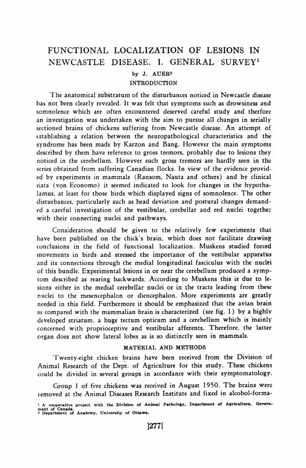
FUNCTIONAL LOCALIZATION OF LESIONS INNEWCASTLE DISEASE. I. GENERAL SURVEY'
by J. AUER2
INTRODUCTION
The anatomical substratum of the disturbances noticed in Newcastle diseasehas not been clearly revealed. It was felt that symptoms such as drowsiness andsomnolence which are often encountered deserved careful study and therforean investigation was undertaken with the aim to pursue all changes in seriallysectioned brains of chickens suffering from Newcastle disease. An attempt ofestablishing a relation between the neuropathological characteristics and thesyndrome has been made by Karzon and Bang. However the main symptomsdescribed by them have reference to gross tremors, probably due to lesions theynoticed in the cerebellum. However such gross tremors are hardly seen in theseries obtained from suffering Canadian flocks. In view of the evidence provid-ed by experiments in mammals (Ransom, Nauta and others) and by clinicaldata (von Economo) it seemed indicated to look for changes in the hypotha-lamus, at least for those birds which displayed signs of somnolence. The otherdisturbances, particularly such as head deviation and postural changes demand-ed a careful investigation of the vestibular, cerebellar and red nuclei togetherwith their connecting nuclei and pathways.
Consideration should be given to the relatively few experiments thathave been published on the chick's brain, which does not facilitate drawingconclusions in the field of functional localization. Mutskens studied forcedmovements in birds and stressed the importance of the vestibular apparatusand its connections through the medial longitudinal fasciculus with the nucleiof this bundle. Experimental lesions in or near the cerebellum produced a symp-tom described as rearing backwards. According to Muskens this is due to le-sions either in the medial cerebellar nuclei or in the tracts leading from thesenuclei to the mesencephalon or diencephalon. More experiments are greatlyneeded in this field. Furthermore it should be emphasized that the avian brainas compared with the mammalian brain is characterized (see fig. 1) by a highlydeveloped striatum, a huge tectum opticum and a cerebellum which is imainlyconcerned with proprioceptive and vestibular afferents. Therefore, the latterorgan does not show lateral lobes as is so distinctly seen in mammals.
MATERIAL AND METHODS
Twenty-eight chicken brains have been received from the Division ofAnimal Research of the Dept. of Agriculture for this study. These chickenscould be divided in several groups in accordance with their symptomatology.
Group 1 of five chickens was received in August 1950. The brains wereiemoved at the Animal Diseases Research Institute and fixed in alcohol-forma-1 A cooperative project with the Divigion of Animal Pathology, Department of Agriculture, Govern-ment of Canada.2 Departnment of Anatomy, University of Ottawa.
]277[

Canadian Journal ofJ278J Comparative Medicine
NEWCASTLE DI.SEASE Vol. XVI, No. SAugust, 1952
line. Specific symptoms for each animal were not available. However a gen-eral statement concerning the symptomatology of the flock, from which thesechickens were a sample, could be obtained. The predominating symptom wasan affection of the neck. Birds would stand or sit normally for some time.Eventually they would move their heads but it was as if they could not bringtheir heads around to a normal position, but they would turn the oppositeway and tremble. Staggering backwards was frequently observed. Such waseasily seen when the birds attempted to eat. Affected birds could not peckfood normally and they often could only get their heads down to the feedsideways. While eating the whole head was buried in the mash.
',,r/ ,J :Fig. 1
'-' ,; DIAGRAM OF AVIAN/ A BRAIN
- _ __ '(Modified after Papez)
-1 1. Bill.2. Nose.
..........4. Forebrain.
5. Cerebellum.
3 ,i b ~~~~~~~~~~~~~~~6.Bulb.
.....7 7. Tectum opticum.
9. Jaw.
on, 9Group 2 of five chickens was received in January 1951. This group was
taken during a natural outbreak among an experimental control flock. Thesymptomatology was clearly indicated this time. Deviation of the head wasthe most conspicuous sign. Furthermore pecking and eating caused the headto be thrown violently backwards. Even the whole bird suffered from rollingbackwards while attempting to eat. The birds seemed to be rather excitable.Somnolence on the other hand was not observed.
Group 3 of thirteen chickens was also received in January 1951. Thiswas a group used in control experiments for vaccine production. Unfortunatelya clear cut symptomatology has not been forthcoming. All the birds died afterparalysis and coma without showing a gradual onset of the disease. This istoo vague a description for the purpose of the present study.
Group 4 of five chicks was obtained in August 1951. The chicks wereexposed to the active virus (intranasal application) and rapidly developedsymptoms which could be observed by the author. As there was a differencein time of the application this group very well displayed a gradual de-velopment of a syndrome which was characterized by somnolence and hypo-

Canadian Journal of NEWCASTLE DISEASE Vol. XVI, No. 8 r'r791Comparative Medicine August, 1952 L"J
motility. The somnolence started with dozing and closed eyes, a picture whichgradually became more severe. The birds could easily be aroused from theirsomnolence but fell back into it after the termination of the external stimulus.Two of the group gradually reached a severe state of paralysis and coma thoughthis was also preceded by somnolence.
'rhe material described above was easily sectioned and the sections werealternately stained with toluidine blue, haematoxylin eosin and Heidenhain'shaematoxylin. The latter stain was performed in order to have data on thenlyelinization for compar.son with normal material.
OBSERVATIONS
All groups show a widespread engorgement of the vessels in the wholebrain. Lymphocytes have invaded the nervous tissues rather uniformly, whilemiany vessels are surrounded by a cuff of lymphocytes. Some brains of group4 show edema. Condensed infiltrates of lymphocytes are present in many areas(see below) while degeneration of ganglion cells is marked. The meninges donot show marked pathological changes.
The hypothalamus, the optic tectum, the midbrain tegmentum, the
Fig. 2. (Left) Perivascular cuffing in metencepha-Ion. (Small figures - 1. Cerebeium, 2 Cuffing,3. Teogmentum). Fig. 3. (Right) Lymphocyte infil-tration in metencephalon. (Small figures - 1. Cerebellum, 2. Infiltration, 3. Somatic nucleus, 4. Teg-mentum hindbrain.)

[2801 Canadian Journal of NEWCASTLE DISEASE Vol. XVI, No.9Comparative Medne August, 192
Fig. 4. Red nucleus in New-castle disease. Cellular exsu-
date. Degenerating cells.
hindbrain and the cerebellum have been extensively studied in group 1, 2 and4. Group 3 is not conducive to conclusions as the onset of the changes is notknown. Furthermore in group 3 the heads of the birds were decalcified inorder to obtain a good picture of the hypothysis. However this has resulted ina poor result of the toluidine blue staining, probably due to change in the pHduring decalcification. From the viewpoint of lesions group 1 and 2 shouldbe combined while group 4 formed a separate entity altogether. Group 1 and 2do not show specific lesions in the hypothalamus except for some lymphocyteaccumulation here and there around the distribution of the vessels. The mid-brain and hindbrain however display degeneration and also extensive lympho-cyte infiltration in many areas.
The degeneration consists of: clumping of the chromatine, excentric nu-cleus, disappearance of the nucleus and neuronophagia. Glia staining has notbeen performed as yet. In view of the cell changes this is highly desirable. How-evcr cuch a technique would not permit alternate staining and therefore a spe-cial group has to be used for this technique.
For this general survey the degenerative signs have been particularly studi-ed in the red nucleus, the nucleus mesencephalicus lateralis of the optic tectum,the nuclei isthmi, the nucleus tecti cerebelli and the vestibular nuclei. Perivas-cular cuffing (fig. 2 see page 279) and lymphocyte infiltration is evident closeto the midline in the midbrain and obviously the fasciculus longitudinalis me-dialis is involved. The functional significance of this also in the light of Mus-kens' work will be discussed below. Fig. 3 (see page 279) illustrates this in-filtration in one of the specimens. Degeneration of the large cells in the red nu-cleus is easily observed. Clumping of the chromatine and cell disappearance isshown in fig. 4. This can be well verified after comparison with the normalred nucleus illustrated in fig. 5. Furthermore it has been noticed that the cere-

Canadian Journal of NEWCASTLE DISEASE Vol. XVI, No. 8 F ])Comparative Medicine .Augt62 [ 2
bellar cortex has undergone definite changes in the form of lymphocyte infil-tration in the molecular layer and all stages of degeneration in the cells of Pur-kinje. Typical bands of lymphocytes were seen radiating from the inner layersto the surface of the cerebellum. It is noteworthy that such radiations have beenrecently described by Karzon and Bang. The nucleus tecti cerebelli has under-gone marked degeneration as is notified in fig. 6 and 7. In fig. 8 a normal cellof this nucleus is shown for comparison. Also the vestibular nuclei have beeninfluenced by the changes. Evidence of important changes in the centers control-ling postural reflexes is amply at hand. The changes also in the area of the fas-ciculus longitudinalis medialis which constitutes connections between the vesti-bular nuclei and the nuclei of some cranial nerves are important. Head deviationvia the accessory nerve nucleus might thus be explained in the cases of group 1-and 2.The sections of group 4 show changes of an entirely different nature, though
widespread lymphocyte invasion is noticed, degeneration could not be detectedanywhere. It should be emphasized however that the mammillary bodies intwo specimens which showed somnolence, were heavily changed by an accu-mulation of lymphocytes. Fig. 9. It is of course difficult to prove that thisinfiltration impairs the normal functions, but in analogy with lesions in themammillary body causing somnolence this conclusion is bound to be made.However a light infiltration in one of the specimens in group 1 and 2 shouldbe a warning that conclusions in this field have to be taken with care. Group
Fig. 5. (Left) Large cells in normal red nucleus. Fig. 6. (Right) Nucleus tecti cerebeill in Newcastledisease. Note infiltration of lvmphocytes and degeneration of cells.

[282] Canadian Journal of[2821 Comparative Medicine NEWCASTLE DISEASE VoL XVI, No. 8August, 1952
Fig. 7. (Left) Perikaryon in Newcastle disease of nucleus tecti cerebelli. Note chromatine clumping.(immersion photo) Fig. 8. (Right) Normal perikaryon in nucleus tecti cerebelli. (immersion photo)
1 and 2 as was explained above were characterized by excitability and there-lore in this case the infiltration has not reached the effect of destruction yet.(fig. 10).
DISCUSSIONIt seems that the relatively small amount of material already allows for
a classification of syndromes. Of these the following stand out:
1. Forced movements e.g. rearing backwards probably caused by the cerebellarlesions, particularly in the nucleus tecti. Pathways from .this nucleus to themidbrain (n. ruber) and to the diencephalon (n. rotundus) have been describ-ed as part of a strio-cerebellar circuit. The strio-cerebellar tract is the otherelement of this circuit and occupies part of the lateral forebrain bundle, a sys-tem replaced by the internal capsule in mammals. Experiments by 'ten Cate('26) and Muskens ('30) where lesions were inflicted in the nujleus tecti pro-bably resulted in forced movements similar to the syndrome in the present ma-terial (group 1 and 2). It is not quite clear yet why such forced movementsoccur mostly when the birds peck and eat. A tract arising in the midbraincourses to the hindbrain and seems to be concerned with pecking movements,according to Papez.2. Head deviation. It is believed that changes close to the midline in the hind-and midbrain are responsible for this deviation. Postural defects are caused

Canadian Journal of NEWCASTLE DISEASE vol. xvs, No. 8 [281Comparative Medicine August, 1962
by impairment of the fasciculus longitudinalis medialis as well as by degene-ration of the vestibular nuclei.
3. Excitability or Somnolence. Group 1 and 2 were very excitable while group4 showed typical somnolence.
The changes in the hypothalamus: vascular engorgement, cuffing andlymphocyte infiltration may very well be responsible for these changes. Itwould appear that the excitability is a sign of irritation in the posterior partof the hypothalamus while the destruction of this area could be followed bysomnolence. Such a conclusion would be quite in accordance with experimentsin mammals and with the pathology of for instance encephalitis lethargicaas described by von Economo. It might be advisable to obtain data on temper-ature and if possible other vegetative signs in birds suffering from this type ofthe disease. The importance of the hypothalamus as a vegetative center waspointed out again in a critical review by the author in 1948. There is nodoubt that the observations briefly mentioned above justify further study onthe functional pathology of Newcastle disease. Glia staining is necessary inorder to follow the process of neuronophagia in the less acute cases of thedisease. The birds should be more closely observed when the disease developsi.e. after they have been injected with the virus as in group 4. It also seems
Fig. 9. (Left) Lymphocyte infiltration in mammil- £. in3nlary body. Somnolent type of Newcastle disease. 1 iFig. 10. (Right) Lymphocyte infiltration in mammil-lary body. Excited type of Newcastle disease. (Small figures - 1. Third ventricle, 2. Engorged vessel,
3. Mammillary body).

[284] Canadian Journal of NEWCASTLE DISEASt Vol. XVI, No. 8Comparative Medicine August, 1952
advantageous to inflict lesions in the brains of normal chickens. Such experi-ments would definitely lead to a better understanding of the function of cer-tain centers. Very little is known about the function of many nuclei, as ex-perimental data are hardly available. As indicated above, the striatum stillneeds a thorough investigation in the material now at hand. This is unfortun-ately hampered by the fact that also little is known of the normal striatum inbirds. (See Ariens Kappers). The study of chickens treated with active andinactive virus is a second part of the project, which, is now under way.
SUMMARY
As anatomical study was undertaken of the brains in several groups ofchickens suffering from different types of Newcastle disease. The principalchanges appeared to be general invasion of lymphocytes, lymphocyte infiltra-tion, vascular engorgement, perivascular cuffing and different stages of neuronaldegeneration. The substratum of at least three main disturbances, i.e. head devia-tion, rearing backwards and somnolence (or excitability) has been found onthe basis of localization of these principal changes.
REFERENCES
KARZON, D. T., and BANG, F. B. The pathogenesis of infection with a virulent (CG 179)and an avirulent (B) strain of Newcastle disease virus in the chicken I. J. of Exp. Med.,93, 3, 267-285, 1951.RANSON, S. W. and INGRAM, W. R. Catalepsy caused by lesions between the mammillarybodies and the third nerve in the cat. Am. J. of Physiol., 101: 609-696, 1932.NAUTA, W. J. H. Hypothalamic regulation of sleep in rats, an experimental study. J. Neuro-physiol., 9, 285-316, 1946.ECONOMO, C. VON. Die Encephalitis lethargica, Wien, Deuticke. 1918.MUSKENS, L. J. J. On tracts and centers involved in the upward and downward associatedmovements of the eye after experiments in birds. J. Comp. Neurol., 50: 289, 1930.TEN CATE, J. Contribution a la physiologie comparee du cervelet. I. Le cervelet du pigeon.Arch. Neerl. de physiol., 11: 1, 1926.PAPEZ, J. W. Comparative Neurology. T. Y. Crowell Co., New York, 1929.AUER, J. The Hypothalamus. Can. J. of Comp. Med., 192-212, July, August, 1948.ARIENS KAPPERS, C. U., HUBER, G. C., and CROSBY, E. C. The comparative anatomy ofthe nervous system of the vertebrates, including man. New York, Macmillan Co. 1936.