Embed Size (px)
Citation preview
Ff
Ia
b
a
ARRAA
KPAPI
1
ltvmtipo
ntmpf
25
0d
Molecular & Biochemical Parasitology 170 (2010) 28–36
Contents lists available at ScienceDirect
Molecular & Biochemical Parasitology
unctional characterization of LIT1, the Leishmania amazonensiserrous iron transporter
smaele Jacquesa, Norma W. Andrewsa,b,∗, Chau Huynha,b
Section of Microbial Pathogenesis, Yale University School of Medicine, New Haven, CT 06510, United StatesDepartment of Cell Biology and Molecular Genetics, University of Maryland, College Park, MD 20742, United States
r t i c l e i n f o
rticle history:eceived 9 October 2009eceived in revised form 7 December 2009ccepted 8 December 2009vailable online 16 December 2009
eywords:romastigotemastigotearasite
a b s t r a c t
Leishmania amazonensis LIT1 was identified based on homology with IRT1, a ferrous iron transporter fromArabidopsis thaliana. �lit1 L. amazonensis are defective in intracellular replication and lesion formationin vivo, a virulence phenotype attributed to defective intracellular iron acquisition. Here we functionallycharacterize LIT1, directly demonstrating that it functions as a ferrous iron membrane transporter fromthe ZIP family. Conserved residues in the predicted transmembrane domains II, IV, V and VII of LIT1 areessential for iron transport in yeast, including histidines that were proposed to function as metal ligandsin ZIP transporters. LIT1 also contains two regions within the predicted intracellular loop that are notfound in Arabidopsis IRT1. Deletion of region I inhibited LIT1 expression on the surface of Leishmaniapromastigotes. Deletion of region II did not interfere with LIT1 trafficking to the surface, but abolished
ron acquisition its iron transport capacity when expressed in yeast. Mutagenesis revealed two motifs within region II,HGHQH and TPPRDM, that are independently required for iron transport by LIT1. D263 was identified as akey residue required for iron transport within the TPPRDM motif, while P260 and P261 were dispensable.Deletion of proline-rich regions within region I and between regions I and II did not affect iron transportin yeast, but in L. amazonensis were not able to rescue the intracellular growth of �lit1 parasites, or theirability to form lesions in mice. These results are consistent with a potential role of the unique intracellular
ar reg
loop of LIT1 in intracellul. Introduction
The parasitic protozoan Leishmania is the causative agent ofeishmaniasis, an increasingly serious human infectious diseasehat ranges from mild, self-healing cutaneous lesions to a lethalisceral form [1]. Individuals become infected with metacyclic pro-astigotes during the bite of an infected sand fly. After being
aken up by macrophages, metacyclic promastigotes are foundnside parasitophosphorous vacuoles (PVs) that have properties ofhagolysosomes [2]. Inside PVs the parasites replicate as amastig-tes, giving rise to the symptoms of leishmaniasis [3].
In order to survive and replicate within acidified PVs, Leishma-ia has to overcome very challenging conditions, which include
he restricted access to essential micronutrients [4]. One suchicronutrient, iron, is critical for the intracellular survival of severalathogens, which depend on high affinity membrane transportersor acquiring this metal [5–7]. Nramp1, a transporter located on late
∗ Corresponding author at: Department of Cell Biology and Molecular Genetics,134 Bioscience Research Building, University of Maryland, College Park, MD 20742-815, United States. Tel.: +1 301 405 9601; fax: +1 301 314 1248.
E-mail address: [email protected] (N.W. Andrews).
166-6851/$ – see front matter © 2009 Elsevier B.V. All rights reserved.oi:10.1016/j.molbiopara.2009.12.003
ulation by Leishmania-specific factors.© 2009 Elsevier B.V. All rights reserved.
endosomal compartments, is thought to restrict pathogen growthby depleting phagosomes in iron [4,8]. Iron is an essential co-factorfor many biologically active molecules, but its acquisition is a majorchallenge under physiological conditions. Ferrous iron (Fe2+) issoluble at neutral pH, but its accumulation must be tightly con-trolled to avoid generation of toxic hydroxyl radicals by the Fentonreaction. For this reason, in aerobic environments iron is largelypresent in the insoluble ferric (Fe3+) form, which relies on carrierproteins such as transferrin, ferritin or lactoferrin for its transportand storage. Reductases thus play a central role in iron acquisi-tion, by converting Fe3+ into Fe2+ for transmembrane transport [9].Ferric iron reductase activity was detected in Leishmania [10], andrecently a plasma membrane molecule with properties of a ferroustransporter was identified for the first time in this parasite [11].
Analysis of the Leishmania genome revealed two identical genesencoding a membrane protein that shares 30% identity with IRT1,an iron regulated transporter from Arabidopsis thaliana [11,12]. Aspreviously shown for IRT1 [13], LIT1 (Leishmania Iron Transporter
1) expression rescued growth of the Saccharomyces cerevisiae�fet3fet4 strain that is defective in iron transport [14,15], and wascapable of stimulating iron import when over-expressed in the par-asites [11]. Also similar to Arabidopsis IRT1 [13], LIT1 was capableof translocating other divalent cations such as manganese, cad-
hemic
mLoittcatm
m[fttZwertt4tdii[iLim
2
2
(stAps
2
sdrd2od6tfwmtac
c
I. Jacques et al. / Molecular & Bioc
ium and zinc, but showed strong preference for iron [11]. TheIT1 protein was only detected in Leishmania amazonensis amastig-tes residing intracellularly within PVs, which are predicted to beron-poor late endosomal compartments [11]. Importantly, dele-ion of the LIT1 gene from the genome of L. amazonensis inhibitedhe parasite’s ability to replicate within macrophages, and to causeutaneous lesions in mice [11]. These findings established LIT1s the first putative ferrous iron transporter to be identified inrypanosomatid parasites, and showed that it is an important deter-
inant of Leishmania virulence.The Leishmania LIT1 belongs to the ZRT-, IRT-like family (ZIP) of
embrane proteins, which includes both zinc and iron transporters16]. Only very limited information is available on the structure,unction, selectivity and localization of ZIP transporters. The A.haliana IRT1 was the first member of this family to be identifiedhrough functional complementation studies in S. cerevisiae [13].IP proteins are predicted to share a similar membrane topology,ith both the amino- and carboxy-terminal ends located on the
xtracellular side of the membrane [11,12,16]. ZIP family proteinsange from 227 to 476 amino acid residues in length, and con-ain alpha helical regions that span the membrane five to eightimes. LIT1 has eight predicted transmembrane domains, and is32 amino acid residues in length [11]. The length variations withinhe ZIP family are due to a variable region between transmembraneomains III and IV, which is predicted to form a loop that extends
nto the cytoplasm [12,16]. Interestingly, the LIT1 variable regions significantly longer than that of IRT1 by 72 amino acid residues11]. In this study, we used site directed mutagenesis as a tool tonvestigate the functional role of this unique region in LeishmaniaIT1, as well as the role of conserved residues that were previouslymplicated in iron transport in other members of the ZIP family of
embrane transporters.
. Materials and methods
.1. Parasites
L. amazonensis promastigotes were cultured in M199 mediaInvitrogen) supplemented with 20% heat-inactivated fetal bovineerum, 5% penicillin–streptomycin, 0.1% hemin (25 mg/ml in 50%riethanolamine), 10 mM adenine, and 5 mM l-glutamine at 26 ◦C.xenic L. amazonensis amastigotes were grown in M199 media sup-lemented with 0.25% glucose, 0.5% trypticase, and 20 mM sodiumuccinate (pH 4.5) at 32 ◦C.
.2. Molecular cloning and mutagenesis to generate LIT1 alleles
Single base pair mutations were generated in the yeast expres-ion vector p426 (URA2+) containing the LIT1 gene [11] by siteirected polymerase chain reaction (PCR) (Stratagene). The PCReaction consisted of the following: 5 �l 10× reaction buffer, 1 �lNTP, 100 ng of plasmid DNA, 125 ng of each oligonucleotide and�l PfuTurbo DNA polymerase (2.5 U/�l) at a final reaction volumef 50 �l. The thermal cycler was programmed as follows: initialenaturation at 95 ◦C for 30 s; 12 cycles at 95 ◦C for 30 s, 55 ◦C for0 s and 68 ◦C for 30 min. The PCR product was digested with DpnIo degrade the methylated parental plasmid, the product was trans-ormed into DH5� cells, and plasmid DNA from selected coloniesas subjected to sequencing to identify clones containing theutagenized construct. Wild type and mutant LIT1 constructs were
ransformed into S. cerevisiae �fet3fet4 as previously described [11]nd selected on minimal essential plates without uracil (the vectoronfers the ability to synthesize uracil).
To create truncated LIT1 proteins, site directed polymerasehain reaction (PCR) was used to amplify upstream and down-
al Parasitology 170 (2010) 28–36 29
stream flanking sequences of the targeted DNA sequence markedfor deletion. PCR products were cloned into pGEM-T Easy Vec-tor (Promega). The sequence downstream of the deleted fragmentwithin LIT1 was digested with BlgII and XbaI, gel purified and lig-ated to the BlgII and XbaI linearized pGEM-T Easy vector carryingthe sequence upstream of the deleted fragment within LIT1. ThepGEM-T Easy vector carrying the truncated LIT1 gene was verifiedthrough sequencing, the truncated gene was excised using BamHIdigestion, ligated into BamHI linearized p426 (URA2+) vector, andagain confirmed by sequencing. To perform parallel studies in Leish-mania, the BamHI excised LIT gene was cloned into BglII digestedpF4X1.4 sat vector (Jena Bioscience, Jena, Germany) to createpF4X1.4-LIT1. Wild type and mutant pF4X1.4-LIT1 were trans-formed into the L. amazonensis �lit1 strain and selected on mediasupplemented with 50 �g/ml nourseothricin [11]. The pF4X1.4 satvector promotes stable chromosomal integration, resulting in highlevel expression (Jena Bioscience).
2.3. Functional complementation studies in yeast
For functional studies in yeast, �fet3fet4 S. cerevisiae (pro-vided by M. Guerinot, Dartmouth College, Hannover, NH) carryingthe Leishmania iron transporter were grown in yeast peptonedextrose (YPD), YPD containing 20 �M bathophenanthroline disul-fonate (BPS) and in selective medium lacking uracil (SCD-URA), asdescribed [11]. The mutant constructs, vector alone or the vectorcontaining the LIT1 gene were placed in �fet3fet4 using the yeastexpression vector p426 (URA2+) that enables yeast to synthesizeuracil. The cells were grown in minimal media lacking uracil (SCD-URA3) for 2 days at 30 ◦C and the cultures were then diluted tooptical densities (OD) of 10, 1, 0.1, and 0.01. Aliquots (5 �l) of thedilution were spotted on both yeast peptone dextrose (YPD) andon agarose plates containing yeast peptone dextrose supplementedwith 20 �M of the iron chelator, bathophenanthroline disulfonate(BPS) (YPD + 20 �M BPS). The plates were incubated for 2 days at30 ◦C and digitally photographed.
2.4. Iron transport assays
Iron uptake in �fet3fet4 was carried out as described [17].Briefly, cells were washed twice in assay buffer containing 10 mMMES, 2% glucose and 1 mM nitriloacetic acid, pH 6.1, resuspendedin ice-cold assay buffer, and 50 �l of the cell suspension was addedto 450 �l of assay buffer containing 1 mM sodium ascorbate and1.2 �l of a 1:10 dilution of 55Fe (82.69 mCi/mg, or 39 mCi/ml;PerkinElmer). The suspension was incubated at 30 ◦C or 0 ◦C for20 min, vortexed, vacuum filtered through Whatman GF/C filters,washed three times with ice-cold SSW buffer (1 mM EDTA, 20 mMNa3-citrate, pH 4.2, 1 mM CaCl2, 5 mM MgSO4 and 1 mM NaCl) [17].The 20 min time point chosen for these assays was found to bewithin the linear 55Fe uptake range in kinetic assays. Radioactivelylabeled cells were detected in a liquid scintillation counter (Wallac1409; PerkinElmer) as described [11].
2.5. Macrophage infection and Leishmania intracellular growthassay
Bone marrow macrophages were isolated from C57BL/6mice (Charles River Laboratories) as previously described [18].Macrophages were seeded at 3.8 × 10⁵ cells per well onto 6 wellplates containing three coverslips per well in RPMI 1640 sup-
plemented with 10% fetal bovine serum containing 5% L cellsupernatant (as a source of M-CSF) at 37 ◦C and 5% CO2. 24 h later3.8 × 105 amastigotes were added to the macrophages for 1 h at34 ◦C and 5% CO2. Cells were washed three times with phosphatebuffered saline containing calcium and magnesium (PBS+/+) and
3 hemic
sfwXwa
2
iw(3ifigwuwi
3
3t
t2trn
FctwfpHrdaDLft
0 I. Jacques et al. / Molecular & Bioc
upplemented with 2% L cell supernatant and 5% fetal bovine serumor the duration of the infection. Infected macrophages were fixedith 2% paraformaldehyde (PFA), permeabilized with 0.5% Triton-100 and stained with DAPI. The number of intracellular parasitesas determined microscopically at 1000× (Zeiss Axiovert 200) inminimum of 300 macrophages per coverslip, in triplicate.
.6. Immunofluorescence microscopy
Exponentially growing L. amazonensis promastigotes were fixedn 2% paraformaldehyde (PFA) overnight at 4 ◦C. The cells were
ashed three times with (PBS+/+), incubated in blocking buffer10% goat serum and 50 mM ammonium chloride in PBS+/+) for0 min, followed by anti-LIT rabbit polyclonal antibodies in block-
ng buffer for 1 h at room temperature. The cells were then washedve times with PBS+/+ and incubated with Alexa Fluor 488 conju-ated goat anti-rabbit antibodies (Invitrogen), washed and stainedith DAPI for 30 min at room temperature. Images were acquiredsing a fluorescence microscope (Zeiss Axiovert 200) equippedith a CCD camera (CoolSNAP HQ; Photometrics) controlled by
maging software (MetaMorph; Molecular Devices Corporation).
. Results
.1. Functional conservation of residues required for ironransport by L. amazonensis LIT1
Residues His-96, His-197, Ser-198, His-224 and Glu-228 of A.
haliana IRT1, which correspond to residues His-108, His-283, Ser-84, His-309, Glu-313 in LIT1 respectively, were previously showno be important for iron transport [19] (Fig. 1, red). These IRT1esidues, postulated to be located on the inner surface of a chan-el formed by the transmembrane domains, are likely candidatesig. 1. Summary of the mutations analyzed for phenotypes in iron transport andellular growth. The figure shows a schematic representation of the Leishmania ironransporter LIT1, with the predicted carboxy- and amino-termini in direct contactith the extracellular medium, and the variable region forming an intracellular loop
acing the cytosolic side of the plasma membrane. Boxed residues correspond to theredicted transmembrane regions I–VIII. The residues in red (H108, E112, D263,283, S284, H309 and E313) are conserved between LIT1 and Arabidopsis IRT1, and
equired for promoting iron acquisition when LIT1 is expressed in the iron transport-eficient �fet3fet4 S. cerevisiae strain. In green (Y382, D263 and D391) are the aminocids that are essential for iron transport in LIT1, but not in IRT1. In orange (D148,153, R157 P259, P260 and S319) are the residues not required for iron transport inIT1. Blue residues outlined in red were designated as regions 1 and 2 and targetedor deletion in this study. HxHxH indicates the histidine-rich motif within region 1hat is essential for iron transport.
al Parasitology 170 (2010) 28–36
for binding metal ions during transport. In order to determine ifthe corresponding residues were also required for iron transportby LIT1, we replaced His-108, His-283, Ser-284, His-309, Glu-313residues by alanine. The mutant alleles were then cloned into ayeast overexpression vector and transformed into the �fet3fet4 S.cerevisiae strain which lacks the FET3 multicopper oxidase requiredfor high affinity Fe2+ transport, and the FET4 low affinity Fe2+ trans-porter [14,15]. The auxotroph strains carrying the mutant plasmidswere then tested for growth in the presence or absence of theiron chelator BPS. As previously reported for IRT1, the mutatedLIT1 alleles failed to rescue the growth phenotype of �fet3fet4under iron limiting conditions (Fig. 2A and B). To confirm that thegrowth failure of these mutant strains was due to loss-of-function,and not to overexpression of the mutated alleles, we carried outiron uptake assays to directly measure transporter function. Unlike�fet3fet4 carrying wild type LIT1, which can acquire 55Fe, yeaststrains carrying the mutated alleles failed to transport 55Fe intra-cellularly (Fig. 2D). In addition to the absolutely conserved residuesdiscussed above, we also investigated the requirement for LIT1 Glu-112, which corresponds to Asp-100 in IRT1, and is predicted to belocated on the extracellular loop between transmembrane domainsII and III. LIT1 carrying a replacement of Glu-112 by alanine failedto rescue the ability of �fet3fet4 to grow in iron limiting conditions(Fig. 2B), and to incorporate 55Fe (Fig. 2D). We also changed LIT1residues Glu-115, Asp-153, Arg-157 and Ser-319 to alanine. Com-plementation tests for �fet3fet4 growth and 55Fe uptake showedthat these residues (of which only Glu-115 is conserved betweenIRT1 and LIT1) did not disrupt function of the Leishmania iron trans-porter (Fig. 2C and D).
3.2. Requirement for amino acids with aromatic and bulky sidegroups at the 382 and 391 positions in LIT1
Residues Tyr-382 and Asp-391, which correspond to Tyr-295and Glu-305 in IRT1, are conserved throughout the ZIP transporterfamily [19] and predicted to reside within transmembrane domainVII. Replacement of these two residues with alanine was reportedto have no effect in the ability of IRT1 to transport iron [19]. Incontrast, the alanine replacements of these residues abolished theability of LIT1 to promote growth in iron-depleted medium, and touptake iron (Fig. 3A and B). However, when Tyr-382 was replaced byphenylalanine, growth and iron transport were restored, suggest-ing a requirement at this location for a residue with an aromaticside chain. We also changed Asp-391 to either Asn-391 or Glu-391,and the results of growth and iron uptake assays showed that LIT1appears to have a preference for a bulky side chain in this position,since the D391N and D391E mutations partially rescued the growthphenotype of �fet3fet4 on iron limiting medium. These strains alsoshowed an intermediate level of 55Fe accumulation within cells(Fig. 3A and B).
These results suggested that LIT1 carrying the Y382A and D391Asubstitutions might have failed to rescue �fet3fet4 iron transportand growth because of defects in protein expression or intra-cellular folding/targeting. To directly investigate this issue, wetransfected �lit1 L. amazonensis promastigotes [11] with LIT1 car-rying the Y382A and D391A mutations. Both mutated transporterswere detected by immunofluorescence on the surface of non-permeabilized �lit1 L. amazonensis (Fig. 3C), in a punctate patternthat is very consistent with the pattern of endogenous LIT1 detectedin intracellular amastigotes [11]. These observations indicate thatthe alanine substitutions did not disrupt the intracellular fold-
ing and plasma membrane trafficking of the transporter. Thus,the Y382 and D391 residues, which are predicted to reside eitherwithin or adjacent to transmembrane VII of LIT1, may be uniquelyrequired for the passing of metal ion substrates across the mem-brane.
I. Jacques et al. / Molecular & Biochemical Parasitology 170 (2010) 28–36 31
Fig. 2. Conserved residues are critical for LIT1 function. The iron transport-deficient �fet3fet4 strain was transformed with wild type LIT1, empty vector or mutantLIT1constructs. Transformed strains were spotted on agar supplemented (YPD + 20 �M BPS) or not (YPD) with the iron chelator, bathophenanthroline disulfonate (BPS),in serial 1:10 dilutions. (A) Wild type LIT1 was able to complement the growth phenotype of �fet3fet4 under iron limit condition, whereas �fet3fet4 transformed with emptyvector failed to do so. (B) The LIT1 conserved amino acids H108A, E112A, H283A, S284A, H309A, E313A were mutagenized to alanine. The transformed strains grew on ironrich agar (YPD) at comparable rates to WT LIT1, but these strains failed to grow under iron-deficient conditions (YPD + 20 �M BPS). (C) �fet3fet4 expressing LIT1 carryingthe mutations E115A, D153A, R157A, S319A grew on both YPD and YPD + 20 �M BPS. (D) The ability of �fet3fet4 carrying wild type LIT1, empty vector, and LIT1 mutantconstructs to accumulate iron 55Fe was measured. LIT1 carrying the H108A, E112A, E115A, H283A, S284A, H309A, E313A, Y382A and D391A mutations did not restore thei r ironi al. The
3
t1arTlpaitpbiwl
ron transport deficiency of �fet3fet4, indicating that these residues are essential foron transport deficiency of �fet3fet4, indicating that these residues are not essenti
.3. Functional role of the cytoplasmic variable region of LIT1
The predicted intracellular loop region of the Leishmania LIT1ransporter is longer than that of IRT1 by 72 amino acids: residues58–211 (53 amino acids, region I) and residues 245–264 (19mino acids, region II) (Fig. 1). We hypothesized that these twoegions might have important roles in regulating LIT1 function.o address this issue, we created two truncated LIT1 constructs:
it1 (�158–211) and lit1 (�245–264) and performed yeast com-lementation experiments. Unlike wild type LIT1, the two deletionlleles (lacking regions I and II) did not complement the �fet3fet4ron transport and growth defect (Fig. 4A and B). One concern washat the large size of region I deletion lit1 (�158–211) could haverevented this truncated protein from reaching the plasma mem-
rane. Indeed, when this construct was expressed in L. amazonensis,t failed to be detected on the cell surface by immunofluorescenceith anti-LIT1 antibodies, in both fixed/non-permeabilized, and
ive promastigotes (Fig. 4C).
transport. In contrast, D148A, D153A, R157A, S319A, D391N or D391E restored thedata represent average ±SD of triplicates.
Residues 251–255 of region II consist of the histidine-rich motifHGHQH. Such motifs found in the variable region between trans-membrane domains III and IV of ZIP family members were proposedto be potential cytoplasmic metal binding domains [20]. This mightexplain why lit1 (�245–264) was not able to complement the�fet3fet4 iron transport defect under iron limiting conditions.To directly verify the contribution of the histidine motif to LIT1function, we generated a construct lacking only the five aminoacids comprising the HxHxH motif. Similar to the lit1 (�245–264)construct, lit1 (�251–255) failed to rescue the growth pheno-type of �fet3fet4 and showed no iron uptake capacity (Fig. 4B).Both lit1 (�245–264) and lit1 (�251–255) mutant proteins wereproperly localized to the plasma membrane of in both fixed/non-permeabilized, and live L. amazonensis (Fig. 4C), indicating that
the iron transport defect is likely to have been a consequenceof inability to of the transporter to bind metal ions through thehistidine-rich motif, and not of improper targeting to the plasmamembrane.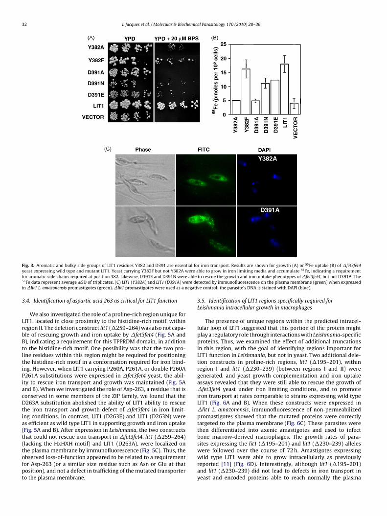
32 I. Jacques et al. / Molecular & Biochemical Parasitology 170 (2010) 28–36
Fig. 3. Aromatic and bulky side groups of LIT1 residues Y382 and D391 are essential for iron transport. Results are shown for growth (A) or 55Fe uptake (B) of �fet3fet4y ere af able5 ere dei egati
3
LrbBtltiPiacDtia(t(tofpt
east expressing wild type and mutant LIT1. Yeast carrying Y382F but not Y382A wor aromatic side chains required at position 382. Likewise, D391E and D391N were5Fe data represent average ±SD of triplicates. (C) LIT1 (Y382A) and LIT1 (D391A) wn �lit1 L. amazonensis promastigotes (green). �lit1 promastigotes were used as a n
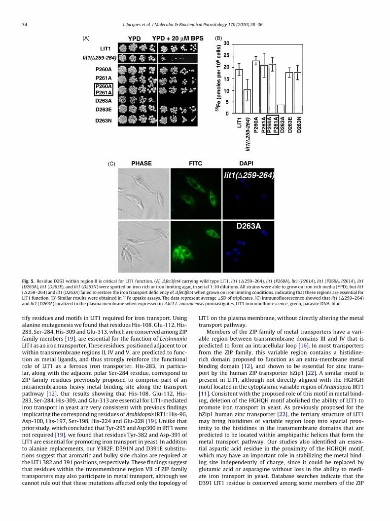
.4. Identification of aspartic acid 263 as critical for LIT1 function
We also investigated the role of a proline-rich region unique forIT1, located in close proximity to the histidine-rich motif, withinegion II. The deletion construct lit1 (�259–264) was also not capa-le of rescuing growth and iron uptake by �fet3fet4 (Fig. 5A and), indicating a requirement for this TPPRDM domain, in additiono the histidine-rich motif. One possibility was that the two pro-ine residues within this region might be required for positioninghe histidine-rich motif in a conformation required for iron bind-ng. However, when LIT1 carrying P260A, P261A, or double P260A261A substitutions were expressed in �fet3fet4 yeast, the abil-ty to rescue iron transport and growth was maintained (Fig. 5And B). When we investigated the role of Asp-263, a residue that isonserved in some members of the ZIP family, we found that the263A substitution abolished the ability of LIT1 ability to rescue
he iron transport and growth defect of �fet3fet4 in iron limit-ng conditions. In contrast, LIT1 (D263E) and LIT1 (D263N) weres efficient as wild type LIT1 in supporting growth and iron uptakeFig. 5A and B). After expression in Leishmania, the two constructshat could not rescue iron transport in �fet3fet4, lit1 (�259–264)lacking the HxHXH motif) and LIT1 (D263A), were localized on
he plasma membrane by immunofluorescence (Fig. 5C). Thus, thebserved loss-of-function appeared to be related to a requirementor Asp-263 (or a similar size residue such as Asn or Glu at thatosition), and not a defect in trafficking of the mutated transportero the plasma membrane.ble to grow in iron limiting media and accumulate 55Fe, indicating a requirementto rescue the growth and iron uptake phenotypes of �fet3fet4, but not D391A. Thetected by immunofluorescence on the plasma membrane (green) when expressedve control; the parasite’s DNA is stained with DAPI (blue).
3.5. Identification of LIT1 regions specifically required forLeishmania intracellular growth in macrophages
The presence of unique regions within the predicted intracel-lular loop of LIT1 suggested that this portion of the protein mightplay a regulatory role through interactions with Leishmania-specificproteins. Thus, we examined the effect of additional truncationsin this region, with the goal of identifying regions important forLIT1 function in Leishmania, but not in yeast. Two additional dele-tion constructs in proline-rich regions, lit1 (�195–201), withinregion I and lit1 (�230–239) (between regions I and II) weregenerated, and yeast growth complementation and iron uptakeassays revealed that they were still able to rescue the growth of�fet3fet4 yeast under iron limiting conditions, and to promoteiron transport at rates comparable to strains expressing wild typeLIT1 (Fig. 6A and B). When these constructs were expressed in�lit1 L. amazonensis, immunofluorescence of non-permeabilizedpromastigotes showed that the mutated proteins were correctlytargeted to the plasma membrane (Fig. 6C). These parasites werethen differentiated into axenic amastigotes and used to infectbone marrow-derived macrophages. The growth rates of para-sites expressing the lit1 (�195–201) and lit1 (�230–239) alleles
were followed over the course of 72 h. Amastigotes expressingwild type LIT1 were able to grow intracellularly as previouslyreported [11] (Fig. 6D). Interestingly, although lit1 (�195–201)and lit1 (�230–239) did not lead to defects in iron transport inyeast and encoded proteins able to reach normally the plasma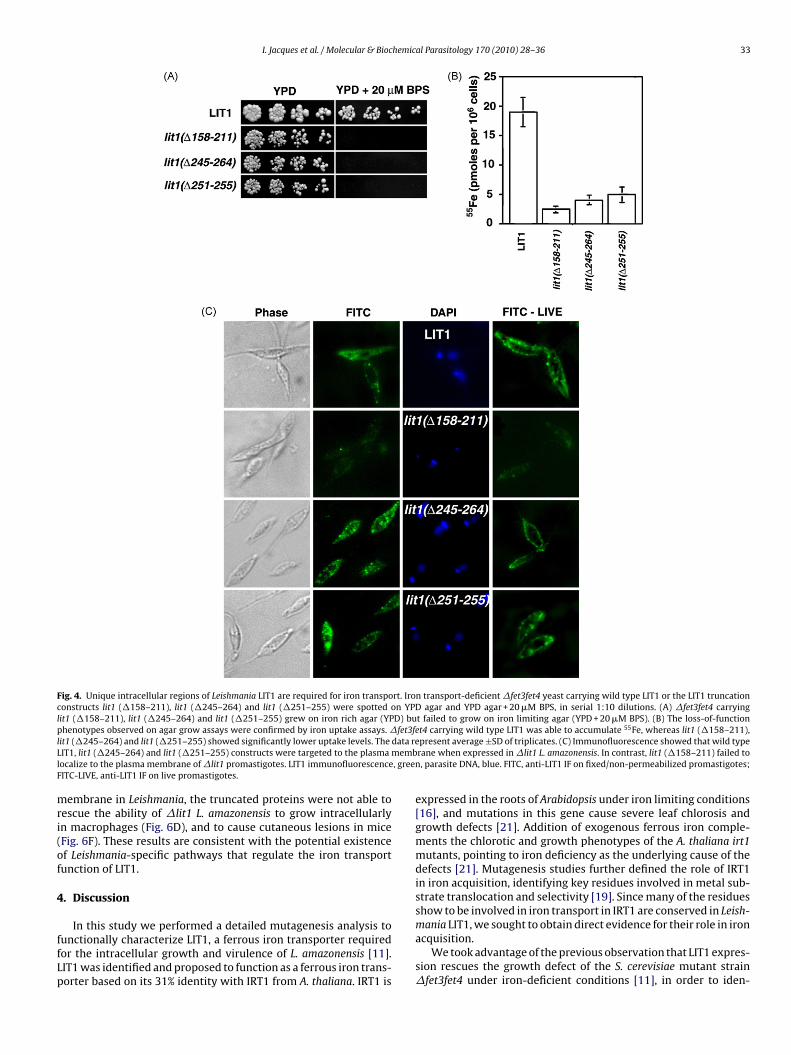
I. Jacques et al. / Molecular & Biochemical Parasitology 170 (2010) 28–36 33
Fig. 4. Unique intracellular regions of Leishmania LIT1 are required for iron transport. Iron transport-deficient �fet3fet4 yeast carrying wild type LIT1 or the LIT1 truncationconstructs lit1 (�158–211), lit1 (�245–264) and lit1 (�251–255) were spotted on YPD agar and YPD agar + 20 �M BPS, in serial 1:10 dilutions. (A) �fet3fet4 carryinglit1 (�158–211), lit1 (�245–264) and lit1 (�251–255) grew on iron rich agar (YPD) but failed to grow on iron limiting agar (YPD + 20 �M BPS). (B) The loss-of-functionp fet3f 55
l data reL membl , greeF
mri(of
4
ffLp
henotypes observed on agar grow assays were confirmed by iron uptake assays. �it1 (�245–264) and lit1 (�251–255) showed significantly lower uptake levels. TheIT1, lit1 (�245–264) and lit1 (�251–255) constructs were targeted to the plasmaocalize to the plasma membrane of �lit1 promastigotes. LIT1 immunofluorescenceITC-LIVE, anti-LIT1 IF on live promastigotes.
embrane in Leishmania, the truncated proteins were not able toescue the ability of �lit1 L. amazonensis to grow intracellularlyn macrophages (Fig. 6D), and to cause cutaneous lesions in miceFig. 6F). These results are consistent with the potential existencef Leishmania-specific pathways that regulate the iron transportunction of LIT1.
. Discussion
In this study we performed a detailed mutagenesis analysis tounctionally characterize LIT1, a ferrous iron transporter requiredor the intracellular growth and virulence of L. amazonensis [11].IT1 was identified and proposed to function as a ferrous iron trans-orter based on its 31% identity with IRT1 from A. thaliana. IRT1 is
et4 carrying wild type LIT1 was able to accumulate Fe, whereas lit1 (�158–211),present average ±SD of triplicates. (C) Immunofluorescence showed that wild typerane when expressed in �lit1 L. amazonensis. In contrast, lit1 (�158–211) failed ton, parasite DNA, blue. FITC, anti-LIT1 IF on fixed/non-permeabilized promastigotes;
expressed in the roots of Arabidopsis under iron limiting conditions[16], and mutations in this gene cause severe leaf chlorosis andgrowth defects [21]. Addition of exogenous ferrous iron comple-ments the chlorotic and growth phenotypes of the A. thaliana irt1mutants, pointing to iron deficiency as the underlying cause of thedefects [21]. Mutagenesis studies further defined the role of IRT1in iron acquisition, identifying key residues involved in metal sub-strate translocation and selectivity [19]. Since many of the residuesshow to be involved in iron transport in IRT1 are conserved in Leish-
mania LIT1, we sought to obtain direct evidence for their role in ironacquisition.We took advantage of the previous observation that LIT1 expres-sion rescues the growth defect of the S. cerevisiae mutant strain�fet3fet4 under iron-deficient conditions [11], in order to iden-
34 I. Jacques et al. / Molecular & Biochemical Parasitology 170 (2010) 28–36
Fig. 5. Residue D263 within region II is critical for LIT1 function. (A) �fet3fet4 carrying wild type LIT1, lit1 (�259–264), lit1 (P260A), lit1 (P261A), lit1 (P260A, P261A), lit1( r, in s( et4 whL resenta zonen
ta2fLwtrlZip2iiApnLtttttc
D263A), lit1 (D263E), and lit1 (D263N) were spotted on iron rich or iron limiting aga�259–264) and lit1 (D263A) failed to restore the iron transport deficiency of �fet3fIT1 function. (B) Similar results were obtained in 55Fe uptake assays. The data repnd lit1 (D263A) localized to the plasma membrane when expressed in �lit1 L. ama
ify residues and motifs in LIT1 required for iron transport. Usinglanine mutagenesis we found that residues His-108, Glu-112, His-83, Ser-284, His-309 and Glu-313, which are conserved among ZIPamily members [19], are essential for the function of LeishmaniaIT1 as an iron transporter. These residues, positioned adjacent to orithin transmembrane regions II, IV and V, are predicted to func-
ion as metal ligands, and thus strongly reinforce the functionalole of LIT1 as a ferrous iron transporter. His-283, in particu-ar, along with the adjacent polar Ser-284 residue, correspond toIP family residues previously proposed to comprise part of anntramembranous heavy metal binding site along the transportathway [12]. Our results showing that His-108, Glu-112, His-83, Ser-284, His-309, and Glu-313 are essential for LIT1-mediated
ron transport in yeast are very consistent with previous findingsmplicating the corresponding residues of Arabidopsis IRT1: His-96,sp-100, His-197, Ser-198, His-224 and Glu-228 [19]. Unlike thatrior study, which concluded that Tyr-295 and Asp300 in IRT1 wereot required [19], we found that residues Tyr-382 and Asp-391 ofIT1 are essential for promoting iron transport in yeast. In additiono alanine replacements, our Y382F, D391N and D391E substitu-
ions suggest that aromatic and bulky side chains are required athe LIT1 382 and 391 positions, respectively. These findings suggesthat residues within the transmembrane region VII of ZIP familyransporters may also participate in metal transport, although weannot rule out that these mutations affected only the topology oferial 1:10 dilutions. All strains were able to grow on iron rich media (YPD), but lit1en grown on iron limiting conditions, indicating that these regions are essential foraverage ±SD of triplicates. (C) Immunofluorescence showed that lit1 (�259–264)
sis promastigotes. LIT1 immunofluorescence, green, parasite DNA, blue.
LIT1 on the plasma membrane, without directly altering the metaltransport pathway.
Members of the ZIP family of metal transporters have a vari-able region between transmembrane domains III and IV that ispredicted to form an intracellular loop [16]. In most transportersfrom the ZIP family, this variable region contains a histidine-rich domain proposed to function as an extra-membrane metalbinding domain [12], and shown to be essential for zinc trans-port by the human ZIP transporter hZip1 [22]. A similar motif ispresent in LIT1, although not directly aligned with the HGHGHmotif located in the cytoplasmic variable region of Arabidopsis IRT1[11]. Consistent with the proposed role of this motif in metal bind-ing, deletion of the HGHQH motif abolished the ability of LIT1 topromote iron transport in yeast. As previously proposed for thehZip1 human zinc transporter [22], the tertiary structure of LIT1may bring histidines of variable region loop into spacial prox-imity to the histidines in the transmembrane domains that arepredicted to be located within amphipathic helices that form themetal transport pathway. Our studies also identified an essen-tial aspartic acid residue in the proximity of the HGHQH motif,
which may have an important role in stabilizing the metal bind-ing site independently of charge, since it could be replaced byglutamic acid or asparagine without loss in the ability to medi-ate iron transport in yeast. Database searches indicate that theD391 LIT1 residue is conserved among some members of the ZIP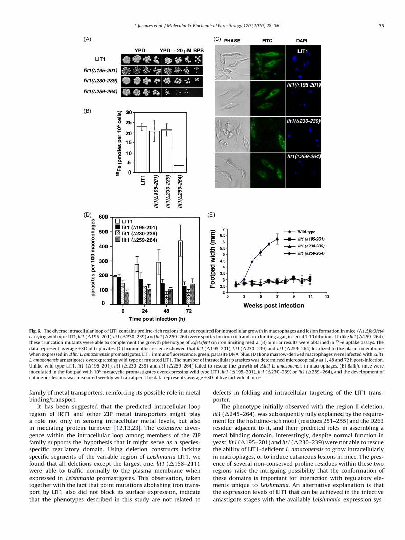
I. Jacques et al. / Molecular & Biochemical Parasitology 170 (2010) 28–36 35
Fig. 6. The diverse intracellular loop of LIT1 contains proline-rich regions that are required for intracellular growth in macrophages and lesion formation in mice. (A) �fet3fet4carrying wild type LIT1, lit1 (�195–201), lit1 (�230–239) and lit1 (�259–264) were spotted on iron rich and iron limiting agar, in serial 1:10 dilutions. Unlike lit1 (�259–264),these truncation mutants were able to complement the growth phenotype of �fet3fet4 on iron limiting media. (B) Similar results were obtained in 55Fe uptake assays. Thedata represent average ±SD of triplicates. (C) Immunofluorescence showed that lit1 (�195–201), lit1 (�230–239) and lit1 (�259–264) localized to the plasma membranewhen expressed in �lit1 L. amazonensis promastigotes. LIT1 immunofluorescence, green, parasite DNA, blue. (D) Bone marrow-derived macrophages were infected with �lit1L f intrU ed toi type Lc e ±SD
fb
raigfssfwetpt
. amazonensis amastigotes overexpressing wild type or mutated LIT1. The number onlike wild type LIT1, lit1 (�195–201), lit1 (�230–239) and lit1 (�259–264) fail
noculated in the footpad with 106 metacyclic promastigotes overexpressing wildutaneous lesions was measured weekly with a caliper. The data represents averag
amily of metal transporters, reinforcing its possible role in metalinding/transport.
It has been suggested that the predicted intracellular loopegion of IRT1 and other ZIP metal transporters might play
role not only in sensing intracellular metal levels, but alson mediating protein turnover [12,13,23]. The extensive diver-ence within the intracellular loop among members of the ZIPamily supports the hypothesis that it might serve as a species-pecific regulatory domain. Using deletion constructs lackingpecific segments of the variable region of Leishmania LIT1, weound that all deletions except the largest one, lit1 (�158–211),
ere able to traffic normally to the plasma membrane whenxpressed in Leishmania promastigotes. This observation, takenogether with the fact that point mutations abolishing iron trans-ort by LIT1 also did not block its surface expression, indicatehat the phenotypes described in this study are not related to
acellular parasites was determined microscopically at 1, 48 and 72 h post-infection.rescue the growth of �lit1 L. amazonensis in macrophages. (E) Balb/c mice wereIT1, lit1 (�195–201), lit1 (�230–239) or lit1 (�259–264), and the development ofof five individual mice.
defects in folding and intracellular targeting of the LIT1 trans-porter.
The phenotype initially observed with the region II deletion,lit1 (�245–264), was subsequently fully explained by the require-ment for the histidine-rich motif (residues 251–255) and the D263residue adjacent to it, and their predicted roles in assembling ametal binding domain. Interestingly, despite normal function inyeast, lit1 (�195–201) and lit1 (�230–239) were not able to rescuethe ability of LIT1-deficient L. amazonensis to grow intracellularlyin macrophages, or to induce cutaneous lesions in mice. The pres-ence of several non-conserved proline residues within these two
regions raise the intriguing possibility that the conformation ofthese domains is important for interaction with regulatory ele-ments unique to Leishmania. An alternative explanation is thatthe expression levels of LIT1 that can be achieved in the infectiveamastigote stages with the available Leishmania expression sys-
3 hemic
tspl
A
oA
R
[
[
[
[
[
[
[
[
[
[
[
[
[between transmembrane domains III and IV of hZip1 are required for zinc
6 I. Jacques et al. / Molecular & Bioc
ems are incompatible with functional complementation. Furthertudies are required to clarify these questions, and to search forutative parasite regulatory factors that might interact intracellu-
arly with the variable region of the LIT1 iron transporter.
cknowledgements
We would like to thank Andrew Flannery for critical readingf the manuscript. This work was supported by NIH grants RO1I067979 and R37 AI034867 to N.W.A. and R37 AI034867S1 to I.J.
eferences
[1] Herwaldt BL. Leishmaniasis. Lancet 1999;354:1191–9.[2] Antoine JC, Prina E, Lang T, Courret N. The biogenesis and properties of the
parasitophorous vacuoles that harbour Leishmania in murine macrophages.Trends Microbiol 1998;6:392–401.
[3] Grimaldi Jr G, Tesh RB. Leishmaniases of the new world: current concepts andimplications for future research. Clin Microbiol Rev 1993;6:230–50.
[4] Fortier A, Min-Oo G, Forbes J, Lam-Yuk-Tseung S, Gros P. Single gene effects inmouse models of host:pathogen interactions. J Leukoc Biol 2005;77:868–77.
[5] Zhou D, Hardt WD, Galan JE. Salmonella typhimurium encodes a putative irontransport system within the centisome 63 pathogenicity island. Infect Immun1999;67:1974–81.
[6] Janakiraman A, Slauch JM. The putative iron transport system SitABCD encodedon SPI1 is required for full virulence of Salmonella typhimurium. Mol Microbiol2000;35:1146–55.
[7] Rodriguez GM. Control of iron metabolism in Mycobacterium tuberculosis.Trends Microbiol 2006;14:320–7.
[8] Jabado N, Jankowski A, Dougaparsad S, Picard V, Grinstein S, Gros P.
Natural resistance to intracellular infections: natural resistance-associatedmacrophage protein 1 (Nramp1) functions as a pH-dependent man-ganese transporter at the phagosomal membrane. J Exp Med 2000;192:1237–48.[9] Andrews NC, Fleming MD, Gunshin H. Iron transport across biologic mem-branes. Nutr Rev 1999;57:114–23.
[
al Parasitology 170 (2010) 28–36
10] Wilson ME, Lewis TS, Miller MA, McCormick ML, Britigan BE. Leishmaniachagasi: uptake of iron bound to lactoferrin or transferrin requires an ironreductase. Exp Parasitol 2002;100:196–207.
11] Huynh C, Sacks DL, Andrews NW. A Leishmania amazonensis ZIP family irontransporter is essential for parasite replication within macrophage phagolyso-somes. J Exp Med 2006;203:2363–75.
12] Eng BH, Guerinot ML, Eide D, Saier Jr MH. Sequence analyses and phylogeneticcharacterization of the ZIP family of metal ion transport proteins. J Membr Biol1998;166:1–7.
13] Eide D, Broderius M, Fett J, Guerinot ML. A novel iron-regulated metal trans-porter from plants identified by functional expression in yeast. Proc Natl AcadSci USA 1996;93:5624–8.
14] Askwith C, Eide D, Van Ho A, et al. The FET3 gene of S. cerevisiae encodes amulticopper oxidase required for ferrous iron uptake. Cell 1994;76:403–10.
15] Dix DR, Bridgham JT, Broderius MA, Byersdorfer CA, Eide DJ. The FET4 geneencodes the low affinity Fe(II) transport protein of Saccharomyces cerevisiae. JBiol Chem 1994;269:26092–9.
16] Guerinot ML. The ZIP family of metal transporters. Biochim Biophys Acta2000;1465:190–8.
17] Eide D, Davis-Kaplan S, Jordan I, Sipe D, Kaplan J. Regulation of iron uptakein Saccharomyces cerevisiae. The ferrireductase and Fe(II) transporter are regu-lated independently. J Biol Chem 1992;267:20774–81.
18] Roy D, Liston DR, Idone VJ, et al. A process for controlling intracellular bacterialinfections induced by membrane injury. Science 2004;304:1515–8.
19] Rogers EE, Eide DJ, Guerinot ML. Altered selectivity in an Arabidopsis metaltransporter. Proc Natl Acad Sci USA 2000;97:12356–60.
20] Kerkeb L, Mukherjee I, Chatterjee I, Lahner B, Salt DE, Connolly EL. Iron-inducedturnover of the Arabidopsis iron-regulated transporter1 metal transporterrequires lysine residues. Plant Physiol 2008;146:1964–73.
21] Vert G, Grotz N, Dedaldechamp F, et al. IRT1, an Arabidopsis transporteressential for iron uptake from the soil and for plant growth. Plant Cell2002;14:1223–33.
22] Milon B, Wu Q, Zou J, Costello LC, Franklin RB. Histidine residues in the region
transport across the plasma membrane in PC-3 cells. Biochim Biophys Acta2006;1758:1696–701.
23] Connolly EL, Fett JP, Guerinot ML. Expression of the IRT1 metal transporter iscontrolled by metals at the levels of transcript and protein accumulation. PlantCell 2002;14:1347–57.