Embed Size (px)
Citation preview

REVIEW Open Access
Functional cellulose-based hydrogels asextracellular matrices for tissue engineeringSayan Deb Dutta1, Dinesh K. Patel2 and Ki-Taek Lim1*
Abstract
Cellulose-based hydrogels are immensely important for tissue engineering. In this review, we attempt to documentthe source, nature, and application of cellulose-based hydrogels as an extracellular matrix for tissue growth andregeneration. Hydrogels can be prepared either from native cellulose, including both bacterial and plant sources orfrom cellulose derivatives, such as methyl cellulose, carboxymethylcellulose, and hydroxypropylmethylcellulose oreven metal ions such as silver. Cellulose-polymer composite (polymers that include natural sources includingchitosan, starch, alginates, collagen, hyaluronic acid, and chitin) are an attractive, inexpensive, and advantageousstructural material that is easy to use. Cellulose-based scaffolding materials are widely used in the regeneration ofvarious tissues, such as bone, cartilage, heart, blood vessel, nerve, and liver, among others. In this review, we discussthe most important applications of cellulosic hydrogels in tissue engineering based on their structuralcompositions.
Keywords: Cellulose, Hydrogels, Scaffolds, Extracellular matrices, Tissue engineering
IntroductionCells communicate with each other either directly viamolecular interactions or through the secretion of differ-ent hormones or mediators which systematically regulatevarious cell functions. Growth factors are also secretedduring cellular crosstalk and may be pro-proliferative oranti-proliferative in nature, being mainly involved in celldifferentiation, migration, adhesion, and gene expression.Natural and synthetic materials may be used as bulkingagents for the binding of various growth factors by mim-icking natural extracellular matrix (ECM) molecularself-assembly via secondary forces, such as ionic orhydrogen bonds, whereas chemical gels are result of co-valent bonds [1–5].Hydrogels have potential applications in various fields
such as agriculture, food, biomaterials, water purification,biomedicine, and pharmaceuticals, among others. [6–8].Hydrogels are primarily made up of natural living tissuerather than synthetic biomaterials, as a result have a highwater content and a soft consistency similar to natural tis-sues [9]. Moreover, the high water content of these
materials contributes to their biocompatibility. Thus,hydrogels can be used as membranes for biosensors[10, 11], in artificial heart and skin [12, 13], contactlenses [14, 15], and drug delivery [3, 6, 16]. Cross-linking synthetic polymer-based hydrogels have beenreported, including poly (ethylene glycol) [17, 18],poly (vinyl alcohol) [18, 19], poly (amido-amine) [20],poly (N-isopropylacrylamide) [21], polyacrylamide [18,22], and poly (acrylic acid) [18, 23].In tissue engineering, hydrogels are the most extensively
used biopolymer due to their highly swollen three- dimen-sional (3D) environment, which is very similar to soft tis-sues and allows for the diffusion of nutrients, growthfactors and cellular waste through the elastic network andfor the regeneration of damaged tissues [13, 18, 24, 25]. Inregenerative medicine, hydrogel used to repair and assistregeneration of various soft and hard tissues, such as cartil-age, bone and vascular tissues [26–28]. Natural hydrogelsinclude the bioprocessing of natural polymer-based mate-rials such as proteins, including collagen, gelatin, and fibrin,and polysaccharides, including alginate chitosan, hyaluronicacid, dextran, and cellulose which are used as extracellularmatrices (ECM).Cellulose is a fibrous, tough, water-insoluble substance,
found in the protective cell walls of plants, particularly in
© The Author(s). 2019 Open Access This article is distributed under the terms of the Creative Commons Attribution 4.0International License (http://creativecommons.org/licenses/by/4.0/), which permits unrestricted use, distribution, andreproduction in any medium, provided you give appropriate credit to the original author(s) and the source, provide a link tothe Creative Commons license, and indicate if changes were made. The Creative Commons Public Domain Dedication waiver(http://creativecommons.org/publicdomain/zero/1.0/) applies to the data made available in this article, unless otherwise stated.
* Correspondence: [email protected] Laboratory, Department of Biosystems Engineering, KangwonNational University, Chuncheon, Republic of KoreaFull list of author information is available at the end of the article
Dutta et al. Journal of Biological Engineering (2019) 13:55 https://doi.org/10.1186/s13036-019-0177-0
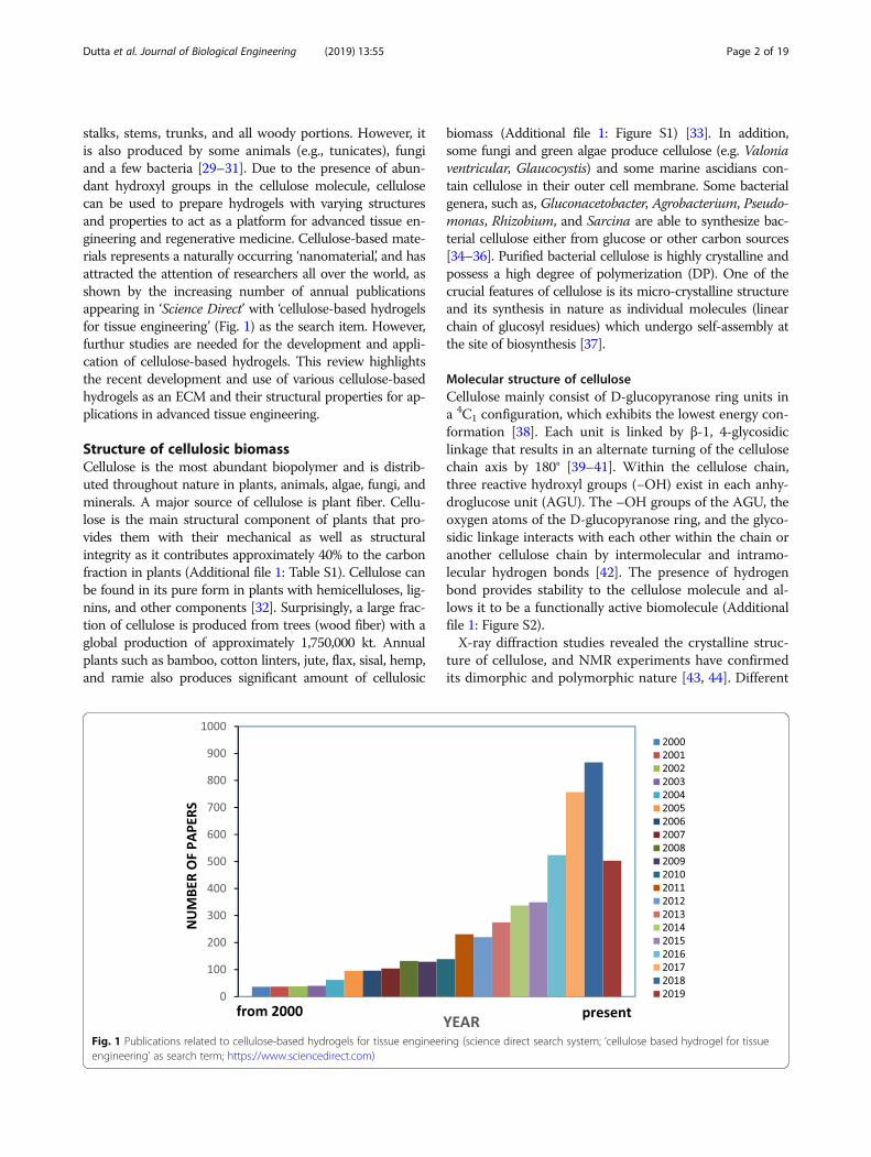
stalks, stems, trunks, and all woody portions. However, itis also produced by some animals (e.g., tunicates), fungiand a few bacteria [29–31]. Due to the presence of abun-dant hydroxyl groups in the cellulose molecule, cellulosecan be used to prepare hydrogels with varying structuresand properties to act as a platform for advanced tissue en-gineering and regenerative medicine. Cellulose-based mate-rials represents a naturally occurring ‘nanomaterial’, and hasattracted the attention of researchers all over the world, asshown by the increasing number of annual publicationsappearing in ‘Science Direct’ with ‘cellulose-based hydrogelsfor tissue engineering’ (Fig. 1) as the search item. However,furthur studies are needed for the development and appli-cation of cellulose-based hydrogels. This review highlightsthe recent development and use of various cellulose-basedhydrogels as an ECM and their structural properties for ap-plications in advanced tissue engineering.
Structure of cellulosic biomassCellulose is the most abundant biopolymer and is distrib-uted throughout nature in plants, animals, algae, fungi, andminerals. A major source of cellulose is plant fiber. Cellu-lose is the main structural component of plants that pro-vides them with their mechanical as well as structuralintegrity as it contributes approximately 40% to the carbonfraction in plants (Additional file 1: Table S1). Cellulose canbe found in its pure form in plants with hemicelluloses, lig-nins, and other components [32]. Surprisingly, a large frac-tion of cellulose is produced from trees (wood fiber) with aglobal production of approximately 1,750,000 kt. Annualplants such as bamboo, cotton linters, jute, flax, sisal, hemp,and ramie also produces significant amount of cellulosic
biomass (Additional file 1: Figure S1) [33]. In addition,some fungi and green algae produce cellulose (e.g. Valoniaventricular, Glaucocystis) and some marine ascidians con-tain cellulose in their outer cell membrane. Some bacterialgenera, such as, Gluconacetobacter, Agrobacterium, Pseudo-monas, Rhizobium, and Sarcina are able to synthesize bac-terial cellulose either from glucose or other carbon sources[34–36]. Purified bacterial cellulose is highly crystalline andpossess a high degree of polymerization (DP). One of thecrucial features of cellulose is its micro-crystalline structureand its synthesis in nature as individual molecules (linearchain of glucosyl residues) which undergo self-assembly atthe site of biosynthesis [37].
Molecular structure of celluloseCellulose mainly consist of D-glucopyranose ring units ina 4C1 configuration, which exhibits the lowest energy con-formation [38]. Each unit is linked by β-1, 4-glycosidiclinkage that results in an alternate turning of the cellulosechain axis by 180° [39–41]. Within the cellulose chain,three reactive hydroxyl groups (−OH) exist in each anhy-droglucose unit (AGU). The –OH groups of the AGU, theoxygen atoms of the D-glucopyranose ring, and the glyco-sidic linkage interacts with each other within the chain oranother cellulose chain by intermolecular and intramo-lecular hydrogen bonds [42]. The presence of hydrogenbond provides stability to the cellulose molecule and al-lows it to be a functionally active biomolecule (Additionalfile 1: Figure S2).X-ray diffraction studies revealed the crystalline struc-
ture of cellulose, and NMR experiments have confirmedits dimorphic and polymorphic nature [43, 44]. Different
Fig. 1 Publications related to cellulose-based hydrogels for tissue engineering (science direct search system; ‘cellulose based hydrogel for tissueengineering’ as search term; https://www.sciencedirect.com)
Dutta et al. Journal of Biological Engineering (2019) 13:55 Page 2 of 19
polymorphs of cellulose are listed in Table 1. Solid-state13C-NMR was used to identify different polymorphs, de-noted as cellulose Iα and Iβ. Cellulose Iβ is naturally oc-curring in plants, whereas cellulose produced byprimitive organisms crystallizes in the Iα form [55].Cellulose chains are arranged in a basic fibrillary unit
or elementary fibrils with a length of 0.1 to 0.2 μm andhave a characteristic lateral dimension of 0.0015 μm to0.0035 μm [56, 57]. Such fibrils are known as cellulose fi-brils. These fibrils are further assembled into microfibrilswith a width of 0.1 μm and a length of 0.1 to 1 μm (Fig.2a). This fibrillary architecture can be found in both na-tive and man-made fibers [39].
Structure of plant cellulose (PC)In the case of plant cell walls, a sheath of amorphous cellu-lose surrounded by a hemicellulose layer covers the micro-fibrils [33]. Fibers from different plants vary in morphologyand dimension. Additional file 1: Figure S3 clearly showsthe variations in the fiber morphologies of cotton (S3a),spruce wood (S3b), and ramie plant (S3c). Surprisingly, allthree plants share a common internal structure made up ofmultiple cell wall layers [58]. During the early growthphase, plant fibers develop a primary cell wall (P layer) thatis much thinner than the secondary wall (S layer) formedon its inner side. Inside the S wall, a tertiary cell wall (Tlayer) is present, which is typically an open, hollow area orlumen-like structure. The cell wall thickness and length ofthe plant fiber are approximately 4–630 μm and 15–30 μm,respectively. The swelling characteristics as well as theirphysical and chemical properties are strongly influenced bythe configuration, composition, and structure of the P layer,which contains microfibrils crisscrossed onto each other tomake a net-like helical structure (S3d-e). The secondarylayer is 3–5 μm in thickness and comprises three sublayers
(S1, S2, and S3) of which S2 is the thickest e (approximately3–5 μm thickness) as shown in Additional file 1: FigureS3d. The S2 layer contains microfibrils arranged in parallel[58–60].
Structure of bacterial cellulose (BC)Bacterial cellulose (BC) can be obtained in pure form.Compared to PC, BC contains no hemicellulose or ligninand only a very small amount of carbonyl and carboxylmoieties are present [61]. BC possesses a high degree ofcrystallinity (above 80%) with a good water retentioncapacity, and an extraordinary mechanical strength, par-ticularly under wet conditions. One important advantageof using BC is its in-situ molding ability, i.e. shapingduring biosynthesis [62]. The culturing and productionof BC is the most important part, although it is also im-portant to maintain the pH of the culture medium, sincea low pH can often led to the accumulation by-products,such as of gluconic, acetic, or lactic acids [63]. Figure 2bclearly shows the structure and formation of bacterialcellulose in Acetobacter xylinum.
Role of extracellular matrix (ECM)ECMs are used in tissue engineering and regenerativemedicine as a natural model for bioactive modifications.Compared to other ECMs, hydrogels have provided op-portunities for the use of a natural ECM as a model fordesigning biomimetic scaffolds.
Structure and composition of ECMThe tissues of the human body contain a significantamount of extracellular space, into which ECM moleculesare secreted by cells to form a large and complex network.The ECM of the extracellular space provides tissue withmechanical strength, organizes cells into specific tissues,
Table 1 Polymorphs of cellulose
Source Cellulosepolymorphs
Features References
Valonia ventricosa1
(bubble algae)Acetobacter xylinum(bacteria)Microdictyon (green algae)Halocynthia (tunicates)
Cellulose I Native cellulose, found in nature, interconvertible, stable. Crystalline forms aretermed as Iα and Iβ. Iα considered as primitive type, while higher plants possess Iβ.
Marchessaultand Sarko, 1967[45][46][47][48]
Halicystis (green algae)2
Mutant strain of A. xylinumCellulose II Obtained from cellulose I, interconvertible, also found in nature. [49]
[50][51]Kuga et al.,1993 [52]
Chemical conversion of Valoniacellulose I and cellulose II
Cellulose III Interconvertible and not found in nature. Two crystalline forms isolated as IIII andIIIII respectively.
[49][50]
Chemical conversion andheating of cellulose IIII and IIIII
Cellulose IV Interconvertible and not found in nature. Two crystalline forms isolated as IVI andIVII respectively.
[53][54]
1highly crystalline cellulose obtained from Valonia2naturally occurring cellulose II
Dutta et al. Journal of Biological Engineering (2019) 13:55 Page 3 of 19

and controls cell behavior and cell differentiation. Twocrucial components of the ECM are proteins and glycans,in particular fibrous proteins (e.g., collagen, laminin, andelastin) and glycosaminoglycans (GAGs) [64, 65]. Fibrousproteins act as a scaffold and provide adhesion to matrixstructure that are initially embedded in GAGs [65]. Thus,cell-matrix adhesions mediate various physiological re-sponses including cell growth, migration, differentiation,survival, tissue organization and matrix remodeling [66].
Function of ECMThe ECM components undergo self-assembly to form acomplex 3D network [18]. Figure 3 shows the role ofECMs in various cellular responses. Cell receptors bindboth soluble and tethered signaling cues from the ECMenvironment. In turn, these receptor-ligand interactions
trigger complex cascades of intracellular enzymatic reac-tions that regulate gene and protein expression and de-fine the fate of a cell in a specific tissue [18, 66]. Cell canalso transmits a signal to actively construct and degradetheir microenvironment. Thus, the ECMs acts as both aspace-filling mechanical scaffold and a bioactive anddynamic environment to mediate cellular functions[64, 65]. However, natural ECMs also provide cellularadhesion, proteolytic degradation and growth factor(GF)- binding [18].
Basic properties of hydrogelsHydrogels are a type of polymer biomaterials with vari-ous properties. In the field of pharmaceutical and bio-medical engineering, hydrogels are very important dueto their in-vivo swelling properties, mechanical strength
Fig. 2 Structure of cellulose and bacterial cellulose. a structure of cellulose fibrils (0.2 μm) and microfibrils (1 μm); b SEM images of Acetobacterxylinum and formation of bacterial cellulose [53] SEM: Scanning electron micrograph
Dutta et al. Journal of Biological Engineering (2019) 13:55 Page 4 of 19

and compatibility with biological tissues, facilitatingbinding (Fig. 4) [68–70].
Mechanical propertiesThe mechanical properties of hydrogels are significant fromboth a pharmaceutical and biomedical point of view [68].The optimum mechanical strength of a hydrogel is an es-sential requirement for its successful implementation as adrug delivery system. The excellent mechanical propertiesof hydrogels allows its physical integrity to be maintaineduntil the cargo molecules are released at a predetermined
rate for a predetermined time. The optimum degree ofcross-linking may lead to a hydrogel with a suitable mech-anical strength. However, by increasing the degree ofcross-linking, a stronger form of the hydrogel can be pre-pared, such as brittle hydrogel that exhibits a decreasedpercentage of elongation [68, 71].
Swelling propertiesHydrogels are polymer-based biomaterials developed by thephysical or chemical linking of polymers. When hydrogelsare exposed to water, they can absorb the water or aqueous
Fig. 3 Schematic representation of the extracellular matrix (ECM). In a natural environment, cells (green) use specific markers (pink) to bind to amechanical support matrix of polysaccharides or hydrogel (yellow) and fibrous proteins (blue). Dissolved proteins like growth factors (purple)enable communication between the cells and matrix-degrading enzymes (black), thus remodeling the matrix [67]
Fig. 4 Advantages of the use of cellulose-based hydrogels for tissue engineering
Dutta et al. Journal of Biological Engineering (2019) 13:55 Page 5 of 19

fluids without dissolving. This swelling continues untilthere is an equilibrium between the water and the polymeris established. On the other hand, the elasticity of this bio-material results from the polymer-polymer interactions thatprevent the water flux inside the hydrogel resulting in astate known as “equilibrium swelling” [72].
BiocompatibilityIn the case of tissue engineering and regenerative medicine,hydrogels must be compatible and non-toxic. Biocompati-bility is a process that deals with the ability of a hydrogel toperform an appropriate host response in a specific applica-tion. Biosafety and bio-functionality are the two keys factorsregulating biocompatibility [73]. Polysaccharide-basedhydrogels are strikingly important among the polymerhydrogels due to the variety of chemical structures andfunctional properties [74, 75]. Hydrogels also act as revers-ible gels with enlargements, such as ionic, H-bonding, orhydrophobic forces which play a crucial role in forming thenetwork [76–78]. The extensive use of hydrogels in the bio-medical field is a direct result of their capacity to hold highamount of water, elasticity, biocompatibility, and non-toxi-city, among others. The swelling properties of hydrogels re-sults from the presence of hydrophilic groups, such as,−OH, −COOH, −CONH2, and -SO3H in polymer chains[79]. Swelling is a crucial property of hydrogels for use inbiomedical applications, such as in wound dressings [80].
Cellulose-based hydrogel productionThe production of cellulose and cellulose-based hydrogelhas many advantages in the biomedical and pharmaceut-ical industries [76]. In addition to plant cellulose (PC)production, microbial cellulose (MC; also known as bac-terial cellulose or BC) production is of great importanceand is normally carried out using Gram-negative bac-teria, such as Acetobacter xylinum [81]. Other bacteriaused to produces cellulose are listed in Table 2. Bacterialcellulose is produced using either static or shaking cul-ture methods. However, the shaking culture method is
more effective than the static culture method; due to theincreased growth of bacteria and the high cellulose yield(Fig. 5) [90]. One of the essential features of bacterialcellulose (BC) is the presence of a fine microfibrillarstructure that is entirely responsible for its high tensilestrength, high crystallinity index, and high degree ofpolymerization. A previous study found that a hydrogelobtained from BC (0.8%) had a good biocompatibilityfor use in tissue remodeling[91]. The study also showedthe high degree of crystallinity of BC around 89% [92], ahigh degree of polymerization [93], and a high specificsurface area (37 m2/g) [94]. Again, BC also showed alarge surface area, high aspect ratio, and low bulk dens-ity, as well as hydrophilicity [76]. For this reason, BC iswidely used in healthcare and medicinal research [95].
Processing of cellulose-based hydrogelsVarious methods have been employed for the productionand processing of hydrogels based on cellulosic mate-rials. Hydrogels can be obtained either directly from na-tive cellulose or from cellulose derivatives [96]. A list ofcellulose derivatives, and their solvents, and processingmethods is presented in Table 3.
Hydrogels obtained from native celluloseA cellulose-based hydrogel can be obtained from a cellu-lose solution through physical cross-linking. Due to thepresence of hydroxyl groups in cellulose, it can easily formcross-linking through hydrogen bonding. The highly ex-tended hydrogen-bonded structure of cellulose results in acompact such that it is not easily dissolved in commonsolvents [113]. Various solvents have been used to dissolvecellulose. Nowadays, new solvents, such as N-methylmor-pholine-N-oxide (NMMO), ionic liquids (ILs), and alkali/urea (or thiourea) aqueous systems have been developedto dissolve cellulose, with important applications in hydro-gel research. However, certain bacterial species are in-volved in the processing of nearly-pure cellulose hydrogels[96]. Many solvent systems are used to obtain hydrogels
Table 2 List of some bacteria producing cellulose
Type of bacteria Example Application References
Gram-negative Acetobacter xylinum Tissue repair material, human tissue substitute or artificial skins; wound dressing [81]; [82]; [83]; [84]
Gluconacetobacter hansenii Medical pads, artificial skins [85]
Acetobacter pasteurianus Medical pads, membranes [86]; [87]
Rhizobium sp. Tissue repair material [82]; [88]
Agrobacterium sp. Tissue repair material [82]; [88]
Aerobacter sp. Tissue repair material [88]
Azotobacter sp. Tissue repair material [88]
Salmonella sp. Tissue repair material [88]
Achromobacter sp. Tissue repair material [88]
Gram-positive Sarcina ventriculi Cell culture, tissue engineering, regenerative medicine [82]; [88]; [89]
Dutta et al. Journal of Biological Engineering (2019) 13:55 Page 6 of 19

from native cellulose. One such systems involves the useof LiCl/DMAc which consists of a mixture of 3 to 15%lithium chloride/LiCl (w/w), dimethylacetamide/DMAc,and 1-methyl-2-pyrrolidinone under specific temperatureconditions (normally less than 150 °C) [114]. Cellulose isthen dissolved in amide and LiCl in the absence of anypolar medium other than amide to obtain hydrogels.However, [99] described the processing of cellulose hydro-gels in bead form via the dropwise addition of cellulose so-lution into DMAc and LiCl to azeotropic methanol orisopropanol as a non-solvent (Fig. 6a). The size of thebeaded hydrogels obtained from this method may variesfrom 100 to 1500 μm [99]. In the LiCl/DMAc system, the
cellulose concentration has been determined to be 7 wt%.The presence of water in the cellulose solution is a criticalfactor for hydrogel production [96]. There have been re-ports of the rapid dissolution of cellulose at roomtemperature (around 25 °C) using solvent system with amixture of dimethylsulfoxide/tertrabutylaluminium fluor-ide trihydrate (DMSO/TBAF) [116]. Due to its ability toform hydrated dipoles in aqueous solution, TBAF is con-sidered as a suitable solvents for cellulose.The NMMO solvent system also provides a method
for the production of regenerated cellulose fibers, films,food casings, membranes, sponges, and beads, amongothers without the formation of hazardous byproducts
Fig. 5 Schematic representation of strategy for BC production [73] BC: bacterial cellulose
Table 3 Summary of some cellulose derivatives and its corresponding hydrogel processing methods
Cellulose/cellulose derivatives Nature of solvents Solventsystems
Corresponding hydrogels preparation methods References
Cellulose form wood Polar solvents NMMO Solution polymerization at 85 °C [97]
Cellulose from cotton pulp Polar solvents LiCl/DMAc Solution polymerization at 75–90 °C [98]; [99]; [100]
Filter paper Ionic solvents [Amim]Cl Solution polymerization at 70 °C, 2 h ([101]; [102])
Tunicate cellulose Alkali aqueoussystems
Alkali/urea Polymerization at −12 to −10 °C, 5–10 min [103]
Cotton linter Alkali aqueoussystems
Alkali/thiourea
Polymerization at −5 °C, 2–10min [104]
Carboxymethylcellulose(CMC)
Polar solvents H2O Solution polymerization, In situ polymerization [105]; [106];[107]
Methyl cellulose (MC) Polar solvents DCM/DMSO Solution polymerization, In situ polymerization [106]; [108];[109]
Hydroxyethyl cellulose (HEC) Polar solvents H2O Solution polymerization, cryogenic treatment [106]; [110]
Hydroxypropyl methyl cellulose(HEMP)
Polar solvents H2O/ethanol Solution polymerization, inverse-phase suspensionpolymerization
[106]; [111]
Cellulose acetate (CA) Polar solvents Acetone/H2O Chemical cross-linking [112]
NMMO N-methylmorpholine-N-oxide, LiCl/DMAc Lithium chloride/dimethylacetamide, [Amim] Cl 1-allyl-3-methylimidazolium chloride, H2O water, DCM/DMSODichloromethane/dimethyl sulfoxide
Dutta et al. Journal of Biological Engineering (2019) 13:55 Page 7 of 19

from cellulose solution [115]. Fiber formation occurs ina dry jet-wet spinning process, taking into account sev-eral physical factors (e.g. nozzle and air-gap dimensions,drew-down ratio, take-up speed) and dopinge character-istics (cellulose DP and concentration, temperature,modifiers) which influence the shaping process and thefinal fibers properties. Tertiary amine oxides are alsocapable of dissolving up to 10% cellulose [117]. A novelmethod has been developed which produces highly con-centrated cellulose, up to 23%, by treating cellulose withNMMO and water [118]. The cellulose fibers generatedusing the NMMO system are of two types: NMMO fiberand viscose fiber. The NMMO fiber is typically round/oval, homogenous/dense, highly amorphous, and crystal-line, as shown in Fig. 6b. On the other hand, viscose fi-bers are lobate, less homogenous, and more or lessamorphous, as indicated in Fig. 6c [119].Ionic liquids (ILs) also served as a suitable solvent for
cellulose and cellulosic materials. Hydrophilic ILs, such as1-butyl-3-methylimidazolium chloride (BMIMCl) and1-allyl-3-methylimidazolium chloride (AMIMCl) are com-monly used to dissolve cellulose at room temperature(around 25 °C) [120, 121]. After treatment with AMIMCl,regenerated cellulose exhibited excellent mechanical prop-erties. Thus, room temperature ILs represents a new andversatile platform for the comprehensive utilization of cel-lulose resources and the manufacturing of novelcellulose-based materials with unique properties [121].Similar to ILs, a cellulose solvent with fast dissolution
was developed using a mixture of precooled (− 12 °C) 7wt% NaOH and 12 wt% urea aqueous solution [[103,122, 123] in]. Native cellulose dissolved within 2 min inNaOH/urea solution. Thus, this alkali/urea solvent sys-tem provides a rapid and convenient method for therapid-rate dissolution of cellulose.
Hydrogels obtained from cellulose derivativesWater-soluble cellulose derivatives are generally biocom-patible, and can therefore be used as thickening agents,binding agents, emulsifiers, film formers, suspension aids,surfactants, lubricants, and stabilizers, and in particular as
additives in the food, pharmaceutical, and cosmetic indus-tries. Selective cellulose derivatives, including methylcellulose (MC), hydroxypropyl cellulose (HPC), hydroxy-propylmethyl cellulose (HPMC), and carboxymethyl cellu-lose (CMC) have been used to fabricate cellulose-basedhydrogels through physical and chemical cross-linking. Inthe case of physically cross-linked gels, no covalent bond-ing formation or breakage takes place, and the cross-linked network is formed through ionic bonding, hydro-gen bonding, or an associative polymer- polymer inter-action [96]. On the other hand, chemical cross-linkedhydrogels are prepared through cross-linking two or morekinds of polymer chains either with a functionalizedcross-linker [124] or under UV irradiation [125]. Physic-ally cross-linked hydrogels are widely used in differentbiomedical fields, including as scaffolds for cell cultures,in cartilage models, and as implants in bone defects [126].Silated-hydroxypropylmethyl cellulose (Si-HPMC)
hydrogels are generally developed for use as scaffold in3D cultures of osteogenic cells, and are suitable for bothin vivo injection and in vitro culturing. However, a previ-ous study presented the use of Si-HPMC hydrogels inosteoblastic survival, proliferation, and differentiationwhen used as a new scaffold and provided a new treat-ment technique after bone replacement surgery [127].MC hydrogels are widely used to mount the surface ofpolystyrene dishes and are used to cultivate human em-bryonic stem cells (hESCs) for the formation of embry-onic bodies (EBs) in liquid suspension cultures [96, 128,129]. The EBs developed from the hESCs are shown toexpress molecular markers specific for representativecells from the three embryonic germ layers, indicatingthe use of MC-coated dish for the large-scale productionof EBs from hESCs as shown in Fig. 7a-c.
Mixed hydrogelsThe mixing or blending of different polymers, such as, acellulose-polymer composite is a desirable, inexpensiveand advantageous method for obtaining novel structuralmaterials [6]. Cellulose (or its derivatives) blended withnatural biodegradable polymers, such as chitin, chitosan
Fig. 6 a Cellulose hydrogel beads with an average size of 467 μm [99], b NMMO fibers, c Viscose fibers [115]NMMO: N-methylmorpholine-N-oxide
Dutta et al. Journal of Biological Engineering (2019) 13:55 Page 8 of 19

[130], starch [131, 132], alginates [133, 134], and hyalur-onic acid [135], has been used to created novel materialsfor specific applications. Some examples include theblending of a cellulose-polymer composite with chitosanfor the removal of heavy metals, with starch for the foodindustry, and with alginates for tissue engineering[Chang, 2011].Cellulose-chitosan hydrogel beads areprepared by
blending cellulose powder to chitosan solution [136].Chitosan is perviously blended with a highly concen-trated carboxymethylated cellulose solution to formphysical hydrogels, which is then cross-linked by irradi-ation [137]. This cellulose-chitosan duplex has beenshown to exert non-diffusible antibacterial properties[128, 129]. A novel microporous hydrogel produced bymixing of cellulose with sodium alginate (SA) solutionand then cross linking with epichlorohydrin. The finalcellulose/SA hydrogels were characterized by solid--state, 13C NMR, wide-angle X-ray diffraction (WXRD),thermo-gravimetric analysis (TGA), scanning electron
microscopy (SEM), rheological measurement, dynamicmechanical analysis (DMA), and swelling test analysesto evaluate the structure and morphology of the hydro-gels (Fig. 8a-c) [138].Currently, polymeric-inorganic hybrid compounds
have been widely used in various fields, such as elec-trical, optical, magnetic, and biological fields, amongothers [138]. A novel method for the incorporation ofinorganic materials and cellulose hydrogels has beenstudied in New Zealand white rabbits with critically-sized bone defects in the distal femoral epiphyses [139].In the experimental process, the researchers used an in-jectable and self-cross-linkable bone substitute (IBS2)composed of Si-HPMC viscous solution (3 wt%) in alka-line medium, supplemented with biphasic calcium phos-phate (BCP) ceramic particles. The diameter of the BCPparticles ranged from 40 to 80 μm. After a number ofweeks, centripetal bone formation was observed near thedefects, with a yield strength that was significantlyhigher than that of the host trabecular bone tissue.
Fig. 7 MC-coated hydrogel dishes for hESCs differentiation. a Original photograph s of the MC Hydrogel-coated in a polystyrene dish at distincttemperatures; b Photograph of a water drop on the surface of the MC hydrogel coated in a polystyrene dish in the dried or hydrated state; cPhotomicrographs of the hESCs cultivated by different methods for distinct periods (magnification 40x). MC: methyl cellulose; hESCs: humanembryonic stem cells; HDC: hanging drop culture; LSC-PS: liquid suspension culture in polystyrene dish; LSC-ULAP: liquid suspension culture inthe Corning Ultralow Attachment plate; LSC-MC/PS: liquid suspension culture in the MC-coated polystyrene dish. Scale bars, 1.0 mm [124, 134]
Dutta et al. Journal of Biological Engineering (2019) 13:55 Page 9 of 19

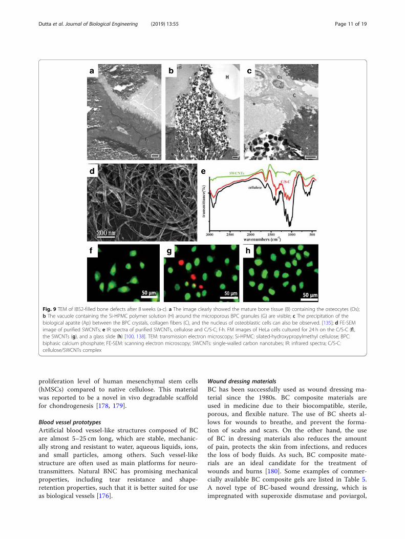
Figure 9a-c shows how bone regeneration occurs afterthe application of Si-HPMC/BCP materials. The use ofBC from Gluconacetobacter hansenii along with a novelcomposite material composed of calcium-deficient hy-droxyapatite (CdHAP) for orthopedic use has been wellcharacterized and described by [140]. On the other hand,[141] reported the use of heparin/cellulose/charcoalcomposites to understand the mechanism and crosstalkamong cells. To study intracellular drug delivery systemsand cellular proliferation, single-walled carbon nano-tubes (SWCNTs) wrapped with cellulose have been ob-served in HeLa cells [101, 102]. Researchers developedSWCNTs with a cellulose solution, dissolved in ionic li-quid 1-butyl-3-methylimidazolium bromide (Fig. 9d-e).Another study showed that long cellulose/SWCNT scaf-folds could promote the growth of HeLa cells, whereasshort cellulose/SWCNT were found to only have a smalleffect on cell proliferation of HeLa cells (Fig. 9f-h).Healthy cells have a green nucleus, uniform chromatin,and an intact cell membrane, whereas necrotic cells orlate apoptotic cells have red nuclei with damaged cellmembranes. Cells cultured on a composite scaffold anda glass slide are healthy with a green nucleus (Fig. 9f andh), however, some cells culture on purified SWCNTs arein the late apoptotic stage (Fig. 9g). Thus, inorganic-based cellulosic hydrogels provide a wide range of appli-cations in the biomedical and tissue engineering field.
Application of cellulose hydrogels in tissueengineeringCellulose-based hydrogels are used in different fields re-lated to tissue engineering. Patterned macroporous (PM)with a diameter larger than 100 μm were introduced topristine 3D nanofibrous BC scaffolds using infrared (IR)micromachining techniques to create an in vitro culturemodel for breast cancer cells (BCs) [142]. PM-BC scaffoldswere found to be promote cellular adhesion, growth, pro-liferation, and infiltration of BCs. A. xylinum BC also pro-motes wound healing as it maintains the wound moist bycontrolling the wound exudates and also heals severesecond-degree burns [143, 144]. Hydroxyethylcellulose
(HEC) and carboxymethyl cellulose sodium salt (CMCNa)cross-linked with hyaluronic acid allow for the prolifera-tion of keratinocytes in an in vitro culture [144]. Bacterialnanocellulose (BNC) has great potential for use as a scaf-fold in tissue engineering, since BC is more effective thanPC, which accounts for BC being the first choice in med-ical and tissue engineering applications.
BC hydrogels in biomedical applicationsBC has promising features due to the similar of its nano-structure and morphology to collagen making BC an at-tractive choice for use in the support and immobilizationof cells. The architecture of BC materials can be engi-neered at range of scales, ranging from the nano to mac-roscale by controlling the biofabrication process. BC fibersare solid and, when used in combination with other bio-compatible materials, produce nanocomposites particu-larly suitable for use in human and veterinary medicine[76]. The applications of BC composite hydrogels in bio-medicine and tissue engineering are listed in Table 4. BCcomposites can also be used in cornea formation after cor-nea surgical treatment, as well as heart and vascular tissueregeneration [148].
Bioactive cartilage implantationSince, BC gels are free from the action of proteolytic en-zymes and reactive oxygen species (ROS), they protectsthe body from carcinogenesis and prevents the appearanceof inflammation. Some examples of cartilage implantscomposed of BC are septum implants, ear implants, andintervertebral discs, among others [176]. Now a days, theuse of bio-mimicking scaffolds has led to the explorationof BC as a potential scaffolding material. A previous studyshowed that BC did not induce the activation of pro- in-flammatory cytokines during in vitro macrophage screen-ing, but rather stimulated the biogenesis of collagen typeII with chondrocytes seeded on BC membranes, indicatingthe suitability of BC as bio-mimicking scaffold [177]. An-other more recent study showed the synthesis ofmodified bacterial cellulose (MBC) from metabolicallyengineered Gluconacetobacter xylinus with a high
Fig. 8 Original photograph (a), SEM image (b), and compressive stress-strain curve (c) of cellulose/SA hydrogel [139] SEM: scanning electronmicroscopy; SA: sodium alginate
Dutta et al. Journal of Biological Engineering (2019) 13:55 Page 10 of 19
proliferation level of human mesenchymal stem cells(hMSCs) compared to native cellulose. This materialwas reported to be a novel in vivo degradable scaffoldfor chondrogenesis [178, 179].
Blood vessel prototypesArtificial blood vessel-like structures composed of BCare almost 5–25 cm long, which are stable, mechanic-ally strong and resistant to water, aqueous liquids, ions,and small particles, among others. Such vessel-likestructure are often used as main platforms for neuro-transmitters. Natural BNC has promising mechanicalproperties, including tear resistance and shape-retention properties, such that it is better suited for useas biological vessels [176].
Wound dressing materialsBC has been successfully used as wound dressing ma-terial since the 1980s. BC composite materials areused in medicine due to their biocompatible, sterile,porous, and flexible nature. The use of BC sheets al-lows for wounds to breathe, and prevent the forma-tion of scabs and scars. On the other hand, the useof BC in dressing materials also reduces the amountof pain, protects the skin from infections, and reducesthe loss of body fluids. As such, BC composite mate-rials are an ideal candidate for the treatment ofwounds and burns [180]. Some examples of commer-cially available BC composite gels are listed in Table 5.A novel type of BC-based wound dressing, which isimpregnated with superoxide dismutase and poviargol,
Fig. 9 TEM of IBS2-filled bone defects after 8 weeks (a-c). a The image clearly showed the mature bone tissue (B) containing the osteocytes (Os);b The vacuole containing the Si-HPMC polymer solution (H) around the microporous BPC granules (G) are visible; c The precipitation of thebiological apatite (Ap) between the BPC crystals, collagen fibers (C), and the nucleus of osteoblastic cells can also be observed. [135]; d FE-SEMimage of purified SWCNTs; e IR spectra of purified SWCNTs, cellulose and C/S-C; f-h. FM images of HeLa cells cultured for 24 h on the C/S-C (f),the SWCNTs (g), and a glass slide (h) [100, 138]. TEM: transmission electron microscopy; Si-HPMC: silated-hydroxypropylmethyl cellulose; BPC:biphasic calcium phosphate; FE-SEM: scanning electron microscopy; SWCNTs: single-walled carbon nanotubes; IR: infrared spectra; C/S-C:cellulose/SWCNTs complex
Dutta et al. Journal of Biological Engineering (2019) 13:55 Page 11 of 19

was found to stimulate the healing of thermal skinburns resulting from acute radiation disease [183].Surprisingly, BC/collagen type I composite was foundto promote the reduction of protease, interleukins,and ROS activity in an in vitro culture study [184].
Surgical implantsBCs and BNCs can be used in the form of tracheotomytubes for reconstructive surgery, such as for artificial heartvalves, and as blood vessels in the form of nanotubes orneurotubes for the regeneration of coronary blood vessel
Table 4 Uses of plant cellulose (PC), microbial cellulose (MC) and bacterial cellulose (BC) composite hydrogels in tissue engineering
Sl.No.
Hydrogel composite Applications References
1 Plant cellulose (PC) purified Tissue engineering and regenerative medicine [145]; Liu et al., 2014 [146]
2 Algal cellulose (AC) Bone tissue and cartilage engineering [147]
3 Bacterial cellulose (BC) purified Bone tissue engineering, cornea treatment, heart and vascular muscleregeneration
[148]
4 Carboxymethyl cellulose (CMC) Drug loading and controlled release of drugs, nucleus pulposus [149]; [148]
5 Polyvinylpyrrolidone (PVP) Soft-tissue replacement wound management [149]
6 Gelatin Wound dressing, tissue regeneration [80]; [150], [151]
7 Starch Reinforcement agent for bionanocomposites [152]
8 Alginate, sodium alginate High strength hydrogel preparation [153]
9 Acrylic acid Burn wound healing [154]
10 Graphene oxide (GO) Biomedicine [155]
11 Vaccarin Cell growth carrier wound dressing [156]
12 Hyaluronic acid (HA) Wound dressing, tissue engineering [157]
13 Chondroitin sulfate (CS) Dental material scaffold Opera et al., 102
14 Calcium phosphate (CP) Bone substitute [158]
15 Ca2+ activated cellulose, cellulose/lactide
Bone tissue engineering [148]
16 2-hydroxyethyl methacrylate(PHEMA)
Contact lenses and optic component for biosensors [159]
17 Polyacrylamide Cartilage replacement [160] & [161]
18 Gellan gum High strength hydrogel for synthetic connective tissue [153]
19 L-carrageenan High strength hydrogel for synthetic connective tissue [153]
20 Hydroxyapatite Bone scaffold substitute, bone tissue engineering [162]; [163]; [164]; [165];[166]
21 Nanohydroxyapatite Bone tissue engineering
22 Polyvinyl alcohol (PVA) Cardiovascular soft tissue replacement, artificial cornea biomaterials ([167]; [168]); [169]; ([170];[171])
23 Polylacitide and glycidylmethacrylate
Skin repair material [172]
24 Collagen Wound dressing for skin regeneration [173]; [148]
20 Silver Antimicrobial wound dressing [174]; [175]
Table 5 Commercially available hydrogel wound dressing contains cellulose or its sodium salt. Most dressings are available in twoforms, either as sheets or as amorphous gels. Products containing silver ions show antimicrobial property
The hydrogel wound dressing (producer) Composition References
IntraSite™ Gel (Smith and Nephew) Carboxymethycellulose sodium (CMCNa), propylene glycol and water
GranuGel™ (ConvaTec) Carboxymethycellulose sodium (CMCNa), Propylene glycol, pectin and water [181], [182]
Purilon Gel™ (ColoPlast) Carboxymethycellulose (CMC), calcium alginate and water
Aquacel Ag™ (ConvaTec) Carboxymethycellulose sodium (CMCNa) and silver ions (1.2%)
Silvercel™ (Johnson and Johnson) Carboxymethycellulose (CMC), silver ions (8%) and calcium alginate
Dutta et al. Journal of Biological Engineering (2019) 13:55 Page 12 of 19
and nerves. Previous studies have found new epithelial celllayers to form over these artificial BC tubes, demonstrat-ing the successful application of BC in tissue implantation[185]. The use of PVA/BC nanocomposites for the re-placement of cardiovascular tissues has also been re-ported, since these would mimic the role of naturalcollagen and elastin (a connective tissue protein that helpsskin to return to its original position [167, 168]
Potential drug delivery materialTransdermal systems can act as an entry gate for BCs intothe domain of drug delivery systems [186]. BC dry filmshave been obtained after the successful immersion ofthese in benzalkonium chloride (an antimicrobial agent).Their subsequent drug loading capacity was found to be0.116mg/cm2 (per unit surface area), and the effect ofdrug was found to last for at least 24 h against Staphylo-coccus aureus and Bacillus subtilis applied to the woundedarea [187]. Silver nanoparticle-coated BC fibers showed99.99% antimicrobial activity against Escherichia coli andS. aureus [164]. Despite these promising results, the appli-cation of BC hydrogels involves certain clinical andpharmacological limitations. However, despite these limi-tations, the complex nanofibrillar structure of BC repre-sents a suitable macromolecular support for the inclusionof drugs, i.e. for use as a drug carrier [188].
Artificial grafting of corneaCorneal disease is a serious health problem that can leadto partial or complete blindness. An estimated 10 mil-lion people have lost their eyesight due to corneal infec-tion or similar diseases. With this in mind, researchersaround the world have developed biomaterials for thetreatment of defective corneas. The properties of bacter-ial cellulose, including its nanoporous structure, and ex-cellent mechanical properties, make it an ideal candidatefor use as an artificial cornea to help maintain the intra-ocular pressure of the eye and re-establish ocular pellu-cidity. The BC/polyvinyl alcohol (BC/PVA) hydrogel hasa water content and light transmittance comparable tothat of natural cornea and was successfully synthesizedand described by Wang et al. for this end.
Dental implantsBC composite hydrogels were prepared from Acetobacterhansenii by [189] for used in dental root canal treatment(RCT) due to intracanal asepsis. Dental RCT is requiredwhen dental caries progress to infection of the dentalpulp. From a materials point of view, BC has superiorproperties compared to plant cellulose (paper points) forthe use in dental RCT. Moreover, research has demon-strated the tissue regeneration of periodontal cells afterthe application of BC hydrogels [190, 191].
Other applicationsBiomimetic scaffolds are of great interest to tissue engin-eering as they supports essential cell functions. BNCscaffolds in combination with soluble collagen-I stimu-late estrogenic differentiation of mesenchymal stem cells(MSCs) [Vielreicher et al., 2018]. The use of cell-derivedECM collagen-I holds good potential, particularly for thetissue engineering of mechanically-challenged tissues.An optimized method for the purification of nano- fibril-lated cellulose (NFC) and hydrogel production fromwood cellulose was described for the development of awound dressing material [192]. Inflammation, autolyticdebridement, granulated tissue formation, and re- epi-thelialization are the processes that generally occur dur-ing wound healing. Wound dressings are designed topromote healing while protecting the wounds from in-fection. This is particularly important in cases of chronicwounds (e.g., ulcers), which fail to heal properly. Since amoist environment encourages rapid healing, hydrogelsare optimal candidates for the development of wounddressings, either as sheets or in an amorphous form[193]. Various types of hydrogel dressings have been pat-ented so far and are currently commercially available(Table 5), based on synthetic or natural polymers, or acombination of these. Among the most recent patents, itis worth citing those describing in situ forming gels (e.g.,based on sprayable formulations [194] and on coalescingnanoparticles [195]), and those exploring radiationcrosslinking as a stabilization technique, which allows toobtain sterile and cross-linked hydrogel films in a sin-gle-step process [196, 197].Scaffold attempts to mimic natural ECMs. The most
common method of tissue engineering includes the use ofbiodegradable scaffolds to support the growth and develop-ment of cells into tissues or by injecting the isolated singlecell suspensions [5]. Cellulose-based scaffolding materialsare widely used to regenerate various tissues, such as bone,cartilage, heart, blood vessel, nerve, and liver, among others.However, the design of scaffolds often involves issues relatedto the need requirement for adequate cell-cell adhesion,cell-cell communication, and cell-ECM communication,which are crucial features of tissue functioning [198]. Toovercome these problems, biodegradable scaffolds havebeen developed. Since, natural polymers are biocompatible,their use allows us to avoid stimulating chronic inflamma-tion or immunological reactions or toxicity. Therefore,hydrogels are used extensively in tissue engineering due totheir high swelling properties and their biocompatibility. Asa result, they can be incorporated the cells of soft tissuesand bioactive molecules via gelling process [199].
Conclusion and future directionsThe current review clearly shows that based explicitly oncellulose biopolymers, hydrogels are a diverse class of
Dutta et al. Journal of Biological Engineering (2019) 13:55 Page 13 of 19

materials that have widespread applications in the fieldof tissue engineering and regenerative medicine. In theseareas, scaffolds played a significant role and have beendeveloped to form temporary, artificial ECMs to supportcell attachment and three-dimensional (3D) tissue for-mation. Due to their high mechanical strength and ther-mostability, bacterial cellulose derivatives are widely usedfor wound dressing and healing, providing a novel methodfor the treatment of epidermal burns. Most interestingly,the work of researchers across the globe in the fields ofcellulose hydrogel development and characterization seemto indicate that hydrogels based on cellulosic biomaterialscould be potential candidates for applications in the fieldof tissue engineering. However, the research outcomes ap-pear somewhat different from the promising predictions.For example, while using hydrogels in bioengineering ap-plications, researchers have encountered a number ofproblems. These include difficulties in the handling, main-tenance, storage of hydrogels, for example, for hydrogelsdesigned using bioprinters, which are not as much mech-anically strong as was theoretically determined. During invitro experiments it was more difficult to sterilize scaffold-ing structures than, for example, the cell culture media.Sterilizing by means of autoclaving can cause the func-tional properties of cellulose-based hydrogels to change.However, their sterilization is necessary since the use ofhydrogels without proper sterilization could be a largesource of contamination during in vivo and in vitro exper-iments in laboratory. Researchers have also often encoun-tered difficulties while loading hydrogels with drugs orcells for controlled drug delivery. Further research intohydrogels will be required for the development of newmethods and protocols in order to overcome these limita-tions. Despite these issues, the use of BC hydrogelscompared to plant-derived or manmade hydrogels is cur-rently on the rise due to the cost-effective production ofBC hydrogels using stirred-tank or static bioreactors.However, more needs to be done to improve plant-derivedcellulosic gel production (PC hydrogels). The use ofcellulose-based hydrogels in tissue engineering has bothadvantages and disadvantages, the latter of which willneed to be resolved before cellulosic hydrogels can bemore widely applied.Researchers are also working to improve our under-
standing of the mechanism behind the molecular inter-action involved in cellulose ECM materials so that, inthe future, materials that mimic natural ECMs in termsof their composition, structural characteristics, andmechanical properties can be developed. The properdevelopment of 3D scaffolding materials could be usedto replace conventional tissue engineering techniques toa great extent. Cellulose- based hydrogels have import-ant applications in tissue engineering due to their highbiocompatibility and environment- friendly properties.
Cellulose-based hydrogels have been recently modifiedusing a nontoxic cross-linking agent or cross-linking treat-ments, to improve the yield of both the final product andthe manufacturing processes. However, further research isneeded to develop more advanced cellulose-based hydro-gels for use in healthcare and medicine.
Additional file
Additional file 1: Table S1. α-Cellulose content of some plant products[197–204]. Figure S1: Source of some naturally occurring cellulose. a.hard wood (beech tree); b. cotton tree; c. bamboo; d. Gluconacetobacterxylinum; e. ascidians. Figure S2. Hydrogen bonding pattern in cellulosemolecule. The hydrogen bonding within or between cellulose moleculesrepresents its crystalline nature while studying through X-ray diffractionor NMR technique. Figure S3. Microphotograph showing variation inmorphology of different fibers. a. twisted cotton fibers; b. tracheids ofspruce wood; c. straight fibers of ramie. Copyright permission from [205];simplified model of plant cell wall. d. structure of S1-S3 layer; e-f. Celluloseassembly with pectin, hemicellulose, and lignin. Copyright permissionfrom ([49]; [206–208]). (DOCX 312 kb)
Abbreviations3D: Three-dimensional; AMIMCl: 1-allyl-3-methylimidazolium chloride;BC: Bacterial cellulose; BCP: Biphasic calcium phosphate; BMIMCl: 1-butyl-3-methylimidazolium chloride; BNC: Bacterial nanocellulose; CdHAP: Calcium-deficient hydroxyapatite; CMC: Carboxymethyl cellulose;CMCNa: Carboxymethylcellulose sodium salt; DMA: Dynamic mechanicalanalysis; DMAc: Dimethylacetamide; DMSO/TBAF: Dimethysulfoxide/tertrabutylaluminium fluoride trihydrate; DP: Dope characteristics;EBs: Embryonic bodies; ECM: Extracellular matrix; GAGs: Glycosaminglycans;HEC: Hydroxyethylcellulose; hESCs: Human embryonic stem cells;HPC: Hydroxypropyl cellulose; HPMC: Hydroxypropylmethyl cellulose;ILs: Ionic liquids; IR: Infrared; LiCl: Lithium chloride; MBC: Modified bacterialcellulose; MC: Methyl cellulose; MSCs: Mesenchymal stem cells;NaOH: Sodium hydroxide; NFC: Nano-fibrillated cellulose; NMMO: N-methylmorpholine-N-oxide; NMR: Nuclear magnetic resonance; PC: Plantcellulose; PM: Patterned macroporous; ROS: Reactive oxygen species;SA: Sodium alginate; SEM: Scanning electron microscopy; Si-HPMC: Silated-hydroxypropylmethyl cellulose; SWCNTs: Single-walled carbon nanotubes;TGA: Thermo-gravimetric analysis; UV: Ultra-violet; WXRD: Wide-angle X-raydiffraction
AcknowledgementsThe authors would like to thank Prof. Lim for his continuous support to writethe manuscript.
FundingThis research was supported by ‘Co-operative Research Program forAgriculture Science and Technology Development (No. PJ012854012017)’,Rural Development Administration, Republic of Korea and ‘Basic ScienceResearch Program’ through the ‘National Research Foundation of Korea’funded by the Ministry of Education (No. 2018R1A6A1A03025582) and the‘National Research Foundation of Korea’ (NRF-2016R1D1 A3B03932921).
Availability of data and materialsNot applicable.
Authors’ contributionsSDD wrote the manuscript. DKP and KTL reviewed the manuscript, edited,and provided feedback. KTL read and approved the final manuscript.
Ethics approval and consent to participateNot applicable.
Consent for publicationNot applicable.
Dutta et al. Journal of Biological Engineering (2019) 13:55 Page 14 of 19

Competing interestsThe authors declare no competing interests.
Publisher’s NoteSpringer Nature remains neutral with regard to jurisdictional claims inpublished maps and institutional affiliations.
Author details1Biorobotics Laboratory, Department of Biosystems Engineering, KangwonNational University, Chuncheon, Republic of Korea. 2The Institute of ForestScience, Kangwon National University, Chuncheon 24341, Republic of Korea.
Received: 24 February 2019 Accepted: 10 May 2019
References1. Silva AKA, Richard C, Bessodes M, Scherman D, Merten OW. Growth factor
delivery approaches in hydrogels. Biomacromolecules. 2009. https://doi.org/10.1021/bm801103c.
2. Peppas NA, Bures P, Leobandung W, Ichikawa H. Hydrogels inpharmaceutical formulations. Eur J Pharm Biopharm. 2000. https://doi.org/10.1016/S0939-6411(00)00090-4.
3. Peppas NA, Mikos AG. Preparation methods and structure of hydrogels. In:Peppas NA, editor. Hydrogels in medicine and pharmacy. Florida: CRC Press;1986. p. 1–27.
4. Stauffer SR, Peppas NA. Poly (vinyl alcohol) hydrogels prepared by freezing-thawing cyclic processing. Polymer. 1992. https://doi.org/10.1016/0032-3861(92)90385-A.
5. Drury JL, Mooney DJ. Hydrogels for tissue engineering: scaffold designvariables and applications. Biomaterials. 2003. https://doi.org/10.1016/j.biomaterials.2010.02.044.
6. Bajpai AK, Shukla SK, Bhanu S, Kankane S. Responsive polymers in controlleddrug delivery. Prog Polym Sci. 2008. https://doi.org/10.1016/j.progpolymsci.2008.07.005.
7. Vinogradov SV, Bronch TK, Kabanov AV. Nanosized cationic hydrogels fordrug delivery preparation, properties and interactions with cells. Adv DrugDeliv Rev. 2002. https://doi.org/10.1016/S0169-409X(01)00245-9.
8. Ostrovidova GU, Makeev AV, Shamtsian MM. Polyfunctional film coatings formedical use. J Mater Sci Eng. 2003. https://doi.org/10.1016/S0928-4931(03)00031-6.
9. Ratner, BD, Hoffman AS. Process of radiation grafting hydrogels ontoorganic polymeric substrates. 1976; US Patent US3939049A.
10. Lee YJ, Braun PV. Tunable inverse opal hydrogel pH sensors. Adv Mater.2003. https://doi.org/10.1002/adma.200304588.
11. Sorber J, Steiner G, Schulz V, Guenther M, Gerlach G, Salzer R, et al.Hydrogel-based piezoresistive pH sensors: Investigations using FT-IRattenuated total reflection spectroscopic imaging. Anal Chem. 2008. https://doi.org/10.1021/ac702598n.
12. Khan F, Tare R, Richard O, Oreffo R, Bradley M. Versatile biocompatiblepolymer hydrogels: scaffolds for cell growth. Angewandate Chemie Int Edn.2009. https://doi.org/10.1002/anie.200804096.
13. Lee KY, Mooney DJ. Hydrogel for tissue engineering. Chem Rev. 2001.https://doi.org/10.1021/cr000108x.
14. Katsoulos C, Karageorgiadis L, Vasileiou N, Mousafeiropoulos T, Asimellis G.Customized hydrogel contact lenses for keratoconus incorporatingcorrection for vertical coma aberration. Ophthalmic Physiol Opt. 2009.https://doi.org/10.1111/j.1475-1313.2009.00645.x.
15. Yasuda H. Biocompatibility of nanofilm-encapsulated silicone and silicone-hydrogel contact lenses. Macromol Biosci. 2006. https://doi.org/10.1002/mabi.200500153.
16. Wu D, Wang T, Lu B, Xu X, Cheng S, Jiang X, et al. Fabrication ofsupramolecular hydrogels for drug delivery and stem cell encapsulation.Langmuir. 2008. https://doi.org/10.1021/la8006876.
17. Nagahama K, Ouchi T, Ohya Y. Temperature-induced hydrogels throughself-assembly of cholesterol-substituted star PEG-b-PLLA copolymers: aninjectable scaffold for tissue engineering. Adv Funct Mat. 2008. https://doi.org/10.1002/adfm.200700587.
18. Zhu J. Bioactive modification of poly (ethylene glycol) hydrogels for tissueengineering. Biomaterials. 2010. https://doi.org/10.1016/j.biomaterials.2010.02.044.
19. Martens PJ, Bryant SJ, Anseth KS. Tailoring the degradation of hydrogelsformed from multivinyl poly (ethylene glycol) and poly (vinyl alcohol)macromers for cartilage tissue engineering. Biomacromolecules. 2003.https://doi.org/10.1021/bm025666v.
20. Ferruti P, Bianchi S, Ranucci E, Chiellini F, Piras AM. Novel agmatine-containing poly (amidoamine) hydrogel as scaffolds for tissue engineering.Biomacromolecules. 2005. https://doi.org/10.1021/bm050210+.
21. Nayak S, Lee H, Chmielewski J, Lyon LA. Folate-mediated cell targeting andcytotoxicity using thermoresponsive microgels. J Am Chem Soc. 2004.https://doi.org/10.1021/ja0474143.
22. Gao D, Xu H, Philbert MA, Kopelman R. Ultrafine hydrogel nanoparticles:synthetic approach and therapeutic application in living cells. AngewandateChemie Int Edn. 2007. https://doi.org/10.1002/ange.200603927.
23. Tomatsu I, Hashidzume A, Harada A. Contrast viscosity changes uponphotoirradiation for mixtures of poly(acrylic acid)-based α-cyclodextrin andazobenzene polymers. J Am Chem Soc. 2006. https://doi.org/10.1021/ja058345a.
24. Kim DG, Seo SW, Cho BK, Lohumi S, Hong SJ, Lee WH. Review of currentapproaches for implementing metabolic reconstruction. J Biosyst Eng. 2018.https://doi.org/10.5307/JBE.2018.43.1.045.
25. Hoffman AS. Hydrogels for biomedical applications. Adv Drug Deliv Rev.2002;43:3–12.
26. Temenoff JS, Mikos AG. Injectable biodegradable materials for orthopedictissue engineering. Biomaterials. 2002. https://doi.org/10.1016/S0142-9612(00)00108-3.
27. Buxton AN, Zhu J, Marchant RE, West JL, Yoo JU, Johnstone B. Design andcharacterization of poly(ethylene glycol) photopolymerizable semi-interpenetrating networks for chondrogenesis of human mesenchymalstem cells. Tissue Eng. 2007. https://doi.org/10.1089/ten.2007.0075.
28. Hahn MS, McHale MK, Wang E, Schmedlen RH, West JL. Physiologic pulsatileflow bioreactor conditioning of poly (ethylene glycol)-based tissueengineered vascular grafts. Ann Biomed Eng. 2007. https://doi.org/10.1007/s10439-006-9099-3.
29. O’Sullivan AC. Cellulose: the structure slowly unravels. Cellulose. 1997;4:173–207.
30. Eichhorn SJ, Young RJ, Davies GR. Modeling crystal and moleculardeformation in regenerated cellulose fibers. Biomacromolecules. 2005.https://doi.org/10.1021/bm049409x.
31. Schurz J. “Trend in polymer science” a bright future for cellulose. ProgPolym Sci. 1999;24:481.
32. Hon DNS. Cellulose and its derivatives: structures, reactions, and medicaluses. In: Dumitriu S, editor. Polysaccharides in medical applications. NewYork: Marcel Dekker; 1996. p. 87–105.
33. Eichhorn SJ, Baillie CA, Zafeiropoulos N, Mwaikambo LY, Ansell MP, DufresneA, et al. Current international research into cellulosic fibers and composites.J Mater Sci. 2001. https://doi.org/10.1023/A:1017512029696.
34. Vandamme EJ, De Baets S, Vanbaelen A. Improved production ofbacterial cellulose and its application potential. Polym Degrad Stab.1998;59:93–9.
35. Jonas R, Farah LF. Production and application of microbial cellulose. PolymDegrad Stab. 1998. https://doi.org/10.1016/S0141-3910(97)00197-3.
36. Dutta SD, Tarafder M, Islam R, Datta BS. Characterization of cellulolyticenzymes of fusarium soli isolates. Biocatal Agri Biotechnol. 2018. https://doi.org/10.1016/j.bcab.2018.03.011.
37. Brown RM Jr, Saxena IM. Cellulose biosynthesis: a model for understandingthe assembly of biopolymers. Plant Physiol Biochem. 2000. https://doi.org/10.1016/S0981-9428(00)00168-6.
38. Rao VSR, Sundararajan PR, Ramakrishnan C, Ramachandran GN.Conformational studies of amylose. In: Ramachandran GN, editor.Conformation of biopolymers (Vol II). London: Academic; 1967. p. 721–37.
39. Krassig HA. Cellulose: structure, accessibility and reactivity. Gordon andBreach Science: Yverdon; 1993.
40. Sorieul M, Dickson A, Hill SJ, Pearson H. Plant fibre: molecular structure andbiomechanical properties, of a complex living material, influencing itsdeconstruction towards a biobased composite. Materials. 2016. https://doi.org/10.3390/ma9080618.
41. Seo YR, Kim JW, Hoon S, Kim J, Chung JH, Lim KT. Cellulose-basednanocrystals: sources and applications via agricultural by-products. J BiosystEng. 2018. https://doi.org/10.5307/JBE.2018.43.1.059.
42. Kondo T. The relationship between intramolecular hydrogen bonds andcertain physical properties of regioselectively substituted cellulose
Dutta et al. Journal of Biological Engineering (2019) 13:55 Page 15 of 19

derivatives. J Polym Sci A Part B: Polym Phys. 1998. https://doi.org/10.1002/(SICI)1099-0488(199703)35:4<717::AID-POLB18>3.0.CO;2-J.
43. Sarko A, Muggli R. Packing analysis of carbohydrates and polysaccharides. III.Valonia cellulose and cellulose II. Macromolecules. 1974. https://doi.org/10.1021/ma60040a016.
44. Atalla RH, VanderHart DL. Native cellulose: a composite of two distinctcrystalline forms. Science. 1984. https://doi.org/10.1126/science.223.4633.283.
45. Marchessault RH, Sarko A. X-ray structureof polysaccharides. In: Wolfrom ML,editor. Advanced Carbohydrate Chemistry. New York: Academic Press; 1967.p. 421–83.
46. Walton AG, Blackwell J. Biopolymers Vol. 22. New York: Academic; 1973. p.468.
47. VanderHart DL, Atalla RH. Studies of microstructure in native cellulosesusing solid state C-13 NMR. Macromolecules. 1984. https://doi.org/10.1021/ma00138a009.
48. Sugiyama J, Persson J, Chanzy H. Combined IR and electron diffractionstudy of the polymorphism of native cellulose. Macromolecules. 1991.https://doi.org/10.1021/ma00009a050.
49. Marrinan M, Mann J. Infrared spectra of the crystalline modifications ofcellulose. J Polym Sci. 1956. https://doi.org/10.1002/pol.1956.120219812.
50. Hayashi J, Sufoka A, Ohkita J, Watanabe S. The conformation of existence ofcellulose IIII, IIIII, IVI and IVII by X-ray method. J Polym Sci Polym Lett Edn.1975. https://doi.org/10.1002/pol.1975.130130104.
51. Nyburg SC. Fibrous macromolecular substances. In: Fieser LF, Fieser M,editors. X-ray analysis of organic structures. New York: Academic; 1961. p.302–14.
52. Kuga S, Takagi S, Brown RM Jr. Native folded-chain cellulose II. Polymer.1993. https://doi.org/10.1016/0032-3861(93)90404-X.
53. Hess K, Kissig H. To note the high-temperature modification of cellulose.Magazine Phys Chem. 1941. https://doi.org/10.1515/zpch-1941-4922.
54. Gardiner ES, Sarko A. Packing analysis of carbohydrates and polysaccharides.16. The crystal structures of celluloses IVI and IVII. Can J Chemistry. 1985.https://doi.org/10.1139/v85-027.
55. Gardner KH, Blackwell J. Structure of native cellulose. Biopolymers. 1974.https://doi.org/10.1002/bip.1974.360131005.
56. Klemm D, Heublein B, Fink HP, Bohn A. Cellulose: fascinating biopolymerand sustainable raw material. Angew Chem Int Ed. 2005. https://doi.org/10.1002/anie.200460587.
57. Shi Z, Zhang Y, Phillips GO, Yang G. Utilization of bacterial cellulose in food.Food Hydrocol. 2014. https://doi.org/10.1016/j.foodhyd.2013.07.012.
58. Ioelovich M, Leykin A. Cellulose as a nanostructured polymer: a shortreview. Bioresources. 2008;3:1403–18.
59. Rytioja J, Hilden K, Yuzon J, Hatakka A, de Vries RP, Makela MR. Plant-polysaccharide-degrading enzymes from basidiomycetes. Microbiol Mol BiolRev. 2014. https://doi.org/10.1128/MMBR.00035-14.
60. Plomion C, Leprovost G, Stokes A. Wood formation in trees. Plant physiol.2001; https://doi.org/10.1104/pp.010816.
61. Schubert S, Schlufter K, Heinze T. Configurations, structures, andmorphologies of cellulose. In: Popa V, editor. Polysaccharides in medicinaland pharmaceutical applications. Shrewsbury: iSmithers; 2011. p. 1–55.
62. Klemm D, Schumann D, Udhardt U, Marsch S. Bacterial synthesized cellulose– artificial blood vessels for microsurgery. Prog Polym Sci. 2001. https://doi.org/10.1016/S0079-6700(01)00021-1.
63. Kongruang S. Bacterial cellulose production by Acetobacter xylinum strainsfrom agricultural waste products. Appl Biochem Biotechnol. 2008. https://doi.org/10.1007/978-1-60327-526-2_70.
64. Scott JE. Extracellular matrix, supramolecular organization and shape. J Anat.1995;187:259–69.
65. Rhodes JM, Simons M. The extracellular matrix and blood vessel formation:not just a scaffold. J Cell Mol Med. 2007. https://doi.org/10.1111/j.1582-4934.2007.00031.x.
66. Cukierman E, Pankov R, Yamada KM. Cell interactions with three-dimensional matrices. Curr Opin Cell Biol. 2002. https://doi.org/10.1016/S0955-0674(02)00364-2.
67. Shastri VP, Forget A. Development of the next generation of syntheticextracellular matrices for 3D cell culture. Lab & More. 2014; http://www.int.laborundmore.com/archive/543411/ Development-of-the-next-generation-of-synthetic-extracellular-matrices-for-3D-cell-culture.html.
68. Das N. Biodegradable hydrogels for controlled drug delivery. In: Mondal IM,editions. Cellulose-based superabsorbent hydrogels. Springer Nature.2018. p.1–41.
69. Prashant PK, Vivek BR, Deepashree ND, Pranav PP. Hydrogels as a drug deliverysystem and applications: a review. Int J Pharm Pharm Sci. 2012;4:1–7.
70. Das N, Bera T, Mukherjee A. Biomaterial hydrogels for different biomedicalapplications. Int J Pharm Bio Sci. 2012;3:586–95.
71. Grassi M, Sandolo C, Perin D, Coviello T, Lapasin R, Grassi G. Structuralcharacterization of calcium alginate matrices by means of mechanical andrelease tests. Molecules. 2009. https://doi.org/10.3390/molecules14083003.
72. De SK, Aluru N, Johnson B, Crone W, Beebe DJ, Moore J. Equilibriumswelling and kinetics of pH-responsive hydrogels: models, experiments, andsimulations. J Microelectromech Syst. 2002. https://doi.org/10.1109/JMEMS.2002.803281.
73. Bryant SJ, Nuttelman CR, Anseth KS. Cytocompatibility of UV and visiblelight photoinitiating systems on cultured NIH/3T3 fibroblasts in vitro. JBiomater Sci Polym Ed. 2000. https://doi.org/10.1163/156856200743805.
74. Akiyoshi K, Deguchi S, Moriguchi N, Yamaguchi S, Sunamoto J. Self-aggregates of hydrophobized polysaccharides in water: formation andcharacteristics of nanoparticles. Macromolecules. 1993. https://doi.org/10.1021/ma00064a011.
75. Morimoto N, Winnik FM, Akiyoshi K. Botryoidal assembly of cholesteryl–pullulan/ poly(nisopropylacrylamide) nanogels. Langmuir. 2007. https://doi.org/10.1021/la0616045.
76. Mohite BV, Koli SH, Patil SV. Bacterial cellulose-based hydrogels: Synthesis,Properties, and Applications. In: Mondal IH, editor. Cellulose-basedsuperabsorbent hydrogels: Springer Nature; 2018. p. 1–22.
77. Fan M, Ma Y, Tan H, Jia Y, Zou S, Guo S, et al. Covalent and injectablechitosan-chondroitin sulfate hydrogels embedded with chitosanmicrospheres for drug delivery and tissue engineering. Mater Sci Eng C.2007. https://doi.org/10.1016/j.msec.2016.09.068.
78. Singh B, Sharma V. Crosslinking of poly(vinylpyrrolidone)/acrylic acid withtragacanth gum for hydrogels formation for use in drug delivery applications.Carbohydr Polym. 2017. https://doi.org/10.1016/j.carbpol.2016.09.086.
79. Hamidi M, Azadi A, Rafiei P. Hydrogel nanoparticles in drug delivery. AdvDrug Deliv Rev. 2008;60:1638–49.
80. Păvăloiu RD, Stoica-Guzun A, Dobre T. Swelling studies of composite hydrogelsbased on bacterial cellulose and gelatin. UPB Sci Bull Ser B. 2015;77:53–62.
81. Mohite BV, Patil SV. A novel biomaterial: bacterial cellulose and its new eraapplications. Biotechnol Appl Biochem. 2014. https://doi.org/10.1002/bab.1148.
82. Ross P, Mayer R, Benziman M. Cellulose biosynthesis and function in bacteria.Microbiol Rev. 1991;55(1):35–58
83. Gromet-Elhanan Z, Hestrin S. Synthesis of cellulose by Acetobacter xylinumVI. Growth on citric acid-cycle intermediates. J Bacteriol. 1963;85:284–92.
84. Rajwade JM, Paknikar KM, Kumbhar JV. Applications of bacterial celluloseand its composites in biomedicine. Appl Microbiol Biotechnol. 2015. https://doi.org/10.1007/s00253-015-6426-3.
85. Jung JY, Park JK, Chang HN. Bacterial cellulose production byGluconacetobacter hansenii in an agitated culture without living non-cellulose producing cells. Enzym Microb Technol. 2005. https://doi.org/10.1016/j.enzmictec.2005.02.019.
86. Yoshino T, Asakura T, Toda K. Cellulose production by Acetobacterpasteurianus on silicone membrane. J Ferment Bioeng. 1996. https://doi.org/10.1016/0922-338X(96)83116-3.
87. Ishihara M, Yamanaka S. Modified bacterial cellulose. 2000. US PatentUS6060289A.
88. Shoda M, Sugano Y. Recent advances in bacterial cellulose production.Biotechnol Bioprocess Eng. 2005. https://doi.org/10.1007/BF02931175.
89. Gatenholm P. Cellulose nanofibriller bioink for 3D bioprinting for cellculturing, tissue engineering and regenerative medicine applications. 2017.US Patent US20170368225A1.
90. Kim JY, Kim JN, Wee YJ, Park DH, Ryu HW. Bacterial cellulose production byGluconacetobacter sp. PKY5 in a rotary biofilm contactor. Appl BiochemBiotechnol. 2007. https://doi.org/10.1007/s12010-007-9077-8.
91. Pita PCDC, Pinto FCM, Lira MMDM, Melo FDAD, Ferreira LM, Aguiar JLDA.Biocompatibility of the bacterial cellulose hydrogel in subcutaneous tissueof rabbits. Acta Cir Bras. 2015. https://doi.org/10.1590/S0102-865020150040000009.
92. Czaja W, Romanovicz D, Brown RM. Structural investigations of microbialcellulose produced in stationary and agitated culture. Cellulose. 2004.https://doi.org/10.1023/B:CELL.0000046412.11983.61.
93. Watanabe K, Tabuchi M, Morinaga Y, Yoshinaga F. Structural features andproperties of bacterial cellulose produced in agitated culture. Cellulose.1998. https://doi.org/10.1023/A:1009272904582.
Dutta et al. Journal of Biological Engineering (2019) 13:55 Page 16 of 19

94. Kim DY, Nishiyama Y, Kuga S. Surface acetylation of bacterial cellulose.Cellulose. 2002. https://doi.org/10.1023/A:1021140726936.
95. Okiyama A, Motoki M, Yamanaka S. Bacterial cellulose IV. Application toprocessed foods. Food Hydrocoll. 1993. https://doi.org/10.1016/S0268-005X(09)80074-X.
96. Chang C, Zhang L. Cellulose-based hydrogels: present status andapplication prospects. Carbohydrate Polym. 2011. https://doi.org/10.1016/j.carbpol.2010.12.023.
97. Zhang Y, Shao H, Wu C, Hu X. Formation and characterization of cellulosemembranes from N-methylmorpholine-N-oxide solution. Macromol Biosci. 2001.https://doi.org/10.1002/1616-5195(20010601)1:4<141::AID-MABI141>3.0.CO;2-J.
98. Saito H, Sakurai A, Sakakibara M, Saga H. Preperation and properties oftransparent cellulose hydrogels. J Appl Polym Sci. 2003. https://doi.org/10.1002/app.13015.
99. De Oliveira WD, Glasser WG. Hydrogels form polysacchardies I Cellulosebeads for chromatographic support. J Appl Polym Sci. 1996. https://doi.org/10.1002/(SICI)1097-4628(19960404)60:1<63::AID-APP8>3.0.CO;2-T.
100. Wang Z, Lu S, Matsumoto Y, Kuga S. Cellulose gel and aerosol from LiCl/DMSO solution. Cellulose. 2012. https://doi.org/10.1007/s10570-012-9651-2.
101. Li L, Lin Z, Yang X, Wan ZZ, Cui SX. A novel cellulose hydrogel preparedfrom its ionic liquid solution. Chin Sci Bull. 2009a. https://doi.org/10.1007/s11434-009-0207-2.
102. Li L, Meng L, Zhang X, Fu C, Lu Q. The ionic liquid-associated synthesis of acellulose/SWCNT complex and its remarkable biocompatibility. J MaterChem. 2009b. https://doi.org/10.1039/b823322e.
103. Cai J, Zhang L. Rapid dissolution of cellulose in LiOH/urea and NaOH/ureaaqueous solution. Macrol Biosci. 2005. https://doi.org/10.1002/mabi.200400222.
104. Liang SM, Zhang L, Li Y, Xu J. Fabrication and properties of cellulosehydrated membrane with unique structure. Macromol Chem Phys. 2007.https://doi.org/10.1002/macp.200600579.
105. Bao Y, Ma J, Li N. Synthesis and swelling behaviors of sodium carboxymethylcellulose-g-poly (AA-co-AM-co-AMPS)/MMT superabsorbent hydrogel.Carbohydr Polym. 2011. https://doi.org/10.1016/j.carbpol.2010.10.061.
106. Bao Y, Ma J, Sun Y. Swelling behaviors of organic/inorganic compositesbased on various cellulose derivatives and inorganic particles. CarbohydrPolym. 2012. https://doi.org/10.1016/j.carbpol.2012.01.003.
107. Feddersen RL, Thorp SN. Chapter 20-sodium carboxymethycellulose. In:Whistler RL, Bemiller JN, editors. Industrial gums: polysaccharides and theirderivatives. 3rd ed. Elsevier: Academic; 1993. https://doi.org/10.1016/B978-0-08-092654-4.50024-3.
108. Bortolin A, Aouadas FA, Mattoso LHC, Ribeiro CR. Nanocomposite PAAm/methyl cellulose/montmorillonite hydrogel: evidence of synergistic effectsfor the slow release of fertilizers. J Agric Food Chem. 2013. https://doi.org/10.1021/jf401273n.
109. Duarte ARC, Gordillo MD, Cardoso MM, Simplicio AL, Durate CMM.Perperation of ethyl cellulose/methyl cellulose blends by supercriticalantisolvent precipitation. Int J Pharm. 2006. https://doi.org/10.1016/j.ijpharm.2005.12.010.
110. Brown W, Henely D. Studies on cellulose derivatives. Part III. Unperturbeddimensions of hydroxyethyl cellulose and other derivatives in aqueous solvents.Macromol Chem Phys. 1964. https://doi.org/10.1002/macp.1964.020750115.
111. Frey MW. Electrospinning cellulose and cellulose derivatives. Polym Rev.2008. https://doi.org/10.1080/15583720802022281.
112. Oliveria VA, Veloso TC, Leao VA, dos Santos CG, Botaro VR. Hydrogels ofcellulose acetate crosslinked with pyromellitic dianhydride-part I: synthesisand swelling kinetics. Quim Nova. 2013. https://doi.org/10.1590/S0100-40422013000100019.
113. Edgar KJ, Buchanan CM, Debenham JS, Rundquist PA, Seiler BD, Shelton MC,et al. Advances in cellulose ester performance and application. Prog PolymSci. 2001;26:1605.
114. Turbak, AF, El-Kafrawy A, Snyder FW, Auerbach AP. 1981. U.S. Patent.4303252.
115. Fink HP, Weigel P, Purz HJ, Ganster J. Structure formation of regeneratedcellulose materials from NMMO-solutions. Prog Polym Sci. 2001;26:1473–524.
116. Heinze T, Dicke R, Koschella A, Klohr EA, Koch W. Effective preparation ofcellulose derivatives in a new simple cellulose solvent. Macromol ChemPhys. 2000. https://doi.org/10.1002/(SICI)1521-3935(20000301)201:6<627::AID-MACP627>3.0.CO;2-Y.
117. Graenacher C, Sallmann R. 1939. US patent 2179181, assigned to society tochemical industry, Basel.
118. McCorsely CC, Varga JK. 1977 US Patent 4142913, assigned to Arizona Inc.119. Fink HP, Purz HJ, Weigel P. Strukturelle Aspekte neuer Cellulosemateriaalien.
Das Paiper. 1997;51:643–52.120. Zhang H, Wu J, Zhang J, He J. 1-allyl-3-methylimidazolium chloride room
temperature ionic liquid: a new and powerful nonderivatizing solvent forcellulose. Macromolecules. 2005. https://doi.org/10.1021/ma0505676.
121. Cao Y, Wu J, Zhang J, Li H, Zhang Y, He J. Room temperature ionic liquids(RTILs): a new and versatile platform for cellulose processing andderivatization. Chem Eng J. 2009. https://doi.org/10.1016/j.cej.2008.11.011.
122. Cai J, Zhang L. Unique gelation behavior of cellulose in NaOH/urea aqueoussolution. Biomacromolecules. 2006a. https://doi.org/10.1021/bm0505585.
123. Cai J, Zhang L. Unique geletion behaviour of cellulose in NaOH/ureaauqeous solution. Biomacromolecules. 2006b. https://doi.org/10.1021/bm0505585.
124. Deng J, He Q, Wu Z, Yang W. Using glycidyl methacrylate as crosslinkingagent to prepare thermosensitive hydrogels by a novel one-step method. JPolym Sci Part A: Polym Chem. 2008. https://doi.org/10.1002/pola.22554.
125. Guo K, Chu CC. Synthesis and characterization of novel biodegradableunsaturated poly (ester amide)/poly (ethylene glycol) diacrylate hydrogels. JPolym Sci Part A: Polym Chem. 2005;43:3932.
126. Bourges X, Weiss P, Daculsi G, Legeay G. Synthesis and general properties ofsilated-hydroxypropyl methylcellulose in prospect of biomedical use. AdvColloid Interf Sci. 2002. https://doi.org/10.1016/S0001-8686(02)00035-0.
127. Trojani C, Weiss P, Michiels JF, Vinatier C, Guicheux J, Daculsi G, et al. Three-dimensional culture and differentiation of human osteogenic cells in aninjectable hydroxypropylmethylcellulose hydrogel. Biomaterials. 2005.https://doi.org/10.1016/j.biomaterials.2005.02.001.
128. Yang MJ, Chen CH, Lin PJ, Huang CH, Chen W, Sung HW. Novel method offorming human embryoid bodies in a polystyrene dish surface-coated witha temperature-responsive methylcellulose hydrogel. Biomacromolecules.2007a. https://doi.org/10.1021/bm0704166.
129. Yang MJ, Chen CH, Lin PJ, Huang CH, Chen W, Sung HW. Novel method offorming human Embryoid bodies in a polystyrene dish surface-coated witha emperature-responsive methylcellulose hydrogel. Biomacromolecules.2007b. https://doi.org/10.1021/bm0704166.
130. Dang DL, Dang VL. Chitosan-carboxymethylcellulose hydrogels as supportsfor cell immobozation. J Macromol Sci: Pure Appl Chem. 1996;33:1875.
131. Faroongsarng D, Sukonrat P. Thermal behavior of water in the selectedstarch-and cellulose-based polymeric hydrogels. Int J Pharm. 2008. https://doi.org/10.1016/j.ijpharm.2007.10.022.
132. Hebeish A, Higazy A, El-Shafei A, Sharef S. Synthesis of carboxymethylcellulose (CMC) and starch-based hybrids and their applications inflocculation and sizing. Carbohydr Polym. 2010. https://doi.org/10.1016/j.carbpol.2009.07.022.
133. Isklan N. Controlled release of insecticide carboxyl from sodium alginate,sodium alginate/gelatin, and sodium alginate/sodium carboxymethylcellulose blend beads cross-linked with glutaraldehyde. J Appl Polym Sci.2005. https://doi.org/10.1002/app.22012.
134. Liang HF, Hong MH, Ho RM, Chung CK, Lin YH, Chen CH, et al. Novelmethod using a temperature-sensitive polymer (methylcellulose) tothermally gel aqueous alginate as a pH-sensitive hydrogel.Biomacromolecules. 2004. https://doi.org/10.1021/bm049813w.
135. Sannino A, Madaghiele M, Conversano F, Mele G, Maffezzoli A, Netti PA, et al.Cellulose derivative-hyaluronic acid-based microporous hydrogels cross-linkedthrough divinyl sulfone to modulate equilibrium sorption capacity and networkstability. Biomacromolecules. 2004. https://doi.org/10.1021/bm0341881.
136. Li N, Bai R. Copper adsorption on chitosan-cellulose hydrogel beads:behaviors and mechanisms. Sep Purif Technol. 2005. https://doi.org/10.1016/j.seppur.2004.08.002.
137. Zhao L, Mitomo H. Adsorption of heavy metal ions from aqueous solutiononto chitosan entrapped CM-cellulose hydrogels synthesized by irradiation.J Appl Polym Sci. 2008. https://doi.org/10.1002/app.28718.
138. Chang C, Peng J, Zhang L, Pang DW. Strongly fluorescent hydrogels withquantum dots embedded in cellulose matrices. J Mater Chem. 2009. https://doi.org/10.1039/B908835K.
139. Fellah BH, Weiss P, Gauthier O, Rouillon T, Piler P, Daculsi G, et al. BoneRepair Using a New Injectable Self-Cross linkable Bone Substitute. J OrthoRes. 2006. https://doi.org/10.1002/jor.20125.
140. Hutchens SA, Benson RS, Evans BR, O’Neill HM, Rawn CJ. Biomimeticsynthesis of calcium-deficient hydroxyapatite in a natural hydrogel.Biomaterials. 2006. https://doi.org/10.1016/j.biomaterials.2006.04.032.
Dutta et al. Journal of Biological Engineering (2019) 13:55 Page 17 of 19

141. Park TJ, Lee SH, Trevor JS, Martin JG, Mousa SA, Snezhkova EA, et al.Heparin-cellulose-charcoal composite for drug detoxification prepared usingroom temperature ionic liquids. Chem Commun. 2008. https://doi.org/10.1039/B809791G.
142. Xiong G, Luo H, Gu F, Zhang J, Hu D, Wan Y. A novel in vitro three-dimensional macroporous scaffolds from bacterial cellulose for culture ofbreast cancer cells. J Biomater Nanobiotechnol. 2013. https://doi.org/10.4236/jbnb.2013.44040.
143. Czaja WK, Young DJ, Kawecki M, Brown RM Jr. The future prospects ofmicrobial cellulose in biomedical applications. Biomacromolecules. 2007.https://doi.org/10.1021/bm060620d.
144. Sannino A, Pappada S, Madaghiele M, Maffezzoli A, Ambrosio L, NicolaisL. Crosslinking of cellulose derivatives and hyaluronic acid with water-soluble carbodiimide. Polymer. 2005. https://doi.org/10.1016/j.polymer.2005.10.048.
145. Modulevsky DJ, Cuerrier CM, Pelling AE. Biocompatibility of subcutaneouslyimplanted plant-derived cellulose biomaterials. PLoS One. 2016. https://doi.org/10.1371/journal.pone.0157894.
146. Liu Y, Wang H, Yu G, Yu Q, Li B, Mu X. A novel approach for the preperationof nanocrystalline cellulose by using phosphotungstic acid. CarbohydrPolym. 2014. https://doi.org/10.1016/j.carbpol.2014.04.040.
147. Bhattarai N, Li Z, Edmondson D, Zhang M. Alginate-based nanofibrousscaffolds: structural, mechanical, and biological properties. Adv Mater. 2006.https://doi.org/10.1002/adma.200502537.
148. Van Vlierberghe S, Dubruel P, Schacht E. Biopolymer-based hydrogels asscaffolds for tissue engineering applications: a review. Biomacromolecules.2011. https://doi.org/10.1021/bm200083n.
149. Shah R, Vyroubal R, Fei H, Saha N, Kitano T, Saha P. Preparation of bacterialcellulose based hydrogels and their viscoelastic behavior. AIP Conf Proc.2015;1662:040007.
150. Nakayama A, Kakugo A, Gong JP, Osada Y, Takai M, Erata T, et al. Highmechanical strength double-network hydrogel with bacterial cellulose. AdvFunct Mater. 2004;14:1124–8.
151. Haroun AA, Gamal-Eldeen A, Harding DRK. Preparation, characterization andin vitro biological study of biomimetic three-dimensional gelatin-montmorillonite/cellulose scaffold for tissue engineering. J Mater Sci MaterMed. 2009;20:2527–40.
152. Woehl MA, Canestraro CD, Mikowski A, Sierakowski MR, Ramos LP, WypychF. Bionanocomposites of thermoplastic starch reinforced with bacterialcellulose nanofibres: effect of enzymatic treatment on mechanicalproperties. Carbohydr Polym. 2010;80:866–73.
153. Khan S, Ul-Islam M, Ikram M, Ullah MW, Israr M, Subhan F, et al. Three-dimensionally microporous and highly biocompatible bacterial cellulose–gelatin composite scaffolds for tissue engineering applications. RSC Adv.2016;6:110840–9.
154. Mohamad N, Amin MCIM, Pandey M, Ahmad N, Rajab NF. Bacterialcellulose/acrylic acid hydrogel synthesized via electron beam irradiation:accelerated burn wound healing in an animal model. Carbohydr Polym.2014;114:312–20.
155. Fang Q, Zhou X, Deng W, Zheng Z, Liu Z. Freestanding bacterial cellulose-graphene oxide composite membranes with high mechanical strength forselective ion permeation. Sci Rep. 2016;6:33185.
156. Qiu Y, Qiu L, Cui J, Wei Q. Bacterial cellulose and bacterial cellulose-vaccarinmembranes for wound healing. Mater Sci Eng C. 2016;59:303–9.
157. de Oliveira SA, da Silva BC, Riegel-Vidotti IC, Urbano A, de Sousa Faria-TischerPC, Tischer CA. Production and characterization of bacterial cellulosemembranes with hyaluronicn acid from chicken comb. Int J Biol Macromol.2017;97:642–53.
158. Daltro P, Daltro G, De Oliveira G, Basmaji P, Guastaldi A. Hydrogel and bacterialcellulose mats behavior with calcium phosphate deposition. In: Front BioengBiotechnol conference abstract: 10th World Biomaterials Congress 17–22 May;2016. https://doi.org/10.3389/conf.FBIOE.2016.01.02697.
159. Li X, Wan W, Panchal CJ. US Patent US894033727, 2015.160. Buyanov AL, Revel’skaya LG, Kuznetzov YP, Shestakova AS. Cellulose–
poly (acrylamide or acrylic acid) interpenetrating polymer networkmembranes for the pervaporation of water–ethanol mixtures. J ApplPolym Sci. 1998;69:761–9.
161. Buyanov AL, Gofman IV, Revel'skaya LG, Khripunov AK, Tkachenko AA.Anisotropic swelling and mechanical behavior of composite bacterialcellulose–poly(acrylamide or acrylamide–sodium acrylate) hydrogels. J MechBehav BioMed Mater. 2010. https://doi.org/10.1016/j.jmbbm.2009.06.001.
162. Ahn SJ, Shin YM, Kim SE, Jeong SI, Jeong JO, Park JS, et al. Characterizationof hydroxyapatite-coated bacterial cellulose scaffold for bone tissueengineering. Biotechnol Bioprocess Eng. 2015;20:948–55.
163. Ran J, Jiang P, Liu S, Sun G, Yan P, Shen X, et al. Constructing multi-component organic/inorganic composite bacterial cellulose-gelatin/hydroxyapatite double-network scaffold platform for stem cell-mediatedbone tissue engineering. Mater Sci Eng C. 2017;78:130–40.
164. Jung R, Kim Y, Kim HS, Jin HJ. Antimicrobial properties of hydrated cellulosemembranes with silver nanoparticles. J Biomater Sci Polym Ed. 2009. https://doi.org/10.1163/156856209X412182.
165. Chen YM, Xi TF, Zheng YD, Wan YZ. In Vitro Cytotoxicity Study of the Nano-Hydroxyapatite/Bacterial Cellulose Nanocomposites. In: Materials Research,Parts 1 and 2. Gu ZW et al., editors. Portland; 2009.p.1011–1016.
166. Muller FA, Muller L, Hofmann I, Greil P, Wenzel MM, Staudenmaier R.Cellulose-based scaffold materials for cartilage tissue engineering.Biomaterials. 2006;27:3955–63.
167. Millon LE, Wan WK. The polyvinyl alcohol-bacterial cellulose system as anew nanocomposite for biomedical applications. J Biomed Mater Res Part BAppl Biomater. 2006a. https://doi.org/10.1002/jbm.b.30535.
168. Millon LE, Wan WK. The polyvinyl alcohol–bacterial cellulose system as anew nano composite for biomedical applications. J Biomed Mater Res PartB. 2006b;79:245–53.
169. Millon LE, Mohammadi H, Wan WK. Anisotropic polyvinyl alcohol hydrogelfor cardiovascular applications. J Biomed Mater Res Part B. 2006;7:305–11.
170. Wang J, Gao C, Zhang Y, Wan Y. Preparation and in vitro characterization ofBC/PVA hydrogel composite for its potential use as artificial cornea biomaterial.Mater Sci Eng C. 2010. https://doi.org/10.1016/j.msec.2009.10.006.
171. Wang J, Gao C, Zhang Y, Wan Y. Preparation and in vitro characterization ofBC/PVA hydrogel composite for its potential use as artificial corneabiomaterial. Mater Sci Eng C. 2010;30:214–8.
172. Lina F, Yue Z, Jin Z, Guang Y. Bacterial cellulose for skin repair materials,chapter 13. In: Fazel RR, editor. Biomedical engineering frontiers andchallenges. London: In Tech; 2011. https://doi.org/10.5772/24323.
173. Moraes PRFDS, Saska S, Barud H, Lima LRD, Martins VDCA, Plepis AMDG, etal. Bacterial cellulose/collagen hydrogel for wound healing. Mater Res.2016;19:106–16.
174. Gupta A, Low WL, Radecka I, Britland ST, Mohd Amin MCI, Martin C.Characterisation and in vitro antimicrobial activity of biosynthetic silver-loaded bacterial cellulose hydrogels. J Micro Encapsul. 2016;33:725–34.
175. Mohite BV, Patil SV. In situ development of nanosilver-impregnated bacterialcellulose for sustainable released antimicrobial wound dressing. J ApplBiomater Funct Mater. 2016;14:e53–8.
176. Avery NC, Sims TJ, Warkup C, Bailey AJ. Collagen cross-linking in porcine mlongissimus lumborum: absence of a relationship with variation in texture atpork weight. Meat Sci. 2007. https://doi.org/10.1016/0309-1740(95)00021-6.
177. Svensson A, Nicklasson E, Harrah T, Panilaitis B, Kaplan DL, Brittberg M, et al.Bacterial cellulose as a potential scaffold for tissue engineering of cartilage.Biomaterials. 2005. https://doi.org/10.1016/j.biomaterials.2004.02.049.
178. Yadav V, Sun L, Panilaitis B, Kaplan DL. In vitro chondrogenesis withlysozyme susceptible bacterial cellulose as a scaffold. J Tissue Eng RegenMed. 2013. https://doi.org/10.1002/term.
179. Andersson J, Stenhamre H, Backdahl H, Gatenholm P. Behaviour of humanchondrocytes in engineered porous bacterial cellulose scaffolds. J BiomedMater Res A. 2010. https://doi.org/10.1002/jbm.a.32784.
180. Shah J, Brown MRJR. Towards electronic paper displays made frommicrobial cellulose. Appl Microbiol Biotechnol. 2005. https://doi.org/10.1007/s00253-004-1756-6.
181. Sannino A, Demitri C, Madaghiele M. Biodegradable cellulose-basedhydrogels: design and applications. Materials. 2009. https://doi.org/10.3390/ma2020353.
182. Barman A, Das M. Cellulose-Based Hydrogels for Pharmaceutical andBiomedical Applications. In: Mondal MIH, editor. Cellulose-basedSuperabsorbent Hydrogels: Springer Nature; 2018. p. 1–28. https://doi.org/10.1007/978-3-319-76573-0_37–1.
183. Legeza VI, Galenko-Yaroshevskii VP, Zinov’ev EV, Paramonov BA, KreichmanGS, Turkovskii II, et al. Effects of new wound dressings on healing of thermalburns of the skin in acute radiation disease. Bull Exp Biol Med. 2004. https://doi.org/10.1007/s10517-005-0029-4.
184. Wiegand C, Elsner P, Hipler UC, Klemm D. Protease and ROS activitiesinfluenced by a composite of bacterial cellulose and collagen type I in vitro.Cellulose. 2006. https://doi.org/10.1007/s10570-006-9073-0.
Dutta et al. Journal of Biological Engineering (2019) 13:55 Page 18 of 19

185. Baptista A, Ferreira I, Borges J. Cellulose-Based Bioelectronic Devices. InTech.2013; p.67–82.
186. Czaja W, Krystynowicz A, Bielecki S, Brown R Jr. Microbial cellulose – thenatural power to heal wounds. Biomaterials. 2006. https://doi.org/10.1016/j.biomaterials.2005.07.035.
187. Wei B, Yang G, Hong F. Preparation and evaluation of a kind of bacterialcellulose dry films with antibacterial properties. Carbohydr Polym. 2011.https://doi.org/10.1016/j.carbpol.2010.12.017.
188. Almeida IF, Pereira T, Silva NHCS, Gomes FP, Silvestre AJD, Freire CSR, et al.Bacterial cellulose membranes as drug delivery systems: an in vivo skincompatibility study. Eur J Pharm biopharma. 2014. https://doi.org/10.1016/j.ejpb.2013.08.008.
189. Yoshino A, Tabuchi M, Uo M, Tatsumi H, Hideshima K, Kondo S, et al.Applicability of bacterial cellulose as an alternative to paper points inendodontic treatment. Acta Biomater. 2013. https://doi.org/10.1016/j.actbio.2012.12.022.
190. Salata LA, Hatton PV, Devlin AJ, Craig GT, Brook IM. In vitro and in vivoevaluation of e-PTFE and alkali-cellulose membranes for guided boneregeneration. Clin Oral Implants Res. 2001. https://doi.org/10.1034/j.1600-0501.2001.012001062.x.
191. Vielreicher M, Kralisch D, Volki S, Sternal F, Arkudas A, Friedrich O. Bacterialnanocellulose stimulates mesenchymal stem cell expansion and formationof stable collagen-I networks as a novel biomaterial in tissue engineering.Scientific Reports. 2018. https://doi.org/10.1038/s41598-018-27760-z.
192. Orlando I, Roy I. Cellulose-based hydrogels for wound healing. In: MondalMIH, editor. Cellulose-based Superabsorbent Hydrogels: Springer Nature;2018. p. 1–18. https://doi.org/10.1007/978-3-319-76573-0_38–1.
193. Winter GD. Formation of the scab and the rate of epithelization ofsuperficial wounds in the skin of the young domestic pig. Nature. 1962.https://doi.org/10.1038/193293a0.
194. Sinko PJ, Stein S, Menjoge AR, Gunaseelan S, Anumolu SN, Navath R.Dressing compositions and methods. Patent WO2008133918, 2008.
195. St. John JV, Moro DG. Hydrogel wound dressing and biomaterials formed insitu and their uses. Patent WO2008070270(A2), 2008.
196. Zhanshan Y, Zhigao R, Ling Y, Shuqin Y, Nankang Z. Medical hydrogel wounddressing and preparation method thereof. Patent CN101293110(A), 2008.
197. Burd A, Tsang MW. Wound healing dressings and methods ofmanufacturing the same. Patent WO2008101417(A1), 2008.
198. LaIuppa JA, McAdams TA, Papoutsakis TE, Miller WM. Culture materials affectex vivo expansion of hematopoietic progenitor cells. Biomed Mater Res.1997. https://doi.org/10.1002/(SICI)1097-4636(19970905)36:3<347::AID-JBM10>3.0.CO;2-B.
199. Cullen B, Watt PW, Lundqvist C, Silcock D, Schmidt RJ, Bogan D, et al. Therole of oxidized regenerated cellulose/collagen in chronic wound repair andits potential mechanism of action. Int J Biochem Cell Biol. 2002. https://doi.org/10.1016/S1357-2725(02)00054-7.
200. Chen W, Yu H, Liu Y, Hai Y, Zhang M, Chen P. Isolation and characterization ofcellulose nanofibers from four plant cellulose fibers using a chemical-ultrasound process. Cellulose. 2011. https://doi.org/10.1007/s10570-011-9497-z.
201. Abe K, Yano H. Comparison of the characteristics of cellulose microfibrilaggregates isolated from fiber and parenchyma cells of Moso bamboo(Phyllostachys pubescens). Cellulose. 2010. https://doi.org/10.1007/s10570-009-9382-1.
202. Kumar PS, Ramakrishnan K, Kirupha SD, Sivanesan S. Thermodynamics andkinetic studies of cadmium adsorption from aqueous onto rice husk. Braz JChem Eng. 2010. https://doi.org/10.1590/S0104-66322010000200013.
203. Paton D. Cellulose from apple tissue: isolation, purification and chemicalmodification. Can Inst Food Sci Technol. 1974;7:61–4.
204. Lu H, Gui Y, Zheng L, Liu X. Morphological, crystalline, thermal andphysicochemical properties of cellulose nanocrystals obtained from sweet potatoresidue. Food Res Int. 2013. https://doi.org/10.1016/j.foodres.2012.10.013.
205. De Morais TE, Correa AN, Manzoli A, de Lima LF, de Oliveria CR. Cellulosenanofibers from white and naturally colored cotton fibers. Cellulose. 2010.https://doi.org/10.1007/s10570-010-9403-0.
206. Israel AU, Obot IB, Umoren SA, Mkpenie V, Asuquo JE. Production ofcellulosic polymers from agricultural waste. E-J Chem. 2008;5:81–5.
207. Caetano NS, VFM S, Mata TM. Valorization of coffee grounds for biodieselproduction. Chem Eng Trans. 2012. https://doi.org/10.3303/CET1226045.
208. Loelovich M, Leykin A. Structural investigation of various cotton fibers andcotton celluloses. BioResources. 2008;3:170–7.
Dutta et al. Journal of Biological Engineering (2019) 13:55 Page 19 of 19


![Cellulose-based hydrogels: Designing concepts, properties ... · their safety and effectiveness [41,42]. In addition to keeping skin dry and preventing diaper rash, the SAP helps](https://img.pdfslide.us/doc/110x75/606aaeb6c193656176046dd6/cellulose-based-hydrogels-designing-concepts-properties-their-safety-and-effectiveness.jpg)